Embed Size (px)
Citation preview
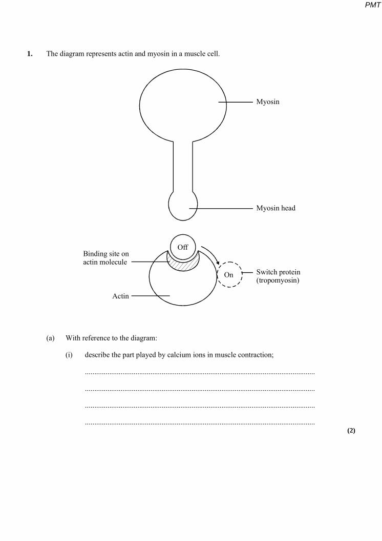
1. The diagram represents actin and myosin in a muscle cell.
Off
On
Myosin
Myosin head
Switch protein(tropomyosin)
Binding site onactin molecule
Actin
(a) With reference to the diagram:
(i) describe the part played by calcium ions in muscle contraction;
...........................................................................................................................
...........................................................................................................................
...........................................................................................................................
........................................................................................................................... (2)
PMT
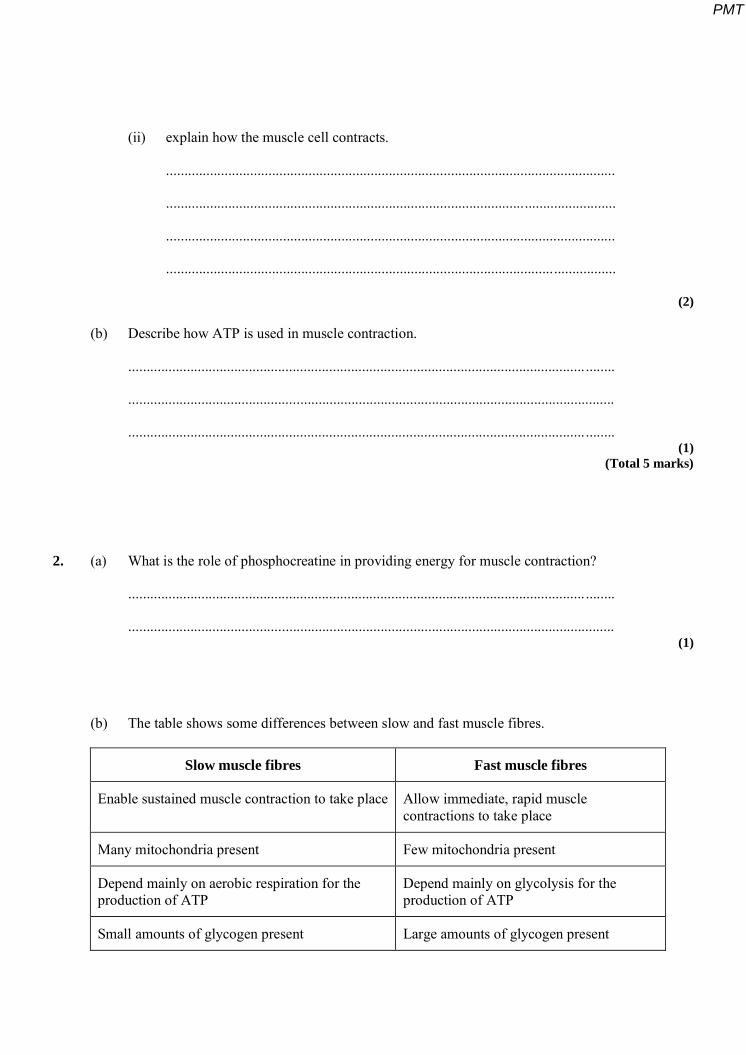
(ii) explain how the muscle cell contracts.
...........................................................................................................................
...........................................................................................................................
...........................................................................................................................
...........................................................................................................................
(2)
(b) Describe how ATP is used in muscle contraction.
............................................................................................................................. ........
.....................................................................................................................................
............................................................................................................................. ........ (1)
(Total 5 marks)
2. (a) What is the role of phosphocreatine in providing energy for muscle contraction?
............................................................................................................................. ........
..................................................................................................................................... (1)
(b) The table shows some differences between slow and fast muscle fibres.
Slow muscle fibres Fast muscle fibres
Enable sustained muscle contraction to take place Allow immediate, rapid muscle contractions to take place
Many mitochondria present Few mitochondria present
Depend mainly on aerobic respiration for the production of ATP
Depend mainly on glycolysis for the production of ATP
Small amounts of glycogen present Large amounts of glycogen present
PMT

(i) Explain the advantage of having large amounts of glycogen in fast muscle fibres.
...........................................................................................................................
...........................................................................................................................
........................................................................................................................... (2)
(ii) Slow muscle fibres have capillaries in close contact. Explain the advantage of this arrangement.
...........................................................................................................................
...........................................................................................................................
...........................................................................................................................
........................................................................................................................... (2)
(Total 5 marks)
3. Muscles contract when some of their cells become shorter in length. This shortening is brought about when myosin and actin filaments in the cytoplasm of muscle cells slide over each other.
Explain how ATP and calcium ions (Ca2+) help the myosin and actin filaments to slide over each other during the shortening of a muscle cell.
ATP ......................................................................................................................... .............
...............................................................................................................................................
............................................................................................................................. ..................
............................................................................................................................. .................. (2)
Ca2+ .......................................................................................................................................
............................................................................................................................. ..................
...............................................................................................................................................
............................................................................................................................. .................. (2)
(Total 4 marks)
PMT
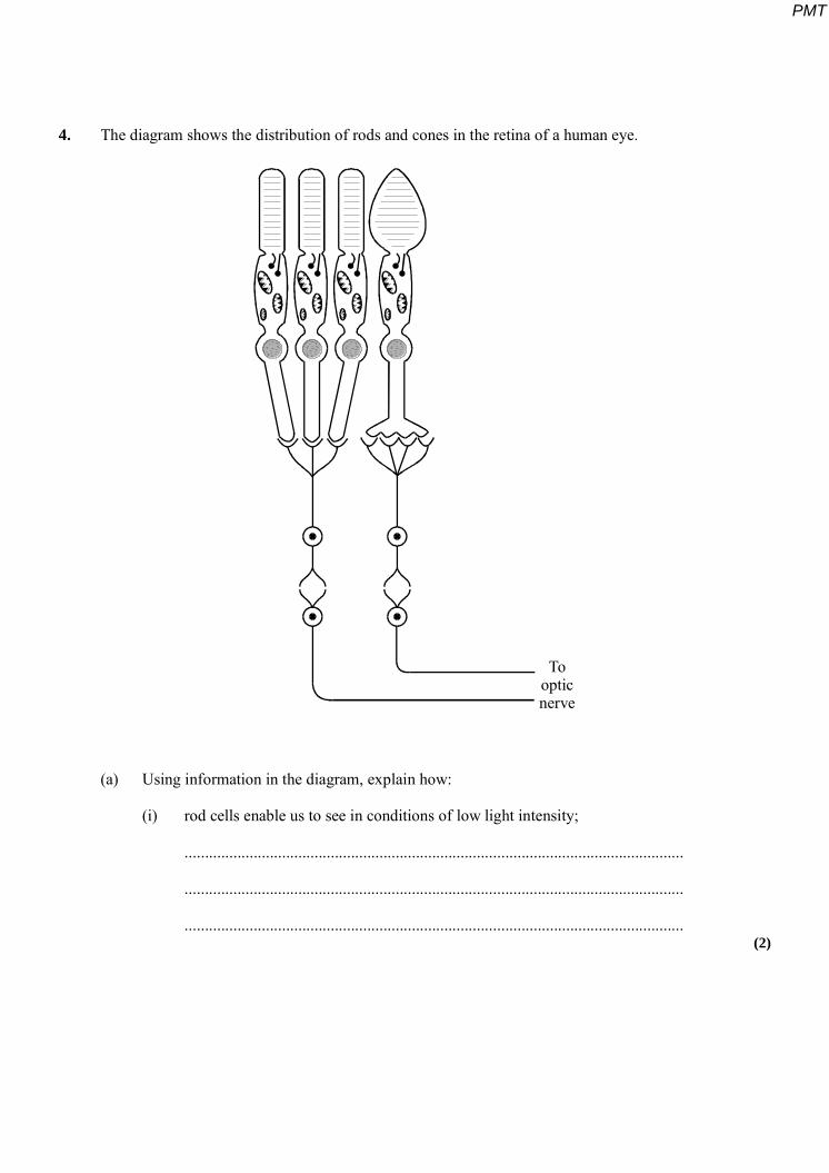
4. The diagram shows the distribution of rods and cones in the retina of a human eye.
Toopticnerve
(a) Using information in the diagram, explain how:
(i) rod cells enable us to see in conditions of low light intensity;
...........................................................................................................................
...........................................................................................................................
........................................................................................................................... (2)
PMT

(ii) cone cells enable us to distinguish between objects close together.
...........................................................................................................................
...........................................................................................................................
...........................................................................................................................
........................................................................................................................... (2)
(b) The graphs show the changes in membrane potential in a presynaptic neurone and a postsynaptic neurone when an impulse passes across a synapse.
+40
+20
0
–20
–40
–60
–800 1 2 3 4
Membranepotential /
m V
Presynaptic neurone
Time / milliseconds
+40
+20
0
–20
–40
–60
–800 1 2 3 4
Membranepotential /
m V
Postsynaptic neurone
Time / milliseconds
PMT
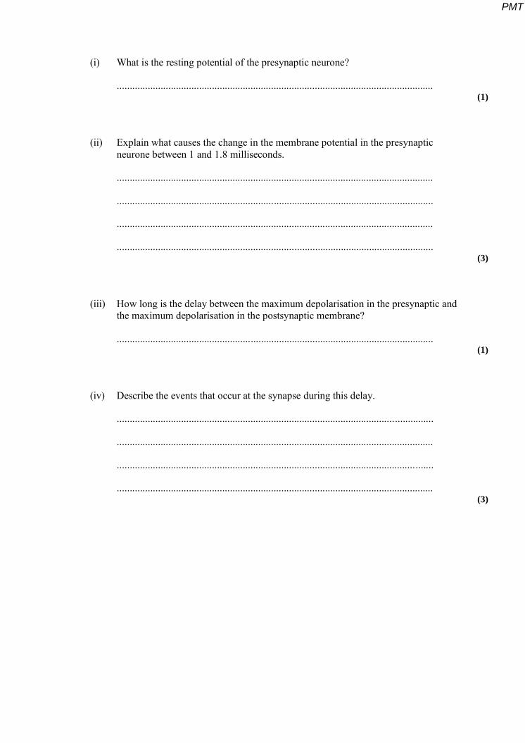
(i) What is the resting potential of the presynaptic neurone?
........................................................................................................................... (1)
(ii) Explain what causes the change in the membrane potential in the presynaptic neurone between 1 and 1.8 milliseconds.
...........................................................................................................................
...........................................................................................................................
...........................................................................................................................
........................................................................................................................... (3)
(iii) How long is the delay between the maximum depolarisation in the presynaptic and the maximum depolarisation in the postsynaptic membrane?
........................................................................................................................... (1)
(iv) Describe the events that occur at the synapse during this delay.
...........................................................................................................................
...........................................................................................................................
...........................................................................................................................
........................................................................................................................... (3)
PMT
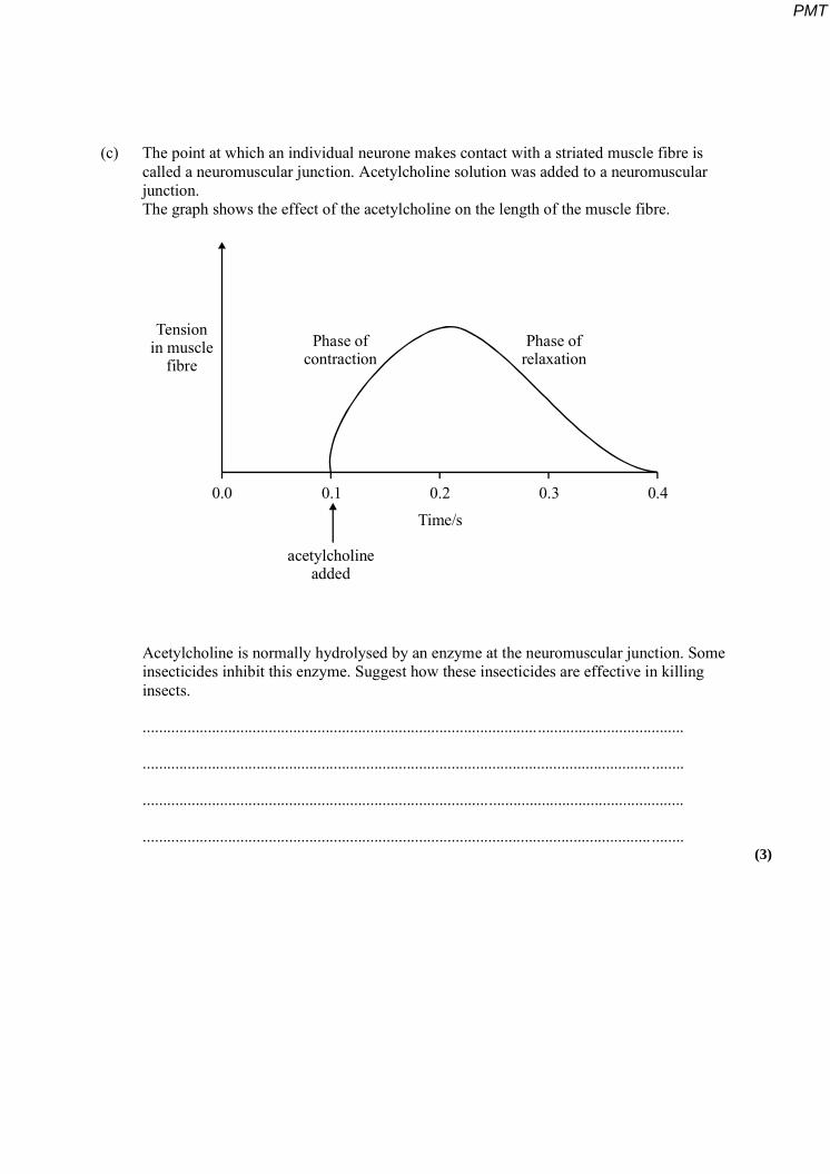
(c) The point at which an individual neurone makes contact with a striated muscle fibre is called a neuromuscular junction. Acetylcholine solution was added to a neuromuscular junction. The graph shows the effect of the acetylcholine on the length of the muscle fibre.
Tensionin muscle
fibrePhase of
contractionPhase of
relaxation
acetylcholineadded
Time/s0.0 0.1 0.2 0.3 0.4
Acetylcholine is normally hydrolysed by an enzyme at the neuromuscular junction. Some insecticides inhibit this enzyme. Suggest how these insecticides are effective in killing insects.
.....................................................................................................................................
............................................................................................................................. ........
.....................................................................................................................................
............................................................................................................................. ........ (3)
PMT
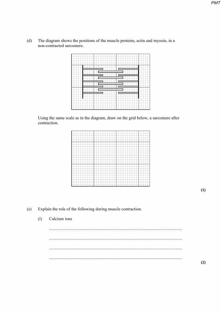
(d) The diagram shows the positions of the muscle proteins, actin and myosin, in a non-contracted sarcomere.
Using the same scale as in the diagram, draw on the grid below, a sarcomere after contraction.
(1)
(e) Explain the role of the following during muscle contraction.
(i) Calcium ions
...........................................................................................................................
...........................................................................................................................
...........................................................................................................................
........................................................................................................................... (2)
PMT
(ii) Mitochondria
...........................................................................................................................
...........................................................................................................................
...........................................................................................................................
........................................................................................................................... (2)
(Total 20 marks)
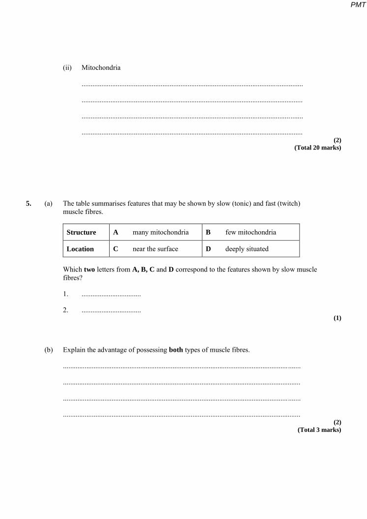
5. (a) The table summarises features that may be shown by slow (tonic) and fast (twitch) muscle fibres.
Structure A many mitochondria B few mitochondria
Location C near the surface D deeply situated
Which two letters from A, B, C and D correspond to the features shown by slow muscle fibres?
1. .................................
2. ................................. (1)
(b) Explain the advantage of possessing both types of muscle fibres.
............................................................................................................................. .......
....................................................................................................................................
............................................................................................................................. .......
.................................................................................................................................... (2)
(Total 3 marks)
PMT
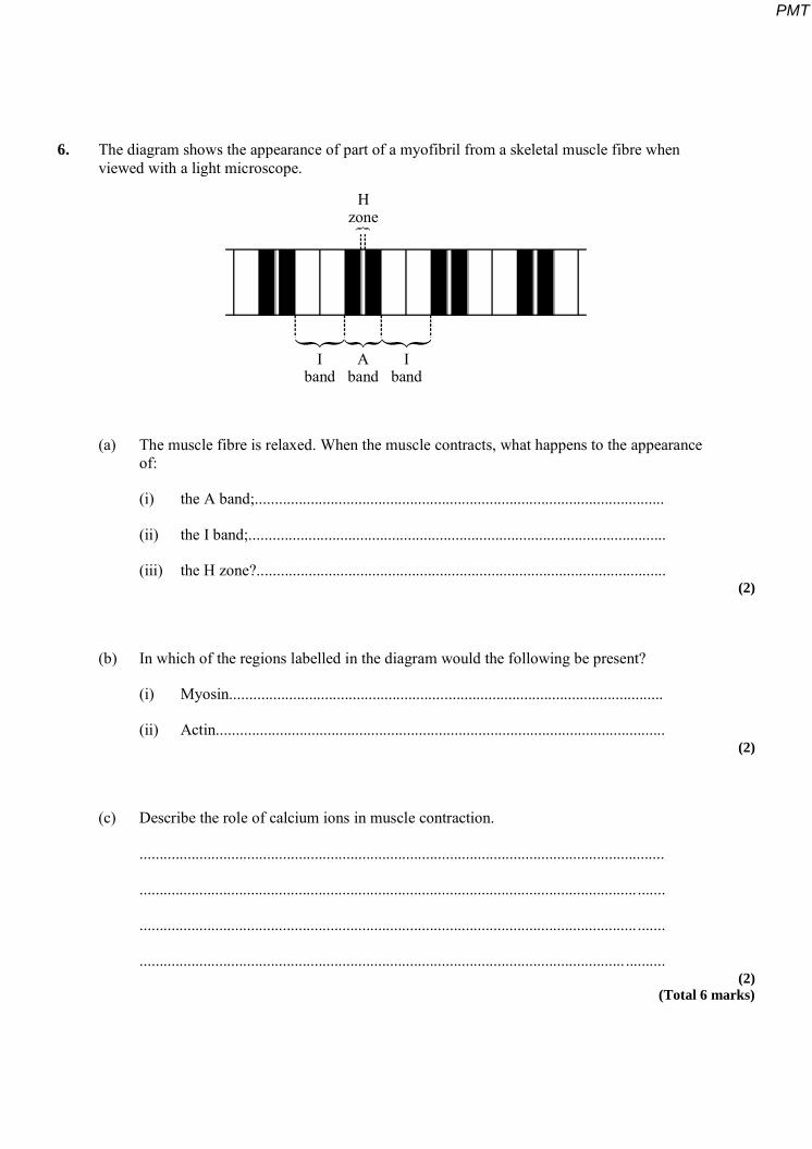
6. The diagram shows the appearance of part of a myofibril from a skeletal muscle fibre when viewed with a light microscope.
Hzone{
Iband
Aband
Iband
(a) The muscle fibre is relaxed. When the muscle contracts, what happens to the appearance of:
(i) the A band;.......................................................................................................
(ii) the I band;.........................................................................................................
(iii) the H zone?....................................................................................................... (2)
(b) In which of the regions labelled in the diagram would the following be present?
(i) Myosin.............................................................................................................
(ii) Actin................................................................................................................. (2)
(c) Describe the role of calcium ions in muscle contraction.
....................................................................................................................................
............................................................................................................................. .......
............................................................................................................................. .......
.......................................................................................................................... .......... (2)
(Total 6 marks)
PMT
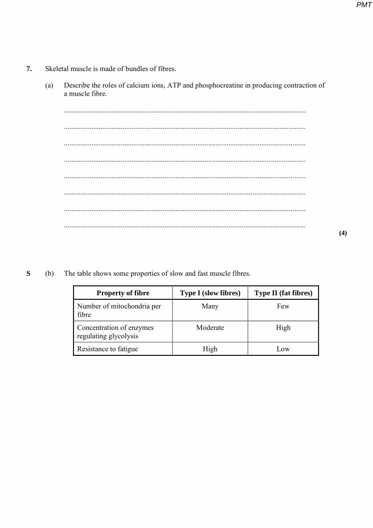
7. Skeletal muscle is made of bundles of fibres.
(a) Describe the roles of calcium ions, ATP and phosphocreatine in producing contraction of a muscle fibre.
............................................................................................................................. .......
........................................................................................................................... .........
............................................................................................................................. .......
....................................................................................................................................
............................................................................................................................. .......
....................................................................................................................................
............................................................................................................................. .......
.................................................................................................................................... (4)
S (b) The table shows some properties of slow and fast muscle fibres.
Property of fibre Type I (slow fibres) Type II (fat fibres)
Number of mitochondria per fibre
Many Few
Concentration of enzymes regulating glycolysis
Moderate High
Resistance to fatigue High Low
PMT

Endurance athletes, such as marathon runners, nearly always have a high proportion of slow fibres in their muscles. Explain the benefit of this.
............................................................................................................................. .......
........................................................................................................................... .........
............................................................................................................................. .......
....................................................................................................................................
............................................................................................................................. .......
....................................................................................................................................
............................................................................................................................. .......
....................................................................................................................................
............................................................................................................................. .......
............................................................................................................................. .......
........................................................................................................................... .........
............................................................................................................................. ....... (6)
(c) During exercise, much heat is generated. Describe the homeostatic mechanisms that restore normal body temperature following vigorous exercise.
............................................................................................................................. .......
....................................................................................................................................
............................................................................................................................. .......
....................................................................................................................................
............................................................................................................................. .......
....................................................................................................................................
............................................................................................................................. .......
....................................................................................................................................
............................................................................................................................. ....... (5)
(Total 15 marks)
PMT
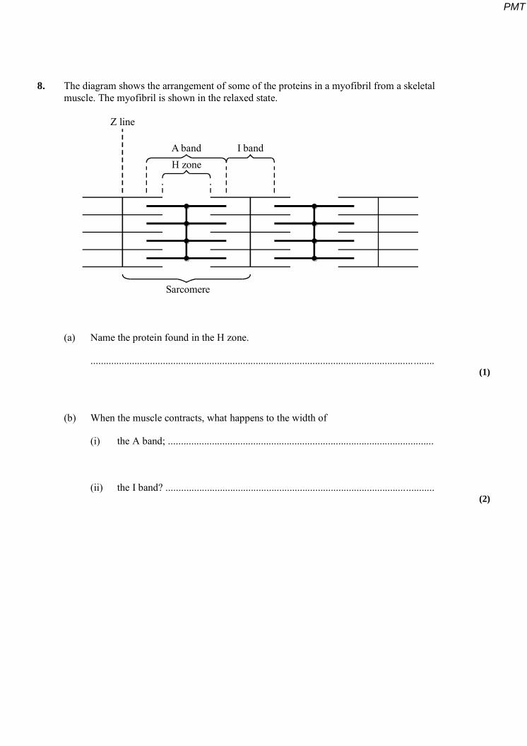
8. The diagram shows the arrangement of some of the proteins in a myofibril from a skeletal muscle. The myofibril is shown in the relaxed state.
Z line
A band I bandH zone
Sarcomere
(a) Name the protein found in the H zone.
............................................................................................................................. ........ (1)
(b) When the muscle contracts, what happens to the width of
(i) the A band; .......................................................................................................
(ii) the I band? ........................................................................................................ (2)
PMT
S (c) The distance between two Z lines in a myofibril is 1.6 m. Calculate the magnification of the diagram. Show your working.
Answer ........................................................................................................................ (2)
(Total 5 marks)
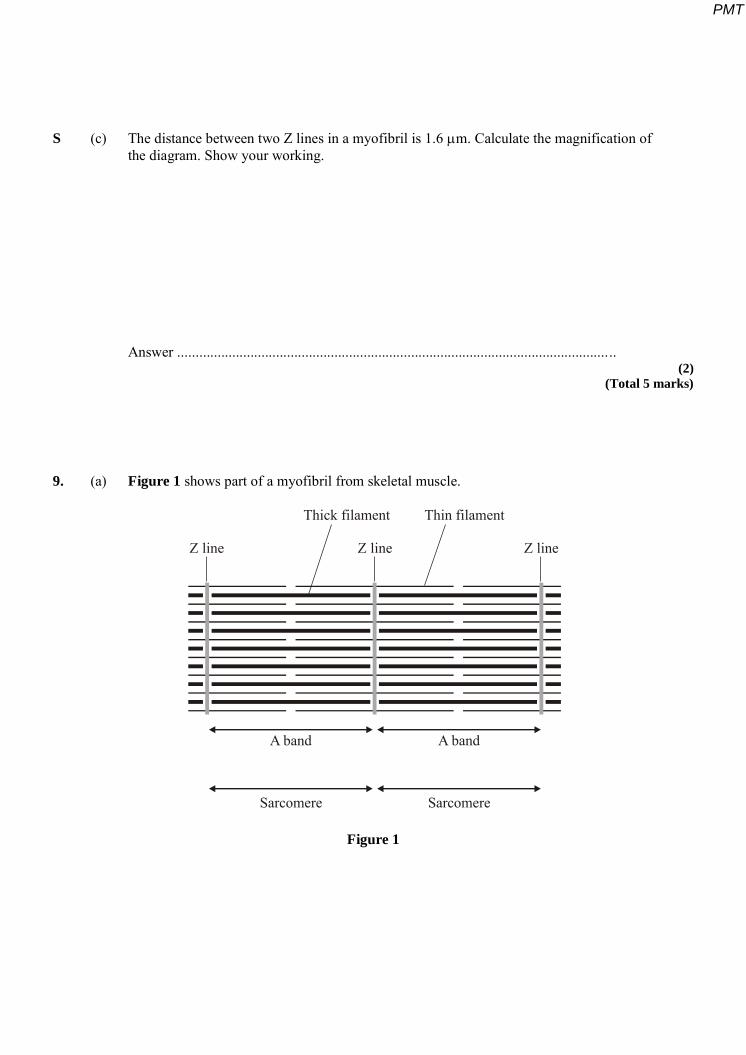
9. (a) Figure 1 shows part of a myofibril from skeletal muscle.
Z line Z lineZ line
Thick filament Thin filament
A band A band
Sarcomere Sarcomere
Figure 1
PMT
(i) Describe two features, visible in the diagram, which show that the myofibril is contracted.
1 .........................................................................................................................
............................................................................................................................
2 ..............................................................................…........................................
............................................................................................................................ (2)
(ii) Explain the role of calcium ions and ATP in bringing about contraction of a muscle fibre.
Calcium ions ..............................................................................................……
............................................................................................................................
............................................................................................................................
ATP ....................................................................................................................
............................................................................................................................
............................................................................................................................ (3)
PMT
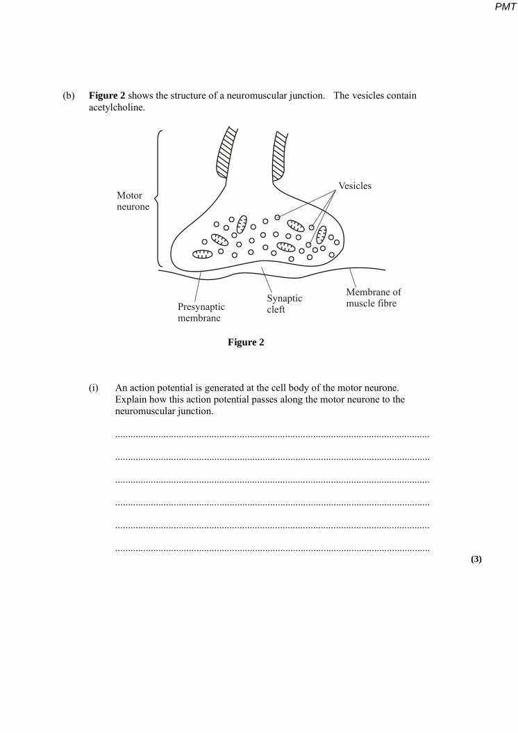
(b) Figure 2 shows the structure of a neuromuscular junction. The vesicles contain acetylcholine.
Motorneurone
Presynapticmembrane
Synapticcleft
Membrane ofmuscle fibre
Vesicles
Figure 2
(i) An action potential is generated at the cell body of the motor neurone. Explain how this action potential passes along the motor neurone to the neuromuscular junction.
............................................................................................................................
............................................................................................................................
............................................................................................................................
............................................................................................................................
............................................................................................................................
............................................................................................................................ (3)
PMT
(ii) When the action potential arrives at the neuromuscular junction, it results in the secretion of acetylcholine into the synaptic cleft. Explain how.
............................................................................................................................
............................................................................................................................
............................................................................................................................
............................................................................................................................
............................................................................................................................
............................................................................................................................ (3)
(c) Between the ages of 20 and 50, 10% of total muscle mass is lost. Between the ages of 50 and 80, a further 40% of the original total muscle mass is lost. Most of the muscle lost consists of fast fibres.
(i) Plot a graph on the grid below to show the percentage of muscle mass remaining between the ages of 20 and 80. Assume that the rate of muscle loss in each age range is constant.
(3)
PMT
(ii) Explain why explosive exercises, such as sprinting and weightlifting, will be more affected by this muscle loss than aerobic exercises, such as jogging.
............................................................................................................................
............................................................................................................................ (1)
(Total 15 marks)
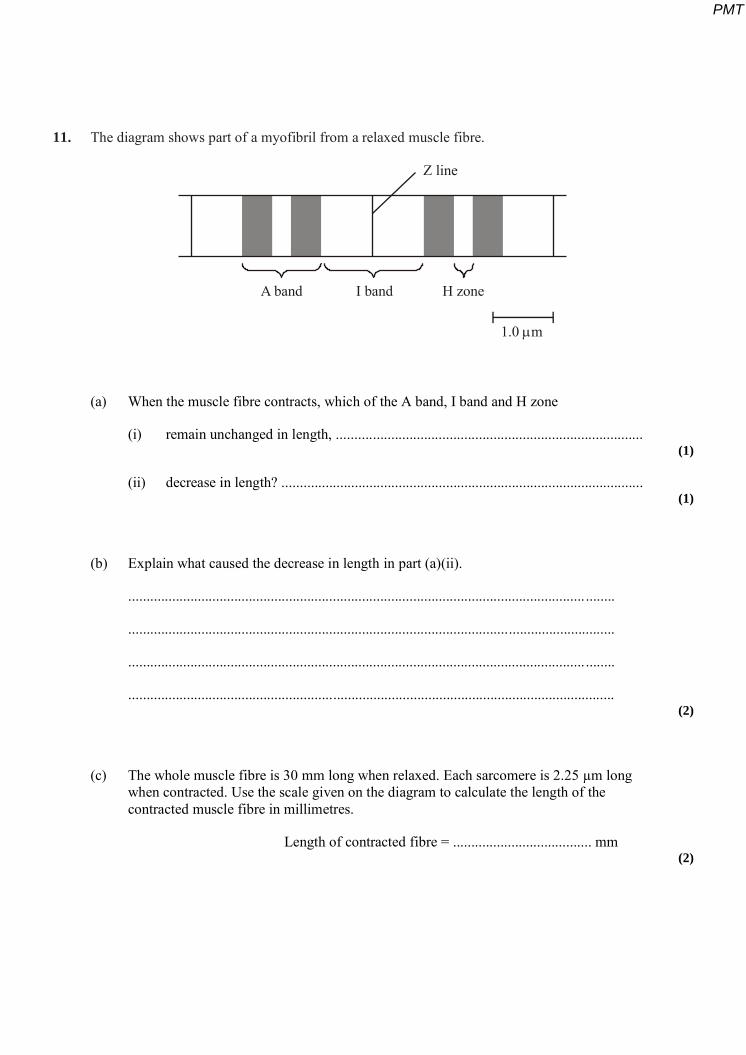
10. The flow chart outlines an investigation to determine from where the calcium ions involved in muscle contraction are released.
Calcium ion transport proteins were isolated from human tissue.
These proteins were injected into a rabbit.
The rabbit formed antibodies to the proteins. These antibodies were collected and labelled with gold particles.
Muscle tissue was treated with the labelled antibodies and examined with an electron microscope. High concentrations of gold particles were observed attached to the sarcoplasmic reticulum.
PMT

S (a) Labelled antibodies and an electron microscope can be used to produce images locating proteins on the surface of organelles, but cannot be used to observe cross bridge cycling in muscle cells. Explain why.
.....................................................................................................................................
............................................................................................................................. ........
.....................................................................................................................................
............................................................................................................................. ........
............................................................................................................................. ........
.......................................................................................................................... ...........
............................................................................................................................. ........
.....................................................................................................................................
............................................................................................................................. ........
..................................................................................................................................... (5)
(b) Describe the role of calcium ions and ATP in muscle contraction.
............................................................................................................................. ........
.....................................................................................................................................
............................................................................................................................. ........
............................................................................................................................. ........
......................................................................................................................... ............
............................................................................................................................. ........
.....................................................................................................................................
............................................................................................................................. ........
.....................................................................................................................................
............................................................................................................................. ........ (5)
(Total 10 marks)
PMT
11. The diagram shows part of a myofibril from a relaxed muscle fibre.
A band I band H zone
1.0 m
Z line
(a) When the muscle fibre contracts, which of the A band, I band and H zone
(i) remain unchanged in length, .................................................................................... (1)
(ii) decrease in length? ................................................................................................... (1)
(b) Explain what caused the decrease in length in part (a)(ii).
............................................................................................................................. ........
.....................................................................................................................................
............................................................................................................................. ........
..................................................................................................................................... (2)
(c) The whole muscle fibre is 30 mm long when relaxed. Each sarcomere is 2.25 µm long when contracted. Use the scale given on the diagram to calculate the length of the contracted muscle fibre in millimetres.
Length of contracted fibre = ...................................... mm (2)
PMT
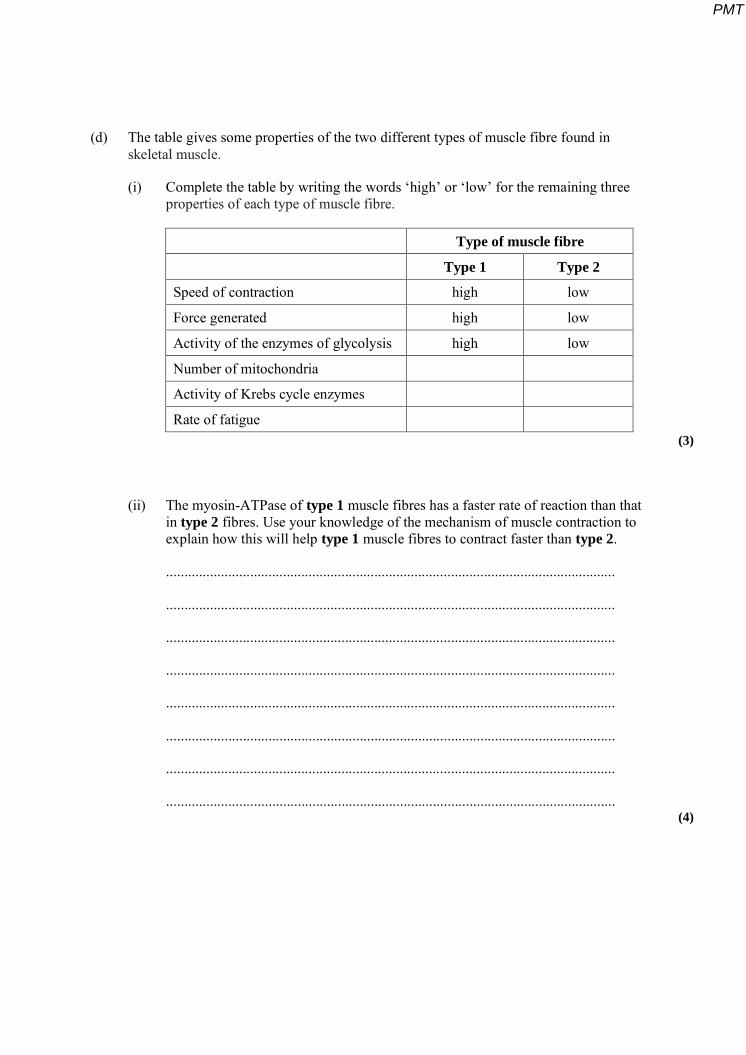
(d) The table gives some properties of the two different types of muscle fibre found in skeletal muscle.
(i) Complete the table by writing the words ‘high’ or ‘low’ for the remaining three properties of each type of muscle fibre.
Type of muscle fibre
Type 1 Type 2
Speed of contraction high low
Force generated high low
Activity of the enzymes of glycolysis high low
Number of mitochondria
Activity of Krebs cycle enzymes
Rate of fatigue (3)
(ii) The myosin-ATPase of type 1 muscle fibres has a faster rate of reaction than that in type 2 fibres. Use your knowledge of the mechanism of muscle contraction to explain how this will help type 1 muscle fibres to contract faster than type 2.
...........................................................................................................................
...........................................................................................................................
...........................................................................................................................
...........................................................................................................................
...........................................................................................................................
...........................................................................................................................
...........................................................................................................................
........................................................................................................................... (4)
PMT

S (iii) The blood leaving an active muscle with a high percentage of type 1 muscle fibres contained a higher concentration of lactate than that leaving a muscle with a high percentage of type 2 muscle fibres. Explain why.
...........................................................................................................................
...........................................................................................................................
...........................................................................................................................
........................................................................................................................... (2)
(Total 15 marks)
12. The diagram shows a neuromuscular junction.
Mitochondrion
Synapticcleft
Axon of neurone
Synaptic knob
Synaptic vesicle
Presynapticmembrane
Sarcoplasm
Musclefibril
Postsynapticmembrane
X
H zone
Lightband
Darkband
1 micrometer
S
(a) (i) Name the neurotransmitter that is released from the synaptic knob.
........................................................................................................................... (1)
PMT
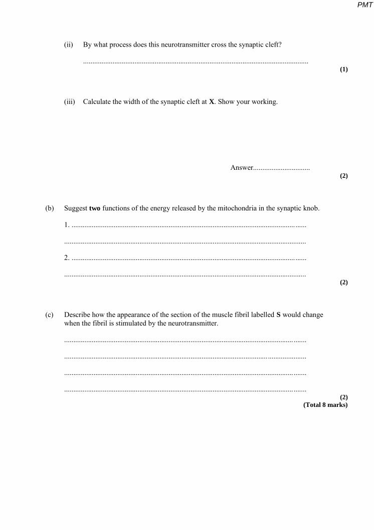
(ii) By what process does this neurotransmitter cross the synaptic cleft?
........................................................................................................................... (1)
(iii) Calculate the width of the synaptic cleft at X. Show your working.
Answer............................... (2)
(b) Suggest two functions of the energy released by the mitochondria in the synaptic knob.
1. .......................................................................................................................... ......
....................................................................................................................................
2. .......................................................................................................................... ......
.................................................................................................................................... (2)
(c) Describe how the appearance of the section of the muscle fibril labelled S would change when the fibril is stimulated by the neurotransmitter.
............................................................................................................................. .......
....................................................................................................................................
............................................................................................................................. .......
............................................................................................................................. ....... (2)
(Total 8 marks)
PMT
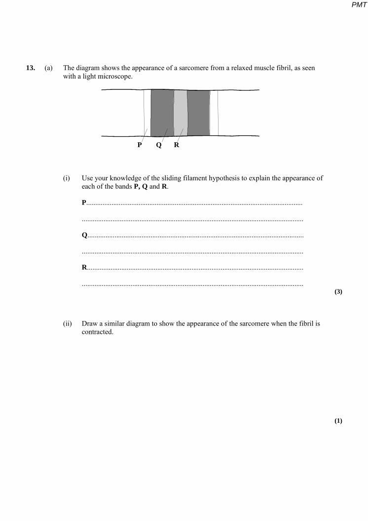
13. (a) The diagram shows the appearance of a sarcomere from a relaxed muscle fibril, as seen with a light microscope.
P Q R
(i) Use your knowledge of the sliding filament hypothesis to explain the appearance of each of the bands P, Q and R.
P........................................................................................................................
...........................................................................................................................
Q........................................................................................................................
...........................................................................................................................
R........................................................................................................................
........................................................................................................................... (3)
(ii) Draw a similar diagram to show the appearance of the sarcomere when the fibril is contracted.
(1)
PMT
(b) Muscles use energy from respiration for contraction. Describe how the energy released in mitochondria during respiration produces contraction of a muscle fibril.
.....................................................................................................................................
............................................................................................................................. ........
.....................................................................................................................................
............................................................................................................................. ........
.....................................................................................................................................
............................................................................................................................. ........
............................................................................................................................. ........
.....................................................................................................................................
............................................................................................................................. ........
..................................................................................................................................... (4)
(Total 8 marks)
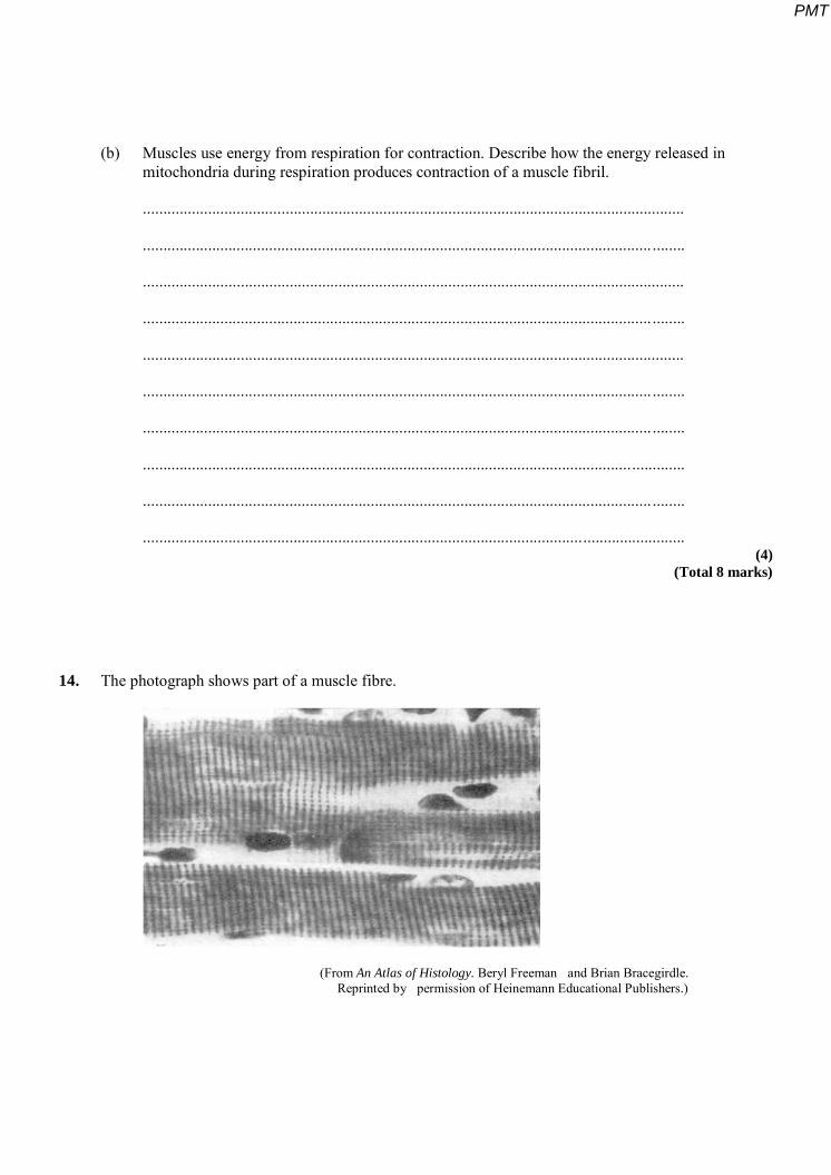
14. The photograph shows part of a muscle fibre.
(From An Atlas of Histology. Beryl Freeman and Brian Bracegirdle. Reprinted by permission of Heinemann Educational Publishers.)
PMT
Give three differences in structure between a muscle fibre and an epithelial cell from the lining of the small intestine.
1 ........................................................................................................................... .................
...............................................................................................................................................
2 ........................................................................................................................... .................
...............................................................................................................................................
3 ........................................................................................................................... .................
............................................................................................................................................. .. (Total 3 marks)
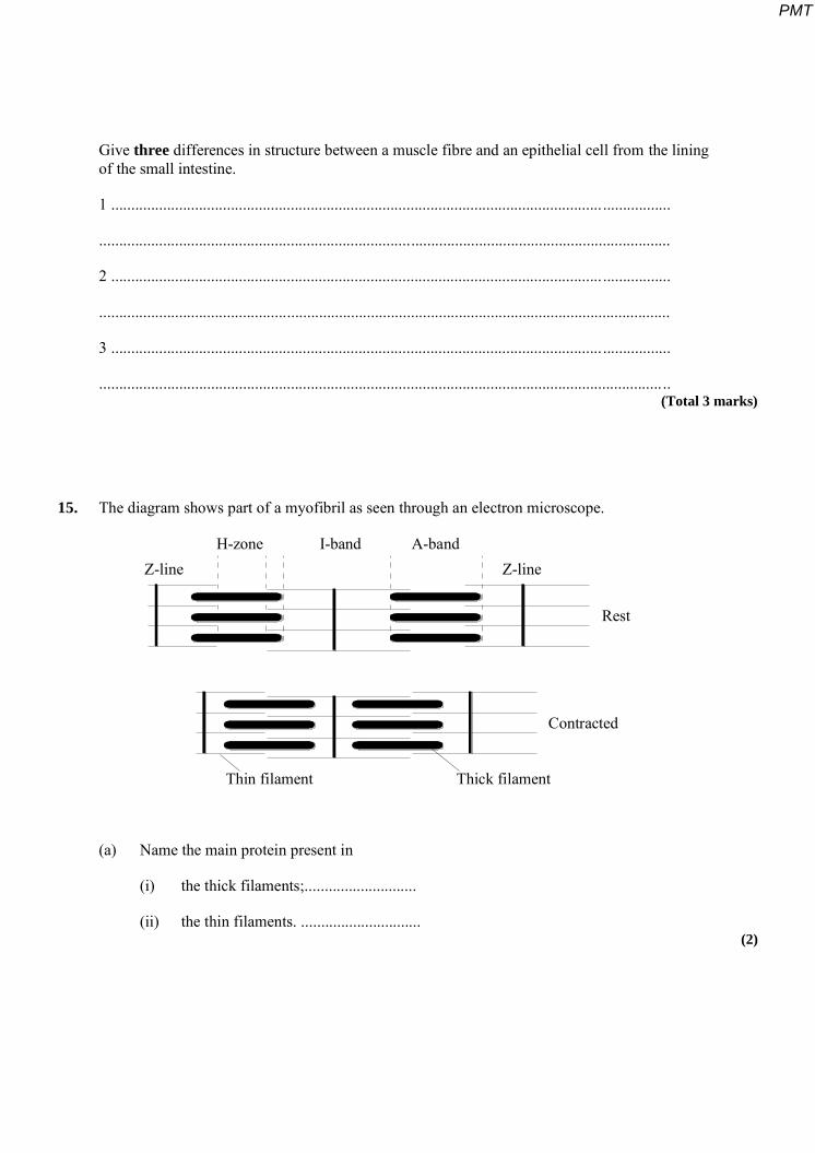
15. The diagram shows part of a myofibril as seen through an electron microscope.
Z-line Z-lineH-zone I-band A-band
Rest
Contracted
Thick filamentThin filament
(a) Name the main protein present in
(i) the thick filaments;............................
(ii) the thin filaments. .............................. (2)
PMT
(b) Describe the mechanism that brings about the change in position of the filaments when the myofibril contracts.
............................................................................................................................. ........
.....................................................................................................................................
............................................................................................................................. ........
................................................................................................................................. ....
............................................................................................................................. ........
............................................................................................................................. ........
.....................................................................................................................................
............................................................................................................................. ........ (4)
(Total 6 marks)
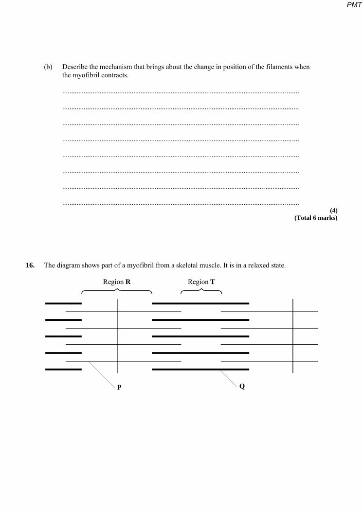
16. The diagram shows part of a myofibril from a skeletal muscle. It is in a relaxed state.
Region R Region T
P Q
PMT
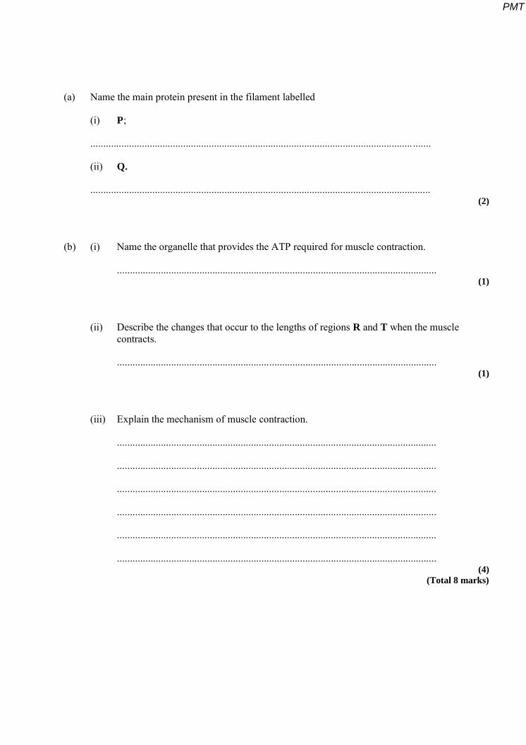
(a) Name the main protein present in the filament labelled
(i) P;
............................................................................................................................. .......
(ii) Q.
.................................................................................................................................... (2)
(b) (i) Name the organelle that provides the ATP required for muscle contraction.
............................................................................................................................ (1)
(ii) Describe the changes that occur to the lengths of regions R and T when the muscle contracts.
............................................................................................................................ (1)
(iii) Explain the mechanism of muscle contraction.
............................................................................................................................
............................................................................................................................
............................................................................................................................
............................................................................................................................
............................................................................................................................
............................................................................................................................ (4)
(Total 8 marks)
PMT
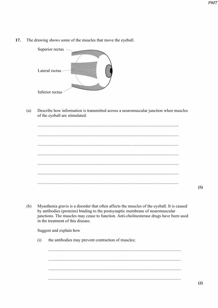
17. The drawing shows some of the muscles that move the eyeball.
Superior rectus
Lateral rectus
Inferior rectus
(a) Describe how information is transmitted across a neuromuscular junction when muscles of the eyeball are stimulated.
....................................................................................................................................
............................................................................................................................. .......
....................................................................................................................................
............................................................................................................................. .......
....................................................................................................................................
............................................................................................................................. .......
.................................................................................................................................... (5)
(b) Myasthenia gravis is a disorder that often affects the muscles of the eyeball. It is caused by antibodies (proteins) binding to the postsynaptic membrane of neuromuscular junctions. The muscles may cease to function. Anti-cholinesterase drugs have been used in the treatment of this disease.
Suggest and explain how
(i) the antibodies may prevent contraction of muscles;
............................................................................................................................
............................................................................................................................
............................................................................................................................
............................................................................................................................ (2)
PMT

(ii) anti-cholinesterase drugs may help in the treatment of myasthenia gravis.
............................................................................................................................
............................................................................................................................
............................................................................................................................
............................................................................................................................ (2)
(Total 9 marks)
18. S Write an essay on the topic below.
The different ways in which organisms use ATP.
In the answer to this question you should bring together relevant principles and concepts
from as many different modules as possible.
Your essay will be marked not only for its scientific accuracy, but also for the selection of
relevant material.
The essay should be written in continuous prose.
The maximum number of marks that can be awarded is:
Scientific content 16
Breadth of knowledge 3
Relevance 3
Quality of Written Communication 3 (Total 25 marks)
PMT
19. Myasthenia gravis is a disease which causes muscular weakness. It develops because of an attack by the body’s own immune system on neuromuscular junctions. The diagram shows a normal neuromuscular junction and one affected by the disease (myasthenic).
Vesicles containingacetylcholine
Acetylcholinereceptors
Acetylcholinesterase
Axon of motor neurone
Membrane ofmuscle cell
MyasthenicNormal
(a) Describe two ways in which a myasthenic neuromuscular junction differs from a normal one and explain how each difference would affect transmissions across the myasthenic neuromuscular junction.
Difference....................................................................................................................
............................................................................................................................. ........
Effect...........................................................................................................................
............................................................................................................................. ........
.....................................................................................................................................
Difference....................................................................................................................
.....................................................................................................................................
Effect....................................................................................................................... ....
............................................................................................................................. ........
.......................................................................................................................... ........... (4)
PMT
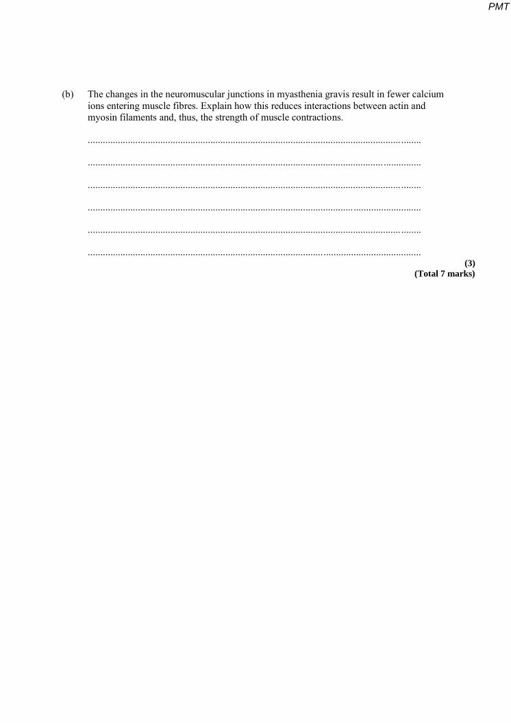
(b) The changes in the neuromuscular junctions in myasthenia gravis result in fewer calcium ions entering muscle fibres. Explain how this reduces interactions between actin and myosin filaments and, thus, the strength of muscle contractions.
............................................................................................................................. ........
.....................................................................................................................................
............................................................................................................................. ........
.....................................................................................................................................
............................................................................................................................. ........
..................................................................................................................................... (3)
(Total 7 marks)
PMT
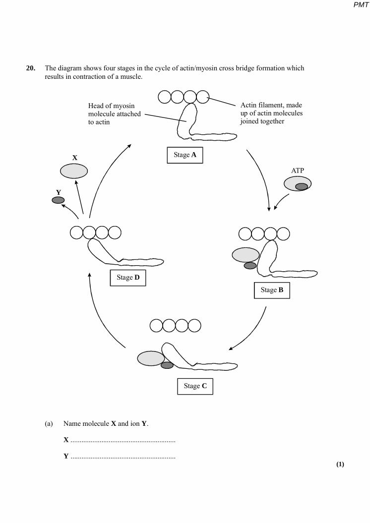
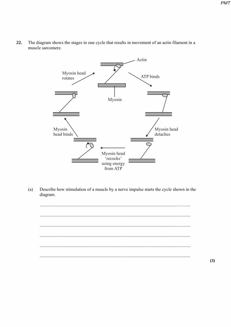
20. The diagram shows four stages in the cycle of actin/myosin cross bridge formation which results in contraction of a muscle.
Stage A
Stage B
Stage C
Stage D
Head of myosinmolecule attachedto actin
Actin filament, madeup of actin moleculesjoined together
X
Y
ATP
(a) Name molecule X and ion Y.
X ..........................................................
Y .......................................................... (1)
PMT
(b) Use the information in the diagram to explain how actin moves past myosin. In your answer, refer to the stages A, B, C and D. (A description of the roles of calcium ions and tropomysin is not required.)
.....................................................................................................................................
............................................................................................................................. ........
.....................................................................................................................................
............................................................................................................................. ........
.....................................................................................................................................
............................................................................................................................. ........
.....................................................................................................................................
............................................................................................................................. ........ (4)
(c) After an animal dies, respiration stops and no more ATP is made. The muscles become rigid and fixed in their length. Use the information in the diagram to suggest an explanation for this.
.....................................................................................................................................
............................................................................................................................. ........
............................................................................................................................. ........ (1)
(Total 6 marks)
PMT
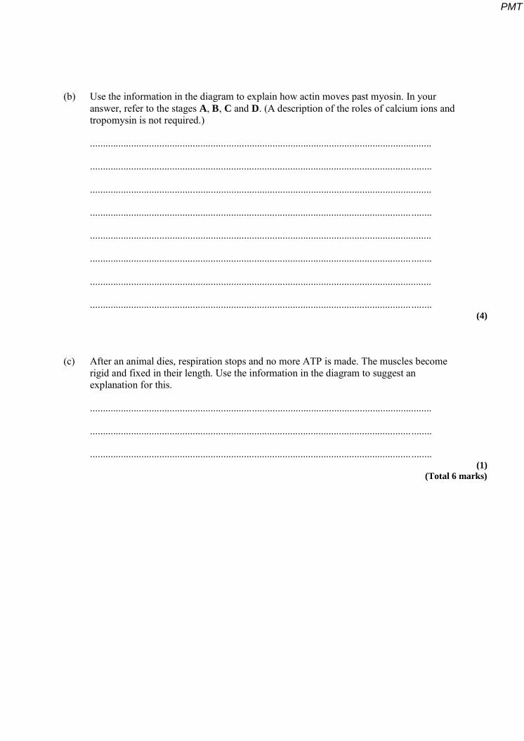
21. Figure 1 shows a diagram of part of a muscle myofibril.
Z ZY
W X
Figure 1
(a) Name the protein present in the filaments labelled W and X.
W ............................................................................................................................. ...
X ................................................................................................................................. (1)
(b) Figure 2 shows the cut ends of the protein filaments when the myofibril was cut at position Y. Figure 3 shows the protein filaments when the myofibril was cut at the same distance from a Z line at a different stage of contraction.
Figure 2 Figure 3
PMT
Explain why the pattern of protein filaments differs in Figure 2 and Figure 3.
............................................................................................................................. ........
.....................................................................................................................................
............................................................................................................................. ........
..................................................................................................................................... (2)
(c) Describe the role of calcium ions in the contraction of a sarcomere.
............................................................................................................................. ........
.....................................................................................................................................
............................................................................................................................. ........
............................................................................................................................. ........
....................................................................................................................... ..............
............................................................................................................................. ........
.....................................................................................................................................
............................................................................................................................. ........ (4)
(Total 7 marks)
PMT
22. The diagram shows the stages in one cycle that results in movement of an actin filament in a muscle sarcomere.
Myosin head‘recocks’
using energyfrom ATP
Myosin
Actin
ATP bindsMyosin headrotates
Myosinhead binds
Myosin headdetaches
(a) Describe how stimulation of a muscle by a nerve impulse starts the cycle shown in the diagram.
............................................................................................................................. ........
.....................................................................................................................................
............................................................................................................................. ........
.....................................................................................................................................
............................................................................................................................. ........
..................................................................................................................................... (3)
PMT
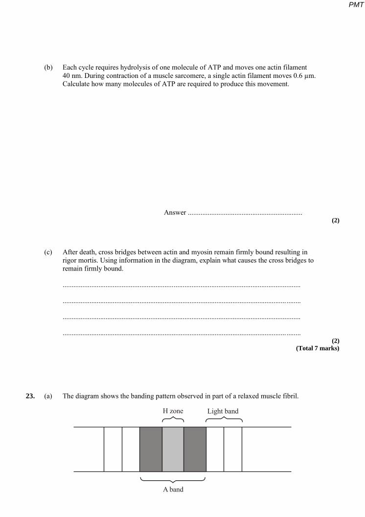
(b) Each cycle requires hydrolysis of one molecule of ATP and moves one actin filament 40 nm. During contraction of a muscle sarcomere, a single actin filament moves 0.6 µm. Calculate how many molecules of ATP are required to produce this movement.
Answer ................................................................ (2)
(c) After death, cross bridges between actin and myosin remain firmly bound resulting in rigor mortis. Using information in the diagram, explain what causes the cross bridges to remain firmly bound.
.....................................................................................................................................
............................................................................................................................. ........
.....................................................................................................................................
............................................................................................................................. ........ (2)
(Total 7 marks)
23. (a) The diagram shows the banding pattern observed in part of a relaxed muscle fibril.
H zone Light band
A band
PMT
(i) Describe what causes the different bands seen in the muscle fibril.
...........................................................................................................................
...........................................................................................................................
...........................................................................................................................
........................................................................................................................... (2)
(ii) Describe how the banding pattern will be different when the muscle fibril is contracted.
...........................................................................................................................
...........................................................................................................................
...........................................................................................................................
........................................................................................................................... (2)
(d) There is an increase in the activity of the enzyme ATPase during muscle contraction. An investigation into muscle contraction involved measuring the activity of ATPase in solutions containing ATP, myosin and different muscle components. The table shows the results.
Solution Contents ATPase activity /
arbitrary units
A ATP, myosin and actin 1.97
B ATP, myosin, actin and tropomyosin 0.54
C ATP, myosin, actin, tropomyosin and calcium ions 3.85
(i) Explain the importance of ATPase during muscle contraction.
...........................................................................................................................
...........................................................................................................................
...........................................................................................................................
........................................................................................................................... (2)
PMT
(ii) Using your knowledge of muscle contraction, explain the difference in the results between
A and B;
...........................................................................................................................
...........................................................................................................................
...........................................................................................................................
........................................................................................................................... (2)
B and C.
...........................................................................................................................
...........................................................................................................................
...........................................................................................................................
........................................................................................................................... (2)
(Total 10 marks)
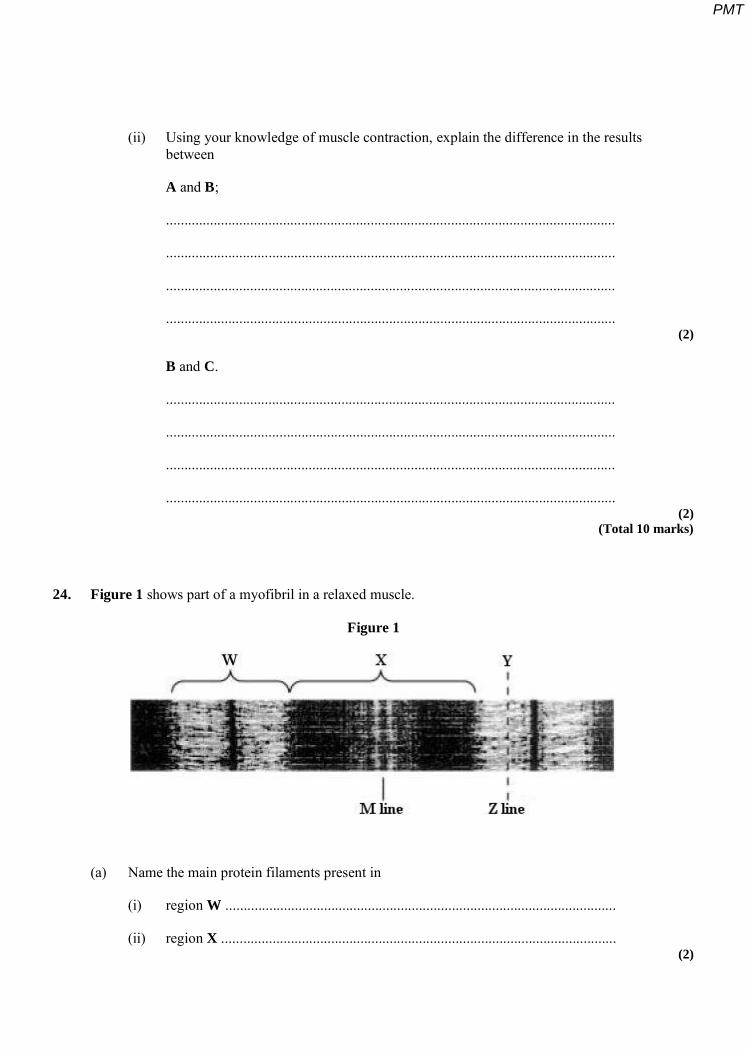
24. Figure 1 shows part of a myofibril in a relaxed muscle.
Figure 1
(a) Name the main protein filaments present in
(i) region W ...........................................................................................................
(ii) region X ............................................................................................................ (2)
PMT
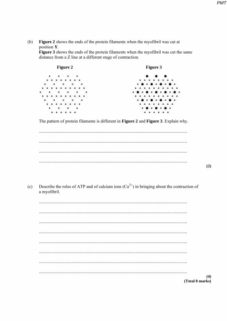
(b) Figure 2 shows the ends of the protein filaments when the myofibril was cut at position Y. Figure 3 shows the ends of the protein filaments when the myofibril was cut the same distance from a Z line at a different stage of contraction.
Figure 2 Figure 3
The pattern of protein filaments is different in Figure 2 and Figure 3. Explain why.
............................................................................................................................. ........
............................................................................................................................. ........
.......................................................................................................................... ...........
............................................................................................................................. ........ (2)
(c) Describe the roles of ATP and of calcium ions (Ca2+) in bringing about the contraction of a myofibril.
............................................................................................................................. ........
....................................................................................................................... ..............
............................................................................................................................. ........
.....................................................................................................................................
............................................................................................................................. ........
.....................................................................................................................................
............................................................................................................................. ........
..................................................................................................................................... (4)
(Total 8 marks)
PMT