Embed Size (px)
Citation preview
Cholesterol metabolism in type 2 diabetes
Piia Simonen
Department of Medicine
University of Helsinki
Helsinki, Finland
Academic dissertation
To be publicly discussed with the permission of the Medical Faculty of the
University of Helsinki, in Auditorium 2, Biomedicum Helsinki,
on December 27th, 2002, at 12 noon
Helsinki 2002
Supervisor Professor Tatu A. Miettinen
Department of Medicine
University of Helsinki
Helsinki, Finland
Reviewers Docent Veikko Salomaa
National Public Health Institute
Helsinki, Finland
and
Docent Hannu Vanhanen
Finnish Heart Association
Helsinki, Finland
Opponent Professor Jussi Huttunen
National Public Health Institute
Helsinki, Finland
ISBN 952-91-5365-1 (nid.)
ISBN 952-10-0826-1 (PDF)
Yliopistopaino
Helsinki 2002

To my family


5
CONTENTS LIST OF ORIGINAL PUBLICATIONS..........................................................................7 ABBREVIATIONS.........................................................................................................8 ABSTRACT ...................................................................................................................9 1. INTRODUCTION .....................................................................................................12 2. REVIEW OF THE LITERATURE ............................................................................15
2.1 Type 2 diabetes .................................................................................................15 2.1.1 Insulin resistance ........................................................................................16 2.1.2 Insulin secretion ..........................................................................................16 2.1.3 Interaction of insulin resistance and beta-cell dysfunction.........................17 2.1.4 The metabolic syndrome.............................................................................18
2.2 Overview of lipoprotein metabolism ..................................................................18 2.3 Lipoprotein metabolism in diabetes...................................................................22 2.4 Obesity...............................................................................................................24
2.4.1 Lipoprotein metabolism in obesity ..............................................................25 2.5 Diabetes and obesity .........................................................................................27 2.6 Cholesterol metabolism .....................................................................................28
2.6.1 Cholesterol absorption................................................................................31 2.6.1.1 Sources of intraluminal cholesterol ......................................................31 2.6.1.2 Luminal events .....................................................................................31 2.6.1.3 Mucosal events ....................................................................................32
2.6.1.3.1 Uptake and re-excretion ................................................................32 2.6.1.3.2 Mucosal cholesterol.......................................................................33
2.6.1.4 Regulation of serum cholesterol level..................................................34 2.6.1.5 Measurement of cholesterol absorption...............................................35 2.6.1.6 Factors affecting cholesterol absorption ..............................................37
2.6.2 Plant sterols and cholestanol......................................................................41 2.6.2.1 Absorption of plant sterols....................................................................42 2.6.2.2 Transport in serum ...............................................................................42
2.6.3 Cholesterol synthesis..................................................................................43 2.6.3.1 Reverse cholesterol transport ..............................................................45
2.6.4 Squalene and demethylated cholesterol precursors ..................................46 2.6.5 Elimination of cholesterol............................................................................47
2.6.5.1 Bile acids ..............................................................................................47 2.6.6 Nuclear receptors and cholesterol metabolism ..........................................49
2.7 Cholesterol synthesis and elimination in obesity ..............................................50 2.8 Cholesterol metabolism in diabetes ..................................................................51
2.8.1 Cholesterol absorption................................................................................51 2.8.2 Regulation of serum cholesterol level.........................................................51 2.8.3 Cholesterol synthesis and excretion...........................................................52 2.8.4 Insulin treatment..........................................................................................53 2.8.5 Summary.....................................................................................................53
2.9 Treatment with weight reduction .......................................................................54 2.9.1 Glucose and lipoprotein metabolism ..........................................................54 2.9.2 Cholesterol metabolism ..............................................................................55
3. AIMS OF THE STUDY ............................................................................................57 4. MATERIALS AND METHODS ...............................................................................59
4.1 Subjects and designs ........................................................................................59 4.1.1 Study I .........................................................................................................59
6
4.1.2 Study II ........................................................................................................60 4.1.3 Study III .......................................................................................................61 4.1.4 Study IV.......................................................................................................62
4.2 Methods .............................................................................................................63 4.2.1 Inclusion criteria measurements.................................................................63 4.2.2 Lipoprotein separation ................................................................................63 4.2.3 Lipids and apolipoproteins ..........................................................................64 4.2.4 Lipoprotein kinetic studies ..........................................................................64 4.2.5 Analysis of cholesterol metabolism ............................................................65
4.2.5.1 Measurement of cholesterol absorption and elimination .....................65 4.2.5.2 Determination of squalene and non-cholesterol sterols ......................66 4.2.5.3 Calculations ..........................................................................................66
4.2.6 Analysis of variables in glucose metabolism..............................................67 4.2.7 Statistical analyses .....................................................................................67
5. RESULTS ................................................................................................................69 5.1 Diabetes and cholesterol metabolism (Study I).................................................69 5.2 Body weight and cholesterol metabolism (Study II) ..........................................72 5.3 Treatment with weight reduction (Study III and IV) ...........................................77
5.3.1 Serum non-cholesterol sterols and squalene during weight reduction (Study III) ..............................................................................................................77 5.3.2 Cholesterol, glucose and lipoprotein metabolism after treatment with weight reduction (Study IV)..................................................................................80
6. DISCUSSION ..........................................................................................................83 6.1 Study population ................................................................................................83 6.2 Measurement of cholesterol metabolism ..........................................................84 6.3 Cholesterol metabolism in diabetes (Study I and II) .........................................86
6.3.1 Cholesterol absorption ................................................................................86 6.3.2 Cholesterol synthesis and excretion...........................................................89
6.4 Cholesterol and lipoprotein metabolism (Study I and II) ...................................89 6.5 Cholesterol and glucose metabolism (Study I and II) ......................................91 6.6 Weight reduction (Study III and IV) ...................................................................92
6.6.1 Chronic caloric restriction (Study III)...........................................................92 6.6.2 Steady state after weight loss (Study IV)....................................................94
6.7 Mechanisms of abnormal cholesterol metabolism in diabetes .........................95 6.7.1 Cholesterol synthesis..................................................................................96 6.7.2 Cholesterol absorption................................................................................97
7. SUMMARY AND CONCLUSIONS .........................................................................99 ACKNOWLEDGEMENTS.........................................................................................104 REFERENCES ..........................................................................................................106

7
LIST OF ORIGINAL PUBLICATIONS
This thesis is based on the following original articles, which are referred to in the text
by their Roman numerals.
I Simonen PP, Gylling HK, Miettinen TA. Diabetes contributes to cholesterol
metabolism regardless of obesity. Diabetes Care 2002; 25:1511-1515.
II Simonen PP, Gylling H, Miettinen TA. Body weight modulates cholesterol
metabolism in non-insulin dependent type 2 diabetics. Obes Res 2002;10:328-
335.
III Simonen P, Gylling H, Miettinen TA. Acute effects of weight reduction on
cholesterol metabolism in obese type 2 diabetes. Clin Chim Acta 2002;316:55-61.
IV Simonen P, Gylling H, Howard AN, Miettinen TA. Introducing a new component of
the metabolic syndrome: low cholesterol absorption. Am J Clin Nutr 2000;72:82-
88.
8
ABBREVIATIONS
ABC adenosine triphosphate-binding cassette transporter ACAT acyl-CoA cholesterol acyltransferase ANOVA analysis of variance Apo apolipoprotein BMI body mass index BSEP bile salt export pump CETP cholesteryl ester transfer protein Cr2O3 chromic oxide CYP7A1 cholesterol 7α-hydroxylase d density ∆ 8 – cholestenol cholestenol ∆ 7 – lathosterol lathosterol DM diabetes group FCR fractional catabolic rate FXR farnesoid X receptor HbA1c glycosylated hemoglobin A1c GLC gas-liquid chromatography HDL high density lipoprotein HMG-CoA 3-hydroxy-3-methylglutaryl-coenzyme A HOMA homeostasis model assessment I-BABP ileal bile-acid binding protein IDL intermediate density lipoprotein LCAT lecithin:cholesterol acyltransferase LDL low density lipoprotein LED low energy diet LRP LDL receptor-related protein LXR liver X receptor MTP microsomal triglyceride transfer protein PLTP plasma phospholipid transfer protein RXR retinoid X receptor SHBG sex hormone binding globulin SR-BI scavenger receptor BI SREBP sterol regulatory element binding protein TR transport rate TRL triglyceride rich lipoproteins VLDL very low density lipoprotein VLED very low energy diet

9
ABSTRACT
Type 2 diabetes is associated with many metabolic disturbances including
hyperinsulinemia, insulin resistance, hyperglycemia, dyslipidemia and obesity. The
alterations occurring in lipoprotein metabolism in diabetes have been described in
detail, but the metabolism of cholesterol and bile acids has been less well
characterized, and the results from the few previous studies are controversial.
Obesity, in addition to predisposing to the development of diabetes, is associated
with abnormal cholesterol metabolism. Accordingly, cholesterol metabolism was
studied in obesity with and without type 2 diabetes, and in type 2 diabetes with and
without overweight. In addition, the effects of weight reduction were studied on
cholesterol and sterol metabolism in a non-stable state during fasting and also in
subsequent steady state after a prolonged follow-up.
Cholesterol absorption and cholesterol and bile acid synthesis were studied in 16
obese (BMI > 30 kg/m2) type 2 diabetic patients and compared to 16 similarly obese
controls to reveal the role of diabetes on cholesterol metabolism. The effects of body
weight on cholesterol metabolism were investigated with 20 normal-weight (BMI ≤
26.0 kg/m2) and 44 overweight (BMI > 26.1 kg/m2) type 2 diabetic patients.
Cholesterol absorption was evaluated with the peroral dual isotope technique and by
quantitating serum ratios of phytosterols and cholestanol to cholesterol, cholesterol
synthesis with sterol balance as well as serum ratios of squalene and precursor
sterols (cholestenol, desmosterol, lathosterol) to cholesterol. In order to clarify the
role of weight reduction in modulating cholesterol metabolism in type 2 diabetes,
parameters associated with cholesterol and sterol metabolism were determined
during weight reduction, and during a 2-year follow-up after weight reduction. Ten
obese type 2 diabetic patients consumed a very low energy diet virtually free of
cholesterol, cholestanol and plant sterols for 3 months, and serum squalene and non-
cholesterol sterol levels were determined before and after the weight reduction
program in a non-steady state situation. Sixteen obese type 2 diabetic patients
consumed a very low energy or low-energy diet for 3 months, after which they
consumed a weight-maintaining diet for up to 2 years. The changes in cholesterol
metabolism were determined by assaying cholesterol absorption efficiency, sterol
balance, and serum sterols after a 2-year follow-up, at a stable, reduced weight level.
10
The efficiency of cholesterol absorption and the amounts of absorbed total, dietary
and biliary cholesterol were lower in the obese type 2 diabetic patients than obese
controls or in normal-weight type 2 diabetic patients. Cholesterol absorption was
similar in both diabetics with normal body weight and obese controls. Fecal
elimination of cholesterol, mainly as neutral sterols and less as bile acids, was
increased, and this enhanced cholesterol synthesis more in obese patients with type
2 diabetes than in obese controls or normal-weight diabetic patients. In addition, fecal
bile acids, the total intestinal cholesterol pool, biliary cholesterol secretion and
cholesterol turnover were significantly higher in obese diabetic patients than normal-
weight diabetic patients, when expressed as mg/d. Moreover, BMI was positively
associated with variables of cholesterol synthesis and negatively with cholesterol
absorption. Cholesterol absorption and synthesis were inversely related in the
diabetic population suggesting that the homeostatic regulation between cholesterol
absorption and synthesis was not distrupted by diabetes.
Serum plant sterols and cholestanol ratios correlated with the cholesterol absorption
efficiency, and those of cholesterol precursor sterols correlated with variables of
cholesterol synthesis and excretion, suggesting that they reflect cholesterol
metabolism similarly as in the non-diabetic population. Indeed, lower cholesterol
absorption and higher synthesis in obese type 2 diabetes was also seen as lower
ratios of serum plant sterols and cholestanol and higher ratios of cholesterol
precursor sterols as compared with normal-weight diabetes.
Serum levels of SHBG were lower and serum insulin higher in obese than in normal-
weight diabetic patients, suggesting that insulin resistance increased with weight.
With high levels of serum insulin and low levels of SHBG, cholesterol absorption was
low and cholesterol synthesis was enhanced in obese diabetes. Serum SHBG was
positively associated with variables of cholesterol absorption and negatively with
cholesterol synthesis. Thus, insulin resistance is related to cholesterol metabolism so
that with increasing insulin resistance, cholesterol absorption is lowered and
synthesis enhanced.
During effective weight reduction, the ratio of cholestanol increased and those of
cholesterol precursor sterols decreased, suggesting that cholesterol absorption was
11
increased and synthesis decreased in a non-steady state situation. Weight reduction
to a steady state caloric balance after the 2-year follow-up increased the efficiency of
lowered baseline cholesterol absorption capacity and the ratios of serum plant sterols
markedly. In addition, the SHBG level increased and serum insulin level decreased,
and SHBG was related to plant sterols and cholestanol after weight reduction. Thus,
with weight reduction, variables related to glucose metabolism improved and
cholesterol absorption increased, and the improvement of insulin resistance possibly
contributed to the enhanced absorption of cholesterol.
In conclusion, type 2 diabetes is associated with low cholesterol absorption and
enhanced cholesterol synthesis, and these alterations in cholesterol metabolism are
not explained by obesity. In addition, body weight, over its entire range, regulates
cholesterol metabolism in type 2 diabetes, so that increasing body weight further
lowers cholesterol absorption. Cholesterol and glucose metabolism are closely
linked, and the regulation of cholesterol metabolism is related to variables reflecting
insulin resistance; the magnitude of the abnormalities in cholesterol absorption and
synthesis possibly indicating the severity of the insulin resistance. The abnormalities
in cholesterol metabolism are not irreversibile, weight reduction is an efficient way to
improve cholesterol metabolism. In addition, the beneficial effects of weight loss on
cholesterol metabolism can be seen rather quickly, even in a non-steady state
situation.
These studies have increased our knowledge of cholesterol metabolism in type 2
diabetes, and also provided new insights into the beneficial effects of weight
reduction as the primary treatment for obese type 2 diabetes.
12
1. INTRODUCTION
Type 2 diabetes is one of the most common endocrine diseases in all populations
throughout the world. Its prevalence has increased in an exponential manner over
the last century. The pathophysiology of type 2 diabetes has become clearer during
recent years, and both insulin resistance and pancreatic beta-cell dysfunction can
affect the development of the disease. Type 2 diabetes is associated with long-term
micro- and macrovascular complications, which account for the overall increased
morbidity and mortality associated with this disease, with the most common causes
of death being cardiovascular diseases (National Diabetes Data Group 1995). Type 2
diabetes is associated with many metabolic disturbances including hyperinsulinemia,
insulin resistance, hyperglycemia, dyslipidemia and obesity, all of which contribute to
the accelerated atherogenesis in diabetes (National Institutes of Health 1985, 1987).
Most patients with type 2 diabetes are obese (American Diabetes Association 1997),
and obesity, as an independent risk factor for the diabetes, also complicates the
management and exacerbates the metabolic abnormalities in diabetes (Maggio and
Pi-Sunyer 1997). Type 2 diabetes, obesity and dyslipidemia are integral parts of the
insulin resistance syndrome (DeFronzo and Ferrannini 1991), and insulin resistance
with or without compensatory hyperinsulinemia provides a possible cause for the
metabolic abnormalities.
Cholesterol is a major component of cell membranes. It is essential for tissue growth
and the production of steroid hormones in the body. Cholesterol is acquired either by
de novo synthesis or by absorption from the diet, and it is eliminated mainly through
biliary secretion into the intestine after conversion to bile acids or secreted into the
bile as cholesterol itself. The amount of cholesterol in the body is regulated by
absorption and endogenous synthesis of cholesterol. The liver is the major organ
responsible for the regulation of total-body cholesterol metabolism. When there is a
reduction of dietary cholesterol, and during bile acid or cholesterol malabsorption,
there is an increase in the hepatic cholesterol synthesis rate, whereas an increase in
dietary cholesterol has an opposite effect, thus compensating for changes that
expand or reduce the tissue pools of cholesterol.
13
The abnormalities occurring in lipoprotein metabolism in type 2 diabetes have been
described in detail (e.g. American Diabetes Association 1993, Evans et al. 1999), but
the metabolism of cholesterol and bile acids has been less extensively
characterized, and the results are controversial. For instance, in some (Bennion and
Grundy 1977, Abrams et al. 1982, Naoumova et al. 1996, Gylling and Miettinen
1997), but not all (Briones et al. 1986) studies, cholesterol synthesis and fecal
excretion as neutral sterols have been increased compared with non-diabetic
subjects. Cholesterol absorption efficiency (Briones et al. 1986, Gylling and Miettinen
1997) and serum plant sterol levels (Sutherland et al. 1992, Gylling and Miettinen
1997), which are indicators of cholesterol absorption, are low in diabetes, even in
subjects with high-normal blood glucose levels (Strandberg et al. 1996). However, in
these earlier studies, the variable degrees of body mass index, the small and
heterogeneous study groups, the different degrees of glucose control, the different
types of dyslipidemia and treatments of type 2 diabetes all complicate the
interpretation of the results, and the actual abnormalities in cholesterol metabolism in
type 2 diabetes remain unclear. In addition, the effects of insulin resistance on
cholesterol metabolism are still largely unexplored.
In obesity, cholesterol synthesis (Miettinen 1971a, Nestel et al. 1973) and turnover
(Nestel et al. 1969) are markedly enhanced and the cholesterol absorption efficiency
is decreased (Miettinen and Gylling 2000). Accordingly, cholesterol metabolism in
type 2 diabetes mimics that observed in obesity. Thus, these results raise the
question whether being overweight alone, though this is frequently associated with
diabetes, is the factor which is responsible for the observed alterations in cholesterol
metabolism.
Weight reduction is considered to be the primary treatment for obese patients with
type 2 diabetes, because of its beneficial effects on glycemic balance, insulin
sensitivity and lipoprotein abnormalities (American Diabetes Association 1999).
Following effective weight reduction in obese patients without diabetes, the high
levels of cholesterol synthesis and fecal excretion of bile acids and neutral sterols
were decreased (Miettinen 1970, Bennion and Grundy 1975, Di Buono et al. 1999),
but, despite acute caloric restriction, the cholesterol absorption percentage remained
unchanged (Kudchodkar et al. 1977). The effects of weight reduction on cholesterol
14
metabolism in diabetes have not been documented. Since weight reduction improves
cholesterol metabolism in obesity, and ameliorates the metabolic abnormalities
associated with diabetes, it could be anticipated also to have beneficial effects on
possible abnormal cholesterol metabolism in type 2 diabetes.
Therefore, this study was conducted to compare cholesterol metabolism in obese
subjects with and without type 2 diabetes, to compare cholesterol metabolism in type
2 diabetes with and without overweight, and to examine the associations between
cholesterol, lipoprotein and glucose metabolism in diabetes. In addition, the effects of
weight reduction were studied on cholesterol and sterol metabolism in a non-steady
state during fasting and in subsequent steady state after a prolonged follow-up
period.
15
2. REVIEW OF THE LITERATURE
2.1 Type 2 diabetes
Type 2 diabetes is one of the most common chronic diseases in the world. It has
been estimated that over 140 million people worldwide currently have diabetes, and
by the year 2025, over 300 million people will have this disease (World Health
Organization 1999). Type 2 diabetes accounts for around 90% of all diabetes cases.
In Finland, there are approximately 150 000 type 2 diabetes patients, and the
incidence has been estimated to increase by 70 % by the year 2010 (DEHKO 2000).
The main factors contributing to the increasing prevalence of type 2 diabetes are
aging of the population, increasing levels of obesity and lack of physical activity.
The most common cause of morbidity and mortality in type 2 diabetes is
cardiovascular disease (National Diabetes Data Group 1995). The risk of coronary
artery disease is two to four fold, higher in women as compared to men, and
following an acute myocardial infarction the risk of death is more than double
compared with a non-diabetic population (Stamler et al. 1993, Haffner et al. 1998,
Miettinen et al. 1998). A diabetic patient without a previous myocardial infarction has
a comparable risk of myocardial infarction as a non-diabetic patient with a previous
infarction (Haffner et al. 1998). There are several known cardiovascular risk factors in
type 2 diabetes including hyperinsulinemia, hypertension, dyslipidemia, insulin
resistance and obesity, all of which are thought to be involved in the accelerated
development of atherosclerosis (National Institutes of Health 1985, 1987).
Type 2 diabetes is defined by elevated glucose levels in blood. The principal
pathophysiological abnormalities include resistance to insulin action combined with a
deficiency in insulin secretion (Taylor et al. 1994, Kahn and Rossetti 1998). These
are present to varying degrees in virtually all patients with the common form of type 2
diabetes. Although the molecular basis of type 2 diabetes is not clear, it has been
speculated to result from genetic defects that cause both insulin resistance and
insulin deficiency. Both of these defects have genetic, environmental and secondary
causes, thus no single gene defect or candidate gene causing the common form of
type 2 diabetes has been found to contribute to the etiology of the disease (Kahn and

16
Porte 2001). The general belief is that type 2 diabetes is a polygenic disorder, which
probably results from several combined gene defects influenced by enviromental
factors, all of which together produce the clinical syndrome.
2.1.1 Insulin resistance
Insulin resistance can be defined as an impaired response to the physiological
effects of insulin occurring in peripheral organs and leading to abnormalities in
glucose, lipid and protein metabolism (Kahn 1994). Over 66 years ago, Himsworth
(1936) observed the phenomenon of insulin sensitivity in some diabetic patients and
suggested that diabetes should be sub-divided into two categories according to the
insulin sensitivity and insensitivity, the latter condition now being classified as type 2
diabetes (non-insulin dependent diabetes). Insulin resistance is present in the
majority of patients with impaired glucose tolerance or type 2 diabetes, and it is also
found in up to 25 % of the general, apparently healthy population (Reaven 1988).
The genetic background as well as many pathological conditions, such as obesity,
contribute to the insulin resistance. Many studies have been performed in order to
find candidate genes and possible mutations and abnormalities in the mechanisms of
insulin action at the molecular level (Groop and Tuomi 1997, Kahn and Porte 2001).
In 1988, Reaven postulated that most individuals with insulin resistance remain non-
diabetic because they are able to compensate for their insulin resistance by secreting
more insulin, and it is claimed that insulin resistance per se does not cause diabetes
as long as the pancreas can secrete more insulin to overcome the insulin resistance
(Porte 1991, Leahy et al. 1992).
2.1.2 Insulin secretion
Pancreatic beta-cell dysfunction causes abnormal secretion of insulin, and
contributes to the pathogenesis of type 2 diabetes. Normal insulin secretion under
basal conditions is phasic, and the hormone is secreted in a pulsatile manner. In non-
diabetic subjects after glucose administration, the rapid and immediate increase in
insulin secretion, lasting approximately 10 minutes, is defined as the first-phase
response, whereas the second-phase insulin secretion is the subsequent sustained
increase in insulin secretion which is slower and lasts longer. The first-phase
secretion of insulin as a response to glucose is lost in type 2 diabetes (Pratley and
Weyer 2001). A secretory defect in the second phase is also characteristic of type 2
17
diabetes, and the ability of glucose to potentiate the effects of other stimulants of
insulin secretion is diminished (Ward et al. 1984, Roder et al. 1998). In addition, the
oscillatory insulin release is abnormal in type 2 diabetes (Lang et al. 1981). There are
anatomic abnormalities in the pancreatic islet cells in type 2 diabetes, but these
cannot account for the beta-cell dysfunctions characteristic of type 2 diabetes (Kahn
and Porte 2001). The etiology of the beta-cell dysfunction of type 2 diabetes is
incompletely understood, it is thought to result from both genetic and environmental
factors (Kahn and Porte 2001, Pratley and Weyer 2001.)
2.1.3 Interaction of insulin resistance and beta-cell dysfunction
Type 2 diabetes is a complex disease, which evolves over many years and
progresses through multiple stages, still today, controversy exists about the precise
sequence of events and primary causes in the natural history of this disease. It is
believed that both insulin resistance and insulin secretion are abnormal before the
onset of frank type 2 diabetes, and the hepatic gluconeogenesis is a late
phenomenon and determines the degree of hyperglycemia. Thus, the following
section is a simplified formula of the progressive evolution of the type 2 diabetes.
Insulin resistance in peripheral organs develops relatively early, leading to an
increased need of insulin in the body to be able to maintain glucose uptake and
utilisation. In order to maintain the balance, pancreatic beta cells increase their rate
of insulin secretion to compensate for the insulin resistance, thus plasma levels of
insulin rise and glucose tolerance remains normal. Nevertheless, the compensation
might not be complete and/or the ability of beta cells to increase insulin secretion
declines, leading to impaired glucose tolerance, and, as the situation evolves,
ultimately to diabetes. In addition, increased hepatic glucose production due to
hepatic insulin resistance and uninhibited lipolysis in adipose tissue causing overflow
of free fatty acids to liver all possibly can accelerate hepatic glucose production
leading to severe hyperglycemia.
Several methods have been developed to quantitate insulin action in patients. These
include clamps, insulin infusion sensitivity tests, measurement of fasting insulin
levels, intravenous glucose tolerance and model assessments (Laakso 1993, Taylor
2001). All of these methods have their limitations, and there is a considerable
18
variation in the complexity and labour intensity of the various methods. The
euglycemic clamp (DeFronzo et al. 1979), “the gold-standard”, is very useful for
intense physiological studies on small numbers of subjects. More recently a new
method was developed, homeostasis model assessment (HOMA) (Matthews et al.
1985), which utilizes computer aided modeling of fasting glucose and insulin
concentrations, and this seems to provide a useful model to assess insulin resistance
and beta-cell function in large epidemiological studies (Bonora et al. 1998, Wallace
and Matthews 2002)
2.1.4 The metabolic syndrome
The term metabolic syndrome consists of a cluster of metabolic disorders, many of
which promote the development of atherosclerosis and increase the risk of
cardiovascular disease events. The major components of the metabolic syndrome
include abdominal obesity, glucose intolerance/type 2 diabetes, dyslipidemia and
hypertension (Hauner 2002). Insulin resistance may lie at the heart of the metabolic
syndrome. During the past few years, evidence has accumulated suggesting that
there are other abnormalities, secondary to insulin resistance and/or compensatory
hyperinsulinemia, that could be added to the cluster of these metabolic events. An
impairment of the fibrinolytic system is now mentioned in extended definitions. In
1988, Reaven suggested that this cluster of abnormalities constituted an important
clinical syndrome, designated as syndrome X (Reaven 1988). The syndrome has
since gained a number of different names including Reaven’s syndrome, insulin
resistance syndrome, metabolic syndrome, chronic cardiovascular risk syndrome;
with no generally accepted definition.
2.2 Overview of lipoprotein metabolism
The following summary of lipoprotein metabolism is based on several references
(e.g. Gotto et al. 1986, Havel and Kane 2001). Fat absorbed from the diet and lipids
synthesized by the liver and adipose tissue must be transported between the various
tissues for utilisation and storage. Since lipids are insoluble in water, they are
transported in plasma as lipoproteins. Lipoprotein particles contain a central core of
non-polar lipids, mainly triglycerides and cholesteryl esters, and a surface monolayer
of polar lipids, mainly phospholipids, apolipoproteins (apo) and free cholesterol.
19
Apolipoproteins, excluding apo B, and free cholesterol are readily water soluble and
thus have high potential to be easily exchanged between lipoprotein particles.
Lipoprotein core lipids and phospholipids need a specific transfer protein in order to
be transferred between lipoproteins but free surface cholesterol is freely
exchangeable.
Based on density, plasma lipoproteins are separated into five major classes, which
have different compositional and functional properties: chylomicrons ( d ~ 0.93 g/ml),
very low density lipoproteins; (VLDL) (d = 0.93-1.006 g/ml), intermediate density
lipoproteins; (IDL) (d = 1.006-1.019 g/ml), low density lipoproteins; (LDL) (d= 1.019-
1.063 g/ml), and high density lipoproteins; (HDL) (d = 1.063-1.210 g/ml). The
lipoprotein particle size is inversely related to their density, describing the amounts of
low-density core lipids and high density apolipoproteins. The core of the two largest
classes, chylomicrons and VLDL, contain mainly triglycerides, and are called
triglyceride rich lipoproteins (TRL).
Chylomicrons are formed in the enterocytes, and they contain mainly the newly
absorbed fatty acids as triglycerides added to smaller amounts of cholesterol esters.
The major protein component is apo B-48, and they contain also the A-
apolipoproteins. After secretion, chylomicrons acquire apolipoproteins C and E from
HDL. These particles are transported via the lymph into blood, where they bind to
lipoprotein lipase on the surface of capillary endothelial cells, leading to rapid
hydrolysis of most of the triglycerides. Some phospholipids and the apolipoproteins A
and C are transferred to HDL resulting in a residual particle called the chylomicron
remnant. The remnants are cleared from blood to the liver by several mechamisms.
Thus, virtually all cholesterol absorbed from the intestine is delivered to the liver. The
cholesterol in hepatocytes can enter metabolic pathways leading to formation of bile
acids, be secreted into bile as such, be incorporated into nascent lipoproteins or be
stored within the cell.
VLDL is formed mainly in hepatocytes, and provides a pathway for export of excess
triglycerides from the liver cells. Triglycerides can be derived from hepatic de novo-
production, from plasma free fatty acids taken up by liver or from chylomicron
remnants. The VLDL particle consists of a large amount of triglycerides and smaller

20
amounts of cholesterol and phospholipids. The major protein component of the
nascent VLDL is apo B-100, and it contains also some C and E apolipoproteins. In
the blood, the triglycerides of VLDL are hydrolyzed in extrahepatic tissues by
lipoprotein lipase leading to smaller, remnant particles including particles isolated as
IDL. The surface components of the remnant particle, including phospholipids, free
cholesterol and soluble apolipoproteins, are transported to HDL facilitated by plasma
phospholipid transfer protein (PLTP)(Tall 1995). VLDL remnants can then interact
with LDLapo B-receptors on hepatocytes via apo E. The remnant particles, which
contain several molecules of apo E, bind effectively to the LDLapo B-receptors and
are rapidly taken up from blood to the hepatocytes for catabolism. Particles with
smaller amounts of apo E remain longer in the blood. These are transformed to IDL
and with further processing by hepatic lipase and the loss of the rest of apo C and E
they can form LDL. In most mammals, the majority of VLDL remnants are rapidly
taken up by liver, and only a small amount is converted via IDL to LDL. In humans, a
much greater fraction of the remnants, perhaps even 50 %, is converted to LDL.
LDL is mainly produced as an end product of the metabolism of VLDL, and it
contains predominantly cholesterol esters added to small amounts of triglycerides,
phospholipids and free cholesterol. LDL is the main carrier of cholesterol in blood
since LDL cholesterol normally accounts for about two-thirds of plasma total
cholesterol. The exclusive apolipoprotein of LDL is apo B-100, one LDL particle
containing one apo B molecule. LDL can be taken up from the circulation into
hepatocytes by LDLapo B- receptors or LDLapo B-receptors on extrahepatic cells.
The binding to the receptors is mediated via recognition of apo B-100. Due to the
relatively low affinity of LDL for the hepatic LDLapo B-receptors, as compared to the
respective affinity of VLDL remnants, LDL circulates in the blood for about three
days. Therefore, an appreciable fraction of blood LDL is taken up by many
extrahepatic tissues via their LDLapo B-receptors. Thus, LDL is the major particle
responsible for transporting cholesterol to peripheral tissues.
Nascent HDL particles are either secreted by the liver or the intestine, or are
assembled in the plasma from products of the catabolism of TRL. During the lipolysis
of TRL in peripheral tissues, their surface components, phospholipids, cholesterol
and apolipoproteins, are transferred to HDL. This is facilitated by PLTP. These
21
components give rise to new HDL particles, or may be incorporated into pre-existing
HDL particles. The major apolipoproteins of HDL are apo A-I and apo A-II. In addition
to being transferred from VLDL and chylomicrons, apolipoproteins may be secreted
as free apolipoproteins, which then acquire lipids via an interaction with the cellular
ATP binding cassette transporter (ABC). In both mechanisms, the discoidal, pre-
beta-HDL particles are formed. The plasma cholesterol–esterifying enzyme lecithin:
cholesterol acyl-transferase (LCAT) circulates bound to these nascent and discoidal
HDLs, and generates cholesteryl esters from free cholesterol. These cholesteryl
esters form the core of the spherical, now mature HDL particle. HDL cholesteryl
esters may be transferred to apo-B containing lipoproteins by cholesteryl ester
transfer protein (CETP) in exchange for triglycerides. The triglycerides of HDL are
hydrolyzed by hepatic lipase. The transfer of triglycerides and other surface
components from the apo-B containing lipoproteins, and the elevation in the core
cholesteryl ester amount due to the function of LCAT both increase the size of the
HDL particle. Conversely the transfer of cholesteryl esters out of HDL by CETP and
hydrolysis of HDL triglycerides and phospholipids by hepatic lipase will reduce the
HDL size. Large HDL particles are often called HDL 2 and the smaller HDL particles
are called HDL 3.
HDL is an important mediator of the reverse cholesterol transport, in which
cholesterol from peripheral tissues is delivered to the liver: pre-beta HDL particles are
specially adapted for mediating free cholesterol efflux from peripheral cells.
Cholesterol is then esterified, generating larger cholesteryl ester rich-HDL particles.
Next, the cholesteryl esters can be removed from the circulation to the liver with apo-
B containing lipoproteins, through selective uptake of special scavenger receptor BI
(SR-BI), or as a part of an HDL particle uptake mechanism. The action of the
different enzymes affecting and remodelling the HDL composition contributes to the
conversion of the mature HDL back to the pre-beta HDL, which is then capable of re-
entering the HDL metabolism circle; thus the removal of cholesterol from the
extrahepatic cells and the flow of the cholesterol to the liver is maintained.
22
2.3 Lipoprotein metabolism in diabetes
Type 2 diabetes is associated with abnormal fasting as well as postprandial
lipoprotein metabolism. The key features of this dyslipidemia are the elevated levels
of triglycerides, the reduced levels of HDL cholesterol, and the increased number of
small, dense LDL particles, called LDL subclass pattern B (Howard 1987, American
Diabetes Association 1993, Reaven et al. 1993, Evans et al. 1999). In contrast, the
levels of total and LDL cholesterol are comparable to those seen in subjects without
diabetes. Studies have shown that a dyslipidemic lipoprotein profile characteristic to
type 2 diabetes precedes the onset of diabetes (Haffner et al. 1990, Mykkänen et al.
1993) and is present in many conditions where only insulin resistance is observed
(American Diabetes Association 1993, Ginsberg 2000).
The mechanism of formation of dyslipidemia in type 2 diabetes remains uncertain,
even though many factors are involved including insulin resistance, hyperinsulinemia,
disturbed fatty acid metabolism and even hyperglycemia (Evans et al. 1999). The
composition and amount of the different lipoproteins are altered. Many studies
demonstrate an overproduction of triglyceride-rich VLDL particles and apolipoprotein
B-100 (e.g. Ginsberg 1987, Howard 1994). The activity of lipoprotein lipase is
diminished leading to a decrease in VLDL catabolism. Despite the expanded VLDL
pool, LDL cholesterol levels may be normal due to increased proportion of VLDL
particles being metabolized without conversion to LDL (Howard 1987) and to the
enhanced fractional catabolic rate of LDL. There is an increased lipid exchange
between triglyceride-rich VLDL and both HDL and LDL, possibly due to increased
activity of CETP and the excess VLDL pool (Elchebly et al. 1996, Ginsberg 2000).
This leads to the decrease of HDL cholesterol and the formation of triglyceride-rich
HDL and LDL particles. In addition, the catabolism of HDL is also increased because
of the overactivity of hepatic lipase (Howard 1994, De Man et al. 1996). This results
in the generation of smaller, more dense lipoprotein particles with abnormal
functions. The fractional catabolic rate (FCR) of apo A-I is increased (Golay et al.
1987) leading to a lower HDL cholesterol level (Brinton et al. 1994).
Lipoprotein particles are also modified by glycosylation in the presence of
hyperglycemia (American Diabetes Association 1993). The clearance of glycated
23
LDL particles is prolonged, and they might be more readily oxidized, also leading to
their increased uptake by macrophages (Witztum et al. 1982, American Diabetes
Association 1993, Bowie et al. 1993).
Insulin resistance is a strong candidate to play a role in evoking these changes:
Dyslipidemia appears to be part of the insulin resistance syndrome with or without
type 2 diabetes (DeFronzo and Ferrannini 1991, American Diabetes Association
1993, Betteridge 1997). The dyslipidemic lipoprotein profile is more severe in insulin-
resistant than in insulin-sensitive type 2 diabetic subjects (Haffner et al. 1999).
Prospective studies have shown that hyperinsulinemia predicts the onset of both
dyslipidemia and diabetes (Haffner et al. 1992). The antilipolytic effect of insulin is
reduced in adipose tissue leading to increased release of fatty acids (Reaven 1988).
Especially when there is the presence of high levels of intra-abdominal fat, liver is
exposed to a large free fatty acid load, which could induce hepatic insulin resistance
(Carey et al. 1996) and provide substrates for increased VLDL production (Björntorp
1991). As a matter of fact, abnormal VLDL production and a deranged activity of
lipoprotein lipase have been linked to insulin resistance (Pollare et al. 1991,
Malmström et al. 1997). In addition, small dense LDL particles have been shown to
be closely related to hypertriglyceridemia in insulin resistance rather than diabetes
per se (Austin and Edwards 1996, Lahdenperä et al. 1996, Syvänne and Taskinen
1997).
The combination of hypertriglyceridemia, and increased numbers of small dense LDL
particles frequently associated with low levels of HDL cholesterol is nowadays called
hypertriglyceridemic hyperapoB. This atherogenic lipoprotein profile is not only seen
in type 2 diabetes, but it is also common in subjects prone to develop diabetes,
subjects with insulin resistance, and subjects with coronary artery disease.
24
2.4 Obesity
The prevalence of obesity everywhere in the world is increasing rapidly (Kuczmarski
et al. 1994, Kuulasmaa et al. 2000). Obesity is the presence of excessive amount of
adipose tissue. It is a physiological response to the environment and behaviour, in
which energy intake exceeds energy output, and the interaction between genotypes
and the environment all contribute to development of obesity.
The body mass index (BMI kg/m2) is commonly used for the assessment of obesity.
The World Health Organization has proposed that BMI from 18.5 kg/m2 to 24.9 kg/m2
is considered as normal (World Health Organization 1998). With respect to the
increased incidence of complications, BMI from 25 kg/m2 to 29.9 kg/m2 is considered
unhealthy, and is defined as overweight. BMI values of 30 kg/m2 and above are
designated as obese. It has been suggested that obesity should be considered as a
disease (World Health Organization 1998, National Institutes of Health 1998).
Numerous studies have shown that elevated body weight, the consequence of
increased body fat, is associated with an increased prevalence of comorbidities,
leading to an elevated risk of death (World Health Organization 1998, National
Institutes of Health 1998, Leibel et al. 2001). An increased mortality rate is
associated with BMI ≥ 30 kg/m2 (National Institutes of Health 1998).There has been
debate regarding the impact of overweight on mortality at BMI from 25 kg/m2 to 30
kg/m2 (Wooley and Wooley 1984, Ernsberger and Haskew 1987, Kassirer and Angell
1998). However, BMI > 28 kg/m2 is associated with a three- to fourfold increase in
overall risk of morbidity (hypertension, dyslipidemia, diabetes), and a two-fold
increase of death (Van Itallie 1985). In cross-sectional studies, there is a progressive
positive correlation between BMI and adiposity-related morbidities (World Health
Organization 1998), and prospective studies show a significant increase in the
incidence of future morbidities when BMI exceeds 27.5 kg/m2 (Sorkin et al. 1994).
Moreover, higher levels of body weight, even within the “normal” range as well as
modest weight gains after 18 years of age, increase greatly the risks of coronary
heart disease and ischemic stroke in middle-aged women (Willett et al. 1995,
Rexrode et al. 1997). Modulating factors such as age, smoking, sex, family history
and physical activity can have an impact on the risks of overweight in any given
individual, and thus the benefits and risks of overweight must be assessed on an
25
individual basis (National Institutes of Health 1998). World Health Organization has
suggested that individuals with BMI > 25 kg/m2 should be considered at-risk for
adiposity-related morbidity (World Health Organization 1998).
Obesity is associated with many metabolic abnormalities such as insulin resistance
with hyperinsulinemia, dyslipidemia, hypertension, cardiovascular diseases and type
2 diabetes (Pi-Sunyer 1993). The risk of development of diabetes increases clearly
as the degree of overweight increases (Van Itallie 1985). In fact, several studies
reveal an increasing risk at relatively low levels of BMI, as well as with even modest
amounts of weight gain after 18 years of age (Chan et al. 1994, Colditz et al. 1995,
Sowers 1995). Most patients with type 2 diabetes are obese (Maggio and Pi-Sunyer
1997), and the dramatic increase in obesity during the past decade has been
accompanied by a 25 % increase in the prevalence of type 2 diabetes (Harris et al.
1998).
The distribution of body fat plays an important role in the obesity-associated health
implications. When body fat is accumulated centrally, e.g., intra-abdominal or visceral
obesity, it is associated with a higher risk of concomitant diseases, metabolic
abnormalities and mortality than more peripheral distribution of body fat or
subcutaneus abdominal fat (Pi-Sunyer 1993, Després 2001). It has been shown that
visceral adiposity increases the risk for hyperinsulinemia and glucose intolerance at a
given BMI (Kaye et al. 1991, Després 1998), and insulin resistance, hyperinsulinemia
and type 2 diabetes are related to increased levels of intra-abdominal fat (Hartz et
al.1983, Haffner et al. 1986). Ohlson et al. (1985) in an 8 year prospective
longitudinal study showed that central obesity imposed an increased risk of
developing diabetes, which was greater than the risk of adiposity per se, a finding
that has been confirmed later by others (Lundgren et al. 1989, Haffner et al. 1991).
2.4.1 Lipoprotein metabolism in obesity
Obesity is often associated with abnormal lipoprotein metabolism. The levels of
triglycerides are higher and HDL cholesterol lower in obese than in lean subjects (Pi-
Sunyer 1993). Total and LDL cholesterol can be elevated, but are also often normal
(Barrett-Connor 1985, Grundy and Vega 1990, Pi-Sunyer 1993). Obesity enhances
the production of apo B-containing lipoproteins (Kesäniemi and Grundy 1983, Egusa
26
et al. 1985, Kesäniemi et al. 1985). However, the plasma cholesterol transport by
LDL appears to increase relatively modestly, probably due to rapid catabolism of LDL
and enhanced removal of VLDL remnants without their conversion to LDL
(Kesäniemi and Grundy 1983, Egusa et al. 1985). Increasing BMI is also associated
with small, dense triglyceride-enriched LDL particles (Krauss et al. 1998).
It is known that the distribution of body fat has a role in lipoprotein metabolism in
obesity. The major accumulation of visceral adipose tissue is characterized by the
most severe metabolic disturbances compared to that is seen with subcutaneus
accumulation of adipose tissue, including fasting hypertriglyceridemia and reduced
HDL cholesterol (Després et al. 1990, Pouliot et al. 1992). Viscerally obese patients
have also an increased proportion of small, dense LDL particles compared to obese
patients lacking visceral body fat accumulation (Tchernof et al. 1996).
The plasma levels of free fatty acids are elevated in obesity and especially in visceral
obesity; this is attributable to their increased elimination from adipose tissue (Jensen
et al. 1989). The increased flux of free fatty acids through the hepatic portal
circulation provides substrates for triglyceride synthesis, and also promotes hepatic
insulin resistance (Björntorp 1991, Grundy 1999, Arner 2001) contributing to the
dyslipidemia.
Many investigations have revealed that hypertriglyceridemia is closely linked to
insulin resistance (Kissebah et al. 1976, Steiner 1994, Després 1998). Some studies
have also shown an association between insulin resistance and small, dense LDL
(Haffner et al. 1995, Austin and Edwards 1996) as well as insulin resistance and low
HDL cholesterol (Karhapää et al. 1994). Obesity is an insulin resistant state (Reaven
1988, Ferrannini et al. 1997) with compensatory hyperinsulinemia providing one
possible cause for the dyslipidemia in obesity.
The detailed mechanisms underlying the dyslipidemia still remain unclear, though
many theories exist. However, the major factors causing/influencing these metabolic
changes are insulin resistance with compensatory hyperinsulinemia and the degree
of intra-abdominal obesity (American Diabetes Association 1993).
27
2.5 Diabetes and obesity
Insulin resistance is a characteristic feature of both type 2 diabetes and obesity. In
the latter, it is acquired due to excessive calorie intake (Sims et al. 1973) with or
without predisposing genetic factors, whereas in the former, inheritance of gene(s)
that confer insulin resistance are involved (DeFronzo and Ferrannini 1991). In insulin
resistance, the normal glucose tolerance is maintained by increased insulin secretion
leading to hyperinsulinemia. In obesity, the compensatory response of insulin
secretion is nearly perfect and glucose tolerance remains normal. In diabetes, a
defect of insulin secretion is present, leading to glucose intolerance and
hyperglycemia. As obesity persists or weight is further gained, the excessive
secretion of insulin cannot be maintained, thus leading to frank diabetes. Even with
inadequate or defective insulin secretion compensatory to the insulin resistance, the
plasma insulin levels remain 1.5- to 2-fold elevated compared with age- and weight-
matched control subjects (DeFronzo 1988, Golay et al. 1988, Haffner et al. 1988,
Reaven et al. 1989, Saad et al. 1989). As the situation evolves, insulin secretion
declines, plasma insulin levels normalize or even fall below normal, and severe
glucose intolerance develops. A prospective follow-up study, in which obese/diabetic
subjects were followed for 6 years, has confirmed the above sequence of events
(Jallut et al. 1990), and a prospective study with Pima Indians have shown similar
results (Saad et al. 1989).
Many studies have shown that the insulin resistance in normal weight type 2 diabetes
patients is of a similar magnitude as in nondiabetic obese patients (Kolterman et al.
1981, Hollenbeck et al. 1984, DeFronzo 1988, Golay et al. 1988). Both diabetes and
obesity are characterized by exhibiting hyperinsulinemia, even though there is a
difference in the plasma insulin concentrations in these two groups. The plasma
insulin response in normal weight type 2 diabetes is higher than in normal weight
controls, but it is significantly decreased compared with nondiabetic obese subjects,
despite a similar magnitude of insulin resistance (DeFronzo and Ferrannini 1991) .
Lipoatrophy predisposes to diabetes, and it is characterized by insulin resistance
probably caused by the absence of fat (Taylor 2001). Gavrilova et al. (2000) have
shown that surgical fat transplantation could reverse hyperglycemia, lower insulin
28
levels and improve insulin sensitivity in lipoatrophic mice, the phenotype of the mice
resembled closely that of humans with severe lipoatrophic diabetes.
Therefore, abnormal quantities of adipose tissue, whether too much or too little
seems to be important in contributing to insulin resistance and increasing the risk for
diabetes mellitus.
2.6 Cholesterol metabolism
Cholesterol is present in every tissue and is transported in plasma lipoproteins either
as free cholesterol or combined to long-chain fatty acids as cholesteryl esters.
Cholesterol is an essential structural component of cell membranes, and it is a key
regulator of membrane fluidity. Cholesterol is also the precursor of all other steroids
in the body, such as corticosteroids, sex hormones, bile acids and vitamin D. The
transport of mainly water-insoluble cholesterol in the circulation is facilitated by
lipoproteins, with LDL being the main carrier.
In humans, cholesterol is acquired from two sources: from the diet and from a cellular
de novo cholesterol synthesis from acetyl-CoA. Virtually all cells containing nucleus
are able to synthesize cholesterol, and cholesterol in any particular tissue is derived
from this de novo synthesis or from the circulating lipoproteins. The liver is the major
organ synthetizing cholesterol in the human body. Cholesterol is eliminated from the
body mainly as cholesterol and bile acids through biliary secretion into the intestine,
from where unabsorbed cholesterol is finally excreted into stools. The main features
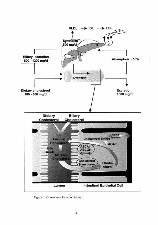
of cholesterol transport in man are presented in Figure 1.
Homeostatic mechanisms maintain the balance between the input of cholesterol from
its two sources, intestinal absorption and de novo synthesis, and its output by two
major hepatic mechanisms, irreversible conversion to bile acids and secretion into
bile as free cholesterol. The liver is the key organ in the metabolism of cholesterol
since it has critical importance in the processes regulating the whole-body cholesterol
homeostasis (cholesterol synthesis, plasma clearance of lipoproteins, bile acid
synthesis, and biliary cholesterol secretion) (Dietschy et al. 1993). Cholesterol
synthesis and dietary cholesterol absorption equals fecal excretion of total steroids in
29
a steady state at the whole body level, and the amount of cholesterol in hepatocytes
(derived from cholesterol synthesis, absorbed from the intestine, produced in other
tissues) is equal to the biliary secretion of lipids. According to the homeostatic
regulation of cholesterol metabolism, low intestinal absorption of cholesterol
upregulates cholesterol synthesis and turnover, whereas an increase in the intestinal
cholesterol flux to the liver suppresses cholesterol synthesis. Therefore, any
reduction of dietary cholesterol, or bile acid or cholesterol malabsorption, will trigger
an increase in hepatic endogenous cholesterol synthesis (Miettinen and Kesäniemi
1986), whereas an increase in dietary cholesterol has an opposite effect.
The sterol balance is defined as the difference between dietary intake and total sterol
excretion. The negative balance implies the removal of cholesterol from the body (the
fecal excretion of total steroids exceeds the dietary intake of cholesterol), whereas a
positive balance indicates that cholesterol is accumulating within the body (dietary
intake of cholesterol exceeds the fecal excretion of total steroids). In steady state,
this balance is usually negative, and numerically equal to the endogenous synthesis
of cholesterol.
31
2.6.1 Cholesterol absorption
2.6.1.1 Sources of intraluminal cholesterol
The intraluminal cholesterol available for absorption originates from three sources;
the diet, bile and turnover of intestinal mucosal epithelium. The daily Western diet
contains 300-500 mg cholesterol, of which a variable proportion is cholesterol esters
(8-19%) (Wilson and Rudel 1994). The amount of biliary cholesterol entering the
intestine daily varies usually from 800 to 1200 mg (Grundy 1983). All biliary
cholesterol is in an unesterified form. The mucosal cell loss and intestinal secretion of
cholesterol provide additional sources of cholesterol to the intestinal cholesterol pool,
though estimates of this contribution are hard to come by. In three patients with total
bile duct obstruction, the intraluminal cholesterol resulting from this source was
estimated to be 250-400 mg daily (Cheng and Stanley 1959). The significance of this
endogenous cholesterol in humans is still unknown, whereas in rats, it was shown to
significantly contribute to the endogenous cholesterol pool (Danielsson 1960). Thus,
in humans, over 1 g of cholesterol enters the intestine daily, and approximately one
half of this amount is absorbed (Grundy and Ahrens 1969, Wilson and Rudel 1994).
2.6.1.2 Luminal events
Cholesterol absorption is intimately linked to the overall process of lipid absorption.
Hydrolysis of dietary fat begins in the stomach, from which the emulsion is delivered
to the lumen of the small intestine. The pancreatic secretions contribute hydrolytic
enzymes, and bile contributes bile salts, which solubilize the hydrolytic end products
of intraluminal fat digestion. Long-chain triglycerides are hydrolysed by pancreatic
lipase, with the hydrolysis being accelerated by colipase (Borgström 1975).
Phospholipids are hydrolyzed at the 2-position by pancreatic phospholipase A2, to
yield lysophospholipid and free fatty acids (Van Deenen et al. 1963). Cholesteryl
esters are hydrolyzed by pancreatic cholesterol esterase (Vahouny and Treadwell
1958, Vahouny et al. 1964). This hydrolysis is important, because only free
cholesterol is efficiently absorbed (Swell et al. 1955, Swell et al. 1960); this
observation has been confirmed later in mice lacking the cholesterol esterase gene
(Howles et al. 1996).
32
Cholesterol is only minimally soluble in aqueous systems, its solubility is dependent
on the solubilizing properties of bile salt solutions (Siperstein et al. 1952, Swell et al.
1958). Mixed bile salt micelles and small unilamellar vesicles are thought to be the
main natural carriers from which the mucosal uptake of lipids is possible (Carey and
Hernell 1992). If there is to be micellar formation and subsequent cholesterol
absorption, there must be bile salts present above the critical micellar concentration
(Hofmann and Small 1967). In the presence of bile salts, more soluble lipids such as
phospholipids, fatty acids and monoglycerides, can increase the solubility of
cholesterol and facilitate the micellar solubilization and therefore they can improve
the absorption of cholesterol. The known requirement of bile acids for intestinal
cholesterol to be absorbed, recently demonstrated with mice lacking an enzyme
essential for bile acid formation (Schwarz et al.1998), is met with this model.
2.6.1.3 Mucosal events
2.6.1.3.1 Uptake and re-excretion
Cholesterol absorption occurs through the intestinal mucosal cells, which cover the
surface of the intestinal villi, and the uptake occurs mainly in the apical region of the
villi (Sylvén and Nordström 1970). The classical model for the molecular mechanism
of cholesterol absorption has been that unesterified cholesterol is shuttled in a mixed
micelle across the unstirred water layer lining the brush border of enterocytes
(Westergaard and Dietschy 1976). Subsequently, the transfer of cholesterol to the
cell surface and inside the cell is mediated by passive diffusion according to the
concentration gradient. More recently, Thurnhofer and Hauser (1990) suggested that
uptake of cholesterol is an active process, and it is protein-mediated, possibly
relating to cholesterol-transfer protein in enterocytes (Thurnhofer et al. 1991).
Scavenger receptor class B type I (SR-BI), a membrane protein located on the brush
border membrane, has been suggested to mediate intestinal cholesterol absorption
by facilitating the uptake of dietary cholesterol from either bile salt micelles or
phospholipid vesicles (Hauser et al. 1998). This receptor has been suggested to play
a role in the absorption of other lipids, such as triglycerides, and even esterified
sterols (Compassi et al. 1995). Prior to SR-BI, a direct role for the pancreatic
cholesterol esterase (i.e carboxyl ester lipase) in intestinal cholesterol absorption was
proposed by Lopez-Candales et al. (1993). However, in knockout mice lacking the
cholesterol esterase gene, the efficiency of intestinal cholesterol absorption was
33
identical to wildtype mice, though intestinal absorption of cholesteryl esters was
impaired (Howles et al.1996). This result has been recently confirmed (Weng et al.
1999).
ABC transporters are integral membrane proteins, which transport various molecules
across the cellular membrane, and are important in cholesterol metabolism (ABCA1)
(Brooks-Wilson et al. 1999, Orso et al. 2000). A study in mice showed that treatment
with retinoid X receptor (RXR) ligand could increase the intestinal expression of
ABCA1 leading to enhanced efflux of cholesterol from enterocytes back to intestinal
lumen (Repa et al. 2000). Further genetic studies have shown that both absorption
and secretion of cholesterol are controlled by ABC transporters, ABCG5 and ABCG8,
which act in concert to pump cholesterol out of cells (Berge et al. 2000, Lee et al.
2001). In the intestine, they re-excrete cholesterol that has entered enterocytes from
the intestinal lumen, thereby limiting cholesterol absorption.
2.6.1.3.2 Mucosal cholesterol
Inside the enterocytes, cholesterol and lipids are esterified, and approximately 75 %
of newly absorbed cholesterol appearing in lymph chylomicrons is in an esterified
form (Wilson and Rudel 1994). Fatty acids and monoglycerides are re-esterified to
form triglycerides and free cholesterol is esterified to form cholesteryl esters. The
monoglyceride pathway is used for the synthesis of chylomicron triglycerides (Field
and Mathur 1995). Acyl CoA:cholesterol acyltransferase (ACAT) is the enzyme
responsible for the esterification of absorbed cholesterol (Purdy and Field 1984,
Chang et al. 1997). More recently, two ACAT enzymes were cloned, ACAT 1 and
ACAT 2, of which ACAT 2 is most likely mainly responsible for esterification of
intestinally absorbed cholesterol (Anderson et al. 1998, Cases et al. 1998, Oelkers et
al. 1998). Accordingly, the role of ACAT 2 in cholesterol absorption is the formation of
cholesteryl esters for packaging into chylomicrons, which prevents the back
diffusion/transport of free cholesterol into intestinal lumen. The effects of ACAT
inhibition on cholesterol absorption have been variable, but most studies have shown
a decrease in the extent of cholesterol absorption (Krause et al. 1993, Wilson and
Rudel 1994). However, with a normal-low cholesterol containing diet, ACAT 2 -
deficient mice had plasma cholesterol similar to those of wildtype mice, but when
they consumed a high fat, high cholesterol diet, their cholesterol absorption was 85
34
% lower than in wildtype mice (Buhman et al. 2000). These results emphasize the
importance of ACAT, especially when high amounts of cholesterol are available for
absorption.
After the esterification, triglycerides and cholesteryl esters are assembled in the
endoplasmic reticulum with other lipids and proteins to form the core of nascent
chylomicrons. The major protein is apo B-48, which is obligatory for chylomicron
formation (Hussain et al. 1996), and which acts in concert with microsomal
triglyceride transfer protein (MTP). The assembly of chylomicron particle is completed
in the Golgi apparatus (Field and Mathur 1995). MTP has a role in rescuing apo B-
48 from intracellular degradation during early lipidation of the protein, this lipidation
process possibly being mediated by MTP (van Greevenbroek et al. 1998). When the
formation is terminated, chylomicrons enter lacteals in the intestinal villi, and are
delivered via the thoracic duct into the bloodstream.
2.6.1.4 Regulation of serum cholesterol level
The cholesterol absorption efficiency has been reported to regulate serum
cholesterol levels in a random population of Finnish males (Kesäniemi and Miettinen
1987, Kesäniemi et al. 1987). The cholesterol absorption efficiency was positively
associated with serum and LDL cholesterol concentrations, and negatively with
cholesterol synthesis (Kesäniemi and Miettinen 1987, Miettinen and Kesäniemi 1989,
Gylling and Miettinen 1989, Miettinen et al. 1990). Studies in hyper- and hypo-
responding nonhuman primates also indicated that cholesterol absorption and
plasma cholesterol level were positively related (Wilson and Rudel 1994). However,
in contrast to these previous studies, the efficiency of cholesterol absorption was not
related to serum total or LDL cholesterol concentrations in two recent studies
(Sehayek et al. 1998a, Bosner et al. 1999). The subjects in the study by Bosner et al.
(1999) were from various ethnic groups with a high interindividual variation in their
percent cholesterol absorption. In the study by Sehayek et al. (1998a), almost two-
thirds of the variation in LDL cholesterol was explained by the dietary cholesterol-
induced change in percentage dietary cholesterol absorption, even though the
relationship between change in percentage dietary cholesterol absorption versus
percent change in LDL cholesterol was non-linear. The use of the plasma isotope
ratio method (Sehayek et al. 1998a, Bosner et al. 1999) determines only the
35
cholesterol absorption percentage, the value is determined from only short period of
time, it yields only a single measure of absorption, and the value of cholesterol
absorption may be dependent on the composition of the test meal. With the
continuous isotope feeding method (Kesäniemi and Miettinen 1987, Kesäniemi et al.
1987, Miettinen and Kesäniemi 1989, Gylling and Miettinen 1989, Miettinen et al.
1990), the cholesterol absorption percentage, amount of cholesterol absorbed and
the intestinal influx of endogenous cholesterol can be determined in a balanced,
constant state providing more steady and consistent values of cholesterol absorption
from day to day during the period of one week. Therefore, the use of different
methods for measuring cholesterol absorption in these earlier studies may explain
the variation in the obtained results.
The role of cholesterol absorption as a regulator of serum HDL cholesterol seems
more evident. Many studies have shown that the cholesterol absorption efficiency as
well as plant sterols, indicators of cholesterol absorption, are positively associated
with serum HDL cholesterol levels. (Miettinen and Kesäniemi 1989, Miettinen et al.
1990, Miettinen and Gylling 2000)
The fractional and absolute absorption of cholesterol correlates negatively with
cholesterol synthesis (Miettinen and Kesäniemi 1989). Accordingly, effective
cholesterol absorption will lower cholesterol synthesis and serum levels of cholesterol
may increase, whereas with ineffective absorption, the overall cholesterol synthesis
is increased (Miettinen et al. 1990). The association between cholesterol synthesis
and serum and LDL cholesterol levels are insignificant in many studies (Gylling and
Miettinen 1988, Miettinen and Kesäniemi 1989, Miettinen et al. 1989, Gylling et al.
1994), suggesting that serum and LDL cholesterol levels are regulated mainly by
cholesterol absorption rather than cholesterol synthesis.
2.6.1.5 Measurement of cholesterol absorption
A variety of different methods have been developed for estimating the absorption of
cholesterol in humans. Most of these methods are based on the use of radioactive
isotopes. One of the earliest methods used was based on the administration of a
single oral or intravenous dose of radioactive 3H- or 14C- , and the absorption of
cholesterol was calculated as the difference between the dietary cholesterol and
36
fecal exogenous neutral steroids (Borgström 1969, Quintao et al. 1971, Sodhi et al.
1974, Samuel et al. 1978). This method has been modified by extending the isotope
administration for several days or even as long as several weeks (Quintao et al.
1971), as well as with the use of intestinal intubation (Grundy and Mok 1977).
Although these methods provide information about cholesterol absorption, they suffer
from some particular disadvantages such as the need for hospitalization in a
metabolic ward and inconvenience to the subject. In addition, only one measurement
of a single dose of radioactive cholesterol may not accurately estimate the mean
absorption over a period of time.
The plasma isotope ratio method, first introduced in rats (Zilversmit 1972), and later
validated in humans (Samuel et al. 1978), is based on giving the reference
compound 3H-cholesterol intravenously simultaneously with an oral dose of 14C-
cholesterol, and the resulting 14C/3H ratio in the plasma gives an estimate of the
percentage absorption of cholesterol from the intestine. Despite the simplicity and
feasibility for outpatient studies, this method measures only absorption percentage of
cholesterol, and the absorption value is determined only from a short period of time,
which possibly is not the true reflection of mean overall absorption.
In the continuous isotope feeding method, developed by Crouse and Grundy (1978),
the subjects receive peroral low-dose 14C-cholesterol and 3H-sitosterol in capsules
three times a day with meals for 7-10 days. The stool is collected on days 3-10. The
ratio of isotopes in feces becomes constant after the first 3 days. The percentage
absorption of cholesterol is calculated from the difference between the dietary
(capsules) and fecal isotope ratios. This measurement gives an accurate value of
cholesterol absorption percentage because of the sufficiently long study period which
allows one to achieve stable state and constancy in the ratio of the isotopes in feces,
and sequential fecal samples diminish the fluctuation in the absorption. In addition,
other advantages include the simple administration of isotope, analysis of fecal
samples is easy, though laborious, the analysis can be repeated daily, and it is
suitable for outpatients. The amount of cholesterol absorbed and the intestinal influx
of endogenous cholesterol can be calculated, if the daily intake of cholesterol and
fecal neutral steroid excretion are also measured. The only drawback is that children
and women of child-bearing age cannot be studied.
37
More recently, the continuous isotope feeding method was modified by using
markers labeled with stable isotopes, deuterated cholesterol, and deuterated
sitostanol, quantified by gas-liquid chromatography-selected ion monitoring
(Lütjohann et al. 1993). This method is claimed to be safe and reproducible without
radioactive exposure. In addition, a nonradioactive modification was developed
based on the plasma isotope ratio method (Zilversmit 1972), in which six extra mass
units of 2H-cholesterol were given orally and 5 extra mass units of 13C were
administered intravenously on day 0: the absorption percentage of cholesterol was
calculated on day 3 from the plasma ratio of the two tracers using gas
chromatography-mass spectrometry with selected ion monitoring (Bosner et al.
1993). The sensitivity of the analysis was further improved by using negative ion
mass spectrometry (Ostlund et al. 1996). Furthermore, the measurement of serum
plant sterols and cholestanol ratios to cholesterol, variables reflecting cholesterol
absorption (Tilvis and Miettinen 1986, Miettinen et al. 1989, Miettinen et al. 1990) by
gas-liquid chromatography provides a less laborious method to evaluate cholesterol
absorption compared to methods based on the use of radioactive or stable isotopes.
2.6.1.6 Factors affecting cholesterol absorption
Despite the close relation between intestinal absorption of cholesterol and dietary
fats as well as the presence of bile acids as requirements for cholesterol absorption,
there are marked differences in the extent of absorption. For instance, the absorption
of dietary fat is over 95 % (Carey et al. 1983), that of cholesterol approximately 50 %,
and the bile acids are almost quantitatively reclaimed (> 95%)(Wilson and Rudel
1994). The cholesterol absorption efficiency between different individuals shows a
high variation, with values ranging widely from 25 % to 80 % (Miettinen and
Kesäniemi 1989, Bosner et al. 1999).
Diet
Results from several studies indicate that the cholesterol absorption efficiency
remains mainly unaltered even though there are daily variations in the dietary
cholesterol content (McMurry et al. 1985, Miettinen and Kesäniemi 1989, Sehayek et
al. 1998a). However, with feeding of extra large amounts of cholesterol, the
cholesterol absorption efficiency becomes decreased (McNamara et al. 1987, Gylling
and Miettinen 1992, Vuoristo and Miettinen 1994). Increasing the amount of dietary
38
cholesterol resulted in substantial overall reductions in the absorption efficiency of
cholesterol, but there was a considerable heterogeneity in the response of different
individuals (Ostlund et al. 1999). Studies in Finnish and American populations have
shown that ordinary cholesterol intake was not significantly related with cholesterol
absorption efficiency (Miettinen and Kesäniemi 1989, Bosner et al. 1999). The
association between cholesterol absorption and hypo- and hyper-responsiveness in
humans (Beynen et al. 1987, Katan and Beynen 1987) might contribute to the results
of dietary cholesterol intake- induced changes in cholesterol absorption. Feeding
increasing amounts of cholesterol to mice led to a decrease in the percentage
absorption of dietary cholesterol and an increase in the biliary cholesterol
concentration, and these two variables were strongly and inversely correlated,
suggesting that the biliary cholesterol secretion may have a role in regulating the
efficiency of dietary cholesterol absorption (Sehayek et al. 1998b). Biliary cholesterol
could saturate the intestinal micelles, preventing the normal absorption of dietary
cholesterol. Other studies in mice (Carter et al. 1997) and monkeys (Rudel et al.
1994) have concluded that cholesterol absorption efficiency is significantly lower,
when the animals are fed a diet containing extra high amounts of cholesterol
compared to feeding a low cholesterol diet.
The results concerning the effects of dietary cholesterol on cholesterol absorption are
not clear. The expanded intestinal cholesterol pool due to high amounts of dietary
cholesterol may contribute to the dilution of labeled cholesterol resulting in its
reduced absorption. The other concern with diets high in cholesterol is the difficulty of
ensuring that all the cholesterol is properly micellar-solubilized in the intestine and
available for absorption. However, most studies reveal that the amount of cholesterol
absorbed is closely related to cholesterol intake, and the increased intake of
cholesterol is associated with an increased mass absorption of cholesterol (Miettinen
and Kesäniemi 1989).
39
Intestinal transit time
There are only limited number of studies concerning the effects of intestinal motility
and transit time on cholesterol absorption. The reduction of small-bowel transit time
by chenodeoxycholic acid (Ponz de Leon et al. 1979) or by metoclopramide (Ponz de
Leon et al. 1982) decreases cholesterol absorption. In addition, the cholesterol
absorption efficiency has been shown to correlate positively with mouth to anus
intestinal transit time (Koivisto and Miettinen 1986). However, the atropine-induced
prolongation of the small-bowel transit time did not enhance dietary cholesterol
absorption in subjects whose cholesterol absorption percentage was normal (Ponz
de Leon et al. 1982). Based on these results, the intestinal transit time seems to be
related to steroid absorption under normal physiological conditions.
Obesity
The cholesterol absorption efficiency is decreased in obesity (Miettinen and Gylling
2000). An inverse correlation between BMI and absorption efficiency has been
reported in a random population of Finnish men (Miettinen and Kesäniemi 1989).
However, in another study, the correlation between BMI and cholesterol absorption
percentage remained insignificant (Bosner et al. 1999). The obesity-related increased
cholesterol synthesis with increased biliary output could contribute to lowered
cholesterol absorption in obesity (Miettinen and Gylling 2000), but the exact
mechanisms are not known.
Age
The effect of age on cholesterol absoption is controversial. Cholesterol absorption
efficiency was lower in 75 year olds compared to 50 year old subjects (Gylling et al.
1994). On the contrary, the relation between age and the serum plant sterol levels,
markers of cholesterol absorption, remained insignificant in another study (Kempen
et al. 1991). In a third study, a positive correlation was found between age and the
amount of dietary cholesterol absorbed, though the respective correlation for the
cholesterol absorption percent was not significant (Bosner et al. 1999)
40
Apo E
The apo E phenotype is closely associated with the cholesterol absorption efficiency,
a finding described first by Kesäniemi et al. (1987). More recent studies have
confirmed this relationship (Gylling et al. 1989, Miettinen et al. 1992). The cholesterol
absorption efficiency was highest in individuals with the E4/4 and E4/3 phenotypes,
lowest with E2/2, and E3/3 was situated in the middle. The serum cholesterol levels
were distributed in a similar manner so that highest levels were found in subjects with
E4/4 or E4/3 phenotypes (Utermann et al. 1977). These results may confirm the role
of cholesterol absorption as the regulator of serum cholesterol levels in a random
sample of male population (Kesäniemi and Miettinen 1987, Kesäniemi et al. 1987). In
studies with individuals consuming low fat-low cholesterol diets, the relation between
apo E and cholesterol absorption efficiency vanished (Miettinen et al. 1992, Bosner
et al. 1999).
Plant sterols
Plant sterols have been used for the treatment of hypercholesterolemia since the
early 1950s because they lower serum cholesterol by inhibiting cholesterol
absorption (Pollak 1953, Lees et al. 1977, Mattson et al. 1982). Further, it was shown
that sitostanol, a saturated derivate of sitosterol, could reduce serum cholesterol
more efficiently than sitosterol in rats (Sugano et al. 1977, Ikeda et al. 1981) and in
humans (Heinemann et al. 1986). In 1993, based on Dr. Miettinen’s hypothesis that a
fat soluble esterified form of stanol would be more physiological and more palatable,
and, accordingly, could inhibit cholesterol absorption and lower serum cholesterol
more efficiently than the crystalline form, stanol ester rich mayonnaise (Vanhanen et
al. 1993) and later rapeseed oil margarine were developed. Subsequently, many
studies have confirmed the effect of stanol ester margarine as a way to safely inhibit
cholesterol absorption and decrease serum total and LDL cholesterol levels in
normo- and hypercholesterolemic individuals (Miettinen et al. 1995, Gylling et al.
1995), postmenopausal women with prior myocardial infarction (Gylling et al. 1997)
as well as in non-insulin dependent diabetes mellitus (Gylling and Miettinen 1994a,
1996a).
Dietary plant sterols interfere with the micellar solubility of cholesterol, which directly
impacts on the incorporation of micellar cholesterol to the absorptive enterocytes
41
(Ikeda and Sugano 1983, Ikeda et al. 1988). Hence, more sterols remain in the
intestinal lumen, subsequently cholesterol absorption is depressed and serum
cholesterol levels decrease. However, the exact mechanisms by which plant sterols
inhibit cholesterol absorption have not been clarified in detail (Björkhem et al. 2001).
2.6.2 Plant sterols and cholestanol
Plant sterols are constituents of plant lipids. The normal human diet contains 200-300
mg/d of plant sterols (Salen et al. 1970), mainly sitosterol (65%), campesterol (32%),
and trace amounts of stigmasterol (3%) (Björkhem et al. 2001). These sterols are
structurally similar to cholesterol except for a difference in their side chain structure.
The side chain of sitosterol contains an ethyl- and campesterol methyl group at the
C-24 position. Stigmasterol has the same structure as sitosterol, except for a double-
bond at C-22.
The human body is unable to synthesize plant sterols, thus all plant sterols are
derived by absorption from the diet. Serum concentrations of plant sterols are
regulated by dietary intake, absorption of these sterols from the intestine, and their
elimination from the body through secretion into bile. A high cholesterol absorption
efficiency is usually associated with high levels in serum of plant sterols, whereas
high cholesterol synthesis, usually in conjunction with low absorption, is associated
with low serum plant sterol levels as well as increased biliary sterol secretion
(Miettinen et al. 1989, Miettinen et al. 1990). The ratios of plant sterols to cholesterol
correlate positively with fractional absorption of dietary cholesterol and negatively
with cholesterol precursor sterols and cholesterol synthesis, and thus measurement
of their ratios to cholesterol can be used as indicators of cholesterol absorption (Tilvis
and Miettinen 1986, Miettinen et al. 1989, Miettinen et al. 1990, Gylling and Miettinen
1997, Miettinen and Gylling 2000).
Cholestanol, a 5α-saturated derivate of cholesterol, is enzymatically formed from its
precursor. Small amounts of cholestanol accompany cholesterol in most tissues.
Dietary intake of cholestanol is normally very small (< 2 mg)(Miettinen et al. 1989),
and its absorption is low, about 9 % (Vuoristo and Miettinen 2000). In addition, the
serum levels of cholestanol remain constant during consumption of a cholestanol
free-diet (Salen and Grundy 1973). Therefore, the body cholestanol is considered to
42
be mainly derived from endogenous cholesterol, its mean daily synthesis being about
12 mg/d (Salen and Grundy 1973). Cholestanol is eliminated from the body through
bile as such or degraded to 5α-bile acids. The accumulation of cholestanol in the
tissues and its high serum levels are seen in cerebrotendinous xanthomatosis, a rare
inherited disease characterized with a defect in bile acid biosynthesis, and in
sitosterolemia (Björkhem et al. 2001), and in cholestasis (Nikkilä et al. 1991).
Measuring the serum cholestanol level is used as an indicator of cholesterol
absorption (Miettinen et al. 1989).
2.6.2.1 Absorption of plant sterols
The intestinal absorption of plant sterols is normally limited. Only < 5 % of ingested
sitosterol is absorbed (Salen et al. 1970), and absorption of campesterol is
approximately 10 % (Tilvis and Miettinen 1986). Despite the high inter-individual
variation of plasma sitosterol concentrations (Kempen et al. 1991), the plasma plant
sterol concentrations seem to be very stable within individuals. The inter-individual
variation in the plant sterol concentrations is genetically determined (Berge et al.
2002). Extremely high concentrations of plant sterols occur in a rare hereditary
disorder called sitosterolemia (phytosterolemia) (Bhattacharyya and Connor 1974), in
which sterol absorption is increased and sterol excretion into bile is decreased
(Miettinen 1980, Björkhem et al. 2001). Mutations in the two ABC transporters,
ABCG5 and ABCG8, have been described as one cause for this disease (Berge et
al. 2000, Lee at al. 2001). Recent studies show that polymorphisms in ABCG8 have
a contributing effect to genetic variation in the serum plant sterol concentrations in
normal, healthy individuals (Berge et al. 2002).
2.6.2.2 Transport in serum
There are only a few studies, in which the distribution of plant sterols and cholestanol
in different serum lipoproteins has been defined. Cholestanol is reported to be
transported similarly to cholesterol mainly in LDL (Salen and Grundy 1973). In
sitosterolemia, plant sterols are mainly transported in LDL and HDL (Bhattacharyya
and Connor 1974), thus following cholesterol distribution. In order to diminish the
effect of varying levels of serum cholesterol on the concentrations of plant sterols,
they are usually standardized and expressed in relation to cholesterol in serum and
lipoproteins as mmol/mol of cholesterol (Vuoristo and Miettinen 2000). In humans

43
when cholesterol absorption is normal, the ratios of plant sterols to cholesterol seem
to accumulate in HDL (Tilvis and Miettinen 1986) this also being seen in rats (Sugano
et al. 1978). In familial hypercholesterolemia patients with increased cholesterol
synthesis due to ileal bypass, the ratios of serum plant sterols to cholesterol in
lipoproteins appear to increase slightly with increasing lipoprotein density (Koivisto
and Miettinen 1988b). In rats, plant sterols are transported to bile only in HDL
(Robins and Fasulo 1997).
2.6.3 Cholesterol synthesis
Cholesterol is synthesized through several intermediates from acetyl-CoA (Figure 2).
The rate-limiting step of cholesterol synthesis is the reduction of 3-hydroxy-3-
methylglutaryl-coenzyme A (HMG-CoA) to mevalonate. This process is catalyzed by
HMG-CoA reductase, the rate-limiting enzyme in the synthesis of cholesterol.
Mevalonate is transformed to squalene, which is converted to lanosterol. Cholesterol
is finally formed from lanosterol after several further steps. The synthetic pathway
from squalene to cholesterol includes several intermediate methyl- and demethyl
sterols released in serum.
Liver is the major organ responsible for cholesterol synthesis. De novo cholesterol
synthesis is strictly regulated by the amount of cholesterol in cells (Brown and
Goldstein 1983). Hepatocytes receive both exogenous and endogenous cholesterol.
Chylomicron remnants transport the absorbed dietary cholesterol from the intestine
to the liver, and circulating lipoproteins, mainly LDL, transport the endogenous
cholesterol back to the liver for further processing. The key to the uptake process of
these particles to the hepatocytes is their binding to the LDLapo B-receptor, thus
mediating the endocytosis (Brown and Goldstein 1986). In addition, LDL receptor-
related protein (LRP) is another member of the LDL receptor gene family, which
binds to chylomicron remnants, probably mediating their uptake (Havel 1998).
Once inside the cell, cholesteryl esters are hydrolyzed in the lysosomes. The level of
free cholesterol mediates a feedback control system, which regulates intracellular
cholesterol homeostasis. This inhibits the HMG-Coa reductase enzyme leading to the
termination of cholesterol synthesis, and it also activates ACAT leading to formation
of cholesteryl esters for storage in the cell. It furthermore suppresses the synthesis of
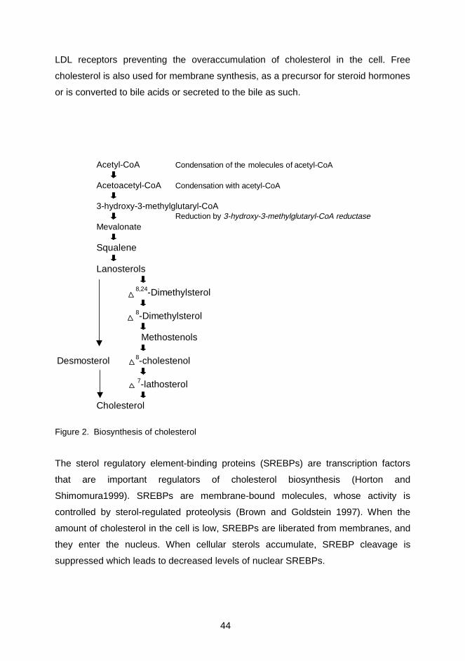
44
LDL receptors preventing the overaccumulation of cholesterol in the cell. Free
cholesterol is also used for membrane synthesis, as a precursor for steroid hormones
or is converted to bile acids or secreted to the bile as such.
Acetyl-CoA Condensation of the molecules of acetyl-CoA
Acetoacetyl-CoA Condensation with acetyl-CoA
3-hydroxy-3-methylglutaryl-CoAReduction by 3-hydroxy-3-methylglutaryl-CoA reductase
Mevalonate
Squalene
Lanosterols
8,24-Dimethylsterol
8-Dimethylsterol
Methostenols
Desmosterol 8-cholestenol
7-lathosterol
Cholesterol
Figure 2. Biosynthesis of cholesterol
The sterol regulatory element-binding proteins (SREBPs) are transcription factors
that are important regulators of cholesterol biosynthesis (Horton and
Shimomura1999). SREBPs are membrane-bound molecules, whose activity is
controlled by sterol-regulated proteolysis (Brown and Goldstein 1997). When the
amount of cholesterol in the cell is low, SREBPs are liberated from membranes, and
they enter the nucleus. When cellular sterols accumulate, SREBP cleavage is
suppressed which leads to decreased levels of nuclear SREBPs.
45
When they gain access to the nucleus, SREBPs activate the genes involved in the
synthesis of cholesterol and fatty acids, and the uptake of cholesterol through
transcription of the LDL receptor enzymes. With respect to the cholesterol
biosynthetic pathway, SREBPs activate the genes encoding HMGCoA reductase,
HMGCoA synthase, farnesyl diphosphate synthase, squalene synthase and
lanosterol demethylase (Brown and Goldstein 1997).
To date, three SREBP isoforms have been identified and characterized, SREBP-1a, -
1c and SREBP-2 (Brown and Goldstein 1997). There are transgenic mice, which
have overexpression of nuclear SREBP-2. This mutation led to marked increases in
the mRNAs of cholesterol biosynthetic enzymes and the rate of cholesterol synthesis
increased 28 –fold (Horton et al. 1998). SREBP-2 is suggested to be the principal
isoform responsible for controlling the cholesterol biosynthetic pathway (Horton and
Shimomura 1999).
2.6.3.1 Reverse cholesterol transport
Lipid-rich HDLs arise from lipid poor precursors or from lipid-free apolipoproteins
(Fielding and Fielding 1995). Reverse cholesterol transport is initiated by the removal
of cholesterol from peripheral tissues, either by passive diffusion to lipid-poor pre-
HDL or receptor-mediated efflux to apo A-I.
The active removal is mediated by ABCA1, an ATP- binding cassette transporter,
which facilitates the addition of phospholipids and cholesterol from the cells to free
apoA-I, initiating the formation of HDL (Schmitz and Langmann 2001). The enzyme
LCAT uses phospholipids and free cholesterol as its substrates and forms cholesteryl
esters. Cholesteryl esters formed within HDL can be delivered to the liver by three
routes.
Cholesteryl esters may be transferred to TRL by CETP (Tall 1993, 1995). These
lipoproteins are further metabolized in the circulation to form lipoprotein remnants.
The uptake of the remnants to the liver is mediated by binding to the hepatic
receptors (LDLapo B-receptors or LRP), proteoglycans and apo E.

46
HDL cholesteryl esters may also be taken up by the liver via a process of selective
lipid uptake. It has been shown recently that this selective uptake is mediated by SR-
BI. Hepatocytes and steroid hormone producing cells express SR-BI. This protein
binds to HDL and mediates the uptake of cholesteryl esters without internalization
and degradation of HDL proteins (Acton et al. 1996, Kozarsky et al. 1997, Rigotti et
al. 1997). In addition to cholesteryl ester uptake, SR-BI might mediate free
cholesterol uptake of HDL to the liver, providing an important source of biliary
cholesterol (Ji et al. 1999, Mardones et al. 2001). Some HDL particles contain apo E,
and can be taken up by hepatocytes via the LDLapo B-receptors (Hennessy et al.
1997). The mechanisms of the HDL particle uptake are not clear, and proteoglycans,
apoproteins and still unknown receptors might also be involved (Ji et al. 1997).
2.6.4 Squalene and demethylated cholesterol precursors
In addition to cholesterol, some of its precursors including squalene and
demethylated sterols, especially ∆8 –cholestenol (cholestenol), desmosterol and ∆7 –
lathosterol (lathosterol), are present in detectable amounts in serum. The sterols are
transported in serum by lipoproteins similarly to cholesterol, such that the highest
amounts are found in LDL. When expressed as ratios to cholesterol, the lathosterol
ratio is highest in VLDL in subjects with increased cholesterol synthesis (Koivisto and
Miettinen 1988a). Most of the squalene is carried in LDL and HDL in fasting human
serum (Miettinen 1982b, Gylling and Miettinen 1994b), but the highest ratio of
squalene to cholesterol (Gylling and Miettinen 1994b) and the newly synthesized
squalene (Goodman 1964) are found in VLDL. Many studies have shown that serum
levels of different precursors to cholesterol are increased under conditions of
increased synthesis of cholesterol, and reduced with reduced synthesis of cholesterol
(Miettinen 1969, 1970, 1981, Miettinen and Koivisto 1983, Miettinen 1985, Vuoristo
and Miettinen 1986). The non-cholesterol precursor sterols correlate positively to
HMG-CoA reductase activity and the cholesterol synthesis rate (Björkhem et al.
1987, Kempen et al. 1988, Miettinen et al. 1990) and negatively to cholesterol
absorption and to the levels of plant sterols in serum (Miettinen et al. 1990). In
addition, treatment with lovastatin reduced the cholesterol synthesis rate and serum
lathosterol concentrations as well as the ratio of lathosterol to cholesterol in serum
(Kempen et al. 1988, Duane 1995). Thus, non-cholesterol sterols have been
demonstrated to reflect the rate of cholesterol synthesis, and thus measurement of

47
their ratios to cholesterol can be used as indicators of cholesterol synthesis. The
serum content of squalene seems to reflect cholesterol synthesis less efficiently
(Miettinen et al. 1990).
2.6.5 Elimination of cholesterol
Liver is the major organ responsible for the removal of cholesterol from the body. The
removal of cellular cholesterol from peripheral tissues to the liver by HDL mediated
extraction occurs by a number of mechanisms including aqueous diffusion, a specific
apoprotein-mediated mechanism as well as facilitated efflux mediated by cell surface
receptors. In addition, some cells are able eliminate intracellular cholesterol by
converting it into more polar metabolites by oxidative mechanisms. These oxysterols
are important as transport forms of cholesterol over cell membranes and the blood-
brain barrier, because they are easily excreted from the cells and transported to the
liver for conversion to bile acids. For instance, cholesterol in the brain is converted
into 24S-hydroxycholesterol which can be transported over the blood-brain barrier
much more rapidly than unmetabolized cholesterol, and taken up by liver from the
circulation for further metabolism (Björkhem and Diczfalusy 2002).
Cholesterol is eliminated from the body primarily by fecal excretion of neutral sterols
and bile acids. Cholesterol is secreted either unchanged or as bile acids into bile, and
biliary secretion of cholesterol is linked with biliary bile acid output (Schersten et al.
1971). Fecal neutral sterols consist mainly of cholesterol and its bacterial metabolites
coprostanol, epicoprostanol and coprostanone, and also variable amounts of
cholesterol precursors and plant sterols, and their coprostanol and coprostanone
bacterial products (Miettinen et al. 1965, McNamara et al. 1981). The daily excretion
rate of cholesterol is 400-1000 mg (Miettinen et al. 1965), mostly being excreted as
its metabolite coprostanol.
2.6.5.1 Bile acids
The formation of bile acids from cholesterol accounts for over 30 % of the cholesterol
removal from the body. During the past few years it has been shown that the initial
step in the formation of the bile acids may start in the extrahepatic tissues (Princen et
al. 1997). However, all the subsequent steps in the formation of the bile acids are
almost exclusively located in the liver. The hepatic conversion of cholesterol into bile

48
acids involves many steps and enzymatic processes. The major, “neutral” pathway
begins with the rate-limiting step of the conversion of cholesterol into 7 α-hydroxy-
cholesterol, catalysed by the cholesterol 7α-hydroxylase (CYP7A1). This enzyme
regulates the overall conversion of cholesterol into bile acids in this pathway, and its
activity is regulated through negative feedback by reabsorbed bile acids returning to
the liver. An alternative pathway, the “acidic pathway”, starts with the 27-
hydroxylation of cholesterol followed by a microsomal 7α –hydroxylation by a specific
oxysterol 7α -hydroxylase (Björkhem and Eggertsen 2001). This pathway is less
responsive to bile acid negative feedback, and is regulated separately from the major
pathway (Björkhem et al. 2001). The neutral pathway seems to be responsible for
over 50 % of the total synthesis of bile acids, whereas acidic pathway starting in the
liver may be responsible for 30-50 % of the total production (Björkhem and Eggertsen
2001). Only a minor amount of the whole bile acid pool is produced by extrahepatic
tissues starting with 27-hydroxylation (Duane and Javitt 1999).
The end products of these pathways are the primary bile acids, cholic acid and
chenodeoxycholic acid. The primary bile acids are secreted into bile as conjugates
with glycine or taurine (3:1). After secretion, these bile acids may be deconjugated
and also dehydroxylated by intestinal bacteria. Deconjugation alters both the physical
and chemical properties of bile acids. Deoxycholic acid is the dehydroxylated product
of cholic acid and lithocholic acid is the corresponding product of chenodeoxycholic
acid. Deoxycholic acid is water soluble and it is partly absorbed, whereas lithocholic
acid is mainly excreted in the feces. From the intestine, bile acids are reabsorbed
and returned to the liver via the portal circulation. Absorbed bile acids are taken up
by hepatocytes and secreted again into bile. This enterohepatic circulation is an
extremely efficient way to conserve bile acids, of which only 2-5% escape the
intestinal absorption to be excreted into feces.
Transformation of cholesterol into bile acids is the major pathway of cholesterol
catabolism, and thus bile acids have an important role in cholesterol homeostasis.
The activity of CYP7A1 is controlled by negative feedback of the reabsorbed bile
acids to guarantee the maintenance of an intact bile acid pool. CYP7A1 enzymatic
activity is influenced by a number of dietary and hormonal factors. A close
49
correlation is found between the activity of HMG-CoA reductase and CYP7A1
(Björkhem and Åkerlund 1988), and their activities usually increase in parallel both in
basal conditions and in situations of bile acid malabsorption (Färkkilä and Miettinen
1990, Björkhem et al. 1997). Enhanced fecal elimination of bile acids due to
interruption of the enterohepatic circulation, e.g. with cholestyramine or surgical
resections, can increase bile acid synthesis, stimulate the expression of hepatic LDL
receptors and increase the synthesis of cholesterol (Everson and Kern 1995), which
is seen also in an elevation in the levels of the cholesterol precursor sterols
(Miettinen 1985, Färkkilä et al. 1988).
Recent studies have shown that the secretion of bile acids and cholesterol from
hepatocytes into bile is controlled by ABC transporters. The bile salt export pump
(BSEP) is a member of the ABC superfamily of transporters (ABCB11). This pump is
located on the canalicular membrane of hepatocytes where it facilitates the transport
of bile acids from the hepatocyte into bile (Schmitz et al. 2000). The secretion of
cholesterol into bile is mediated by two other ABC transporters, ABCG5 and ABCG8
(Goldstein and Brown 2001).
2.6.6 Nuclear receptors and cholesterol metabolism
Nuclear receptors function as ligand-activated transcription factors that regulate the
expression of target genes. Two nuclear hormone receptors involved in the
regulation of cholesterol homeostasis are liver X receptor (LXR) and farnesoid X
receptor (FXR). The natural ligands for LXR and FXR are oxysterols and bile acids
(Accad and Farese 1998, Russell 1999). Ligand-activated LXR or FXR both form
heterodimers with the RXR in order to modulate and control transcriptional activity of
several important genes regulating cholesterol metabolism.
Three genes coding ABC transporters (ABCA1, ABCG5 and ABCG8) which are
involved in the absorption of cholesterol, are the proposed target genes of LXR
(Berge et al. 2000, Repa et al. 2000, Lee et al. 2001). In addition, genes coding
CYP7A1, (enzyme onvolved in bile acid synthesis), and SREBP-1c (biosynthesis of
fatty acids) are activated by the LXR-RXR heterodimer (Repa and Mangelsdorf 1999,
Goldstein and Brown 2001). FXR is highly expressed in liver where it appears to
function as a bile acid sensor. The genes encoding BSEP and ileal bile-acid binding

50
protein (I-BABP) (mediates the re-uptake of bile acids from the intestinal lumen) are
target genes for FXR (Edwards et al. 2002). In addition, the FXR-RXR heterodimer
represses CYP7A1 transcription (Lu et al. 2000).
Therefore, nuclear receptors are important in regulating cholesterol metabolism by
controlling the transcription of several important genes involved in cholesterol and
bile acid metabolism.
2.7 Cholesterol synthesis and elimination in obesity
Cholesterol synthesis (Miettinen 1971a, Liu et al. 1975, Nestel et al. 1973) and
turnover Nestel et al. 1969) are increased in obesity. The serum levels of methyl and
demethyl sterols, cholesterol precursor sterols, and squalene are elevated in
obesity, reflecting increased synthesis of cholesterol (Miettinen 1970, Liu et al. 1975,
Nestel and Kudchodkar 1975, Miettinen and Gylling 2000). Some of the excess
cholesterol is produced by the enlarged amount of adipose tissue (Miettinen and
Tilvis 1981), though adipose tissue-synthesized cholesterol does not account for all
of the enhanced synthesis of cholesterol in obesity (Schreibman and Dell 1975,
Miettinen and Tilvis 1981). Increased cholesterol synthesis in liver seems to be
responsible for most of the excess cholesterol produced in obesity, associated with
increased fatty acid transport to the liver and generally enhanced lipogenesis (Nestel
and Goldrick 1976). It has been shown that the activity of hepatic HMG-CoA
reductase is increased in obesity (Angelin et al. 1982). More recently, in a study of
liver biopsies obtained from obese and non-obese subjects, the activity and mRNA
level of HMG-CoA reductase were markedly higher in the liver of obese than non-
obese subjects (Ståhlberg et al. 1997).
In addition to enhanced cholesterol synthesis, the elimination of cholesterol to feces
is enhanced through its increased secretion into bile and also because of the
increased formation of bile acids (Miettinen 1971a, Nestel et al. 1973, Bennion and
Grundy 1975, Miettinen and Gylling 2000). In fact, the activity and mRNA level of
CYP7A1 are clearly increased in obese vs non-obese subjects (Ståhlberg et al.
1997). The increased secretion of cholesterol into bile increases the risk for gallstone
formation in obesity (Miettinen 1987).
51
2.8 Cholesterol metabolism in diabetes
Type 2 diabetes is associated with many metabolic disturbances, including
abnormalities in glucose and lipid metabolism. The alterations in lipoprotein
metabolism have been studied and described in detail earlier, and thus are well
known (American Diabetes Association 1993, Evans 1999). The study of cholesterol
metabolism and bile acids in diabetes has been somewhat neglected, perhaps
because of the more common and obvious abnormality in plasma lipids. There are
some earlier studies describing cholesterol metabolism in diabetes, though the
results are not always consistent.
2.8.1 Cholesterol absorption
A few studies have shown that the cholesterol absorption efficiency might be low in
hypercholesterolemic type 2 diabetes (Gylling and Miettinen 1997) or in markedly
hypertriglyceridemic diabetes subjects (Briones et al. 1986). However, in the latter
study, of 40 diabetic subjects, 22 were receiving insulin treatment at the time of the
study, and 6 were insulin –dependent diabetics. Low plant sterol concentrations in
type 2 diabetes have been described in two studies (Sutherland et al. 1992, Gylling
and Miettinen 1997). The cholesterol absorption efficiency and serum plant sterol
levels are higher in type 2 diabetic subjects with coronary artery disease compared to
those without this disease (Gylling and Miettinen 1996b). In addition, cholesterol
absorption efficiency and serum levels of plant sterols and cholestanol are low in
nondiabetic subjects with high-normal blood glucose levels (Strandberg et al. 1996).
2.8.2 Regulation of serum cholesterol level
There are only few studies describing the regulation of serum cholesterol level in type
2 diabetes, but the number of study populations in these reports is are very small.
The serum cholesterol level seems to be regulated in type 2 diabetes similarly as in
the non-diabetic population by the homeostasis between cholesterol absorption,
synthesis and LDL catabolism (Gylling and Miettinen 1997). The serum LDL
cholesterol level was inversely related to cholesterol synthesis and FCR of LDL apo
B (Gylling and Miettinen 1996b, 1997), and FCR of LDL apo B also correlated
positively with the lathosterol ratio (Gylling and Miettinen 1997). However, LDL
cholesterol did not correlate significantly with cholesterol absorption. Based on these
52
results obtained with a few study subjects, the connection between cholesterol
absorption and serum LDL levels seen in the normal population, may not necessarily
occur in type 2 diabetics.
2.8.3 Cholesterol synthesis and excretion
Bile acid and cholesterol synthesis and cholesterol excretion as neutral sterols are
increased in non-obese, mildly hypercholesterolemic (Gylling and Miettinen 1997),
mildly hypertriglyceridemic non-obese (Abrams et al. 1982, Naoumova et al. 1996),
markedly hypertriglyceridemic (Briones et al. 1986) and markedly overweight
(Bennion and Grundy 1977) patients with type 2 diabetes. The ratios of cholesterol
precursor sterols to cholesterol in serum were higher in diabetics than in controls
(Gylling and Miettinen 1997). The serum level of insulin and lathosterol were
correlated positively only in the diabetic but not in control subjects (Sutherland et al.
1992). Scoppola et al. (1995) found that cholesterol synthesis, measured by urinary
mevalonic acid excretion, was higher in normolipidemic, non-obese diabetics than
controls. In addition, urinary mevalonate excretion was significantly and positively
correlated with glycated hemoglobin concentrations (HbA1c) in non-obese
normolipidemic type 2 diabetics, suggesting that there is enhanced cholesterol
synthesis with a lower degree of blood glucose control (Scoppola et al. 2001). On the
contrary, by using sterol balance measurements, Briones et al. (1986) found no
difference in cholesterol synthesis between diabetics and controls, when the lipid
profile was normal. However, 12 out of the 19 diabetic subjects were receiving insulin
treatment, and 4 were type 1 diabetics. Andersen et al. (1986) found that the net
steroid balance (total cholesterogenesis) was almost twice as high in diet-treated
type 2 diabetics than in controls, and overweight was not a confounding factor.
Cholesterol synthesis, indicated by both the cholesterol precursor ratios to
cholesterol and by sterol balance data were similar in type 2 diabetes patients with
or without coronary artery disease (Gylling and Miettinen 1996b). Cholesterol
synthesis and serum lathosterol and desmosterol levels were increased in
nondiabetic subjects with high-normal blood glucose level (Strandberg et al. 1996),
53
and one-third of a randomly selected non-diabetic hypertriglyceridemic population
(n=53) had an increased serum lathosterol concentration (Asplund-Carlson et al.
1999).
2.8.4 Insulin treatment
There is no consensus on the effect of insulin treatment on cholesterol metabolism in
type 2 diabetes. Enhanced bile acid and cholesterol synthesis was decreased, and
gallbladder bile was more saturated after insulin therapy in six obese diabetics, and
therefore the net sterol balance was changed (Bennion and Grundy 1977). In five
non-obese diabetics, the fecal bile acid excretion decreased and fecal neutral steroid
excretion increased with insulin therapy, so that the total sterol balance was not
changed (Saudek and Brach 1978). In addition to the controversial results obtained,
there were many differences in the protocols of these two studies, such as the
duration of the insulin treatment, characteristics of the study populations and their
caloric intake. In another study with 14 non-obese diabetics, the increased synthesis
rates of cholesterol and bile acids remained elevated after a short treatment with
insulin (Abrams et al. 1982). The formation and turnover of bile acids and the fecal
excretion of neutral steroids were similar in insulin-treated diabetics and controls, 6
out of 11 diabetics being type 1 diabetics (Andersen et al. 1986). In a study with 6
non-obese type 2 diabetics with higher baseline plasma mevalonic acid
concentrations than in 6 controls, the mevalonic acid concentrations decreased with
hyperinsulinemic clamp in both groups, but significantly so only in the control group
(Naoumova et al. 1996). Intensive insulin therapy decreased the high baseline
cholesterol synthesis in ten non-obese normolipidemic type 2 diabetic subjects, when
measured by the urinary excretion of mevalonic acid (Scoppola et al. 1995).
Moreover, the increased plasma levels of mevalonic acid decreased markedly with a
3-h euglycemic hyperinsulinemic clamp in these same patients.
2.8.5 Summary
Increased synthesis and decreased absorption of cholesterol have been reported in
mildly hypercholesterolemic (Gylling and Miettinen 1997), and hypertriglyceridemic
(Briones et al. 1986) type 2 diabetes subjects without insulin therapy. However,
considering the earlier cholesterol metabolic studies, variable degrees of BMI, small
and heterogeneous study groups, different degrees of glucose control, different types

54
of dyslipidemia and diabetes treatment as well as controversy in results complicate
the interpretation of these results, and abnormalities of cholesterol metabolism in
type 2 diabetes remain somewhat unclear.
2.9 Treatment with weight reduction
Obesity, especially abdominal obesity, enhances insulin resistance and is a risk
factor for the development of type 2 diabetes. Many studies have shown that weight
reduction, even a modest degree of sustained weight loss, can reduce the risk for
developing type 2 diabetes as well as impaired glucose tolerance progressing to
frank diabetes compared to obese diabetics not losing weight (Pi-Sunyer 1996,
Leibel et al. 2001). In addition, the reduction in plasma lipids in obese subjects with
type 2 diabetes has been suggested to be the cause of the reversibility of insulin
resistance after weight loss, rather than the weight loss per se (Mingrone et al.
1997).
2.9.1 Glucose and lipoprotein metabolism
Dattilo and Kris-Etherton (1992) have quantified the effects of weight loss on plasma
lipids by meta-analysis of 70 studies; every 1 kg decrease in weight was associated
with a reduction of total cholesterol by 0.05 mmol/l, LDL cholesterol by 0.02 mmol/l,
triglyceride levels by 0.015 mmol/l, increase in HDL cholesterol by 0.009 mmol/l, as
long as the weight reduction was maintained. In addition, HDL cholesterol decreased
by 8% or 0.007 mmol/l while subjects were actively losing weight in short term
interventions.
Weight reduction is considered as the primary treatment for obese type 2 diabetic
subjects, because of its beneficial effects for the improvement of metabolic
abnormalities associated with this disorder (American Diabetes Association 1999).
Weight loss has been shown to reduce fasting plasma glucose and insulin
concentrations (Henry et al. 1985, 1986, Wing et al. 1987), decrease insulin
resistance (Henry et al.1986, Henry and Gumbiner 1991) and improve insulin
secretion (Henry et al. 1986, Gumbiner et al. 1990). Many studies have shown that
weight reduction lowers serum triglyceride levels, total and LDL cholesterol and free
fatty acid concentrations, and converts LDL particles to a more normal pattern
55
(Gumbiner et al. 1998, American Diabetes Association 1999). HDL cholesterol
increases with weight reduction (Pi-Sunyer 1996), even though the composition of
the diet, the amount of weight loss, and the time after the weight reduction seem to
contribute to the changes in the HDL cholesterol level (Henry and Gumbiner 1991).
Even energy restriction independent of the weight loss can have marked effects on
the glycemic control (Wing et al. 1994), and several studies have shown improved
glycemic control within 10 days of imposing energy restriction before any significant
weight loss could be achieved (Henry et al. 1985, Bosello et al. 1997). The
improvements in the glycemic and lipid abnormalities have been observed even with
modest weight reductions, ~ 5 % of the initial weight, and these confer long-term
benefits (Wing et al. 1987, Pi-Sunyer 1996). In conclusion, with energy restriction and
weight reduction, even with modest weight loss, the glycemic and lipoprotein
metabolism is improved, and some of these changes occur quite rapidly. However,
obesity is an accentuating factor for abnormal lipoprotein metabolism in type 2
diabetes, but not the only cause, and therefore dyslipidemia is not fully corrected with
weight reduction. Triglyceride levels may remain moderately elevated, low HDL
levels often persist, and small, dense LDL particles can be retained (Garg et al.
1992).
2.9.2 Cholesterol metabolism
There are only a few studies of weight reduction on cholesterol metabolism in
general and their results are inconsistent. In addition, in type 2 diabetes, the effects
of calorie restriction and weight reduction on cholesterol metabolism are largely
unknown. In non-diabetic subjects, weight reduction reduces the fecal excretion of
neutral and acidic steroids (Miettinen 1968a, 1970, 1971a, Bennion and Grundy
1975, Kudchodkar et al. 1977), and lowers effectively cholesterol synthesis
(Miettinen 1968a, 1970, 1971a, Bennion and Grundy 1975, Kudchodkar et al. 1977),
and serum methyl sterol values (Miettinen 1968b, 1971b). Di Buono et al. (1999)
studied cholesterol biosynthesis by measuring the incorporation of deuterium from
body water into free cholesterol in the erythrocyte membrane over 24 hours. They
found that endogenous cholesterol synthesis was suppressed immediately after the
modest weight reduction achieved with energy restriction. Griffin et al. (1998) studied
the relationship of postprandial glucose and insulin to cellular cholesterol synthesis in
thirteen subjects, characterized with an increased postprandial cholesterol synthesis.
56
Cellular cholesterol synthesis was measured by quantitating the amount of 14C-
acetate incorporated into human peripheral blood mononuclear leucocytes. After an
8% weight reduction to a new-weight stable condition, insulin resistance and insulin
secretion fell in seven subjects (3 diabetic and 4 non-diabetic), and their cholesterol
synthesis no longer increased postprandially. In six subjects (3 diabetic, 3 non-
diabetic), however, despite the occurence of a similar weight loss, insulin resistance,
insulin secretion and postprandiallly enhanced cholesterol synthesis did not change.
Acute caloric restriction did reduce cholesterol synthesis and total fecal bile acids,
even though cholesterol absorption percentage remained unchanged (Kudchodkar et
al. 1977). Bennion and Grundy (1975) also studied the effects of chronic caloric
restriction on cholesterol and bile acid metabolism, and found that the outputs of
cholesterol, bile acids and phospholipids, and the bile acid pool size were reduced,
and in addition, cholesterol synthesis and fecal excretion of cholesterol were also
reduced. In another study, cholesterol output and bile acid secretion were decreased
in six obese subjects after they consumed a hypocaloric diet for four weeks (Mazzella
et al. 1995). In a recent study, the fractional cholesterol synthesis rates were lower
after the subjects consumed a diet containing low amounts of fat and/or energy
compared with a diet containing an unlimited amount of fat and energy (Raeini-Sarjaz
et al. 2001).
The effects of weight reduction on cholesterol metabolism in type 2 diabetes have not
been documented in detail. There are only two studies in which the study population
included type 2 diabetic patients; in the first study, two out of three subjects were
diabetic (Miettinen 1971a), and in the second study, six out of thirteen subjects were
diabetic (Griffin et al. 1998).
These earlier studies which were performed mainly in non-diabetic subjects indicate
that cholesterol synthesis and fecal excretion of bile acids and neutral sterols are
reduced with weight reduction in subjects still actively losing weight or on a stable,
reduced weight level regardless of whether some weight loss, even modest, is
achieved by total fasting or caloric restriction. However, the effect of weight loss on
cholesterol absorption has not been documented.
57
3. AIMS OF THE STUDY
The risk of coronary artery disease is high in type 2 diabetes, and the frequent
metabolic disturbances associated with diabetes including dyslipidemia,
hyperinsulinemia, insulin resistance, hyperglycemia and obesity can contribute to the
accelerated atherogenesis. Cholesterol metabolism has not been characterized in
detail in type 2 diabetes, and very little is known about the interrelation of cholesterol
and glucose metabolism in diabetes. The metabolism of cholesterol is disturbed in
obesity, and the abnormalities resemble those seen in a few studies with type 2
diabetic subjects. It is not known whether overweight, which is frequently associated
with diabetes, is responsible for the alterations observed in the few studies of
cholesterol metabolism in type 2 diabetes, or whether diabetes has any independent
role in regulating cholesterol metabolism. Accordingly, type 2 diabetes with obesity
may be associated with more profound alterations of cholesterol metabolism.
Weight reduction is the primary means to treat overweight diabetic subjects, because
of its beneficial effects on glycemic balance, insulin sensitivity and lipoprotein
abnormalities. The effects of weight reduction on cholesterol metabolism remain
unclear in diabetes. Studies of obesity without diabetes have indicated that
cholesterol synthesis decreases with weight loss. Therefore, treatment by weight
reduction might change cholesterol metabolism in type 2 diabetes as well. However,
the role of effective weight reduction in cholesterol and sterol metabolism acutely
after the weight loss is not known, nor the stability of these changes during prolonged
follow-up. Thus, the objectives of the present study were
• to investigate the relations of cholesterol, lipoprotein and glucose metabolism
in type 2 diabetes.
• to evaluate the independent effect of type 2 diabetes on cholesterol
metabolism, when obesity is not a confounding factor, comparing obese
patients with and without diabetes with each other.
• to investigate the effect of body weight on cholesterol metabolism in type 2
diabetes, comparing type 2 diabetes patients with and without overweight with
each other.

58
• to investigate cholesterol metabolism, reflected by serum squalene and non-
cholesterol sterols, during chronic weight reduction in type 2 diabetes, and
• to investigate the effects of weight reduction on cholesterol, glucose and
lipoprotein metabolism in type 2 diabetes after a two year follow-up in a stable,
weight maintaining eucaloric phase in type 2 diabetes.

59
4. MATERIALS AND METHODS
4.1 Subjects and designs
The type 2 diabetic patients for the studies were recruited on the basis of their reply
to an advertisement in a newspaper, from health centers of Helsinki, and from the
Outpatient Department of Helsinki University Central Hospital. The diagnosis of type
2 diabetes was based on repeated fasting blood glucose level ≥ 7 mmol/l (World
Health Organization 1999). The age of the subjects ranged 41 to 74 years. None of
the subjects were using insulin therapy or lipid lowering medication. Exclusion criteria
were also the presence of diabetic nephropathy and neuropathy, abnormal thyroid,
liver or kidney function tests, gastrointestinal disease, unstable angina pectoris,
recent myocardial infarction or invasive coronary treatment within one year. All
women were postmenopausal and not taking hormone replacement therapy. All
subjects volunteered for the studies and gave an informed consent. The study
protocols had been accepted by the Ethics Committee of Department of Medicine,
University of Helsinki. The studies were conducted according to the principles of the
Declaration of Helsinki.
Table 1. Characteristics of the study populations. Variables Study I Study II Study III Study IV DM Controls DM DM DM DM
Number of subjects 16 16 20 44 10 16 Males/females 13/3 10/6 16/4 34/10 8/2 13/3 Age, years 52.2±1.8 50.8±0.5 62.1±2.0 59.0±1.2 51.5±2.8 52.3±1.8 BMI, kg/m2 32.2±1.0 33.3±0.9 24.1±0.4* 31.0±0.5 31.7±0.9 31.7±0.6 Blood glucose, mmol/l 8.4±0.6* 4.6±0.2 8.5±0.5 8.6±0.4 6.9±0.5 8.4±0.6 Apo E phenotype E2/E3/E4 (n) 1/9/6 2/10/4 3/11/6 8/19/17 1/5/4 1/9/6 Values are mean±SE. * for the difference between groups in the individual studies (Study I, II, III and IV). For apo E phenotype, E2 = phenotypes 2/3 and 2/4; E3 = phenotype 3/3; E4 = phenotypes 4/3 and 4/4.
4.1.1 Study I
To determine the effect of type 2 diabetes on cholesterol metabolism, the study
population consisted of obese type 2 diabetic subjects (DM group) and an obese
non-diabetic control group (Table 1). The DM group comprised sixteen obese type 2
diabetes patients, thirteen men and three women. Their BMI was > 30 kg/m2 , and
the mean age was 52±2 (SE) years. Diabetes was recently diagnosed (< 2 years).
Diabetes was treated with diet in 10 patients, 3 had glibenclamide and 3 had a
combination therapy of glibenclamide and biguanide. The control group was recruited

60
from random population-based age-cohorts of 50 year old men and women. It
consisted of sixteen obese, healthy normoglycemic subjects, of which ten were men
and six were women. Their BMI and age were similar to the DM group. Their health
status was determined with medical examination and laboratory tests.
The DM group consumed a low-fat, low-cholesterol diet, whereas the controls
consumed their normal ad libitum home diet. The subjects visited the Outpatient
Department twice a week. During the week, the subjects kept a food record for seven
days. In order to measure absorption and fecal elimination of cholesterol, they took a
marker capsule, the ingredients of which are given in detail in the methods section,
three times a day with their regular meals during the 7-day period. A three-day stool
collection was performed during the last days of the week. The absorption, synthesis
and fecal excretion of cholesterol and bile acids were quantitated from these stool
samples.
A blood sample was taken at the beginning and at the end of the study, both after 12-
hour fasting, from which serum lipids, lipoproteins and blood glucose were analyzed.
4.1.2 Study II
The modulating effect of body weight on cholesterol metabolism was investigated in
type 2 diabetic groups with and without overweight (Table 1). The total study
population consisted of 64 DM patients, 51 men and 13 women with a mean age
60±1 years, and BMI of 28.8±0.5 kg/m2 (range from 20.7 to 40.1 kg/m2). The
diagnosis of diabetes had been made within 5 years. Diabetes was treated with diet
in 32 subjects, with sulphonylureas in 14 subjects, with biguanides in 5 subjects, and
with a combination of these two drugs in 13 subjects. Twenty-two patients were
taking beta-blocking therapy, 25 calcium channel blockers and 6 diuretics.
The subjects were divided into two study groups according to their BMI. Those 20
subjects with BMI ≤ 26.0 kg/m2 formed a normal-weight group, and 44 subjects with
BMI > 26.1 kg/m2 formed the overweight group. The distribution of the different
medical treatments were similar between the study groups. The medication and the
diet were kept unchanged during the study week.
61
The subjects consumed their normal home diet during the study. The study protocol
was similar as in Study I. The methods and the procedure to measure cholesterol
synthesis and absorption, and fecal composition of bile acids, neutral steroids and
non-cholesterol sterols were performed similarly as in Study I. Two fasting blood
samples were taken one week apart. Serum lipids, lipoproteins, non-cholesterol
sterols, squalene, insulin, blood glucose and serum sex hormone binding glodulin
(SHBG), an indicator of insulin resistance (Haffner 1996), were analysed from these
samples.
4.1.3 Study III
The changes in cholesterol metabolism reflected by serum squalene and non-
cholesterol sterols were studied before and during extensive long-term weight
reduction, on a non-steady state, in obese patients with type 2 diabetes (Table 1).
The study population consisted of 8 men and 2 women with a mean age of 51.5± 2.8
years and BMI > 30 kg/m2. Diagnosis of diabetes had been made within 2 years.
Diabetes had been treated with diet in 8 subjects, one had glibenclamide and one
had a combination therapy with glibenclamide and biguanide. Four subjects used a
combination therapy of beta- and calcium channel blockers, and three used beta-
blockers as a hypotensive medication.
The study consisted of two periods. The first, run-in period, lasted for six weeks. The
subjects consumed their normal home diet, and the baseline studies were performed.
The second period, the effective weight reduction, lasted for three months. During
this period, the subjects consumed daily only 3 servings of a very low energy diet
(VLED), which consisted of 97 kJ of energy per day (Cambridge Diet, Howard
Foundation, Cambridge, UK). One serving consisted of 14.2 g of protein, 15.0 g of
carbohydrates, 2.7 g of fat and essential minerals, trace nutrients and vitamins. The
hypoglycemic medication was discontinued during the low calorie period.
For the baseline studies and at the end of the weight reduction period, two fasting
blood samples were taken, a week a part. Serum lipids, lipoproteins, squalene and
non-cholesterol sterols, blood glucose, serum insulin and SHBG were analysed from
serum samples obtained before and at the end of the low calorie period. The amount
of sterols of the VLED serving were also quantitated.
62
4.1.4 Study IV
This study was performed to investigate the effects of weight reduction on
cholesterol-, glucose- and lipoprotein metabolism in type 2 diabetes after a two year
follow-up in a stable, weight maintaining eucaloric phase (Table 1). Sixteen type 2
diabetic subjects participated in the study. The study population consisted of 13 men
and 3 women with a mean age 52.3±1.8 years and BMI > 30 kg/m2. Diabetes was
recently diagnosed ( < 2 years).
Ten subjects were randomly selected to intensive weight reduction with a VLED for 3
moths, called the VLED group, and six subjects formed the diet group. In VLED
group, diabetes was treated with diet in 8 subjects, one used glibenclamide, and one
had a combination therapy of glibenclamide and biguanide. In the diet group, 2
subjects were on diet, 2 had glibenclamide and 2 had combination therapy. Four
subjects in the VLED group and two in the diet group used a combination of beta-
and calcium channel blockers, and three control subjects had beta-blockers alone,
and this medication was kept unchanged during the study.
The study lasted for two years, and it consisted of three periods. During the first, the
run-in period, which lasted for six weeks, the subjects were randomized, and
baseline metabolic studies were performed. The second, the effective weight
reduction period lasted for 3 months. The VLED group ingested daily 3 servings of
the same very low energy diet as used in Study III. The diet group was advised to
consume a low fat, low cholesterol diet. Biguanides were discontinued, and the
glibenclamide dose was adjusted to keep blood glucose < 7 mmol/l. The third period
continued after the 3-month weight reduction and lasted up to 2 years. Both groups
consumed a weight maintenance diet, in which the daily energy balance was zero,
each subject individually advised by a dietician.
Initially, before the low-calorie diet, and at the end of the study, at about two years
after active weight reduction, metabolic and kinetic studies of lipoproteins were
performed and blood glucose, serum insulin and SHBG were measured from fasting
blood samples. The methods and the procedure to measure cholesterol synthesis
and absorption, and fecal composition of bile acids, neutral steroids and non-
cholesterol sterols were performed similarly as in Studies I and II. LDL and HDL
63
turnover studies were performed after the stool collection. During these studies,
which lasted for two weeks, serum lipids, lipoproteins, apolipoproteins A-I, A-II and B-
100, and serum non-cholesterol sterols and squalene were analysed four times from
fasting serum samples.
The baseline serum sterols and the fecal samples were frozen and analyzed together
with the 2-year samples to avoid the interassay variation. The freezing procedure
does not alter the sterol concentrations or the compositions; e.g. the interassay
coefficient of variation for serum campesterol was 4.8 %. The lipoprotein assays
were performed in fresh serum samples. The interassay coeffiecient of variation was
e.g. for LDL cholesterol 6.1 %. Therefore, the laboratory procedures did not confound
the obtained results.
4.2 Methods 4.2.1 Inclusion criteria measurements
The diagnosis of diabetes was confirmed by measuring blood glucose levels at least
from two fasting blood samples. Serum total, HDL cholesterol, serum triglycerides,
variables describing liver (alanine aminotransferase) and kidney functions
(creatinine) and thyroid hormones (thyroxine) were also quantitated from these blood
samples. LDL cholesterol was calculated by Friedewald formula (Friedewald et al.
1972). Specimens of urine were analysed with commercial dipstics.
The health status of the subjects participating in the studies was determined in a
medical examination, which included also weight, height, waist-hip ratio, and blood
pressure measurements.
4.2.2 Lipoprotein separation
Lipoproteins were separated from fasting blood samples by ultracentrifugation into
density classes: VLDL < 1.006 g/ml, IDL 1.006-1.019 g/ml, LDL 1.019-1.063 g/ml,
and HDL 1.063-1.210 g/ml (Havel et al. 1955, Lipid Research Clinics Program 1974).
For this purpose, 2x3.6 ml of plasma was overlayered with a salt solution of density
1.006 g/ml and centrifuged in a Ti 50.4 rotor (Beckman Instruments) at 35 000 rpm
for 18 hours. Lipoproteins of less than solvent density were concentrated in a layer at
the top of the tubes, thus VLDL was isolated by aspiration of the top supernatant. A
64
salt solution of density 1.019 g/ml was added to the remaining infranate, centrifuged
at same rpm for 18 hours, and IDL was isolated by aspiration. The LDL was isolated
with salt solution of density 1.151 g/ml, centrifuged 35 000 rpm for 20 hours,
aspirated, and then the remaining infranatant was isolated as HDL.
4.2.3 Lipids and apolipoproteins
Total and free cholesterol, triglycerides and phospholipids from serum and different
lipoproteins were analyzed enzymatically with commercial kits (Boehringer
Diagnostica, Mannheim, Germany; Wako Chemicals GmbH, Neuss, Germany).
Apolipoproteins A-I, A-II and B were quantitated with commercial kits, in which an
immunochemical assay is based on immunoprecipitation of antiserum reagent and
reference sample (Orion Diagnostica, Espoo, Finland). Apolipoprotein E phenotypes
(six different main phenotypes, E2/2, E2/3, E2/4, E3/3, E3/4, and E4/4) were
determined electrophoretically by isoelectric focusing from serum (Havekes et al.
1987).
4.2.4 Lipoprotein kinetic studies
LDL and HDL turnover studies were performed in Study IV in order to elucidate the
production and clearance of these lipoproteins.
50 ml of fasting plasma was drawn for these kinetic studies. Autologous LDL and
HDL were separated by serial density preparative ultracentrifugations. LDL apo B
was obtained by method described by Bilheimer et al. (1972), and apolipoprotein A-I
was isolated from HDL (Gylling et al. 1992). Briefly; guanidine hydrochloride was
added to HDL, solution was incubated, after which it was dialyzed four times against
dialysis solution containing 150 mmol/l NaCl, 10mmol/l Tris-chloride, 0.01 % EDTA,
pH 8.0. The HDL solution was then density-adjusted to 1.12 g/ml using NaBr, and
ultracentrifugated. The protein content of the infranatant contained pure apo A -I, thus
the bottom 1.5 ml of the sample was extracted and extensively dialysed aganist 150
mmol/l NaCl, 0,01 % EDTA, pH 7.4.
Apo A-I was iodinated with 125I, and LDL apo B with 131I by a modification of the
iodine-monochloride method (McFarlane 1958, Bilheimer et al. 1972). Three days
before injection the subjects started to take peroral potassium iodide to maximize the
65
safety of the thyroid gland. Approximately 1 mg of the labeled autologous LDL apo B
and apo A-I were mixed with 5 % human serum albumin, filtered, and injected
simultaneously. The total amount of radioactivity did not exceed 60 µCi.
After the injection, blood samples of 10 ml were collected for 14 days and counted.
The die-away curves were constructed from plasma for 125I - HDL and 131I - LDL.
FCR for LDL apo B and HDL apo A-I were determined using a two-pool model
(Matthews 1957). This model assumes the existence of an intravascular pool in
dynamic equilibrium with an extravascular pool. Accordingly, both new inputs and
exits of apolipoproteins occur from the intravascular pool. Transport rate (TR) was
calculated by multiplying FCR by the pool size. The pool size was the apolipoprotein
plasma concentration multiplied by plasma volume, which was calculated to be 4.5%
of body weight. FCR describes the rate at which lipoprotein is removed from the
circulation, and it is expressed in pools/day. TR describes the production rate of the
lipoprotein in the circulation, and it is expressed in mg/kg/d.
4.2.5 Analysis of cholesterol metabolism
4.2.5.1 Measurement of cholesterol absorption and elimination
The determination of cholesterol absorption was performed by using the continuous
isotope feeding method described by Crouse and Grundy (1978). For this, the study
subjects consumed a capsule containing 0.14 Ci of 14C-cholesterol, 0.18 Ci of 3H-
sitostanol, and 200 mg of chromic oxide (Cr2O3) three times a day, one capsule with
each of three major meals, during the 7-day period. The ratio of the isotopes in feces
becomes constant after the first 3 days. Feces were collected during the last 3 days
for the 7-day period. Cholesterol absorption efficiency was calculated from the
difference of the isotope ratios (14C -cholesterol/3H-sitostanol ratio) between the
capsule administered and the feces samples collected. Cr2O3 was applied to evaluate
the fecal flow (Bolin et al. 1952). Fecal cholesterol as fecal neutral sterols
(cholesterol, coprostanol and coprostanone) and bile acids, and plant sterols (and
their coprostanols and coprostanones), were quantitated by gas-liquid
chromatography (GLC) from non-saponifiable material as described earlier (Grundy
et al. 1965, Miettinen et al. 1965, Miettinen 1982a), correcting fecal flow by Cr2O3
measurement. The measurement of fecal plant sterols indicate the amount of dietary
plant sterol intake. Fecal analyses were carried out in studies I, II and IV. In order to
66
diminish the effect of varying body weight on variables of cholesterol metabolism, the
values are standardized and expressed by mg/kg/d. The use of nonstandardized
values (mg/d) are separately noted.
4.2.5.2 Determination of squalene and non-cholesterol sterols
Non-cholesterol sterols and squalene were quantitated by GLC from nonsaponifiable
serum material on a 50 m long Ultra 1 R SE-30 capillary column (Hewlett-Packard,
Delaware, Little Falls, Wilmington, USA) (Miettinen and Koivisto 1983). This
procedure measures total cholesterol, cholesterol precursors: squalene, cholestenol,
desmosterol and lathosterol, and plant sterols, campesterol and sitosterol, and
cholestanol from serum. In this procedure, 100 µl 5α-cholestane, an internal
standard, was added to 200 µl of serum samples. The samples were saponified with
99,5% of ethanol and potassium-hydroxide (10M) (9:1, volume:volume) solution.
Non-saponified lipids were extracted by hexane. The lipid extracts were silylated, and
sterol fractions were isolated and quantitated with GLC as trimethylsilyl derivates.
The sterol composition of the VLED serving was also analysed with GLC.
Serum squalene and non-cholesterol sterols are mainly carried in lipoproteins
similarly to cholesterol, and their concentrations are highly dependent on serum and
lipoprotein cholesterol levels. To eliminate the effect of variation in serum and
lipoprotein cholesterol levels, the squalene and non-cholesterol sterol values are
expressed in terms of 102 x mmol/mol of cholesterol, i.e ratios to serum total and
different lipoprotein cholesterol. When concentrations of compounds are described
these are stated separately.
4.2.5.3 Calculations
The dietary intakes of cholesterol, fat and calories were calculated from the 7-day
food record (Knuts et al. 1991).
Cholesterol synthesis was obtained as the difference between fecal steroids (neutral
and acidic) of cholesterol origin and dietary cholesterol.
Total intestinal cholesterol flux (pool) was the fecal neutral sterols of cholesterol
origin divided by (1-fractional cholesterol absorption).
67
Biliary cholesterol secretion (flux) was measured as total intestinal cholesterol flux
minus dietary cholesterol.
The absorbed mass of total, dietary and biliary cholesterol was equal to the
respective fluxes multiplied by cholesterol absorption efficiency.
Cholesterol turnover was obtained as the sum of cholesterol synthesis and dietary
cholesterol absorbed.
All of the above values represent daily means and are expressed as mg/kg/d of body
weight. In special circumstances they are also expressed as mg/d.
The ratios of lathosterol to campesterol and sitosterol were calculated by dividing the
concentrations of lathosterol by those of campesterol and sitosterol. Those ratios
were considered to indicate cholesterol synthesis (Study II).
4.2.6 Analysis of variables in glucose metabolism
Blood glucose was analyzed enzymatically with the hexokinase method, and HbA1c
with high-pressure liquid chromatography. Serum insulin was assayed with
radioimmunoassay (Phadeseph Insulin RIA, Pharmacia and Upjohn, Uppsala,
Sweden) and serum SHBG with fluoro-immunoassay (Delfia SHBG, Wallac, Turku,
Finland), using commercial kits.
The ratios of SHBG to insulin were calculated by dividing the SHBG concentration by
the insulin concentration in order to evaluate any changes in variables describing
glucose metabolism after weight reduction.
4.2.7 Statistical analyses
Statistical analysis of data were performed with Microsoft Excel version 6 and the
Biomedical Data Processing Program (BMDP Statistical Software, Inc., Los Angeles,
California, USA). Differences between the study groups for independent continuous
variables were tested with two-tailed t-tests, and for dichotomous variables with Chi-
square tests. The differences within the groups of repeated measures were tested
with paired t-tests. With three or more independent groups of observations, testing
was performed using one way analysis of variance (ANOVA). Correlations between
the continuous variables were analysed by calculating Pearson’s correlation
coefficient or Spearman’s rank correlation coefficient. Most variables were normally
68
distributed. In the case of skewed distributions, transformation to symmetric
distribution was obtained by taking logarithms or non-parametric correlation
coefficient was calculated.
A stepwise regression analysis was performed to examine the potential role of BMI,
the variables of glucose metabolism and cholesterol absorption as independent
explanatory variables in the synthesis of cholesterol in Study II. The analysis was
carried out using cholesterol synthesis as the dependent variable and BMI,
percentage of cholesterol absorption, serum SHBG, blood glucose, dietary
cholesterol intake, and LDL cholesterol as the independent variables.
The appropriate sample size for the Study IV was calculated assuming the achieved
weight reduction of 5 kg, α=0.05, and β=0.2, i.e., the sample size was expected to
give an 80 % power to detect a weight reduction of 5 kg at the level p < 0.05.
The value p < 0.05 was considered as statistically significant. Values of the
continuous variables were expressed as mean±SE.
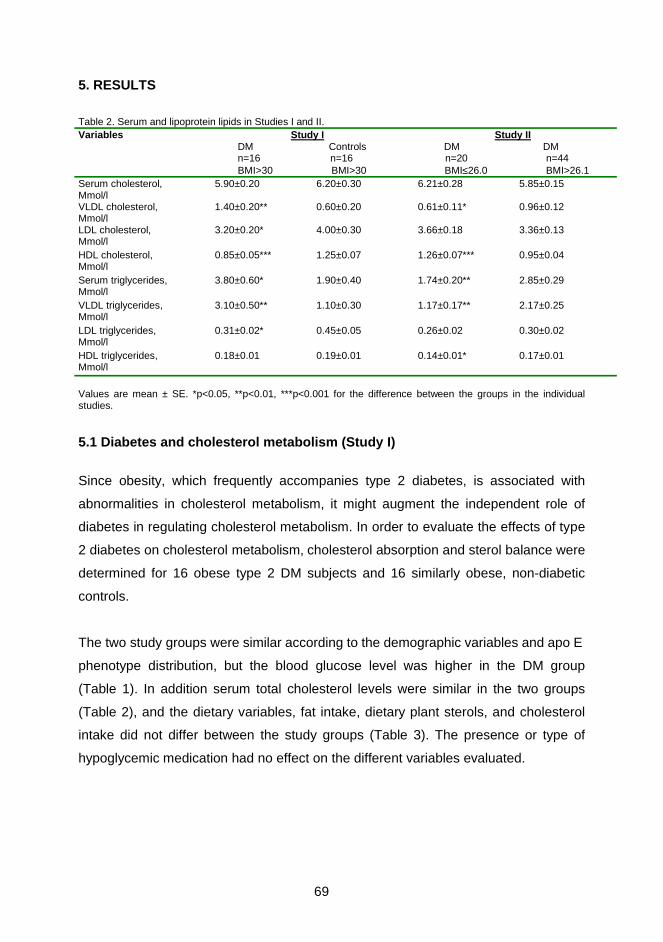
69
5. RESULTS
Table 2. Serum and lipoprotein lipids in Studies I and II. Variables Study I Study II DM Controls DM DM n=16 n=16 n=20 n=44 BMI>30 BMI>30 BMI≤26.0 BMI>26.1 Serum cholesterol, Mmol/l
5.90±0.20 6.20±0.30 6.21±0.28 5.85±0.15
VLDL cholesterol, Mmol/l
1.40±0.20** 0.60±0.20 0.61±0.11* 0.96±0.12
LDL cholesterol, Mmol/l
3.20±0.20* 4.00±0.30 3.66±0.18 3.36±0.13
HDL cholesterol, Mmol/l
0.85±0.05*** 1.25±0.07 1.26±0.07*** 0.95±0.04
Serum triglycerides, Mmol/l
3.80±0.60* 1.90±0.40 1.74±0.20** 2.85±0.29
VLDL triglycerides, Mmol/l
3.10±0.50** 1.10±0.30 1.17±0.17** 2.17±0.25
LDL triglycerides, Mmol/l
0.31±0.02* 0.45±0.05 0.26±0.02 0.30±0.02
HDL triglycerides, Mmol/l
0.18±0.01 0.19±0.01 0.14±0.01* 0.17±0.01
Values are mean ± SE. *p<0.05, **p<0.01, ***p<0.001 for the difference between the groups in the individual studies.
5.1 Diabetes and cholesterol metabolism (Study I)
Since obesity, which frequently accompanies type 2 diabetes, is associated with
abnormalities in cholesterol metabolism, it might augment the independent role of
diabetes in regulating cholesterol metabolism. In order to evaluate the effects of type
2 diabetes on cholesterol metabolism, cholesterol absorption and sterol balance were
determined for 16 obese type 2 DM subjects and 16 similarly obese, non-diabetic
controls.
The two study groups were similar according to the demographic variables and apo E
phenotype distribution, but the blood glucose level was higher in the DM group
(Table 1). In addition serum total cholesterol levels were similar in the two groups
(Table 2), and the dietary variables, fat intake, dietary plant sterols, and cholesterol
intake did not differ between the study groups (Table 3). The presence or type of
hypoglycemic medication had no effect on the different variables evaluated.
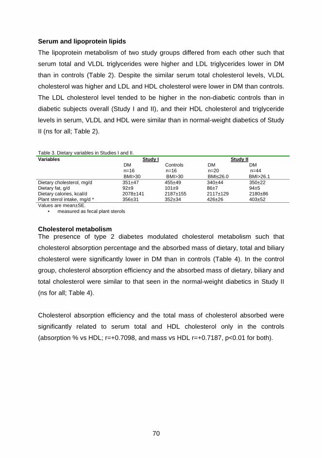
70
Serum and lipoprotein lipids
The lipoprotein metabolism of two study groups differed from each other such that
serum total and VLDL triglycerides were higher and LDL triglycerides lower in DM
than in controls (Table 2). Despite the similar serum total cholesterol levels, VLDL
cholesterol was higher and LDL and HDL cholesterol were lower in DM than controls.
The LDL cholesterol level tended to be higher in the non-diabetic controls than in
diabetic subjects overall (Study I and II), and their HDL cholesterol and triglyceride
levels in serum, VLDL and HDL were similar than in normal-weight diabetics of Study
II (ns for all; Table 2).
Table 3. Dietary variables in Studies I and II. Variables Study I Study II DM Controls DM DM n=16 n=16 n=20 n=44 BMI>30 BMI>30 BMI≤26.0 BMI>26.1 Dietary cholesterol, mg/d 351±47 455±49 340±44 350±22 Dietary fat, g/d 92±9 101±9 86±7 94±5 Dietary calories, kcal/d 2078±141 2187±155 2117±129 2180±86 Plant sterol intake, mg/d * 356±31 352±34 426±26 403±52 Values are mean±SE.
• measured as fecal plant sterols
Cholesterol metabolism The presence of type 2 diabetes modulated cholesterol metabolism such that
cholesterol absorption percentage and the absorbed mass of dietary, total and biliary
cholesterol were significantly lower in DM than in controls (Table 4). In the control
group, cholesterol absorption efficiency and the absorbed mass of dietary, biliary and
total cholesterol were similar to that seen in the normal-weight diabetics in Study II
(ns for all; Table 4).
Cholesterol absorption efficiency and the total mass of cholesterol absorbed were
significantly related to serum total and HDL cholesterol only in the controls
(absorption % vs HDL; r=+0.7098, and mass vs HDL r=+0.7187, p<0.01 for both).
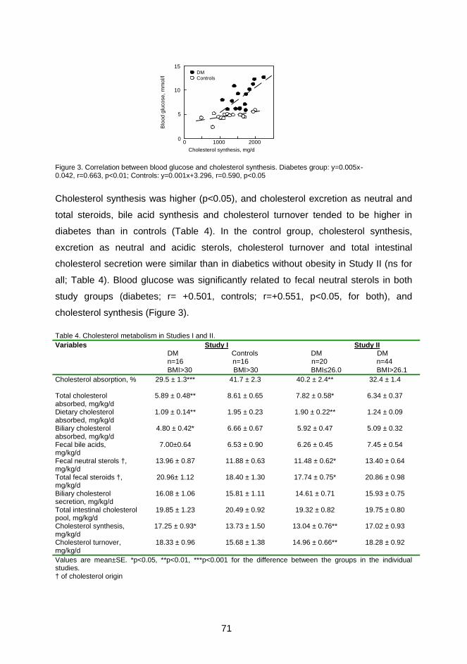
71
Figure 3. Correlation between blood glucose and cholesterol synthesis. Diabetes group: y=0.005x- 0.042, r=0.663, p<0.01; Controls: y=0.001x+3.296, r=0.590, p<0.05
Cholesterol synthesis was higher (p<0.05), and cholesterol excretion as neutral and
total steroids, bile acid synthesis and cholesterol turnover tended to be higher in
diabetes than in controls (Table 4). In the control group, cholesterol synthesis,
excretion as neutral and acidic sterols, cholesterol turnover and total intestinal
cholesterol secretion were similar than in diabetics without obesity in Study II (ns for
all; Table 4). Blood glucose was significantly related to fecal neutral sterols in both
study groups (diabetes; r= +0.501, controls; r=+0.551, p<0.05, for both), and
cholesterol synthesis (Figure 3).
Table 4. Cholesterol metabolism in Studies I and II. Variables Study I Study II DM Controls DM DM n=16 n=16 n=20 n=44 BMI>30 BMI>30 BMI≤26.0 BMI>26.1 Cholesterol absorption, %
29.5 ± 1.3*** 41.7 ± 2.3 40.2 ± 2.4** 32.4 ± 1.4
Total cholesterol absorbed, mg/kg/d
5.89 ± 0.48** 8.61 ± 0.65 7.82 ± 0.58* 6.34 ± 0.37
Dietary cholesterol absorbed, mg/kg/d
1.09 ± 0.14** 1.95 ± 0.23 1.90 ± 0.22** 1.24 ± 0.09
Biliary cholesterol absorbed, mg/kg/d
4.80 ± 0.42* 6.66 ± 0.67 5.92 ± 0.47 5.09 ± 0.32
Fecal bile acids, mg/kg/d
7.00±0.64 6.53 ± 0.90 6.26 ± 0.45 7.45 ± 0.54
Fecal neutral sterols †, mg/kg/d
13.96 ± 0.87 11.88 ± 0.63 11.48 ± 0.62* 13.40 ± 0.64
Total fecal steroids †, mg/kg/d
20.96± 1.12 18.40 ± 1.30 17.74 ± 0.75* 20.86 ± 0.98
Biliary cholesterol secretion, mg/kg/d
16.08 ± 1.06 15.81 ± 1.11 14.61 ± 0.71 15.93 ± 0.75
Total intestinal cholesterol pool, mg/kg/d
19.85 ± 1.23 20.49 ± 0.92 19.32 ± 0.82 19.75 ± 0.80
Cholesterol synthesis, mg/kg/d
17.25 ± 0.93* 13.73 ± 1.50 13.04 ± 0.76** 17.02 ± 0.93
Cholesterol turnover, mg/kg/d
18.33 ± 0.96 15.68 ± 1.38 14.96 ± 0.66** 18.28 ± 0.92
Values are mean±SE. *p<0.05, **p<0.01, ***p<0.001 for the difference between the groups in the individual studies. † of cholesterol origin
0
5
10
15
0 1000 2000
DMControls
Cholesterol synthesis, mg/d
Blo
od g
luco
se, m
mol
/l
72
Taken together, cholesterol absorption was lower and cholesterol synthesis was
higher in obese subjects with diabetes than in those without diabetes, suggesting that
diabetes modulates cholesterol metabolism more than would be the case if the
subjects were merely obese.
5.2 Body weight and cholesterol metabolism (Study II) Since obesity is known to exacerbate the abnormalities of lipoprotein metabolism in
type 2 diabetes, it might modulate cholesterol metabolism in diabetes as well. To
evaluate whether weight interferes with cholesterol metabolism, cholesterol
absorption and cholesterol and bile acid synthesis were studied in 64 type 2 diabetic
subjects with different weights, ranging from lean to overweight values. The study
population was divided by BMI into normal-weight and overweight subgroups. The
distributions of age, gender, blood glucose (Table 1), blood pressure and medication
did not differ between the two study groups, and the dietary intakes of plant sterols,
cholesterol, fat and calories were also similar (Table 3). In addition, age and HbA1c
were not related with any of the variables of cholesterol metabolism.
Despite the similar blood glucose levels, the serum level of SHBG was significantly
higher, and the serum insulin level lower in the normal-weight than in the overweight
group (p<0.001, for both) (Table 5). The ratio of serum SHBG to insulin was four
times higher in the normal-weight than in the overweight group.
Table 5. Variables of glucose metabolism in Studies II, III and IV. In Study III, variables are at baseline and during weight reduction. In Study IV, variables are at baseline and 2 years after weight reduction (follow-up). Variables Study II Study III Study IV DM (n=20) DM (n=44) DM (n=10) DM (n=16) BMI≤26.0 BMI>26.1 BMI>30 During BMI>30 Baseline weight loss Baseline After follow-up Blood glucose, mmol/l 8.5±0.5 8.6±0.4 6.9±0.5 5.3±0.4*** 8.4±0.6 7.2±0.5**
SHBG, nmol/l 45.6±5.1 30.9±2.2*** 24.7±4.7 45.4±5.8*** 24.7±4.7 26.6±2.4
Serum insulin, mU/l 10.2±1.0 20.0±1.4*** 18.0±1.7 7.3±0.8*** 19.7±1.2 15.1±1.7
SHBG / serum insulin 6.24±1.75 1.44±0.19*** 1.26±0.19 8.54±3.10* 1.27±0.18 2.11±0.35*
Values are mean±SE. *p<0.05, **p<0.01, ***p<0.001 for difference between the groups in Study II, baseline and during weight loss in Study III, and baseline and after follow-up in Study IV.
Serum and lipoprotein lipids and sterols
Serum total and LDL cholesterol were similar in the two study groups, but the VLDL
cholesterol was higher, and the HDL cholesterol lower in the overweight than in the
normal-weight group (Table 2). Serum total, VLDL and HDL triglycerides were
73
significantly lower in the normal-weight group vs overweight group. HDL and VLDL
cholesterol and serum and lipoprotein triglyceride levels were similar in the normal-
weight DM group compared to the respective values in the obese subjects without
diabetes in Study I (ns for all; Table 2).
The serum campesterol, sitosterol and cholestanol ratios were markedly higher,
whereas squalene and the precursor sterol ratios (except lathosterol) were lower in
the normal-weight than in the overweight group (Table 6). Lathosterol/campesterol
(1.02±0.14 vs 1.47±0.11 p<0.05) and lathosterol/sitosterol ratios were lower in the
normal-weight than overweight subjects.
Table 6. Serum squalene and non-cholesterol sterol ratios to cholesterol (102 x mmol/mol of cholesterol) in Studies II, III and IV. In Study III, variables are at baseline and during weight reduction. In Study IV, variabl es are at baseline and 2 years after weight reduction (follow-up). Variables Study II Study III Study IV DM (n=20) DM (n=44) DM (n=9) DM (n=10) BMI≤26.0 BMI>26.1 BMI>30 During BMI>30 Baseline weight loss Baseline After follow-up Cholestanol 108.0±4.6 89.1±3.8** 95.6±4.1 125.1±5.6*** 85.0±4.0 95.0±5.0
Campesterol 223.5±17.3 182.9±10.7 154.5±15.7 126.3±11.5 162.0±14.0 197.0±14.0*
Sitosterol 121.9±8.6 95.9±4.6** 84.1±9.2 79.2±7.4 87.5±5.0 103.0±8.0*
Squalene 33.4±2.6 42.2±2.9* 47.4±3.2 58.7±13.2 nd nd
Cholestenol 18.9±1.8 28.7±1.7*** 20.4±3.0 15.7±1.2 24.1±1.7 22.3±1.2
Desmosterol 84.7±4.8 116.2±12.2* 82.4±4.7 66.3±4.5*** 136.0±29.0 109.0±15.0
Lathosterol 195.5±17.8 233.7±12.0 207.8±14.9 159.2±10.4* 226.0±8.0 218.0±10.0
Values are mean±SE. *p<0.05, **p<0.01, ***p<0.001 for the difference between groups in Study II, baseline and during weight loss in Study III, and baseline and follow-up in Study IV. nd =not determined.
Cholesterol metabolism
Cholesterol absorption efficiency was higher (p<0.01) in the normal-weight subjects
compared to the overweight subjects (Table 4, Figure 4). The amounts of absorbed
total and dietary cholesterol were higher in normal-weight subjects, but the mass of
absorbed biliary cholesterol showed no significant difference between the groups.
The variables of cholesterol absorption in the normal-weight diabetes group were
similar to the respective values in obese subjects without diabetes in Study I (ns for
all; Table 4).
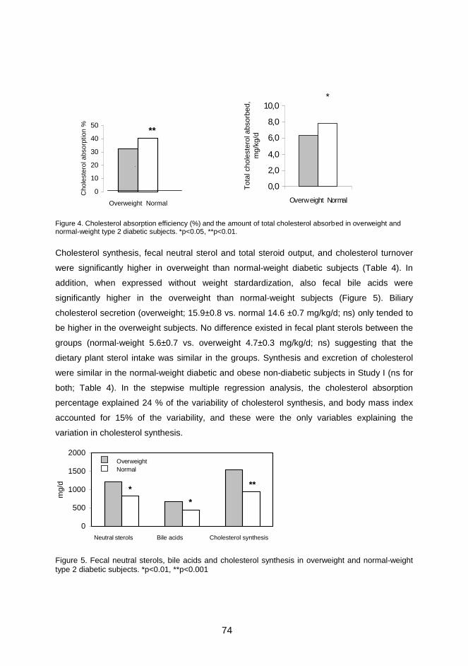
74
Figure 4. Cholesterol absorption efficiency (%) and the amount of total cholesterol absorbed in overweight and normal-weight type 2 diabetic subjects. *p<0.05, **p<0.01.
Cholesterol synthesis, fecal neutral sterol and total steroid output, and cholesterol turnover
were significantly higher in overweight than normal-weight diabetic subjects (Table 4). In
addition, when expressed without weight stardardization, also fecal bile acids were
significantly higher in the overweight than normal-weight subjects (Figure 5). Biliary
cholesterol secretion (overweight; 15.9±0.8 vs. normal 14.6 ±0.7 mg/kg/d; ns) only tended to
be higher in the overweight subjects. No difference existed in fecal plant sterols between the
groups (normal-weight 5.6±0.7 vs. overweight 4.7±0.3 mg/kg/d; ns) suggesting that the
dietary plant sterol intake was similar in the groups. Synthesis and excretion of cholesterol
were similar in the normal-weight diabetic and obese non-diabetic subjects in Study I (ns for
both; Table 4). In the stepwise multiple regression analysis, the cholesterol absorption
percentage explained 24 % of the variability of cholesterol synthesis, and body mass index
accounted for 15% of the variability, and these were the only variables explaining the
variation in cholesterol synthesis.
Figure 5. Fecal neutral sterols, bile acids and cholesterol synthesis in overweight and normal-weight type 2 diabetic subjects. *p<0.01, **p<0.001
0
500
1000
1500
2000
mg/
d
Neutral sterols Bile acids Cholesterol synthesis
OverweightNormal
**
**
0
10
20
30
40
50
Overweight Normal
Cho
lest
erol
abs
orpt
ion
%
**
*
0,0
2,0
4,0
6,0
8,0
10,0
Overweight Normal
Tot
al c
hole
ster
ol a
bsor
bed,
m
g/kg
/d
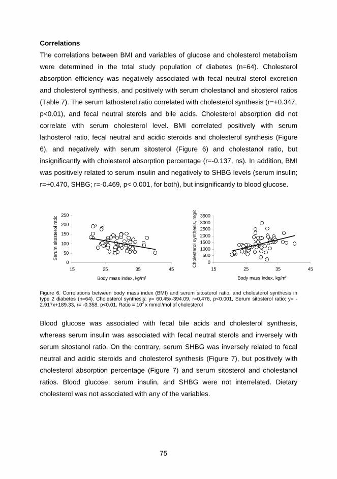
75
Correlations
The correlations between BMI and variables of glucose and cholesterol metabolism
were determined in the total study population of diabetes (n=64). Cholesterol
absorption efficiency was negatively associated with fecal neutral sterol excretion
and cholesterol synthesis, and positively with serum cholestanol and sitosterol ratios
(Table 7). The serum lathosterol ratio correlated with cholesterol synthesis (r=+0.347,
p<0.01), and fecal neutral sterols and bile acids. Cholesterol absorption did not
correlate with serum cholesterol level. BMI correlated positively with serum
lathosterol ratio, fecal neutral and acidic steroids and cholesterol synthesis (Figure
6), and negatively with serum sitosterol (Figure 6) and cholestanol ratio, but
insignificantly with cholesterol absorption percentage (r=-0.137, ns). In addition, BMI
was positively related to serum insulin and negatively to SHBG levels (serum insulin;
r=+0.470, SHBG; r=-0.469, p< 0.001, for both), but insignificantly to blood glucose.
Figure 6. Correlations between body mass index (BMI) and serum sitosterol ratio, and cholesterol synthesis in type 2 diabetes (n=64). Cholesterol synthesis: y= 60.45x-394.09, r=0.476, p<0.001, Serum sitosterol ratio: y= -2.917x+189.33, r= -0.358, p<0.01. Ratio = 102 x mmol/mol of cholesterol
Blood glucose was associated with fecal bile acids and cholesterol synthesis,
whereas serum insulin was associated with fecal neutral sterols and inversely with
serum sitostanol ratio. On the contrary, serum SHBG was inversely related to fecal
neutral and acidic steroids and cholesterol synthesis (Figure 7), but positively with
cholesterol absorption percentage (Figure 7) and serum sitosterol and cholestanol
ratios. Blood glucose, serum insulin, and SHBG were not interrelated. Dietary
cholesterol was not associated with any of the variables.
0
50
100
150
200
250
15 25 35 45
Body mass index, kg/m2
Ser
um s
itost
erol
rat
io
0
5001000
15002000
25003000
3500
15 25 35 45
Body mass index, kg/m2
Cho
lest
erol
syn
thes
is, m
g/d
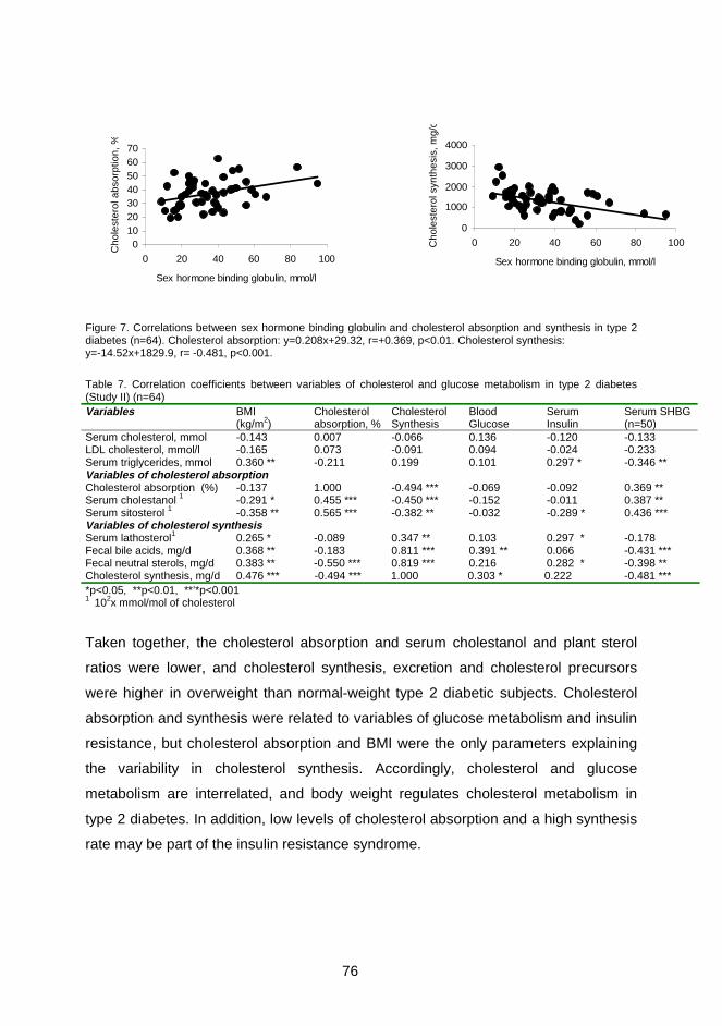
76
Figure 7. Correlations between sex hormone binding globulin and cholesterol absorption and synthesis in type 2 diabetes (n=64). Cholesterol absorption: y=0.208x+29.32, r=+0.369, p<0.01. Cholesterol synthesis: y=-14.52x+1829.9, r= -0.481, p<0.001.
Table 7. Correlation coefficients between variables of cholesterol and glucose metabolism in type 2 diabetes (Study II) (n=64) Variables
BMI (kg/m2)
Cholesterol absorption, %
Cholesterol Synthesis
Blood Glucose
Serum Insulin
Serum SHBG (n=50)
Serum cholesterol, mmol -0.143 0.007 -0.066 0.136 -0.120 -0.133 LDL cholesterol, mmol/l -0.165 0.073 -0.091 0.094 -0.024 -0.233 Serum triglycerides, mmol 0.360 ** -0.211 0.199 0.101 0.297 * -0.346 ** Variables of cholesterol absorption Cholesterol absorption (%) -0.137 1.000 -0.494 *** -0.069 -0.092 0.369 ** Serum cholestanol 1 -0.291 * 0.455 *** -0.450 *** -0.152 -0.011 0.387 ** Serum sitosterol 1 -0.358 ** 0.565 *** -0.382 ** -0.032 -0.289 * 0.436 *** Variables of cholesterol synthesis Serum lathosterol1 0.265 * -0.089 0.347 ** 0.103 0.297 * -0.178 Fecal bile acids, mg/d 0.368 ** -0.183 0.811 *** 0.391 ** 0.066 -0.431 *** Fecal neutral sterols, mg/d 0.383 ** -0.550 *** 0.819 *** 0.216 0.282 * -0.398 ** Cholesterol synthesis, mg/d 0.476 *** -0.494 *** 1.000 0.303 * 0.222 -0.481 *** *p<0.05, **p<0.01, **’*p<0.001 1 102x mmol/mol of cholesterol
Taken together, the cholesterol absorption and serum cholestanol and plant sterol
ratios were lower, and cholesterol synthesis, excretion and cholesterol precursors
were higher in overweight than normal-weight type 2 diabetic subjects. Cholesterol
absorption and synthesis were related to variables of glucose metabolism and insulin
resistance, but cholesterol absorption and BMI were the only parameters explaining
the variability in cholesterol synthesis. Accordingly, cholesterol and glucose
metabolism are interrelated, and body weight regulates cholesterol metabolism in
type 2 diabetes. In addition, low levels of cholesterol absorption and a high synthesis
rate may be part of the insulin resistance syndrome.
010203040506070
0 20 40 60 80 100
Sex hormone binding globulin, mmol/l
Cho
lest
erol
abs
orpt
ion,
%
0
1000
2000
3000
4000
0 20 40 60 80 100
Sex hormone binding globulin, mmol/l
Cho
lest
erol
syn
thes
is, m
g/d
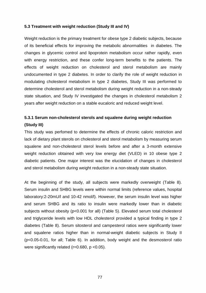
77
5.3 Treatment with weight reduction (Study III and IV)
Weight reduction is the primary treatment for obese type 2 diabetic subjects, because
of its beneficial effects for improving the metabolic abnormalities in diabetes. The
changes in glycemic control and lipoprotein metabolism occur rather rapidly, even
with energy restriction, and these confer long-term benefits to the patients. The
effects of weight reduction on cholesterol and sterol metabolism are mainly
undocumented in type 2 diabetes. In order to clarify the role of weight reduction in
modulating cholesterol metabolism in type 2 diabetes, Study III was performed to
determine cholesterol and sterol metabolism during weight reduction in a non-steady
state situation, and Study IV investigated the changes in cholesterol metabolism 2
years after weight reduction on a stable eucaloric and reduced weight level.
5.3.1 Serum non-cholesterol sterols and squalene during weight reduction
(Study III)
This study was perfomed to determine the effects of chronic caloric restriction and
lack of dietary plant sterols on cholesterol and sterol metabolism by measuring serum
squalene and non-cholesterol sterol levels before and after a 3-month extensive
weight reduction obtained with very low energy diet (VLED) in 10 obese type 2
diabetic patients. One major interest was the elucidation of changes in cholesterol
and sterol metabolism during weight reduction in a non-steady state situation.
At the beginning of the study, all subjects were markedly overweight (Table 8).
Serum insulin and SHBG levels were within normal limits (reference values, hospital
laboratory:2-20mU/l and 10-42 nmol/l). However, the serum insulin level was higher
and serum SHBG and its ratio to insulin were markedly lower than in diabetic
subjects without obesity (p<0.001 for all) (Table 5). Elevated serum total cholesterol
and triglyceride levels with low HDL cholesterol provided a typical finding in type 2
diabetes (Table 8). Serum sitosterol and campesterol ratios were significantly lower
and squalene ratios higher than in normal-weight diabetic subjects in Study II
(p<0.05-0.01, for all; Table 6). In addition, body weight and the desmosterol ratio
were significantly related (r=0.680, p <0.05).

78
Weight reduction
Dietary intake
VLED was well-tolerated. The daily intake contained about 19 % of the prefasting
dietary calories. The diet contained only minimal amounts of sterols, because
analysis of 15 servings with GLC revealed that the daily intake of cholesterol was
11.4±2.0 mg/d, campesterol 4.1±0.3 mg/d, sitosterol 11.3±0.8 mg/d, and stigmasterol
3.7±0.3 mg/d. The dietary intakes of lathosterol, cholestanol, campestanol, sitostanol,
avenasterol and squalene were all < 1 mg/d. Thus, the dietary intake of cholesterol
was about 3 % and plant sterols, campesterol and sitosterol, 4% of the prefasting
values measured in Study IV.
Body weight and glucose metabolism
Body weight was reduced by 15.5 ± 1.7 kg, (range 8.5-25 kg), the reduction being
16.8±1.5% from the baseline weight (Table 8). Despite the effective weight reduction,
several subjects were still overweight, their mean BMI was 26.3±0.7 kg/m2. Blood
glucose was lowered by 23 %, HbA1c by 8%, and serum insulin by 59%. Serum
SHBG increased by 108%, and its ratio to blood glucose and serum insulin were
increased (Table 5). All of these changes were highly significant (p<0.05-0.001, for
all).
Serum lipids and lipoproteins
Serum total cholesterol was reduced by 21 % and triglycerides by 45 % from the
baseline values (p< 0.001, for both; Table 8). The HDL cholesterol level remained
unchanged, but its ratio to serum total cholesterol was increased by 34 % (p<0.001).
Table 8. Weight, BMI, variables of glucose metabolism and serum and lipoprotein lipids at baseline and during weight reduction in type 2 diabetes (n=10) Variables Baseline During weight reduction Weight, kg 92.3 ± 4.8 76.7 ± 4.2 * BMI, kg/m2 31.7 ± 0.9 26.3 ± 0.7 * Blood glucose, mmol/l 6.9 ± 0.5 5.3 ± 0.4 * SHBG, nmol/l 24.7 ± 4.7 45.4 ± 5.8 * Serum insulin, mU/l 18.0 ± 1.7 7.3 ± 0.8 * SHBG / serum insulin 1.26 ± 0.19 8.54 ± 3.10 # Serum cholesterol , mmol/l 5.83 ± 0.24 4.52 ± 0.13 * Serum triglycerides, mmol/l 2.60 ± 0.26 1.40 ± 0.17 * HDL cholesterol, mmol/l 0.96 ± 0.08 0.99 ± 0.07 Values are mean±SE. # p < 0.05 , * p < 0.001 by paired t-test. Reference values from hospital laboratory: serum insulin 2-20 mU/l, SHBG 10-42 nmol/l.

79
Squalene and non-cholesterol sterols
The desmosterol and lathosterol ratios to cholesterol decreased significantly,
whereas the ratio of serum squalene increased by 24 % (ns) (Table 6). The serum
cholestanol concentration tended to increase (203.4±14.7 µg/dl vs. 209.4±15.5 µg/dl,
ns), whereas the decrease in campesterol and sitosterol concentrations was only
significant for campesterol (330.3±39.6 µg/dl vs. 208.2±18.2 µg/dl, p < 0.01). As
measured as ratios to cholesterol, the increase in cholestanol ratio was highly
significant (p <0.001), while the ratios of campesterol and sitosterol only tended to
decrease (-12% and –4 %, ns for both) (Table 6).
The cholestanol ratio was markedly higher after the weight reduction than the
respective ratio in diabetic subjects with normal weights (Table 6). In addition,
cholestenol, desmosterol and lathosterol ratios were lower after weight loss in these
still overweight subjects compared to the normal-weight diabetic subjects.
Correlations
The change in body weight tended to be positively associated with that of the
desmosterol ratio (r=0.630, p=0.06) and inversely with final the SHBG level (r=-0.776,
p < 0.01). A significant inverse association existed between the change in serum
sitosterol and the baseline (r=-0.752) and final (r=-0.742, p< 0.05, for both) body
weight. In addition, the ratios of serum lathosterol and cholestanol were interrelated
(r=-0.823, p<0.01) after the weight reduction, and the change in serum cholestanol
correlated inversely with the final serum insulin level (r=-0.689, p<0.05).
Taken together, the restriction of calories and dietary sterols improved markedly the
control of diabetes. It also decreased serum cholesterol precursor sterols and
increased serum cholestanol suggesting that cholesterol absorption was increased
and synthesis decreased. Serum values of plant sterols only tended to decrease
despite their minimal dietary intake. Accordingly, cholesterol metabolism is improved
during weight reduction in a non-steady state situation, and the changes in glucose
and cholesterol metabolism are interrelated.
80
5.3.2 Cholesterol, glucose and lipoprotein metabolism after treatment with
weight reduction (Study IV)
This study examined the effects of weight reduction on glucose, cholesterol and
lipoprotein metabolism after a 2-year follow-up in type 2 diabetes. Cholesterol
absorption efficiency was measured with peroral dual isotopes and cholesterol
synthesis with sterol balance. The 3-month weight reduction was obtained by very-
low-energy diet (VLED; n=10 diabetic subjects) or by low-energy diet (LED; n=6
diabetic subjects). Age, sex, BMI, serum lipids, serum sterols and variables of
cholesterol metabolism were similar in these two groups at the baseline. The
presence or type of hypoglycemic therapy had no effect on the distribution of BMI,
serum glucose, SHBG, serum sterols, and variables of cholesterol metabolism. There
was no significant difference in serum lipids and metabolic variables between women
and men, and the 16 subjects were analyzed as an aggregate.
At the beginning of the study, the subjects were markedly overweight (Table 9). The
subjects had a similar dyslipidemic lipoprotein profile and serum levels of insulin and
SHBG as in Study III. Blood glucose was related to bile acid and cholesterol
synthesis, and to serum insulin concentration (r=0.558, p<0.05). The serum insulin
level also correlated with fecal neutral sterols and cholesterol synthesis. Serum
SHBG was positively associated with body weight and the cholesterol absorption
efficiency (r=+0.575, p<0.05) but not with blood glucose or serum insulin.
Two-year follow-up
All subjects completed the study without side effects, and VLED was well tolerated.
After the two year follow up, the mean body weight was 6±1 kg (6.4 %) lower than at
baseline (Table 9). The changes in body weight ranged from –18 kg to +1 kg. Most of
the subjects were still characterized as obese, because the mean BMI was 30.3±0.8
kg/m2. The daily intakes of energy, cholesterol, and plant sterols, as expressed by
mg/kg/day, were similar, but the fat intake was significantly lower than at the
baseline.
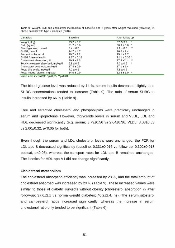
81
Table 9. Weight, BMI and cholesterol metabolism at baseline and 2 years after weight reduction (follow-up) in obese patients with type 2 diabetes (n=16) Variables Baseline After follow-up
Weight, (kg) 93.2 ± 3.7 87.2±3.2 * BMI, (kg/m2) 31.7 ± 0.6 30.3 ± 0.8 * Blood glucose, mmol/l 8.4 ± 0.6 7.2 ± 0.5 ** SHBG, nmol/l 24.7 ± 4.7 26.6 ± 2.4 Serum insulin, mU/l 19.7 ± 1.2 15.1 ± 1.7 SHBG / serum insulin 1.27 ± 0.18 2.11 ± 0.35 * Cholesterol absorption, % 29.5 ± 1.3 37.6 ±2.1 ** Total cholesterol absorbed, mg/kg/d 5.9 ± 0.5 7.3 ± 0.6 * Cholesterol synthesis, mg/kg/d 17.3 ± 0.9 17.1 ± 1.4 Fecal bile acids, mg/kg/d 7.0 ± 0.6 7.8 ± 0.9 Fecal neutral sterols, mg/kg/d 14.0 ± 0.9 12.5 ± 1.0 * Values are mean±SE. *p<0.05, **p<0.01.
The blood glucose level was reduced by 14 %, serum insulin decreased slightly, and
SHBG concentrations tended to increase (Table 9). The ratio of serum SHBG to
insulin increased by 66 % (Table 9).
Free and esterified cholesterol and phospholipids were practically unchanged in
serum and lipoproteins. However, triglyceride levels in serum and VLDL, LDL and
HDL decreased significantly (e.g. serum; 3.79±0.56 vs 2.64±0.36, VLDL; 3.08±0.53
vs 2.00±0.32, p<0.05 for both).
Even though the serum and LDL cholesterol levels were unchanged, the FCR for
LDL apo B decreased significantly (baseline; 0.331±0.016 vs follow-up; 0.302±0.018
pools/d, p<0.05), whereas the transport rates for LDL apo B remained unchanged.
The kinetics for HDL apo A-I did not change significantly.
Cholesterol metabolism
The cholesterol absorption efficiency was increased by 28 %, and the total amount of
cholesterol absorbed was increased by 23 % (Table 9). These increased values were
similar to those of diabetic subjects without obesity (cholesterol absorption % after
follow-up; 37.6±2.1 vs normal-weight diabetes; 40.2±2.4, ns). The serum sitosterol
and campesterol ratios increased significantly, whereas the increase in serum
cholestanol ratio only tended to be significant (Table 6).
82
The serum SHBG was positively related to the ratios of sitosterol (r=+0.779, p<0.01),
campesterol (r=+ 0.669) and cholestanol (r=+0.516, p<0.05 for both). The reduction
of body weight was associated with the changes in campesterol (r=-0.582) and
sitosterol ratios (r=-0.647, p<0.05 for both).
The synthesis of bile acids and cholesterol remained practically unchanged despite
the significant decrease in fecal neutral sterol excretion (Table 9). The ratios of
cholesterol precursor sterols, cholestenol, desmosterol and lathosterol, only tended
to decrease (Table 6). The significant baseline correlations between serum insulin,
blood glucose, and variables of cholesterol synthesis and excretion were not
significant after the follow-up.
Therefore, cholesterol absorption (i.e. absolute and relative absorption and the serum
ratios of plant sterols and cholestanol) markedly increased after weight reduction
suggesting that weight reduction tended to normalize cholesterol metabolism in type
2 diabetes. Hyperinsulinemia and insulin resistance also improved after weight
reduction. Variables reflecting insulin resistance were related to variables reflecting
cholesterol absorption after weight reduction. Thus, the cholesterol absorption
efficiency and insulin resistance are interrelated, and the efficacy of cholesterol
absorption seems to be an inverse indicator of insulin resistance.
83
6. DISCUSSION
6.1 Study population
The study population provided a homogenous and large-enough population to
elaborate cholesterol metabolism in type 2 diabetes. The mean age of the subjects
was 56 years varying from 41 to 74 years. All women participating in the studies were
postmenopausal and did not use any hormone replacement therapy. The variables of
cholesterol, glucose and lipoprotein metabolism did not differ between the genders,
and therefore male and female subjects in the individual studies could be analyzed
together. Gender is known to affect lipoprotein metabolism e.g. women have
generally higher HDL cholesterol levels as compared to men. However, HDL
cholesterol levels did not differ between women and men in the present study, in
which the number of women participants was quite small.
Diabetes had been recently diagnosed, and blood glucose levels were comparable
among the diabetic patients. None of the subjects were receiving insulin therapy or
using any lipid-lowering medication. The patients with diabetes did not have any
detectable diabetic nephro- or neuropathy, and they had normal hepatic, pancreatic,
thyroid and intestinal functions. Therefore, the subjects were in good glycemic control
and did not suffer any diabetic complications. The presence or type of hypoglycemic
medication did not affect the variables of cholesterol and glucose metabolism. In
addition, the use of hypotensive treatment was not a confounding factor.
The dietary variables, which could be assumed to affect cholesterol metabolism, i.e.,
cholesterol and fat intake, type of fat and dietary plant sterols, including stanols, were
similar in the different studies. In addition, the study groups were well comparable
with regard to serum and LDL cholesterol levels and apo E phenotype distribution
suggesting that there were no obvious confounding factors. In the case-control study
I, the subjects with and without diabetes were similarly obese and differed from each
other only in their blood glucose level.
Cholesterol metabolism was compared between the different studies (e.g Study I and
II), even though it was not originally in the protocol. Since the factors possibly
84
affecting cholesterol metabolism (e.g. demographic and dietary variables and apo E
phenotype distribution) did not differ between the two studies, except for age, these
comparisons could be performed. In addition, age did not correlate with any variables
of diet and cholesterol metabolism, suggesting that it was not a confounding factor.
6.2 Measurement of cholesterol metabolism
The measurement of cholesterol absorption requires differentiation of endogenous
from exogenous steroids, which can be achieved by using labeled cholesterol
administered both orally and intravenously. The oral dose labels exogenous
cholesterol and its steroid products, whereas the intravenous dose labels
endogenous steroids. With or without simultaneous measurement of cholesterol
excretion, the absorption of dietary cholesterol can be calculated after a leveling
period. Several methods have been developed for the determination of dietary
cholesterol absorption, and these methods are described in detail in the review of the
literature section on pages 35-37.
In the present study, the absorption of cholesterol was measured with the continuous
isotope feeding method, which is based on peroral administration of labeled
cholesterol and an unabsorbable reference compound. A reduction of the labeled
cholesterol/marker ratio in stools reveals the absorption percent of cholesterol. The
measurement of cholesterol absorption with this method gives an accurate value of
the absorption because the study period is long enough to achieve steady state and
constancy of ratio of the isotopes in feces, and sequential fecal samples diminish the
fluctuation in the absorption. In addition, other advantages include the simple
administration of isotope, analysis of fecal samples is easy, though laborious, the
analysis is possible to be repeated daily, and it is suitable for outpatients.
Furthermore, the administration of the isotopes in capsules with the regular meals of
the study subjects gives the accurate absorption of cholesterol during normal home
conditions in everyday life, thus preventing the possible confounding effect of a
special test meal. The possible recirculation of absorbed isotopic cholesterol into
fecal samples is not a confounding factor, since the fecal isotope ratio remains
constant for 10 days after administration of the isotope, and the percent absorption
obtained with the present method compared with that of other methods is similar up

85
to 14 days after isotope administration. Moreover, the amount of cholesterol
absorbed and the intestinal influx of endogenous cholesterol can be calculated, if the
daily intake of cholesterol and fecal neutral sterol excretion are also measured.
A more simplified way to measure cholesterol absorption is the plasma isotope ratio
method, which is based on the simultaneous oral and intravenous administration of
labeled cholesterol. Although this method has provided considerable information
about cholesterol absorption, it has several drawbacks. It estimates only the
cholesterol absorption percentage, the value is determined from only a short period
of time, it yields only a single measurement of absorption, and the value of
cholesterol absorption may be dependent on the composition of the test meal. In
addition, as the method is based on the determination of the ratio of the two isotopes
in plasma after days of equilibration in the rapidly miscible pool of body cholesterol,
the applicability of this method in subjects with different metabolic conditions or in
malabsorption syndromes is uncertain; i.e. in conditions where cholesterol synthesis
and elimination is enhanced, the metabolic fate of intravenously administered
cholesterol tracers is largely unknown. In these conditions the turnover of the rapidly
miscible cholesterol pool is faster and thus invalidates the complete equilibration of
the two tracers, resulting bias in the estimation of the cholesterol absorption
percentage.
The measurement of serum squalene and non-cholesterol sterol ratios to cholesterol
from serum samples by gas-liquid chromatography provides an efficient, easily
reproducible and less laborious method to estimate cholesterol absorption and
synthesis. The sterol balance technique, in which assaying total-body sterol pool in a
steady state by calculating the difference between fecal steroids of cholesterol origin
and dietary cholesterol intake, provides a direct and accurate method to determine
the endogenous biosynthesis of cholesterol.
In the present study, serum plant sterol and cholestanol ratios correlated with the
cholesterol absorption efficiency, and the ratios of serum cholesterol precursor
sterols with cholesterol synthesis. Accordingly, serum non-cholesterol sterols reflect
cholesterol metabolism in a similar manner to the non-diabetic population (Miettinen
1970, Miettinen and Koivisto 1983, Tilvis and Miettinen 1986, Vuoristo and Miettinen
86
1986, Kempen et al. 1988, Miettinen et al. 1989, Miettinen et al. 1990, Miettinen and
Gylling 2000), and can be used in the evaluation of cholesterol absorption and
synthesis. In addition, serum squalene was higher in subjects with elevated
cholesterol synthesis compared with those of lower synthesis suggesting that in this
diabetic population, serum squalene was also a marker of cholesterol synthesis rate.
6.3 Cholesterol metabolism in diabetes (Study I and II) 6.3.1 Cholesterol absorption
Low cholesterol absorption has been described earlier in a limited number of type 2
diabetes patients with overweight and hypercholesterolemia (Gylling and Miettinen
1997), or those with hypertriglyceridemia (Briones et al. 1986), and in obesity
(Miettinen and Gylling 2000). One of the most interesting observations in the present
study was the lower cholesterol absorption in obese type 2 diabetes than in normal-
weight type 2 diabetes, or when compared with the respective similarly obese, non-
diabetic state. In addition, absorption of cholesterol was similar in normal-weight type
2 diabetic patients and obese non-diabetic patients. These results suggest that
diabetes with some unknown mechanism inhibits the absorption of cholesterol. In
addition, obesity in diabetes still attenuates the efficiency of sterol absorption.
Diet
The effect of dietary cholesterol on cholesterol absorption efficiency is controversial
according to earlier results. It may remain mainly unaltered despite high amounts of
dietary cholesterol (McMurry et al. 1985, Miettinen and Kesäniemi 1989, Sehayek et
al. 1998a) or decreased (McNamara et al. 1987, Gylling and Miettinen 1992, Vuoristo
and Miettinen 1994). In some studies, cholesterol intake was not significantly related
to cholesterol absorption efficiency (Miettinen and Kesäniemi 1989, Bosner et al.
1999). More recent studies have shown that with a modest increase in dietary
cholesterol, there was a decline in the cholesterol absorption efficiency (Ostlund et al.
1999). Along these lines, the absorption percent in the present study might have
been even higher in the control group, and the difference in cholesterol absorption
efficiency between the groups even larger. However, the dietary cholesterol intake
was not associated with the variables of cholesterol, lipoprotein or glucose
metabolism. Moreover, the dietary cholesterol intake and cholesterol absorption
87
efficiency were not interrelated, if anything, the relationship was negative. Therefore,
it could be argued that the dietary cholesterol intake was not a confounding factor in
the present studies.
Extra amounts of dietary fiber added to the regular diet lower the cholesterol
absorption percentage in normal (Salvioli et al. 1985) and hypercholesterolemic
subjects (Everson et al. 1992). However, the association between fiber intake and
the absorption percentage of cholesterol remained negative in subjects on their
normal diet (Miettinen and Kesäniemi 1989, Kesäniemi et al. 1990, Bosner et al.
1999). In the present study, the subjects consumed their normal, regular home diet
without extra fiber intake, and did not consume any additional products of gel forming
or viscous fibers.
Plant sterols and their stanol esters are known to reduce cholesterol absorption (e.g.
Grundy et al. 1969, Mattson et al. 1982, Vanhanen et al. 1993, Vanhanen et al.
1994). Fecal plant sterols were similar in all study groups suggesting they had a
similar dietary plant sterol intake. In addition, none of the subjects consumed
functional foods consisting of plant stanols and this was confirmed by analysing their
levels of fecal stanols.
Apolipoprotein E
Apo E 2/3/4 phenotype is closely associated with intestinal absorption efficiency of
cholesterol (Kesäniemi et al. 1987, Miettinen et al. 1992), and the polymorphism of
apo E may be one reason for the high interindividual variabily in cholesterol
absorption (Miettinen and Kesäniemi 1989). However, the similar distribution of apo
E phenotypes between the study groups suggests that the results obtained in
cholesterol metabolism were not affected by the apo E phenotype.
Intestinal motility
Gastrointestinal motility disorders are common in patients with diabetes, and their
orocaecal transit time is prolonged (Rayner et al. 2001). In addition to the autonomic
neuropathy resulting in motility disorders, hyperglycemia has inhibitory effects on
gastrointestinal motility to which hyperinsulinemia is contributing (Byrne et al. 1998).
In healthy subjects without diabetes, intestinal transit time influences cholesterol
88
absorption such that a short transit time appears to reduce the cholesterol absorption
efficiency (Ponz de Leon et al. 1982, Koivisto and Miettinen 1986). In the present
study, diabetes had been recently diagnosed, and subjects did not have any diabetic
complications suggesting normal/near normal gastrointestinal motility and transit
time. In study II, despite the similar blood glucose levels, the normal-weight type 2
diabetic patients had a greater cholesterol absorption than overweight diabetic
patients. Moreover, it could have been anticipated that the possible prolonged
intestinal transit time should have enhanced the absorption efficiency of cholesterol
in normal-weight diabetes vs obese nondiabetes. However, this was not the case, as
their cholesterol absorption efficiencies were similar. Therefore, intestinal motility and
intestinal transit time were not confounding factors in the present study.
Clinical conditions and statins
Many diseases and organic dysfunctions, e.g. pancreatic insufficiency, liver, gastric
or ileal diseases or dysfunctions, especially celiac disease, alter cholesterol
metabolism by inducing cholesterol malabsorption (e.g. Vuoristo et al. 1988, Vuoristo
et al. 1992). Hypothyreosis, in which cholesterol synthesis and elimination (Abrams
and Grundy 1981), and intestinal motility are impaired, may influence cholesterol
absorption. During the initial stage of this study, the possibility of some kind of
disease or condition possibly affecting cholesterol metabolism in the subjects was
ruled out. Therefore, clinical conditions did not have any effect on the results in the
present study.
Statins, HMG-CoA inhibitors, in addition to inhibiting cholesterol biosynthesis, have
been found to interfere with cholesterol absorption probably due to the reduction in
biliary cholesterol secretion. Statin treatment caused a significant reduction in
cholesterol absorption of patients with familial hypercholesterolemia (Miettinen 1991,
Vanhanen et al. 1992), but not in nonfamilial mildly hypercholesterolemic patients
(Vanhanen and Miettinen 1995). Since the use of statin therapy might interfere with
cholesterol absorption in type 2 diabetes as well, the use of statins or any
hypolipidemic drug was one of the exclusion criteria in the present studies.
89
6.3.2 Cholesterol synthesis and excretion
It has been observed earlier in a limited number of diabetic subjects that cholesterol
and bile acid synthesis and fecal elimination of cholesterol are increased compared
with controls (Bennion and Grundy 1977, Abrams et al. 1982, Briones et al. 1986,
Scoppola et al. 1995, Naoumova et al. 1996, Gylling and Miettinen 1997). In all of
these previous studies, the subjects were obese and /or hypertriglyceridemic, and
some had insulin treatment. However, no difference was found in cholesterol
synthesis between diabetic patients and controls, when the lipid profile was normal
(Briones et al. 1986). The results from the present study confirm that cholesterol
synthesis and excretion are higher in obese than normal-weight type 2 diabetes, and
even higher than in obese non-diabetic subjects. Furthermore, the synthesis and
excretion of cholesterol is similar in diabetes with normal body weight to non-diabetic
state with obesity. Accordingly, diabetes per se seems to upregulate cholesterol
synthesis, and obesity further enhances those diabetes-induced alterations.
The constancy of total body cholesterol is maintained by balancing dietary and biliary
cholesterol absorption and endogenous cholesterol synthesis with bile acid synthesis
and excretion of biliary cholesterol. In addition, there is a homeostatic regulation
between cholesterol absorption and synthesis such that a low intestinal absorption of
cholesterol will upregulate cholesterol synthesis and turnover. The interrelation of the
variables of cholesterol absorption and synthesis in the present study suggests that
cholesterol homeostasis is not impaired in type 2 diabetes. Furthermore, the
homeostasis is preserved despite the effects of varying body weight.
6.4 Cholesterol and lipoprotein metabolism (Study I and II)
Hypertriglyceridemia with low HDL cholesterol, and moderately elevated serum total
and LDL cholesterol is the typical lipoprotein profile in an insulin resistant state
(DeFronzo and Ferrannini 1991, American Diabetes Association 1993), this being
also a finding in the present study. However, patients with obese type 2 diabetes
were characterized with higher triglyceride contents in serum and VLDL and lower
HDL cholesterol than in obese non-diabetic patients or in normal-weight diabetes.
Many factors are involved in the development of dyslipidemia, including insulin
resistance with compensatory hyperinsulinemia, disturbed fatty acid metabolism, and
90
hyperglycemia (Evans et al. 1999). Furthermore, the dyslipidemic lipoprotein profile is
more severe in insulin-resistant than insulin sensitive type 2 diabetes (Haffner et al.
1999), suggesting that dyslipidemia is aggravated by the increasing degree of insulin
resistance. The insulin resistance in type 2 diabetics with normal weight is of similar
magnitude as in the nondiabetic obese state (Defronzo 1988, Golay et al. 1988).
Therefore, the more aggravated dyslipidemic lipoprotein profile observed in obese
type 2 diabetes in the present study suggests that insulin resistance was increased
with increasing body weight.
In the non-diabetic population, earlier studies have reported an association between
serum total, LDL and HDL cholesterol level, and cholesterol absorption efficiency
(Gylling and Miettinen 1989, Miettinen and Kesäniemi 1989, Miettinen et al.1990),
suggesting that the higher the cholesterol absorption, the higher the serum
cholesterol level. However, not all studies support this observation (Sehayek et al.
1998a, Bosner et al. 1999). The controversy of the results of these studies may be
explained by the use of different methods for measuring cholesterol absorption, since
the plasma isotope ratio method (Sehayek et al. 1998a, Bosner et al. 1999)
determines cholesterol absorption from only a short period of time yielding only a
single measure of absorption. Thus, the value of cholesterol absorption may be
dependent of the composition of the test meal. On the other hand, the continuous
isotope feeding method (Gylling and Miettinen 1989, Miettinen and Kesäniemi 1989,
Miettinen et al. 1990) determines cholesterol absorption over a longer period of time
in a balanced, constant state resulting in more steady and consistent values from day
to day.
The lack of associations between cholesterol synthesis and serum total and LDL
cholesterol level is consistent in nondiabetic population (Gylling and Miettinen 1988,
Miettinen and Kesäniemi 1989, Miettinen et al. 1989, Gylling et al. 1994). However,
earlier studies in a limited number of type 2 diabetic patients, the connection between
cholesterol absorption and LDL cholesterol levels is inconsistent (Gylling and
Miettinen 1997), whereas LDL cholesterol and cholesterol synthesis have been
inversely linked (Gylling and Miettinen 1996b, 1997). In the present study, neither
cholesterol absorption nor cholesterol synthesis were associated with serum or
lipoprotein cholesterol levels in diabetic patients suggesting that the association

91
between serum lipids and cholesterol metabolism was more complicated than in non-
diabetic subjects.
6.5 Cholesterol and glucose metabolism (Study I and II)
In non-diabetic men with high glucose levels, cholesterol absorption is lower than in
low-normal glucose men (Strandberg et al. 1996) suggesting that increasing glucose
concentrations occur in parallel with decreasing cholesterol absorption and
increasing cholesterol synthesis. However, in the present study, the low cholesterol
absorption in obese vs normal-weight diabetic patients with similar blood glucose
levels, and the comparable cholesterol absorption between persons with normal-
weight diabetes and obese controls do not support the role of hyperglycemia as the
only modulator of cholesterol metabolism. In addition, even though blood glucose
and cholesterol synthesis were interrelated in an univariate model, the independent
effects of blood glucose on cholesterol metabolism attenuated in the regression
models.
The serum variables reflecting glucose metabolism and insulin resistance were
related to variables reflecting cholesterol metabolism. With high levels of serum
insulin and low levels of SHBG, suggesting more aggravated insulin resistance,
cholesterol absorption is low and synthesis is enhanced, whereas with low insulin
and high SHBG levels, cholesterol absorption is high and cholesterol synthesis low.
As a result, the regulation of cholesterol metabolism seems to be closely linked to
insulin resistance. In addition, since the cholesterol absorption percentage and BMI
were the only variables significantly explaining the variability of cholesterol synthesis,
the effects of insulin resistance on cholesterol metabolism may be mediated by BMI
in diabetes. The almost 2-fold higher serum insulin levels, and the 32 % lower serum
SHBG values in diabetes with obesity compared with normal-weight diabetic patients
showed increased insulin resistance as weight increased. Therefore, the
abnormalities in cholesterol metabolism reflect the magnitude of insulin resistance in
a steady state, the efficacy of cholesterol absorption being an inverse and the rate of
cholesterol synthesis being a direct index of insulin resistance.
92
6.6 Weight reduction (Study III and IV)
6.6.1 Chronic caloric restriction (Study III)
The increase of the ratios of serum cholestanol and the respective decrease of the
cholesterol precursor sterols during effective weight reduction in type 2 diabetes
suggest that cholesterol absorption was increased and synthesis decreased in this
acute non-steady state situation. In addition, the negative correlation between
increased cholestanol and decreased lathosterol indicated that decreased synthesis
during weight reduction was associated with an increased cholesterol absorption
efficiency. This suggests that normal homeostasis of cholesterol metabolism is
sustained even in calorie non-steady state.
Actual mechanisms for the increase in serum cholestanol during weight reduction are
not known. The serum level of cholestanol can be regulated by its biliary secretion,
intestinal absorption or production from cholesterol (Björkhem et al. 2001). Serum
cholestanol is very consistently positively related to cholesterol absorption or
inversely to cholesterol synthesis, but usually not associated with the synthesis of
bile acids (Miettinen et al. 1989). Dietary intake of cholestanol is normally very small
(Miettinen et al. 1989), and in the present study virtually zero, its absorption from the
intestine low (Vuoristo and Miettinen 2000), and its serum levels remain constant
during consumption of a cholestanol free diet (Salen and Grundy 1973). Thus, the
mechanism for the relation of serum cholestanol to cholesterol absorption is also
unclear. Extensively high serum cholestanol values are found in primary biliary
cirrhosis (Nikkilä et al. 1991), and cholestatic conditions increase the serum
cholestanol level in a corresponding manner (Hakala et al. 1996). However, in the
present series, no signs of cholestasis were found during weight reduction.
A diet with the lowest possible amount of plant sterols is used in treating a patient
with sitosterolemia, after which the plasma plant sterol levels usually decrease
rapidly (Björkhem et al. 2001). In the present study, serum plant sterol ratios only
tended to decrease despite the virtually phytosterol-free diet for 3 months. In fact, the
dietary intake of plant sterols was only about 4 % of the prefasting amount. Since
plant sterols are not synthesized in human tissues, the serum levels of plant sterols
are regulated by dietary intake, absorption efficiency of sterols and biliary sterol

93
secretion. As for the minimal dietary intake in the present study, the levels of serum
plant sterols depended on their absorption efficiency, release from lost adipose tissue
and biliary secretion. In normal individuals, the fractional turnover rate of sitosterol is
more rapid than cholesterol (Salen et al. 1970) possibly because of restricted
intestinal absorption of sitosterol combined with its rapid excretion into bile. Due to
the rapid turnover rate, plant sterols do not normally accumulate in the organs, the
greatest amounts are normally found in the liver, adrenal glands, ovaries and testes
in experimental animals (Subbiah and Kuksis 1973, Sugano et al. 1978). Even
though plant sterols are not accumulated in the adipose tissue to any great extent,
the high amounts of adipose tissue in obesity might conceal their accumulation. In
addition, shrinkage of adipose tissue by calorie restriction results in accumulation of
cholesterol and other sterols in adipocytes (Tilvis and Miettinen 1979), release of
sterols being slower than that of triglyceride fatty acids. Therefore, the slow
mobilization of plant sterols from the shrinken adipocytes during weight reduction
may partly prevent the decrease of serum levels. Moreover, reduced synthesis and
biliary secretion of cholesterol due to caloric restriction might have decreased the
intestinal dilution of plant sterols, increasing their micellar solubilization and thus
facilitating the absorption of the minimal amounts of dietary and biliary plant sterols.
Therefore, the release of plant sterols from the adipose tissue together with the
markedly improved efficiency of cholesterol and sterol absorption prevented the
significant decrease in serum plant sterols ratios, despite their minimal dietary intake.
Even though serum desmosterol and lathosterol ratios were reduced, suggesting
diminished cholesterol synthesis, serum squalene was not decreased, it even tended
to increase. Squalene is both absorbed from the diet and synthesized in adipose
tissue, liver and skin. It is not known whether there is an interregulation between
different tissue pools and the serum level of squalene. The serum level of squalene is
usually low, and adipose tissue is rich in squalene, though most of it is metabolically
inactive (80% of total), very slowly released from the adipocytes if at all (Tilvis et al.
1978). After weight reduction due to intestinal bypass, adipose tissue squalene
concentrations increased so that the peak values were reached 12 months after the
operation, although adipose tissue cholesterol reduced after 6 months (Tilvis and
Miettinen 1979). According to the present results, the unchanged serum squalene
levels after the 17 % reduction of body weight might suggest that either the elapsed
94
time was not long enough to reflect changes in squalene metabolism or the
metabolism of squalene in adipose tissue was separate from the hepatic pool.
Therefore, squalene seems to be a poor marker of cholesterol synthesis in the
nonsteady situation.
6.6.2 Steady state after weight loss (Study IV)
Earlier studies have shown that weight reduction can normalize cholesterol
metabolism in obesity without diabetes so that cholesterol and bile acid syntheses
and fecal excretion of neutral sterols are reduced with weight reduction, even with
caloric restriction, measured in a non-steady or in a steady state (Miettinen 1970,
1971a, 1971b, Bennion and Grundy 1975, Kudchodkar et al. 1977, Di Buono et al.
1999). The results from the present study show that in type 2 diabetes during steady
state, cholesterol absorption (i.e. absolute or relative absorption and serum plant
sterol and cholestanol ratios to cholesterol) were markedly improved after weight
reduction so that diminished excretion of cholesterol in feces as neutral sterols was
balanced by increased absorption. Accordingly, weight reduction tended to normalize
cholesterol metabolism, even though cholesterol synthesis still remained higher and
absorption lower than in a random population of similar age (Miettinen et al. 1990).
These results point to the reversibility of the abnormalities of cholesterol metabolism
in diabetes. In a previous study, enhanced post-prandial cholesterol synthesis,
measured by quantitating the amount of 14C-acetate incorporated into human
peripheral blood mononuclear leucocytes, did not change with weight reduction, if
there was no decrease in insulin resistance (Griffin et al. 1998). Accordingly, despite
the modest weight loss achieved in the present study, the subjects were still obese
and probably insulin resistant. It seems that even a modest decrease in weight is be
enough to diminish insulin resistance, which then could lead to enhancement of
cholesterol absorption, whereas a more profound improvement in insulin resistance
is needed to see a change in cholesterol synthesis.
Weight reduction in type 2 diabetes improves hyperinsulinemia and insulin resistance
(Henry et al. 1986, Henry and Gumbiner 1991), which were also observed in the
present study on the basis of serum glucose, insulin, and SHBG concentrations. After
weight reduction, the serum SHBG was significantly associated with variables of
cholesterol absorption, but not with the variables of cholesterol synthesis. In addition,

95
weight reduction and the final steady state body weight were inversely related to the
changes of the variables reflecting cholesterol absorption, but not to the variables
expressing cholesterol synthesis. These associations suggest that weight reduction-
induced improvements in insulin resistance and cholesterol absorption are
interrelated. Furthermore, the efficacy of cholesterol absorption can also inversely
indicate the changes in insulin resistance. Even though the rate of cholesterol
synthesis may indicate insulin resistance in a steady state, it does not reflect insulin
resistance in a new steady state after weight reduction.
6.7 Mechanisms of abnormal cholesterol metabolism in diabetes
The question then arises, what is initially responsible for the altered cholesterol
metabolism in diabetes? The results from the present study suggest that type 2
diabetes modulates cholesterol absorption and synthesis, and it is not only due to
obesity alone. However, also body weight can affect cholesterol metabolism in
diabetes and thus the possible effects of insulin resistance on cholesterol metabolism
are mediated also by BMI. Accordingly, diabetes seems either to up-regulate
cholesterol synthesis or down-regulate cholesterol absorption compared with what
occurs in the respective non-diabetic state throughout the weight scale. Obesity
enhances the diabetes-induced alterations, but the homeostasis between cholesterol
absorption and synthesis is still preserved regardless of weight also in type 2
diabetes.
Since the efficient weight loss in diabetes improves cholesterol absorption efficiency
and markers of insulin resistance, and these changes are closely interrelated,
cholesterol absorption efficiency might be the variable being affected primarily. In
addition, based on the multivariate regression analyses, the variability of cholesterol
synthesis was explained by cholesterol absorption and BMI. However, the blood
glucose level is significantly related to cholesterol synthesis, whereas the respective
relation to absorption of cholesterol remains insignificant. Even though the variability
of cholesterol synthesis is not independently explained by blood glucose, the role of
cholesterol synthesis as a primarily regulated variable can not be ruled out.
According to the cholesterol homeostasis theory, when cholesterol absorption is
decreased, cholesterol synthesis is increased, whereas with enhanced synthesis of
96
cholesterol, its absorption decreases. Does the insulin resistance per se affect
intestinal sterol transport, or is it the up-regulated cholesterol synthesis that
predominates? Both situations are considered separately in the following section.
6.7.1 Cholesterol synthesis
The synthesis of cholesterol will be upregulated if the hepatic cholesterol pool is
diminished. This could be due to depressed hepatic cholesterol influx from the
tissues, from intestine, or due to increased cholesterol output through bile as bile
acids or neutral steroids, or increased VLDL synthesis.
Considering first the hepatic influx of cholesterol from tissues, the HDL cholesterol
was within normal limits in the non-obese diabetic patients but lowered in the obese
diabetic patients, suggesting that reverse cholesterol transport might be impaired in
obesity. However, even if it were, its significance in upregulating cholesterol
synthesis remains open. In addition, because of the small amounts of cholesterol
carried through this pathway (Tall et al. 2001), this would be unlikely to be sufficient
to up-regulate cholesterol synthesis.
Second, insulin resistant fat cells release large amounts of free fatty acids into the
circulation, which are taken up by the liver. Lipoprotein synthesis is increased,
followed by the assembly and secretion of large amounts of VLDL and this could be
observed in the obese type 2 diabetic group. The similar serum total and VLDL
triglyceride levels in obese controls and normal-weight diabetic patients suggest that
despite the higher amounts of adipose tissue in obesity, the increased flux of free
fatty acids to the liver is similar. It can be speculated that the adipose tissue and its
fat cells are more prone to insulin resistance in type 2 diabetes. It is not known,
whether the enhanced flux of free fatty acids to the liver and increased lipogenesis is
able to activate sterol regulatory element binding protein 2 (SREBP), which is needed
to be up-regulated to augment cholesterol synthesis (Horton and Shimomura 1999).
However, in the present study, the variables of cholesterol synthesis were not
associated with serum or VLDL triglyceride levels. Accordingly, the role of enhanced
lipogenesis in increasing cholesterol synthesis remains open.
97
Third, the enhanced production of apo-B containing lipoproteins in obesity is
associated with their rapid catabolism and enhanced removal of the remnants
(Kesäniemi and Grundy 1983, Egusa et al. 1985). The production rates of LDL apo-B
were similar between non-obese type 2 diabetic patients and non-diabetic controls,
whereas the LDL apo-B removal was higher in diabetic subjects (Gylling and
Miettinen 1997). Since LDL cholesterol levels were higher in the obese controls than
obese diabetic patients, it could be speculated that the probably enhanced
production of LDL apo B in both groups due to obesity was compensated by their
higher removal in the diabetic group. Therefore, the flux of cholesterol to the liver via
the LDL receptor pathway compensated by the increased LDL apo B synthesis was
not thought to be sufficient to up-regulate cholesterol synthesis.
6.7.2 Cholesterol absorption
The fourth possibility is that cholesterol synthesis is increased in obese type 2
diabetes because of diminished hepatic influx of cholesterol due to decreased
cholesterol absorption from the intestine. Even though the relative and absolute
absorption (mg/kg/d) of cholesterol was lower in the obese type 2 diabetic subjects,
the amount of cholesterol entering the liver was similar to that in non-obese diabetic
subjects because of the larger amount of cholesterol entering the intestine from bile.
It has been shown in transgenic mice that biliary cholesterol concentration is
inversely and almost linearly correlated with the percentage of cholesterol absorption
(Sehayek et al. 1998b), suggesting that large amounts of biliary cholesterol are able
to saturate the micellar cholesterol pool resulting in poor absorption of cholesterol
and other sterols. In subjects without diabetes, similar negative correlations were
found for all biliary lipid concentrations, even though the percentage of absorption
was only insignificantly negatively related to intestinal cholesterol pool (Miettinen and
Gylling 2000), similar to findings in the present diabetic population. In addition, biliary
cholesterol secretion did not differ in diabetic or non-diabetic subjects of differing
body weight.
The question remains, why was cholesterol absorption lower in the obese type 2
diabetic subjects compared with obese non-diabetes or with normal-weight diabetes?
Elevated cholesterol synthesis in obesity increases the biliary secretion of
cholesterol, expanding the intestinal cholesterol pool (Miettinen and Gylling 2000).
98
The large intestinal cholesterol pool may dilute the labeled cholesterol contributing to
the reduced cholesterol absorption efficiency. In addition, increased biliary lipids may
prevent the entry of labeled dietary cholesterol from the oil phase to the micellar
phase, reducing the absorption of labeled cholesterol in obese subjects. The low
relative and absolute (mg/kg/d) absorption of cholesterol was probably not due to
dilution of the isotope in the larger intestinal cholesterol pool in obese diabetic
subjects, because serum plant sterols and cholestanol ratios gave the same result,
both being lower in obese type 2 diabetes. Certainly dietary plant sterols can also be
diluted in the large intestinal cholesterol mass, slightly lowering their absorption, and
effective biliary cholesterol and sterol secretion could have also contributed to their
low serum levels. However, despite the increased fecal neutral sterol elimination, the
bile acid synthesis, biliary cholesterol secretion and intestinal cholesterol pool tended
to be similar in the obese and normal-weight diabetic subjects. In addition, there were
increases in cholesterol absorption efficiency, and the serum levels of plant sterols
and cholestanol after weight reduction which were not accompanied by any change
in intestinal cholesterol pool, suggesting that improved insulin resistance with slightly
decreased body weight, enhanced mucosal capacity to absorb cholesterol and
sterols.
The question must be asked, whether ABC transporters play a role in regulating
sterol absorption in diabetes and obesity, i.e. in the insulin resistance state? A high
fat diet increases mRNA expression of the ABCG5 and ABCG8 genes in the liver and
intestine of mice (Berge et al. 2000), inhibiting the absorption of sterols. On the
contrary, in phytosterolemia, their expression is depressed by mutations (Berge et al.
2000, Lee et al. 2001), resulting in major absorption of sterols and high serum plant
sterol levels. Recent studies have shown that polymorphisms in ABCG8 have a
contributing effect to the variation in serum plant sterol concentrations in normal,
healthy individuals (Berge et al. 2002). Therefore, insulin resistance may play a role
in regulating the expression of these ABC transporter genes. It could be argued that
type 2 diabetes alone, especially when associated with obesity, could increase the
expression of these genes in the liver and intestine similarly to a high fat diet in mice,
resulting in low absorption and effective biliary secretion of cholesterol and plant
sterols.

99
7. SUMMARY AND CONCLUSIONS
Type 2 diabetes is associated with abnormalities in glucose and lipoprotein
metabolism, which contribute to accelerated atherosclerosis. Cholesterol metabolism
in diabetes has been less clearly documented, and the results are controversial.
Obesity, in addition to favoring to the development of diabetes, is associated with
abnormal cholesterol metabolism. Therefore, the aim of this study was to investigate,
first, the metabolism of cholesterol and bile acids in diabetes, and second, whether
body weight modulates cholesterol metabolism in diabetes. In addition, the
relationships between cholesterol, lipoprotein and glucose metabolism were
evaluated. Furthermore, the effects of weight reduction on cholesterol and sterol
metabolism in a non-stable state as well as in a steady state after a prolonged follow-
up were studied.
Cholesterol metabolism was studied in 16 obese (BMI > 30 kg/m2) type 2 diabetic
patients compared to 16 similarly obese non-diabetic controls to discover the role of
diabetes in cholesterol metabolism. Second, the effects of body weight on cholesterol
metabolism in diabetes were investigated in patients with normal-weight (n=20) and
overweight (n=44) patients with type 2 diabetes.
Cholesterol absorption was evaluated with the peroral dual isotope technique and by
quantitating serum ratios of phytosterols and cholestanol to cholesterol, and
cholesterol synthesis with sterol balance and by quantitating the serum ratios of
squalene and precursor sterols (cholestenol, desmosterol, lathosterol) to cholesterol.
Cholesterol absorption efficiency and the amounts of absorped total, dietary and
biliary cholesterol were lower in the obese diabetic patients than obese non-diabetic
controls or normal-weight diabetic patients. Cholesterol absorption was similar in
diabetes with normal body weight to obese non-diabetes. The differences in
cholesterol absorption were not due to hyperglycemia because of lower cholesterol
absorption efficiency with similar blood glucose levels in obese diabetic as in normal-
weight diabetic subjects, and also because of comparable cholesterol absorption
between normal-weight diabetic and obese non-diabetic subjects. Fecal elimination
of cholesterol was increased, mainly as neutral sterols rather than bile acids,
100
enhancing cholesterol synthesis more in patients with obese type 2 diabetes than
obese non-diabetic controls or normal-weight diabetes. In addition, fecal bile acids,
the total intestinal cholesterol pool, biliary cholesterol secretion and cholesterol
turnover were significantly higher in obese diabetics compared to normal-weight
diabetics when expressed as mg/d. The variables expressing the synthesis and
excretion of cholesterol and bile acids were quite similar in obese non-diabetic and
normal-weight diabetic subjects. The ratios of serum plant sterols, sitosterol and
campesterol, and cholestanol to cholesterol, indicators of cholesterol absorption,
were higher, and those of the cholesterol precursors, markers of cholesterol
synthesis, were lower in normal-weight than obese diabetic patients.
In the diabetic population, BMI was positively associated with variables of cholesterol
synthesis and negatively with cholesterol absorption. Since the study populations did
not differ with respect to the possible confounding factors affecting cholesterol
metabolism, e.g. age, gender, apo E phenotype distribution and dietary intakes of
cholesterol and plant sterols, the lower cholesterol absorption and higher synthesis in
obese type 2 diabetics were probably due to diabetes, which, with some unknown
mechanism, can modulate cholesterol metabolism. It can be speculated that diabetes
either inhibits the absorption of cholesterol and sterols leading to compensatory
increase in cholesterol synthesis, or that it upregulates cholesterol synthesis per se.
Furthermore, obesity enhances these diabetes-induced alterations.
There is a homeostatic regulation between cholesterol absorption and synthesis in
the normal population such that with a low cholesterol absorption, the synthesis of
cholesterol increases. The variables of cholesterol absorption and synthesis
(including also squalene and non-cholesterol sterols) were interrelated in diabetic
population, suggesting that cholesterol homeostasis was not disturbed in type 2
diabetes. In addition, the negative correlation between cholestanol and lathosterol
during weight reduction suggested that normal homeostasis of cholesterol
metabolism was sustained also in the calorie non-steady state. Serum plant sterols
and cholestanol were correlated with cholesterol absorption efficiency, and
cholesterol precursor sterols were correlated with cholesterol synthesis measured
with sterol balance, indicating that serum non-cholesterol sterols reflected cholesterol
metabolism in a manner similar to that in the non-diabetic population. The higher
101
serum levels of squalene in those subjects with higher cholesterol synthesis
compared to those with lower synthesis could indicate that serum squalene levels
reflect cholesterol synthesis rate in diabetes, which is different from the situation in
the non-diabetic population.
Serum levels of SHBG, a marker of insulin resistance, and its ratio to serum insulin
were higher and serum insulin level lower in normal-weight than obese diabetes,
despite the similar blood glucose levels, suggesting that insulin resistance in diabetes
increased with weight. Furthermore, BMI was positively associated with serum insulin
and negatively with SHBG levels.
Hypertriglyceridemia with low HDL cholesterol, and moderately elevated serum total
and LDL cholesterol was characteristic for the diabetic patients and obese non-
diabetic subjects. Serum total cholesterol levels did not differ in the subjects with or
without diabetes regardless of their body weight despite their differences in
cholesterol metabolism. However, obese type 2 diabetic patients had higher
triglyceride contents in serum and VLDL, and lower HDL cholesterol than subjects
with obese non-diabetes or normal-weight diabetes. Thus, this more aggravated
dyslipidemic lipoprotein profile may be related to the higher magnitude of insulin
resistance in diabetic patients with obesity. The serum or lipoprotein cholesterol
levels were not associated with cholesterol absorption or synthesis in diabetes
suggesting that the association between serum lipids and cholesterol metabolism
was more complicated than in non-diabetes.
Serum cholestanol levels increased and serum cholesterol precursor levels
decreased with effective weight reduction in type 2 diabetes measured in a non-
steady state situation. In addition, weight reduction to a steady state caloric balance
after a 2-year follow up increased low baseline cholesterol absorption efficiency to
the same level as in normal-weight diabetic patients. In addition, ratios of serum plant
sterols and cholestanol to cholesterol were higher, and those of cholesterol
precursors lower than at baseline at this new, steady state weight level. Thus, the
abnormalities of cholesterol metabolism in diabetes are reversibile, and the weight
reduction is an efficient way to improve cholesterol metabolism. The increased
102
cholesterol absorption might be due to the improved insulin resistance with some
mechanism leading to enhanced intestinal mucosal capacity to absorb cholesterol.
The serum variables reflecting glucose metabolism and insulin resistance were
related to the variables reflecting cholesterol metabolism. When there were high
levels of serum insulin and low levels of SHBG, cholesterol absorption was low and
its synthesis enhanced, whereas with low insulin and high SHBG levels, cholesterol
absorption was high and cholesterol synthesis low. As a result, the regulation of
cholesterol metabolism seemed to be closely linked to indicators of insulin
resistance. Since the results from multivariate analysis showed that the percentage
of cholesterol absorption and BMI were the only variables significantly explaining the
variability of cholesterol synthesis, the effects of insulin resistance on cholesterol
metabolism may be mediated by BMI. When the subjects under-went weight
reduction, the variables of glucose metabolism were improved and cholesterol
absorption was increased, with these changes being related to each other. In
addition, weight reduction correlated with the changes in variables of cholesterol
absorption, but not with cholesterol synthesis, suggesting that improved insulin
resistance leads to enhanced absorption of cholesterol, and that the efficacy of
cholesterol absorption was an inverse indicator of insulin resistance.
In conclusion, type 2 diabetes is associated with abnormalities of cholesterol
metabolism, which are not explained either by hyperglycemia or obesity. However,
body weight, through its entire range, can regulate cholesterol metabolism in
diabetes so that in conjunction with the increasing insulin resistance evoked by
obesity, the cholesterol absorption became lowered and cholesterol synthesis
increased. Thus, the regulation of cholesterol metabolism seems to be closely linked
to insulin resistance. Despite the disturbances in cholesterol metabolism, the total-
body homeostasis between cholesterol absorption and synthesis is not disturbed.
The abnormalities in cholesterol metabolism are not irreversible, and weight
reduction, even a modest sustained weight loss, is an efficient way to improve
cholesterol metabolism by increasing cholesterol absorption. In addition, the
beneficial effects of weight loss on cholesterol metabolism can be seen rather
rapidly, even in a non-steady calorie state.

103
Taken together, low cholesterol absorption and high synthesis seem to be part of the
insulin resistance syndrome. The exact molecular mechanisms for the modulating
effect of insulin resistance on the intracellular trafficking of cholesterol in the liver and
intestinal cells are unknown. It could be assumed that insulin resistance in type 2
diabetes, and its higher magnitude especially when associated with obesity, could
modulate the expression of genes regulating cholesterol metabolism resulting in low
absorption, more effective biliary secretion and elevated synthesis of cholesterol, and
low levels of serum plant sterols.

104
ACKNOWLEDGEMENTS
This study was carried out during the years 1997-2002 in the laboratory of Professor
Tatu A. Miettinen in the Department of Medicine, Division of Internal Medicine at the
University of Helsinki. I wish to thank Professors Pertti Pentikäinen and Reijo Tilvis,
the former and present head of the Division, for providing me excellent research
facilities.
I want to express my deepest gratitude to my supervisor, Professor Tatu A. Miettinen,
for the opportunity to join his research group and for introducing me to the fascinating
world of science. Professor Miettinen’s unique and profound knowledge of
cholesterol metabolism has been a fundamental basis of this thesis. I am extremely
indebted to him for sharing his knowledge and experience, for his endless flow of
new ideas and for all encouragement and guidance during these years.
Docent Veikko Salomaa and Docent Hannu Vanhanen are sincerely acknowledged
for reviewing carefully the manuscript and for their valuable comments and
constructive criticism.
I want to thank Doctor Ewen MacDonald for excellent and rapid editing of the
language of this thesis, and Maarit Närhi for skillfully processing the paperwork of this
study despite the rapid descent of my deadline. I also thank Paula Paqvalin, who has
played a vital role in processing my official paperwork.
I want to express my warmest gratitude to all of the personnel at the Scientific
Laboratory, who have participated in this study and made this possible to
accomplish. I would especially like to highly acknowledge Leena Kaipiainen, for
patiently teaching me the laboratory techniques and helping in any practical
problems, Anne Honkonen for cheerfully managing the stream of study participants
and for her technical assistance, Pia Hoffström for irreplaceable computer aiding, and
Orvokki Ahlroos, Taina Nieminen, Ritva Nissilä and Leena Saikko for their skillfulness
with laboratory analyses. My thanks also go to Marja Aarnio, Soile Huumonen, Raija
Selivuo and Kaiju Sysikumpu for their secretarial and practical help.

105
I have had a pleasure to work with Doctors Anna Ketomäki, Nina Lindbohm,
Radhakrishnan Rajaratnam and Heikki Relas, all of whom are appreciated for their
help and wonderful company. I also thank Anna Höckerstedt for friendship and for
providing an excellent company and some unforgettable moments during congress
trips around the world.
I owe my sincere thanks to my outstanding colleagues and friends from the Medical
Faculty, University of Tampere, an honourable mention to Mervi Hakanen, Anu
Mustakari, Nanna Sarvilinna and Hanna Viskari. Their company, positive attitudes,
and sense of humour has been crucial for my well-being during the years as a
medical student in Tampere and at the same time as a researcher in Helsinki. I also
want to thank Docent Olli Jaakkola for being my tutor in Tampere.
I wish to thank all my dear friends for encouraging me with this work, for sharing my
life outside the research, and most importantly, for being my friends.
I owe my most sincere thanks to my parents, Helena and Olli, whose never failing
support and encouragement has been extremely precious and important. Helena’s
help and advise has been essential in accomplishing this work. I also want to thank
my sister, Katariina, for keeping up my spirits during these, sometimes arduous years
of research. I extend my thanks to my grandparents, Terttu and late Martti, for their
endless encouragement and interest in my work.
This work was financially supported by the Aarne and Aili Turunen Foundation, the
Research Foundation of Orion Corporation, The Finnish Medical Society Duodecim,
the Emil Aaltonen Foundation, the Foundation for Diabetes Research, the Maud
Kuistila Memorial Foundation, and The Howard Foundation, Cambridge, UK, all of
which I acknowledge with gratitude.
Helsinki, November 2002 Piia Simonen

106
REFERENCES
Abrams JJ, Grundy SM. Cholesterol metabolism in hypothyroidism and hyperthyroidism in man. J Lipid Res 1981;22:323-338. Abrams JJ, Ginsberg H, Grundy SM. Metabolism of cholesterol and plasma triglycerides in nonketotic diabetes mellitus. Diabetes 1982; 31: 903-910. Accad M, Farese RV Jr. Cholesterol homeostasis: a role for oxysterols. Curr Biol 1998;8:R601-604. Acton S, Rigotti A, Landschulz KT, Xu S, Hobbs HH, Krieger M. Identification of scavenger receptor SR-BI as a high density lipoprotein receptor. Science 1996;271:518-520. American Diabetes Association. Detection and management of lipid disorders in diabetes. Diabetes Care 1993;16:828-834. American Diabetes Association Expert Committee: Report of the Expert Committee on the diagnosis and classification of diabetes mellitus. Diabetes Care 1997;20:1183-1197. American Diabetes Association. Management of dyslipidemia in adults with diabetes. Diabetes Care 1999;22 Suppl 1:S56-S59. Andersen E, Hellström P, Hellström K. Cholesterol and bile acid metabolism in middle-aged diabetics. Diabete Metab 1986;12:261-267. Anderson RA, Joyce C, Davis M, Reagan JW, Clark M, Shelness GS, Rudel LL. Identification of a form of acyl-CoA:cholesterol acyltransferase specific to liver and intestine in nonhuman primates. J Biol Chem 1998;273:26747-26754. Angelin B, Backman L, Einarsson K, Eriksson L, Ewerth S. Hepatic cholesterol metabolism in obesity: activity of microsomal 3-hydroxy-3-methylglutaryl coenzyme A reductase. J Lipid Res 1982;23:770-773. Arner P. Free fatty acids – do they play a central role in type 2 diabetes? Diabetes, Obesity and Metabolism 2001;3(Suppl. 1):S11-S19. Asplund-Carlson A, Lund E, Björkhem I, Carlson LA. Studies in hypertriglyceridaemia. VI:Serum lathosterol concentration is raised in hypertriglyceridaemic non-diabetic males with hyperinsulinaemia. J Inter Med 1999;245:247-252. Austin MA, Edwards KL. Small, dense low density lipoproteins, the insulin resistance syndrome and noninsulin-dependent diabetes. Curr Opin Lipidol 1996;7:167-171. Barrett-Connor EL. Obesity, atherosclerosis, and coronary artery disease. Ann Intern Med 1985;103:1010-1019. Bennion LJ, Grundy SM. Effects of obesity and caloric intake on biliary lipid metabolism in man. J Clin Invest 1975;56:996-1011. Bennion LJ, Grundy SM. Effects of diabetes mellitus on cholesterol metabolism in man. N Engl J Med 1977;296:1365-1371.
107
Berge KE, Tian H, Graf GA, Yu L, Grishin NV, Schultz J, Kwiterovich P, Shan B, Barnes R, Hobbs HH. Accumulation of dietary cholesterol in sitosterolemia caused by mutations in adjacent ABC transporters. Science 2000;290:1771-1775. Berge KE, von Bergmann K, Lutjohann D, Guerra R, Grundy SM, Hobbs HH, Cohen JC. Heritability of plasma noncholesterol sterols and relationship to DNA sequence polymorphism in ABCG5 and ABCG8. J Lipid Res 2002;43:486-494. Betteridge DJ. LDL heterogeneity: implications for atherogenicity in insulin resistance and NIDDM. Diabetologia 1997;40 Suppl 2:S149-151. Beynen AC, Katan MB, Van Zutphen LF. Hypo- and hyperresponders: individual differences in the response of serum cholesterol concentration to changes in diet. Adv Lipid Res 1987;22:115-117. Bhattacharyya AK, Connor WE. Beta-sitosterolemia and xanthomatosis. A newly described lipid storage disease in two sisters. J Clin Invest 1974;53:1033-1043. Bilheimer DW, Eisenberg S, Levy RI. The metabolism of very low density lipoprotein proteins. I. Preliminary in vitro and in vivo observations. Biochim Biophys Acta 1972;260:212-221. Björkhem I, Miettinen T, Reihnér E, Ewerth S, Angelin B, Einarsson K. Correlation between serum levels of some cholesterol precursors and activity of HMG-CoA reductase in human liver. J Lipid Res 1987;28:1137-1143. Björkhem I, Åkerlund JE. Studies on the link between HMG-CoA reductase and cholesterol 7 alpha-hydroxylase in rat liver. J Lipid Res 1988;29:136-143. Björkhem I, Lund E, Rudling M. Coordinate regulation of cholesterol 7 alpha-hydroxylase and HMG-CoA reductase in the liver. Subcell Biochem 1997;28:23-55. Björkhem I, Eggertsen G. Genes involved in initial steps of bile acid synthesis. Curr Opin Lipidol 2001;12:97-103. Björkhem I, Boberg KM, Leitersdorf E. Inborn errors in bile acid biosynthesis and storage of sterols other than cholesterol. In: The metabolic and molecular bases of inherited disease. Scriver CR, Beaudet AL, Sly WS, Valle D, eds. McGraw-Hill, New York 2001; 2961-2988. Björkhem I, Diczfalusy U. Oxysterols: friends, foes, or just fellow passengers? Arterioscler Thromb Vasc Biol 2002;22:734-742. Björntorp P. Metabolic implications of body fat distribution. Diabetes Care 1991;14:1132-1143. Bolin DW, King RP, Klosterman EW. A simplified method for the determination of chromic oxide (Cr2O3 ) when used as an index substance. Science 1952;116:634-635. Bonora E, Kiechl S, Willeit J, Oberhollenzer F, Egger G, Targher G, Alberiche M, Bonadonna RC, Muggeo M. Prevalence of insulin resistance in metabolic disorders: the Bruneck Study. Diabetes 1998;47:1643-1649. Borgström B. Quantification of cholesterol absorption in man by fecal analysis after feeding of a single isotope-labeled meal. J Lipid Res 1969;10:331-337.
108
Borgström B. On the interactions between pancreatic lipase and colipase and the substrate and the importance of bile salts. J Lipid Res 1975;16:411-417. Bosello O, Armellini F, Zamboni M, Fitchet M. The benefits of modest weight loss in type 2 diabetes. Int J Obesity 1997;21, suppl. 1:S10-S13. Bosner MS, Ostlund RE Jr., Osofisan O, Grosklos J, Fritschle C, Lange LG. Assessment of percent cholesterol absorption in humans with stable isotopes. J Lipid Res 1993;34:1047-1053. Bosner MS, Lange LG, Stenson WF, Ostlund RE Jr. Percent cholesterol absorption in normal women and men quantified with dual isotopic tracers and negative ion mass spectrometry. J Lipid Res 1999;40:302-308. Bowie A, Owens D, Collins P, John son A, Tomkin GH. Glycosylated low density lipoprotein is more sensitive to oxidation: implications for the diabetic patient? Atherosclerosis 1993;102:63-67. Brinton EA, Eisenberg S, Breslow JL. Human HDL cholesterol levels are determined by apoA-I fractional catabolic rate, which correlates inversely with estimates of HDL particle size. Effects of gender, hepatic and lipoprotein lipases, triglyceride and insulin levels, and body fat distribution. Arterioscler Thromb 1994;14:707-720. Briones ER, Steiger DL, Palumbo PJ, O’Fallon WM, Langworthy AL, Zimmerman BR, Kottke BA. Sterol excretion and cholesterol absorption in diabetics and nondiabetics with and without hyperlipidemia. Am J Clin Nutr 1986;44:353-361. Brooks-Wilson A, Marcil M, Clee SM, Zhang L-H, Roomp K, van Dam M, Yu L, Brewer C, Collins JA, Molhuizen HO, Loubser O, Ouelette BF, Fichter K, Ashbourne-Excoffon KJ, Sensen CW, Scherer S, Mott S, Denis M, Martindale D, Frochlich J, Morgan K, Koop B, Pimstone S, Kastelein JJ, Genest J, Hayden MR. Mutations in ABC1 in Tangier disease and familiar high-density lipoprotein deficiency. Nat Genet 1999;22:336-345. Brown MS, Goldstein JL. Lipoprotein receptors in the liver. Control signals for plasma cholesterol traffic. J Clin Invest 1983;72:743-747. Brown MS, Goldstein JL. A receptor –mediated pathway for cholesterol homeostasis. Science 1986;232:34-47. Brown MS, Goldstein JL. The SREBP pathway: regulation of cholesterol metabolism by proteolysis of a membrane-bound transcription factor. Cell 1997;89:331-340. Buhman KK, Accad M, Novak S, Choi RS, Wong JS, Hamilton RL, Turley S, Farese RV Jr. Resistance to diet-induced hypercholesterolemia and gallstone formation in ACAT2-deficient mice. Nat Med 2000;6:1341-1347. Byrne MM, Pluntke K, Wank U, Schirra J, Arnold R, Göke B, Katschinski M. Inhibitory effects of hyperglycaemia on fed jejunal motility: potential role of hyperinsulinaemia. Eur J Clin Invest 1998;28:72-78. Carey MC, Small DM, Bliss CM. Lipid digestion and absorption. Annu Rev Physiol 1983;45:651-677. Carey MC, Hernell O. Digestion and absorption of fat. Semin Gastrointest Dis 1992;3:189-208.
109
Carey DG, Jenkins AB, Campbell LV, Freund J, Chisholm DJ. Abdominal fat and insulin resistance in normal and overweight women: Direct measurements reveal a strong relationship in subjects at both low and high risk of NIDDM. Diabetes 1996;45:633-638. Carter CP, Howles PN, Hui DY. Genetic variation in cholesterol absorption efficiency among inbred strains of mice. J Nutr 1997;127:1344-1348. Cases S, Novak S, Zheng Y-W, Myers HM, Lear SR, Sande E, Welch CB, Lusis AJ, Spencer TA, Krause BR, Erickson SK, Farese RV Jr. ACAT-2, a second mammalian acyl-CoA:cholesterol acyltransferase. Its cloning, expression, and characterization. J Biol Chem 1998; 273:26755-26764. Chan JM, Rimm EB, Colditz GA, Stampfer MJ, Willett WC. Obesity, fat distribution, and weight gain as risk factors for clinical diabetes in men. Diabetes Care 1994;17:961-969. Chang TY, Chang CC, Cheng D. Acyl-coenzyme A:cholesterol acyltransferase. Annu Rev Biochem 1997;66:613-638. Cheng SH, Stanley MM. Secretion of cholesterol by intestinal mucosa in patients with complete common bile duct obstruction. Proc Soc Biol Med 1959;104:223-225. Colditz GA, Willett WC, Rotnitzky A, Manson JE. Weight gain as a risk factor for clinical diabetes mellitus in women. Ann Intern Med 1995;122:481-486. Compassi S, Werder M, Boffelli D, Weber FE, Hauser H, Schulthess G. Cholesteryl ester absorption by small intestinal brush border membrane is protein-mediated. Biochemistry 1995;34:16473-16482. Crouse JR, Grundy SM. Evaluation of a continuous isotope feeding method for measurement of cholesterol absorption in man. J Lipid Res 1978;19:967-971. Danielsson H. On the origin of the neutral fecal sterols and their relation to cholesterol metabolism in the rat. Acta Physiol Scand 1960;48:364-372. Dattilo AM, Kris-Etherton PM. Effects of weight reduction on blood lipids and lipoproteins: a meta-analysis. Am J Clin Nutr 1992; 56:320-328. DeFronzo RA, Tobin JD, Andres R. Glucose clamp technique: a method for quantifying insulin secretion and resistance. Am J Physiol 1979;237:E214-223. DeFronzo RA. Lilly lecture 1987: The triumvirate: beta-cell, muscle, liver. A collusion responsible for NIDDM. Diabetes 1988;37:667-687. DeFronzo RA, Ferrannini E. Insulin resistance: A multifaceted syndrome responsible for NIDDM, obesity, hypertension, dyslipidemia, and atherosclerotic cardiovascular disease. Diabetes Care 1991;14:173-194. DEHKO- Diabeteksen ehkäisyn ja hoidon kehittämisohjelma 2000-2010. Suomen Diabetesliitto ry. Tampere 2000. De Man FH, Cabezas MC, Van Barlingen HH, Erkelens DW, de Bruin TW. Triglyceride-rich lipoproteins in non-insulin-dependent diabetes mellitus: post-prandial metabolism and relation to premature atherosclerosis. Eur J Clin Invest 1996;26:89-108.
110
Després JP, Moorjani S, Lupien PJ, Tremblay A, Nadeau A, Bouchard C. Regional distribution of body fat, plasma lipoproteins, and cardiovascular disease. Arteriosclerosis 1990;10:497-511. Després JP. The insulin resistance-dyslipidemic syndrome of visceral obesity: effect on patients’ risk. Obes Res 1998;6 Suppl 1:8S-17S. Després JP. Health consequences of visceral obesity. Ann Med 2001;33:534-541. Di Buono M, Hannah JS, Katzel LI, Jones PJH. Weight loss due to energy restriction suppresses cholesterol biosynthesis in overweight, mildly hypercholesterolemic men. J Nutr 1999;129:1545-1548. Dietschy JM, Turley SD, Spady DK. Role of liver in the maintenance of cholesterol and low density lipoprotein homeostasis in different animal species, including humans. J Lipid Res 1993;34:1637-1659. Duane WC. Serum lathosterol levels in human subjects reflect changes in whole body cholesterol synthesis induced by lovastatin but not dietary cholesterol. J Lipid Res 1995;36:343-348. Duane WC, Javitt NB. 27-hydroxycholesterol: production rates in normal human subjects. J Lipid Res 1999;40:1194-1199. Edwards PA, Kast HR, Anisfeld AM. BAREing it all: the adoption of LXR and FXR and their roles in lipid homeostasis. J Lipid Res 2002;43:2-12. Egusa G, Beltz WF, Grundy SM, Howard BV. Influence of obesity on the metabolism of apolipoprotein B in humans. J Clin Invest 1985;76:596-603. Elchebly M, Porokhov B, Pulcini T, Berthezene F, Ponsin G. Alterations in composition and concentration of lipoproteins and elevated cholesteryl ester transfer in non-insulin dependent diabetes mellitus (NIDDM). Atherosclerosis 1996;123:93-101. Ernsberger P, Haskew P. Health implications of obesity: an alternative view. J Obes Weight Regul 1987;6:58-137. Evans M, Khan N, Rees A. Diabetic dyslipidaemia and coronary heart disease: new perspectives. Curr Opin Lipidol 1999;10:387-391. Everson GT, Daggy BP, McKinley C, Story JA. Effects of psyllium hydrophilic mucilloid on LDL-cholesterol and bile acid synthesis in hypercholesterolemic men. J Lipid Res 1992;33:1183-1192. Everson GT, Kern F Jr. Bile acid and cholesterol homeostasis. In: Gastroenterology. Haubrich WS, Schaffner F, Berk JE, eds. W.B Saunders Company 1995; 984-996. Ferrannini E, Natali A, Bell P, Cavallo-Perin P, Lalic N, Mingrone G. Insulin resistance and hypersecretion in obesity. European Group for the Study of Insulin Resistance (EGIR). J Clin Invest 1997;100:1166-1173. Field FJ, Mathur SN. Intestinal lipoprotein synthesis and secretion. Prog Lipid Res 1995;34:185-198.
111
Fielding CJ, Fielding PE. Molecular physiology of reverse cholesterol transport. J Lipid Res 1995:36:211-228. Friedewald WT, Levy RI, Fredrickson DS. Estimation of the concentration of low-density lipoprotein cholesterol in plasma, without use of the preparative ultracentrifuge. Clin Chem 1972;18:499-502. Färkkilä MA, Tilvis RS, Miettinen TA. Raised plasma cholesterol precursors in patients with gut resections. Gut 1988;29:188-195. Färkkilä M, Miettinen TA. Lipid metabolism in bile acid malabsorption. Ann Med 1990;22:5-13. Garg A, Grundy SM, Koffler M. Effect of high carbohydrate intake on hyperglycemia, islet function, and plasma lipoproteins in NIDDM. Diabetes Care 1992;15:1572-1580. Gavrilova O, Marcus-Samuels B, Graham D, Kim JK, Shulman GI, Castle AL, Vinson C, Eckhaus M, Reitman ML. Surgical implantation of adipose tissue reverses diabetes in lipoatrophic mice. J Clin Invest 2000;105:271-278. Ginsberg HN. Very low density lipoprotein metabolism in diabetes mellitus. Diabetes Metab Rev 1987;3:571-589. Ginsberg HN. Insulin resistance and cardiovascular disease. J Clin Invest 2000;106:453-458. Golay A, Zech L, Shi M-Z, Chiou Y-AM, Reaven GM, Chen Y-DI. High density lipoprotein (HDL) metabolism in noninsulin-dependent diabetes mellitus: measurement of HDL turnover using tritiated HDL. J Clin Endocrinol Metab 1987;65:512-518. Golay A, Felber JP, Jequier E, DeFronzo RA, Ferrannini E. Metabolic basis of obesity and noninsulin-dependent diabetes mellitus. Diabetes Metab Rev 1988;4:727-747. Goldstein JL, Brown MS. Molecular medicine: The cholesterol quartet. Science 2001:292:1310-1312. Goodman DS. Squalene in human and rat blood plasma. J Clin Invest 1964;43:1480-1485. Gotto AM Jr., Pownall HJ, Havel RJ. Introduction to the plasma lipoproteins. Methods Enzymol 1986;128:3-41. Griffin M, Frazer A, Johnson A, Collins P, Owens D, Tomkin GH. Cellular cholesterol synthesis-The relationship to post-prandial glucose and insulin following weight loss. Atherosclerosis 1998;138:313-318. Groop LC, Tuomi TM. Non-insulin-dependent diabetes mellitus – A collision between thrifty genes and an affluent society. Ann Med 1997;29:37-53. Grundy SM, Ahrens EH Jr, Miettinen TA. Quantitative isolation and gas-liquid chromatographic analysis of total fecal bile acids. J Lipid Res 1965;6:397-410. Grundy SM, Ahrens EH Jr. Measurements of cholesterol turnover, synthesis, and absorption in man, carried out by isotope kinetic and sterol balance methods. J Lipid Res 1969;10:91-107.

112
Grundy SM, Ahrens EH Jr, Davignon J. The interaction of cholesterol absorption and cholesterol synthesis in man. J Lipid Res 1969;10:304-315. Grundy SM, Mok HYI. Determination of cholesterol absorption in man by intestinal perfusion. J Lipid Res 1977;18:263-271. Grundy SM. Absorption and metabolism of dietary cholesterol. Annu Rev Nutr 1983;3:71-96. Grundy SM, Vega GL. Causes of high blood cholesterol. Circulation 1990;81:412-427. Grundy SM. Hypertriglyceridemia, insulin resistance, and the metabolic syndrome. Am J Cardiol 1999;83:25F-29F. Gumbiner B, Polonsky KS, Beltz WF, Griver K, Wallace P, Brechtel G, Henry RR. Effects of weight loss and reduced hyperglycemia on the kinetics of insulin secretion in obese non-insulin dependent diabetes mellitus. J Clin Endocrinol Metab 1990;70:1594-1602. Gumbiner B, Low CC, Reaven PD. Effects of a monounsaturated fatty acid-enriched hypocaloric diet on cardiovascular risk factors in obese patients with type 2 diabetes. Diabetes Care 1998;21:9-15. Gylling H, Miettinen TA. Serum noncholesterol sterols related to cholesterol metabolism in familial hypercholesterolemia. Clin Chim Acta 1988;178:41-50. Gylling H, Miettinen TA. Absorption and metabolism of cholesterol in familial hypercholesterolaemia. Clin Sci 1989;76:297-301. Gylling H, Kuusi T, Vanhanen H, Miettinen TA. Apolipoprotein E phenotype and cholesterol metabolism in familial hypercholesterolemia. Atherosclerosis 1989;80:27-32. Gylling H, Miettinen TA. Cholesterol absorption and synthesis related to low density lipoprotein metabolism during varying cholesterol intake in men with different apoE phenotypes. J Lipid Res 1992;33:1361-1371. Gylling H, Vega GL, Grundy SM. Physiologic mechanisms for reduced apolipoprotein A-I concentrations associated with low levels of high density lipoprotein cholesterol in patients with normal plasma lipids. J Lipid Res 1992;33:1527-1539. Gylling H, Strandberg T, Tilvis R, Miettinen TA. Regulation of serum cholesterol level in middle-aged and elderly men. Relation of cholesterol absorption and synthesis to lipoprotein metabolism. Arterioscler Thromb 1994;14:694-700. Gylling H, Miettinen TA. Serum cholesterol and cholesterol and lipoprotein metabolism in hypercholesterolaemic NIDDM patients before and during sitostanol ester-margarine treatment. Diabetologia 1994a;37:773-780. Gylling H, Miettinen TA. Postabsorptive metabolism of dietary squalene. Atherosclerosis 1994b;106:169-178. Gylling H, Siimes MA, Miettinen TA. Sitostanol ester margarine in dietary treatment of children with familial hypercholesterolemia. J Lipid Res 1995;36:1807-1812. Gylling H, Miettinen TA. Effects of inhibiting cholesterol absorption and synthesis on cholesterol and lipoprotein metabolism in hypercholesterolemic non-insulin-dependent diabetic men. J Lipid Res 1996a;37:1776-1785.

113
Gylling H, Miettinen TA. Cholesterol absorption and lipoprotein metabolism in type II diabetes mellitus with and without coronary artery disease. Atherosclerosis 1996b;126:325-332. Gylling H, Radhakrishnan R, Miettinen TA. Reduction of serum cholesterol in postmenopausal women with previous myocardial infarction and cholesterol malabsorption induced by dietary sitostanol ester margarine. Circulation 1997;96:4226-4231. Gylling H, Miettinen TA. Cholesterol absorption, synthesis, and LDL metabolism in NIDDM. Diabetes Care 1997;20:90-95. Haffner SM, Stern MP, Hazuda HP, Rosenthal M, Knapp JA, Malina RM. Role of obesity and fat distribution in non-insulin-dependent diabetes mellitus in Mexican Americans and non-Hispanic whites. Diabetes Care 1986;9:153-161. Haffner SM, Stern MP, Hazuda HP, Mitchell BD, Patterson JK. Increased insulin concentrations in nondiabetic offspring of diabetic parents. N Engl J Med 1988;319:1297-1301. Haffner SM, Stern MP, Hazuda HP, Mitchell BD, Patterson JK. Cardiovascular risk factors in confirmed prediabetic individuals. Does the clock for coronary heart disease start ticking before the onset of clinical diabetes? JAMA 1990;263:2893-2898. Haffner S, Mitchell B, Hazuda HP, Stern M. Greater influence of central distribution of adipose tissue on incidence of non-insulin-dependent diabetes in women than men. Am J Clin Nutr 1991;53:1312-1317. Haffner SM, Valdez RA, Hazuda HP, Mitchell BD, Morales PA, Stern MP. Prospective analysis of the insulin-resistance syndrome (syndrome X). Diabetes 1992;41:715-722. Haffner SM, Mykkänen L, Robbins D, Valdez R, Miettinen H, Howard BV, Stern MP, Bowsher R. A preponderance of small dense LDL is associated with specific insulin, proinsulin and the components of the insulin resistance syndrome in non-diabetic subjects. Diabetologia 1995;38:1328-1336. Haffner SM. Sex hormone-binding protein, hyperinsulinemia, insulin resistance and noninsulin-dependent diabetes. Horm Res 1996;45:233-237. Haffner SM, Lehto S, Rönnemaa T, Pyörälä K, Laakso M. Mortality from coronary heart disease in subjects with type 2 diabetes and in nondiabetic subjects with and without prior myocardial infarction. N Engl J Med 1998;339:229-234. Haffner SM, D’Agostino R Jr., Mykkänen L, Tracy R, Howard B, Rewers M, Selby J, Savage PJ, Saad MF. Insulin sensitivity in subjects with type 2 diabetes: relationship to cardiovascular risk factors: the Insulin Resistance Atherosclerosis Study. Diabetes Care 1999;22:562-568. Hakala K, Vuoristo M, Miettinen TA. Serum cholesterol, cholesterol precursors and plant sterols in different inflammatory bowel diseases. Digestion 1996;57:83-89. Harris MI, Flegal KM, Cowie CC, Eberhardt MS, Goldstein DE, Little RR, Wiedmeyer HM, Byrd-Holt DD. Prevalence of diabetes, impaired fasting glucose, and impaired glucose tolerance in US adults. The Third National Health and Nutrition Examination Survey, 1988-94. Diabetes Care 1998;21:518-524.
114
Hartz AJ, Rupley DC Jr., Kalkhoff RD, Rimm AA. Relationship of obesity to diabetes: influence of obesity level and body fat distribution. Prev Med 1983;12:351-357. Hauner H. Insulin resistance and the metabolic syndrome-a challenge of the new millenium. Eur J Clin Nutr 2002;56 Suppl 1:S25-29. Hauser H, Dyer JH, Nandy A, Vega MA, Werder M, Bieliauskaite E, Weber FE, Compassi S, Gemberli A, Boffelli D, Wehrli E, Schulthess G, Phillips MC. Identification of a receptor mediating absorption of dietary cholesterol in the intestine. Biochemistry 1998;37:17843-17850. Havekes LM, de Knijff P, Beisiegel U, Havinga J, Smit M, Klasen E. A rapid micromethod for apolipoprotein E phenotyping directly in serum. J Lipid Res 1987;28:455-463. Havel RJ, Eder HA, Bragdon JH. The distribution and chemical composition of ultracentrifugally separated lipoproteins in human serum. J Clin Invest 1955;34:1345-1353. Havel RJ. Receptor and non-receptor mediated uptake of chylomicron remnants by the liver. Atherosclerosis 1998;141 Suppl 1:S1-7. Havel RJ, Kane JP. Introduction: Structure and metabolism of plasma lipoproteins. In: The metabolic and molecular bases of inherited disease. Scriver CR, Beaudet AL, Sly WS, Valle D, eds. McGraw-Hill, New York 2001; 2705-2716. Heinemann T, Leiss O, von Bergmann K. Effect of low-dose sitostanol on serum cholesterol in patients with hypercholesterolemia. Atherosclerosis 1986;61:219-223. Hennessy LK, Kunitake ST, Jarvis M, Hamilton RL, Endeman G, Protter A, Kane JP. Isolation of subpopulations of high-density lipoproteins: three particle species containing apoE and two species devoid of apoE that have affinity for heparin. J Lipid Res 1997;38:1859-1868. Henry RR, Scheaffer L, Olefsky JM. Glycemic effects of intensive caloric restriction and isocaloric refeeding in noninsulin dependent diabetes mellitus. J Clin Endocrinol Metab 1985;61:917-925. Henry RR, Wallace P, Olefsky JM. Effects of weight loss on mechanisms of hyperglycemia in obese non-insulin-dependent diabetes mellitus. Diabetes 1986;35:990-998. Henry RR, Gumbiner B. Benefits and limitations of very-low-calorie diet therapy in obese NIDDM. Diabetes Care 1991;14:802-823. Himsworth H. Diabetes mellitus: a differentiation into insulin-sensitive and insulin-insensitive types. Lancet 1936;1:127-130. Hofmann AF, Small DM. Detergent properties of bile salts: correlation with physiological function. Annu Rev Med 1967;18:333-376. Hollenbeck CB, Chen Y-DI, Reaven GM. A comparison of the relative effects of obesity and non-insulin-dependent diabetes mellitus on in vivo insulin-stimulated glucose utilization. Diabetes 1984;33:622-626. Horton JD, Shimomura I, Brown MS, Hammer RE, Goldstein JL, Shimano H. Activation of cholesterol synthesis in preference to fatty acid synthesis in liver and adipose tissue of
115
transgenic mice overproducing sterol regulatory element-binding protein-2. J Clin Invest 1998;101:2331-2339. Horton JD, Shimomura I. Sterol regulatory element-binding proteins: activators of cholesterol and fatty acid biosynthesis. Curr Opin Lipidol 1999;10:143-150. Howard BV. Lipoprotein metabolism in diabetes mellitus. J Lipid Res 1987;28:613-628. Howard BV. Lipoprotein metabolism in diabetes. Curr Opin Lipidol 1994;5:216-220. Howles PN, Carter CP, Hui DY. Dietary free and esterified cholesterol absorption in cholesterol esterase (bile salt-stimulated lipase) gene-targeted mice. J Biol Chem 1996;271:7196-7202. Hussain MM, Kanscha RK, Zhou Z, Luchoomun J, Zu H, Bakillah A. Chylomicron assembly and catabolism: role of apolipoproteins and receptors. Biochim Biophys Acta 1996;1300:151-170. Ikeda I, Kawasaki A, Samezima K, Sugano M. Antihypercholesterolemic activity of beta-sitostanol in rabbits. J Nutr Sci Vitaminol 1981;27:243-251. Ikeda I, Sugano M. Some aspects of mechanism of inhibition of cholesterol absorption by beta-sitosterol. Biochim Biophys Acta 1983;732-651-658. Ikeda I, Tanaka K, Sugano M, Vahouny GV, Gallo LL. Inhibition of cholesterol absorption in rats by plant sterols. J Lipid Res 1988;29:1573-1582. Jallut D, Golay A, Munger R, Frascarolo P, Schutz Y, Jequier E, Felber JP. Impaired glucose tolerance and diabetes in obesity: a 6-year follow-up study of glucose metabolism. Metabolism 1990:39;1068-1075. Jensen MD, Haymond MW, Rizza RA, Cryer PE, Miles JM. Influence of body fat distribution on free fatty acid metabolism in obesity. J Clin Invest 1989;83:1168-1173. Ji ZS, Dichek HL, Miranda RD, Mahley RW. Heparan sulfate proteoglycans participate in hepatic lipase and apolipoprotein E-mediated binding and uptake of plasma lipoproteins, including high density lipoproteins. J Biol Chem 1997; 272:31285-31292. Ji Y, Wang N, Ramakrishnan R, Sehayek E, Huszar D, Breslow JL, Tall AR. Hepatic scavenger receptor BI promotes rapid clearance of high density lipoprotein free cholesterol and its transport into bile. J Biol Chem 1999;274:33398-33402. Kahn CR. Banting lecture. Insulin action, diabetogenes, and the cause of type II diabetes. Diabetes 1994;43:1066-1084. Kahn BB, Rossetti L. Type 2 diabetes – who is conducting the orchestra? Nat Genet 1998;20:223-225. Kahn SE, Porte D Jr. β-cell dysfunction in type 2 diabetes: pathophysiological and genetic bases. In: The metabolic and molecular bases of inherited disease. Scriver CR, Beaudet AL, Sly WS, Valle D, eds. McGraw-Hill, New York 2001; 1407-1431. Karhapää P, Malkki M, Laakso M. Isolated low HDL cholesterol. An insulin-resistant state. Diabetes 1994;43:411-417.

116
Kassirer JP, Angell M. Losing weight: an ill-fated New Year’s resolution. N Engl J Med 1998;338:52-54. Katan MB, Beynen AC. Characteristics of human hypo- and hyperresponders to dietary cholesterol. Am J Epidemiol 1987;125:387-399. Kaye SA, Folsom AR, Sprafka JM, Prineas RJ, Wallace RB. Increased incidence of diabetes mellitus in relation to abdominal adiposity in older women. J Clin Epidemiol 1991;44:329-334. Kempen HJM, Glatz JFC, Gevers Leuven JA, van der Voort HA, Katan MB. Serum lathosterol concentration is an indicator of whole-body cholesterol synthesis in humans. J Lipid Res 1988;29:1149-1155. Kempen HJ, de Knijff P, Boomsma DI, van der Voort HA, Gevers Leuven JA, Havekes L. Plasma levels of lathosterol and phytosterols in relation to age, sex, anthropometric parameters, plasma lipids, and apolipoprotein E phenotype in160 Dutch families. Metabolism 1991;40:604-611. Kesäniemi YA, Grundy SM. Increased low density lipoprotein production associated with obesity. Arteriosclerosis 1983;3:170-177. Kesäniemi YA, Beltz WF, Grundy SM. Comparisons of metabolism of apolipoprotein B in normal subjects, obese patients, and patients with coronary artery disease. J Clin Invest 1985;76:586-595. Kesäniemi YA, Miettinen TA. Cholesterol absorption efficiency regulates plasma cholesterol level in the Finnish population. Eur J Clin Invest 1987;17:391-395. Kesäniemi YA, Ehnholm C, Miettinen TA. Intestinal cholesterol absorption efficiency in man is related to apoprotein E phenotype. J Clin Invest 1987;80:578-581. Kesäniemi YA, Tarpila S, Miettinen TA. Low vs high dietary fiber and serum, biliary and fecal lipids in middle-aged men. Am J Clin Nutr 1990;51:1007-1012. Kissebah AH, Alfarsi S, Adams PW, Wynn V. Role of insulin resistance in adipose tissue and liver in the pathogenesis of endogenous hypertriglyceridaemia in man. Diabetologia 1976;12:563-571. Knuts L-R, Rastas M, Haapala P. Micro-Nutrica. Version 1.0. Helsinki, Finland : Kansaneläkelaitos (National Social Security Institute); 1991. Koivisto P, Miettinen TA. Adaptation of cholesterol and bile acid metabolism and vitamin B12 absorption in the long-term follow-up after partial ileal bypass. Gastroenterology 1986;90:984-990. Koivisto PVI, Miettinen TA. Increased amounts of cholesterol precursors in lipoproteins after ileal exclusion. Lipids 1988a;23:993-996. Koivisto PVI, Miettinen TA. Effect of ileal exclusion on lipoprotein sitosterol in familial hypercholesterolaemia. Scand J Clin Lab Invest 1988b;48:193-198. Kolterman OG, Gray RS, Griffin J, Burstein P, Insel J, Scarlett JA, Olefsky JM. Receptor and postreceptor defects contribute to the insulin resistance in noninsulin-dependent diabetes mellitus. J Clin Invest 1981;68:957-969.

117
Kozarsky KF, Donahee MH, Rigotti A, Iqbal SN, Edelman ER, Krieger M. Overexpression of the HDL receptor SR-BI alters plasma HDL and bile cholesterol levels. Nature 1997;387:414-417. Krause BR, Anderson M, Bisgaier CL, Bocan T, Bousley R, Dehart P, Essenburg A, Hamelehle K, Homan R, Kieft K, McNally W, Stanfield R, Newton RS. In vivo evidence that the lipid-regulating activity of the ACAT inhibitor CI-976 in rats is due to inhibition of both intestinal and liver ACAT. J Lipid Res 1993;34:279-294. Krauss RM, Winston M, Fletcher BJ, Grundy SM. Obesity: impact on cardiovascular disease. Circulation 1998;98:1472-1476. Kuczmarski RJ, Flegal KM, Campbell SM, Johnson CL. Increasing prevalence of overweight among US adults: The National Health and Nutrition Examination Surveys, 1960 to 1991. JAMA 1994;272:205-211. Kudchodkar BJ, Harbhajan SS, Mason DT, Borhani NO. Effects of acute caloric restriction on cholesterol metabolism in man. Am J Clin Nutr 1977;30:1135-1146. Kuulasmaa K, Tunstall-Pedoe H, Dobson A, Fortmann S, Sans S, Tolonen H, Evans A, Ferrario M, Tuomilehto J. Estimation of contribution of changes in classic risk factors to trends in coronary-event rates across the WHO MONICA Project populations. Lancet 2000;355:675-687. Laakso M. How good a marker is insulin level for insulin resistance? Am J Epidemiol 1993;137:959-965. Lahdenperä S, Syvänne M, Kahri J, Taskinen M-R. Regulation of low-density lipoprotein particle size distribution in NIDDM and coronary disease: importance of serum triglycerides. Diabetologia 1996;39:453-461 . Lang DA, Matthews DR, Burnett M, Turner RC. Brief, irregular oscillations of basal plasma insulin and glucose concentrations in diabetic man. Diabetes 1981;30:435-439. Leahy JL, Bonner-Weir S, Weir GC. Beta-cell dysfunction induced by chronic hyperglycemia. Current ideas on mechanism of impaired glucose-induced insulin secretion. Diabetes Care 1992;15:442-455. Lee MH, Lu K, Hazard S, Yu H, Shulenin S, Hidaka H, Kojima H, Allikmets R, Sakuma N, Pegoraro R, Srivastava AK, Salen G, Dean M, Patel SB. Identification of a gene, ABCG5, important in the regulation of the dietary cholesterol absorption. Nat Genet 2001;27:79-83. Lees AM, Mok HY, Lees RS, McCluskey MA, Grundy SM. Plant sterols as cholesterol –lowering agents: clinical trials in patients with hypercholesterolemia and studies of sterol balance. Atherosclerosis 1977;28:325-338. Leibel RL, Streamson CC, Rosenbaum M. Obesity. In: The metabolic and molecular bases of inherited disease. Scriver CR, Beaudet AL, Sly WS, Valle D, eds. McGraw-Hill, New York 2001; 3965-4028. Lipid Research Clinics Program. Manual of laboratory operations, Lipid Research Clinic Program. Washington, DC: DHEW Publication NO NIH/75-628, 1974; 51-59.

118
Liu GCK, Ahrens EH Jr., Schreibman PH, Samuel P, McNamara DJ, Crouse JR. Measurement of cholesterol synthesis in man by isotope kinetics of squalene. Proc Natl Acad Sci USA 1975;72:4612-4616. Lopez-Candales A, Bosner MS, Spilburg CA, Lange LG. Cholesterol transport function of pancreatic cholesterol esterase: directed sterol uptake and esterification in enterocytes. Biochemistry 1993;32:12085-12089. Lu TT, Makishima M, Repa JJ, Schoonjans K, Kerr TA, Auwerx J, Mangelsdorf DJ. Molecular basis for feedback regulation of bile acid synthesis by nuclear receptors. Mol Cell 2000;6:507-515. Lundgren H, Bengtsson C, Blohme G, Lapidus L, Sjostrom L. Adiposity and adipose tissue distribution in relation to incidence of diabetes in women: results from a prospective population study in Gothenburg, Sweden. Int J Obes 1989;13:413-423. Lűtjohann D, Meese CO, Crouse III JR, von Bergmann K. Evaluation of deuterated cholesterol and deuterated sitostanol for measurement of cholesterol absorption in humans. J Lipid Res 1993;34:1039-1046. Maggio CA, Pi-Sunyer FX. The prevention and treatment of obesity: application to type 2 diabetes. Diabetes Care 1997;20:1744-1766. Malmström R, Packard CJ, Caslake M, Bedford D, Steward P, Yki-Järvinen H, Shepherd J, Taskinen MR. Defective regulation of triglyceride metabolism by insulin in the liver in NIDDM. Diabetologia 1997;40:454-462. Mardones P, Quinones V, Amigo L, Moreno M, Miquel JF, Schwarz M, Miettinen HE, Trigatti B, Krieger M, VanPatten S, Cohen DE, Rigotti A. Hepatic cholesterol and bile acid metabolism and intestinal cholesterol absorption in scavenger receptor class B type I-deficient mice. J Lipid Res 2001;42:170-180. Matthews CME. The theory of tracer experiments with 131I-labeled plasma proteins. Phys Med Biol 1957;2:36-53. Matthews DR, Hosker JP, Rudenski AS, Naylor BA, Treacher DF, Turner RC. Homeostasis model assessment: insulin resistance and beta-cell function from fasting plasma glucose and insulin concentrations in man. Diabetologia 1985;28:412-419. Mattson FH, Grundy SM, Crouse JR. Optimizing the effect of plant sterols on cholesterol absorption in man. Am J Clin Nutr 1982;35:697-700. Mazzella G, Cipolla A, Villanova N, Polimeni C, Sipahi A, Parini P, Fusaroli P, Festi D, Roda E. Changes in biliary lipid secretion and cholic acid kinetics induced by diet, diet plus simvastatin and diet plus ursodeoxycholic acid in obese subjects. Ital J Gastroenterol 1995;27:441-445. McFarlane AS. Efficient trace-labeling of proteins with iodine. Nature 1958;28:53-57. McMurry MP, Connor WE, Lin DS, Cerqueira MT, Connor SL. The absorption of cholesterol and the sterol balance in the Tarahumara Indians of Mexico fed cholesterol-free and high cholesterol diets. Am J Clin Nutr 1985;41:1289-1298. McNamara DJ, Proia A, Miettinen TA. Thin-layer and gas-liquid chromatographic identification of neutral steroids in human and rat feces. J Lipid Res 1981;22:474-484.
119
McNamara DJ, Kolb R, Parker TS, Batwin H, Samuel P, Brown CD, Ahrens EH Jr. Heterogeneity of cholesterol homeostasis in man. Response to changes in dietary fat quality and cholesterol quantity. J Clin Invest 1987;79:1729-1739. Miettinen H, Lehto S, Salomaa V, Mahonen M, Niemelä M, Haffner SM, Pyörälä K, Tuomilehto J. Impact of diabetes on mortality after the first myocardial infarction. Diabetes Care 1998;21:69-75. Miettinen TA, Ahrens EH, Grundy SM. Quantitative isolation and gas-liquid chromatographic analysis of total dietary and fecal neutral steroids. J Lipid Res 1965;6:411-424. Miettinen TA. Fecal steroid excretion during weight reduction in obese patients with hyperlipidemia. Clin Chim Acta 1968a;19:341-344. Miettinen TA. Lanosterol and other methyl sterols in serum of fed and fasted human subjects. Ann Med Exp Fenn 1968b;46:172-176. Miettinen TA. Serum squalene and methyl sterols as indicators of cholesterol synthesis in vivo. Life Sci 1969;8:713-721. Miettinen TA. Detection of changes in human cholesterol metabolism. Ann Clin Res 1970;2:300-320. Miettinen TA. Cholesterol production in obesity. Circulation 1971a;44:842-850. Miettinen TA. Serum methyl sterols and their distribution between major lipoprotein fractions in different clinical conditions. Ann Clin Res 1971b;3:264-271. Miettinen TA. Phytosterolaemia, xanthomatosis and premature atherosclerotic arterial disease: a case with high plant sterol absorption, impaired sterol elimination and low cholesterol synthesis. Eur J Clin Invest 1980;10:27-35. Miettinen TA. Effects of bile acid feeding and depletion on plasma and biliary squalene, methyl sterols and lathosterol. In: Bile acids and lipids. Paumgartner G, Stiehl A, Gerok W, eds. MTP Press, Lancaster 1981; 255-262. Miettinen TA, Tilvis R. Cholesterol synthesis and storage in adipose tissue. Int J Obes 1981;5:613-618. Miettinen TA. Gas-liquid chromatographic determination of fecal neutral sterols using a capillary column. Clin Chim Acta 1982a;124:245-248. Miettinen TA. Diurnal variation of cholesterol precursors squalene and methyl sterols in human plasma lipoproteins. J Lipid Res 1982b;23:466-473. Miettinen TA, Koivisto P. Non-cholesterol sterols and bile acid production in hypercholesterolaemic patients with ileal bypass. In: Bile acids and cholesterol in health and disease. Paumgartner G, Stiehl A, Gerok W, eds. MTP Press, Lancaster 1983; 183-187. Miettinen TA. Cholesterol precursors and their diurnal rhythm in lipoproteins of patients with jejuno-ileal bypass and ileal dysfunction. Metabolism 1985;34:425-430. Miettinen TA, Kesäniemi YA. Cholesterol balance and lipoprotein metabolism in man. In: Bile acids and atherosclerosis. Grundy SM, ed. Raven Press, New York 1986;15:113-115.

120
Miettinen TA. Bile acid metabolism. In: Atherosclerosis. Biology and clinical science. Olsson AG ed. Churchill Livingstone, Longman Group 1987; 281-289. Miettinen TA, Tilvis RS, Kesäniemi YA. Serum cholestanol and plant sterol levels in relation to cholesterol metabolism in middle-aged men. Metabolism 1989;38:136-140. Miettinen TA, Kesäniemi YA. Cholesterol absorption: regulation of cholesterol synthesis and elimination and within-population variations of serum cholesterol levels. Am J Clin Nutr 1989;49:629-635. Miettinen TA, Tilvis RS, Kesäniemi YA. Serum plant sterols and cholesterol precursors reflect cholesterol absorption and synthesis in volunteers of a randomly selected male population. Am J Epidemiol 1990;131:20-31. Miettinen TA. Inhibition of cholesterol absorption by HMG-CoA reductase inhibitor. Eur J Clin Pharmacol 1991;40[Suppl 1]:S19-S21. Miettinen TA, Gylling H, Vanhanen H, Ollus A. Cholesterol absorption, elimination and synthesis related to LDL kinetics during varying fat intake in men with different apoprotein E phenotypes. Arterioscler Thromb 1992;12:1044-1052. Miettinen TA, Puska P, Gylling H, Vanhanen H, Vartiainen E. Reduction of serum cholesterol with sitostanol-ester margarine in a mildly hypercholesterolemic population. N Engl J Med 1995;333:1308-1312. Miettinen TA, Gylling H. Cholesterol absorption efficiency and sterol metabolism in obesity. Atherosclerosis 2000;153:241-248. Mingrone G, DeGaetano A, Greco AV, Capristo E, Benedetti G, Castagneto M, Gasbarrini G. Reversibility of insulin resistance in obese diabetic patients: role of plasma lipids. Diabetologia 1997;40:599-605. Mykkänen L, Kuusisto J, Pyörälä K, Laakso M. Cardiovascular disease risk factors as predictors of type 2 (non-insulin-dependent) diabetes mellitus in elderly subjects. Diabetologia 1993;36:553-559. Naoumova RP, Cummings MH, Watts GF, Rendell NB, Taylor GW, Sönksen PH, Thompson GR. Acute hyperinsulinaemia decreases cholesterol synthesis less in subjects with non-insulin-dependent diabetes mellitus than in non-diabetic subjects. Eur J Clin Invest 1996;26:332-340. National Diabetes Data Group: Diabetes in America. 2nd ed. Harris MI, Cowie CC, Stern MP, Boyko EJ, Reiber GE, Bennett PH, eds. Washington, DC, U.S. Govt. Printing Office, 1995 (NIH publ. no. 95-1468). National Institutes of Health Consensus Development Conference Statement. Health implications of obesity. Ann Intern Med 1985;103:1073-1077. National Institutes of Health. Consensus development conference on diet and exercise in non-insulin dependent diabetes mellitus. Diabetes Care 1987;10:639-644. National Institutes of Health. Clinical guidelines on the identification, ivaluation, and treatment of overweight and obesity in adults: The Evidence Report. Obes Res 1998;6 Suppl 2:51S-209S.
121
Nestel PJ, Whyte HM, Goodman DS. Distribution and turnover of cholesterol in humans. J Clin Invest 1969;48:982-991. Nestel PJ, Schreibman PH, Ahrens EH Jr. Cholesterol metabolism in human obesity. J Clin Invest 1973;52:2389-2397. Nestel PJ, Kudchodkar B. Plasma squalene as an index of cholesterol synthesis. Clin Sci Mol Med 1975;49:621-624. Nestel P, Goldrick B. Obesity: changes in lipid metabolism and the role of insulin. Clin Endocrinol Metab 1976;5:313-335. Nikkilä K, Höckerstedt K, Miettinen TA. High cholestanol and low campesterol-to sitosterol ratio in serum of patients with primary biliary cirrhosis before liver transplantation. Hepatology 1991;13:663-669. Oelkers P, Behari A, Cromley D, Billheimer JT, Sturley SL. Characterization of two human genes encoding acyl coenzyme A:cholesterol acyltransferase-related enzymes. J Biol Chem 1998;273:26765-26771. Ohlson LO, Larsson B, Svardsudd K, Welin L, Eriksson H, Wilhelmsen L, Björntorp P, Tibblin G. The influence of body fat distribution on the incidence of diabetes mellitus. 13.5 years of follow-up of the participants in the study of men born in 1913. Diabetes 1985;34:1055-1058. Orso E, Broccardo C, Kaminski WE, Böttcher A, Liebisch G, Drobnik W, Götz A, Chambenoit O, Diederich W, Langmann T, Spruss T, Luciani M-F, Rothe G, Lackner KJ, Chimini G, Schmitz G. Transport of lipids from golgi to plasma membrane is defective in tangier disease patients and Abc1-deficient mice. Nat Genet 2000;24:192-196. Ostlund RE Jr., Hsu F-F, Bosner MS, Stenson WF, Hachey DL. Quantification of cholesterol tracers by gas liquid chromatography/negative ion chemical ionization mass spectrometry. J Mass Spectrom 1996;31:1291-1296. Ostlund RE Jr., Bosner MS, Stenson WF. Cholesterol absorption efficiency declines at moderate dietary doses in normal human subjects. J Lipid Res 1999;40:1453-1458. Pi-Sunyer FX. Medical hazards of obesity. Ann Intern Med 1993;119:655-660. Pi-Sunyer FX. Weight and non-insulin dependent diabetes mellitus. Am J Clin Nutr 1996;63(suppl):426S-429S. Pollak OJ. Reduction of blood cholesterol in man. Circulation 1953;7:702-706. Pollare T, Vessby B, Lithell H. Lipoprotein lipase activity in skeletal muscle is related to insulin sensitivity. Arterioscler Thromb 1991;11:1192-1203. Ponz de Leon M, Carulli N, Loria P, Iori R, Zironi F. The effect of chenodeoxycholic acid (CDCA) on cholesterol absorption. Gastroenterology 1979;77:223-230. Ponz de Leon M, Iori R, Barbolini G, Pompei G, Zaniol P, Carulli N. Influence of small-bowel transit time on dietary cholesterol absorption in human beings. N Engl J Med 1982;307:102-103. Porte D Jr. Banting lecture 1990. Beta-cells in type II diabetes mellitus. Diabetes 1991;40:166-180.

122
Pouliot MC, Després JP, Nadeau A, Moorjani S, Prud’Homme D, Lupien PJ, Tremblay A, Bouchard C. Visceral obesity in men. Associations with glucose tolerance, plasma insulin, and lipoprotein levels. Diabetes 1992;41:826-834. Pratley RE, Weyer C. The role of impaired early insulin secretion in the pathogenesis of Type II diabetes mellitus. Diabetologia 2001;44:929-945. Princen HMG, Post SM, Twisk J. Regulation of bile acid synthesis. Curr Pharm Design 1997;3:59-84. Purdy BH, Field FJ. Regulation of acylcoenzyme A cholesterol acyltransferase and 3-hydroxy-3-methylglutaryl coenzyme A reductase activity by lipoproteins in the intestine of parabiont rats. J Clin Invest 1984;74:351-357. Quintao E, Grundy SM, Ahrens EH Jr. An evaluation of four methods for measuring cholesterol absorption by the intestine in man. J Lipid Res 1971;12:221-232. Raeini-Sarjaz M, Vanstone CA, Papamandjaris AA, Wykes LJ, Jones PJ. Comparison of the effect of dietary fat restriction with that of energy restriction on human lipid metabolism. Am J Clin Nutr 2001;73:262-267. Rayner CK, Samson M, Jones KL, Horowitz M. Relationship of upper gastrointestinal motor and sensory function with glycemic control. Diabetes Care 2001;24:371-381. Reaven GM. Banting lecture 1988. Role of insulin resistance in human disease. Diabetes 1988;37:1595-1607. Reaven GM, Hollenbeck CB, Chen YD. Relationship between glucose tolerance, insulin secretion, and insulin action in non-obese individuals with varying degrees of glucose tolerance. Diabetologia 1989;32:52-55. Reaven GM, Chen YD, Jeppesen J, Maheux P, Krauss RM. Insulin resistance and hyperinsulinemia in individuals with small, dense low density lipoprotein particles. J Clin Invest 1993;92:141-146. Repa JJ, Mangelsdorf DJ. Nuclear receptor regulation of cholesterol and bile acid metabolism. Curr Opin Biotechnol 1999;10:557-563. Repa JJ, Turley SD, Lodaccaro J-MA, Medina J, Li L, Lustig K, Shan B, Heyman RA, Dietschy JM, Mangelsdorf DJ. Regulation of absorption and ABC1-mediated efflux of cholesterol by RXR heterodimers. Science 2000;289:1524-1529. Rexrode KM, Hennekens CH, Willett WC, Colditz GA, Stampfer MJ, Rich-Edwards JW, Speizer FE, Manson JE. A prospective study of body mass index, weight change, and risk of stroke in women. JAMA 1997;277:1593-1545. Rigotti A, Trigatti BL, Penman M, Rayburn H, Hetz J, Krieger M. A targeted mutation in the murine gene encoding the high density lipoprotein (HDL) receptor scavenger receptor class B type I reveals its key role in HDL metabolism. Proc Natl Acad Sci USA 1997;94:12610-12615. Robins SJ, Fasulo JM. High density lipoproteins, but not other lipoproteins, provide a vehicle for sterol transport to bile. J Clin Invest 1997;99:380-384.
123
Roder ME, Porte D Jr., Kahn SE. Disproportionately elevated proinsulin levels reflect the degree of impaired B-cell secretory capacity in patients with noninsulin-dependent diabetes mellitus. J Clin Endocrinol Metab 1998;83:604-608. Rudel L, Deckelman C, Wilson M, Scobey M, Anderson R. Dietary cholesterol and downregulation of cholesterol 7 alpha-hydroxylase and cholesterol absorption in African green monkeys. J Clin Invest 1994;93:2463-2472. Russell DW. Nuclear orphan receptors control cholesterol catabolism. Cell 1999;97:539-542. Saad MF, Knowler WC, Pettitt DJ, Nelson RG, Mott DM, Bennett PH. Sequential changes in serum insulin concentration during development of non-insulin-dependent diabetes. Lancet 1989;1:1356-1359. Salen G, Ahrens EH Jr., Grundy SM. Metabolism of beta-sitosterol in man. J Clin Invest 1970;49:952-967. Salen G, Grundy SM. The metabolism of cholestanol, cholesterol, and bile acids in cerebrotendinous xanthomatosis. J Clin Invest 1973;52:2822-2835. Salvioli G, Lugli R, Pradelli JM. Cholesterol absorption and sterol balance in normal subjects receiving dietary fiber or ursodeoxycholic acid. Dig Dis Sci 1985;30:301-307. Samuel P, Crouse JR, Ahrens EH Jr. Evaluation of an isotope ratio method for measurement of cholesterol absorption in man. J Lipid Res 1978;19:82-93. Saudek CD, Brach E. Cholesterol metabolism in diabetes. I. The effect of diabetic control on sterol balance. Diabetes 1978;27:1059-1064. Schersten T, Nilsson S, Cahlin E, Filipson M, Brodin-Persson G. Relationship between the biliary excretion of bile acids and the excretion water, lecithin, and cholesterol in man. Eur J Clin Invest 1971;1:242-247. Schmitz G, Kaminski WE, Orsó E. ABC transporters in cellular lipid trafficking. Curr Opin Lipidol 2000;11:493-501. Schmitz G, Langmann T. Structure, function and regulation of the ABC1 gene product. Curr Opin Lipidol 2001;12:129-140. Schreibman PH, Dell RB. Human adipocyte cholesterol. Concentration, localization, synthesis, and turnover. J Clin Invest 1975;55:986-993. Schwarz M, Russell DW, Dietschy JM, Turley SD. Marked reduction in bile acid synthesis in cholesterol 7α-hydroxylase-deficient mice does not lead to diminished tissue cholesterol turnover or to hypercholesterolemia. J Lipid Res 1998;39:1833-1843. Scoppola A, Testa G, Frontoni S, Maddaloni E, Gambardella S, Menzinger G, Lala A. Effects of insulin on cholesterol synthesis in type II diabetes patients. Diabetes Care 1995;18:1362-1369. Scoppola A, Montecchi FR, Menzinger G, Lala A. Urinary mevalonate excretion rate in type 2 diabetes: role of metabolic control. Atherosclerosis 2001;156:357-361. Sehayek E, Nath C, Heinemann T, McGee M, Seidman CE, Samuel P, Breslow JL. U-shape relationship between change in dietary cholesterol absorption and plasma lipoprotein
124
responsiveness and evidence for extreme interindividual variation in dietary cholesterol absorption in humans. J Lipid Res 1998a;39:2415-2422. Sehayek E, Ono JG, Shefer S, Nguyen LB, Wang N, Batta AK, Salen G, Smith JD, Tall AR, Breslow JL. Biliary cholesterol excretion: A novel mechanism that regulates dietary cholesterol absorption. Proc Natl Acad Sci 1998b;95:10194-10199. Sims EAH, Danforth E Jr., Horton ES, Bray GA, Glennon JA, Salans LB. Endocrine and metabolic effects of experimental obesity in man. Recent Prog Horm Res 1973;29:457-496. Siperstein MD, Chaikoff IL, Reinhard WO. C14-cholesterol. V. Obligatory function of bile acids in intestinal absorption of cholesterol. J Biol Chem 1952;198:111-114. Sodhi HS, Kudchodkar BJ, Varughese P, Duncan D. Validation of the ratio method for calculating absorption of dietary cholesterol in man. Proc Soc Exp Biol Med 1974;145:107-111. Sorkin JD, Muller D, Andres R. Body mass index and mortality in Seventh-day Adventist men. A critique and re-analysis. Int J Obes Relat Metab Disord 1994;18: 752-754. Sowers JR. Modest weight gain and the development of diabetes: another perspective. Ann Intern Med 1995;122:548-549. Stamler J, Vaccaro O, Neaton JD, Wentworth D. Diabetes, other risk factors, and 12-yr cardiovascular mortality for men screened in the Multiple Risk Factor Intervention Trial. Diabetes Care 1993;16:434-444. Steiner G. Hyperinsulinaemia and hypertriglyceridaemia. J Intern Med Suppl 1994;736:23-26. Strandberg TE, Salomaa V, Vanhanen H, Miettinen TA. Associations of fasting blood glucose with cholesterol absorption and synthesis in nondiabetic middle-aged men. Diabetes 1996;45:755-761. Ståhlberg D, Rudling M, Angelin B, Björkhem I, Forsell P, Nilsell K, Einarsson K. Hepatic cholesterol metabolism in human obesity. Hepatology 1997;25:1447-1450. Subbiah MT, Kuksis A. Differences in metabolism of cholesterol and sitosterol following intravenous injection in rats. Biochem Biophys Acta 1973;306:95-105. Sugano M, Morioka H, Ikeda I. A comparison of hypocholesterolemic activity of beta-sitosterol and beta-sitostanol of rats. J Nutr 1977;107:2011-2019. Sugano M, Morioka H, Kida Y, Ikeda I. The distribution of dietary plant sterols in serum lipoproteins and liver subcellular fractions of rats. Lipids 1978;13:427-432. Sutherland WH, Scott RS, Lintott CJ, Robertson MC, Stapely SA, Cox C. Plasma non-cholesterol sterols in patients with non-insulin dependent diabetes mellitus. Horm Metab Res 1992;24:172-175. Swell L, Boiter TA, Field H Jr., Treadwell CR. Absorption of dietary cholesterol esters. Am J Physiol 1955;180:129-132. Swell L, Trout EC Jr., Hopper JR, Field H Jr., Treadwell CR. Specific function of bile salts in cholesterol absorption. Proc Soc Exp Biol Med 1958;98:174-176.

125
Swell L, Field H Jr., Treadwell CR. Absorption of cholesterol-4-C14 oleate. Proc Soc Exp Biol Med 1960;103:263-266. Sylvén C, Nordström C. The site of absorption of cholesterol and sitosterol in the rat small intestine. Scand J Gastroenterol 1970;5:57-63. Syvänne M, Taskinen MR. Lipids and lipoproteins as a coronary risk factors in non-insulin dependent diabetes mellitus. Lancet 1997;350 Suppl 1:SI20-23. Tall AR. Plasma cholesteryl ester transfer protein. J Lipid Res 1993;34:1255-1274. Tall A. Plasma lipid transfer proteins. Annu Rev Biochem 1995;64:235-257. Tall AR, Wang N, Mucksavage P. Is it time to modify the reverse cholesterol transport model? J Clin Invest 2001;108:1273-1275. Taylor SI, Accili D, Imai Y. Insulin resistance or insulin deficiency. Which is the primary cause of NIDDM? Diabetes 1994;43:735-740. Taylor SI. Insulin action, insulin resistance, and type 2 diabetes mellitus. In: The metabolic and molecular bases of inherited disease. Scriver CR, Beaudet AL, Sly WS, Valle D, eds. McGraw-Hill, New York 2001; 1433-1469. Tchernof A, Lamarche B, Prud’Homme D, Nadeau A, Moorjani S, Labrie F, Lupien PJ, Després JP. The dense LDL phenotype. Association with plasma lipoprotein levels, visceral obesity, and hyperinsulinemia in men. Diabetes Care 1996;19:629-637. Thurnhofer H, Hauser H. Uptake of cholesterol by small intestinal brush border membrane is protein mediated. Biochemistry 1990;29:2142-2148. Thurnhofer H, Schnabel J, Betz M, Lipka G, Pidgeon C, Hauser H. Cholesterol-transfer protein located in the intestinal brush-border membrane. Partial purification and characterization. Biochim Biophys Acta 1991;1064:275-286. Tilvis RS, Kovanen PT, Miettinen TA. Release of newly synthesized squalene, methyl sterols and cholesterol from human adipocytes in the presence of lipoproteins. Scand J Clin Lab Invest 1978;38:83-87. Tilvis R, Miettinen TA. Effects of weight reduction on squalene, methyl sterols and cholesterol and on their synthesis in human adipose tissue. Eur J Clin Invest 1979;9:155-160. Tilvis RS, Miettinen TA. Serum plant sterols and their relation to cholesterol absorption. Am J Clin Nutr 1986;43:92-97. Utermann G, Hees M, Steinmetz A. Polymorphism of apolipoprotein E and occurrence of dysbetalipoproteinaemia in man. Nature 1977;269:604-607. Vahouny GV, Treadwell CR. Absorption of cholesterol esters in the lymph-fistula rat. Am J Physiol 1958;195:516-520. Vahouny GV, Borja CR, Treadwell CR. Absorption and esterification of micellar free and esterified cholesterol 4- 14C. Arch Biochem Biophys 1964;106:440-446.
126
Van Deenen LL, de Hass GH, Heemskerk CH. Hydrolysis of synthetic mixed acid phosphatides by phospholipase A from human pancreas. Biochim Biophys Acta 1963;67:295-304. van Greevenbroek MM, Robertus-Teunissen MG, Erkelens DW, de Bruin TW. Participation of the microsomal triglyceride transfer protein in lipoprotein assembly in CaCo-2 cells: interaction with saturated and unsaturated dietary fatty acids. J Lipid Res 1998;39:173-185. Vanhanen H, Kesäniemi YA, Miettinen TA. Pravastatin lowers serum cholesterol, cholesterol-precursor sterols, fecal steroids, and cholesterol absorption in man. Metabolism 1992;41:588-595. Vanhanen HT, Blomqvist S, Ehnholm C, Hyvönen M, Jauhiainen M, Torstila I, Miettinen TA. Serum cholesterol, cholesterol precursors, and plant sterols in hypercholesterolemic subjects with different apoE phenotypes during dietary sitostanol ester treatment. J Lipid Res 1993;34:1535-1544. Vanhanen HT, Kajander J, Lehtovirta H, Miettinen TA. Serum levels, absorption efficiency, faecal elimination and synthesis of cholesterol during increasing doses of dietary sitostanol esters in hypercholesteroleamic subjects. Clin Sci 1994;87:61-67. Vanhanen HT, Miettinen TA. Cholesterol absorption and synthesis during pravastatin, gemfibrozil and their combination. Atherosclerosis 1995;115:135-146. Van Itallie TB. Health implications of overweight and obesity in the United States. Ann Intern Med 1985;103:983-988. Vuoristo M, Miettinen TA. Serum cholesterol precursor sterols in coeliac disease; effects of gluten free diet and cholestyramine. Gut 1986;27:1312-1319. Vuoristo M, Tilvis R, Miettinen TA. Serum plant sterols and lathosterol related to cholesterol absorption in coeliac disease. Clin Chim Acta 1988;174:213-224. Vuoristo M, Väänänen H, Miettinen TA. Cholesterol malabsorption in pancreatic insufficiency: effects of enzyme substitution. Gastroenterology 1992;102:647-655. Vuoristo M, Miettinen TA. Absorption, metabolism, and serum concentrations of cholesterol in vegetarians: effects of cholesterol feeding. Am J Clin Nutr 1994;59:1325-1331. Vuoristo M, Miettinen TA. Absorption and malabsorption of cholesterol. In: Fat digestion and absorption. Christophe AB, De Vriese S, eds. AOCS Press, Champaign, IL. 2000; 244-275. Ward WK, Bolgiano DC, McKnight B, Halter JB, Porte D Jr. Diminished B-cell secretory capacity in patients with noninsulin-dependent diabetes mellitus. J Clin Invest 1984;74:1318-1328. Wallace TM, Matthews DR. The assessment of insulin resistance in man. Diabete Med 2002;19:527-543. Weng W, Li L, Van Bennekum AM, Potter SH, Harrison EH, Blaner WS, Breslow JL, Fisher EA. Intestinal absorption of dietary cholesteryl ester is decreased but retinyl ester absorption is normal in carboxyl ester lipase knockout mice. Biochemistry 1999;38:4143-4149.

127
Westergaard H, Dietschy JM. The mechanism whereby bile acid micelles increase the rate of fatty acid and cholesterol uptake into the intestinal mucosal cell. J Clin Invest 1976;58:97-108. Willett WC, Manson JE, Stampfer MJ, Colditz GA, Rosner B, Speizer FE, Hennekens CH. Weight, weight change, and coronary heart disease in women. Risk within the “normal” weight range. JAMA 1995;273:461-465. Wilson MD, Rudel LL. Review of cholesterol absorption with emphasis on dietary and biliary cholesterol. J Lipid Res 1994;35:943-955. Wing RR, Koeske R, Epstein LH, Nowalk MP, Goodling W, Becker D. Long-term effects of modest weight loss in type 2 diabetic patients. Arch Intern Med 1987;147:1749-1753. Wing RR, Blair EH, Bononi P, Marcus MD, Watanabe R, Bergman RN. Caloric restriction per se is a significant factor in improvements in glycemic control and insulin sensitivity during weight loss in obese NIDDM patients. Diabetes Care 1994;17:30-36. Witztum JL, Mahoney EM, Branks MJ, Fisher M, Elam R, Steinberg D. Nonenzymatic glycosylation of low-density lipoprotein alters its biologic activity. Diabetes 1982;31:283-291. Wooley SC, Wooley OW. Should obesity be treated at all? In: Eating and Its Disorders. Stunkard AJ, Stellar E, eds. Raven Press, New York 1984;185-192. World Health Organization. Obesity:preventing and managing the global epidemic. In : Report of a World Health Organization Consultation on Obesity, Geneva, 3-5 June, 1997. Geneva, Switzerland: World Health Organization;1998:1-276. World Health Organization. Report of a WHO Consultation, Definition and Classification of Diabetes Mellitus and its complications. Part I: diagnosis and classification of diabetes mellitus. Geneva 1999. Zilversmit DB. A single blood sample dual isotope method for the measurement of cholesterol absorption in rats. Proc Soc Exp Biol Med 1972;140:862-865.