Embed Size (px)
Citation preview
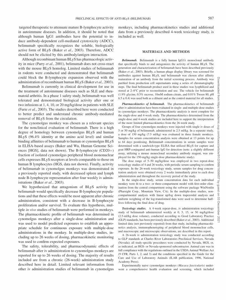
Chronic Administration of Belimumab, a BLyS Antagonist,Decreases Tissue and Peripheral Blood B-LymphocytePopulations in Cynomolgus Monkeys: Pharmacokinetic,
Pharmacodynamic, and Toxicologic Effects
Wendy G. Halpern,*,1,2 Patrick Lappin,†,3 Thomas Zanardi,†,3 Wendy Cai,*,2 Marta Corcoran,*,2
John Zhong,*,2 and Kevin P. Baker*,2
*Human Genome Sciences, Inc., Rockville, Maryland 20850; and †Charles River Laboratories Preclinical Services, Nevada, Sparks, Nevada 89431
Received December 19, 2005; accepted February 23, 2006
The tolerability, pharmacodynamic effects, and pharmacoki-
netics of belimumab (LymphoStat-B) were evaluated in cyno-
molgus monkeys. Belimumab is a fully human IgG1l antibody
directed against B-lymphocyte stimulator (BLyS) protein. BLyS is
a TNF family member that supports B-lymphocyte maturation
and survival and has been implicated in the pathogenesis of auto-
immune diseases and B-lymphocyte malignancies. Belimumab
was developed to antagonize BLyS activity in autoimmune dis-
eases and B-lymphocyte malignancies, where undesirable effects
of B-lymphocyte activity may cause or contribute to disease.
Pharmacodynamic effects of belimumab were monitored by
immunophenotyping of peripheral blood. Pathology end points,
including tissue immunophenotyping, are described after 13
and 26 weeks of treatment and after a 34-week treatment-free
(recovery) period. Belimumab was safe and well tolerated in
repeat-dose toxicology studies at 5–50 mg/kg for up to 26 weeks.
Monkeys exposed to belimumab had significant decreases in pe-
ripheral blood B lymphocytes by 13 weeks of exposure, continuing
into the recovery period, despite total lymphocyte counts similar
to the controls. There were concomitant decreases in spleen and
lymph node B-lymphocyte representation after 13 or 26 weeks
of treatment with belimumab. Microscopically, monkeys treated
with belimumab for 13 or 26 weeks had decreases in the number
and size of lymphoid follicles in the white pulp of the spleen. All
findings were generally reversible within a 34-week recovery pe-
riod. These data confirm the specific pharmacologic activity of
belimumab in reducing B lymphocytes in the cynomolgus monkey.
The favorable safety profile and lack of treatment-related infec-
tions also support continued clinical development of belimumab.
Key Words: agents—pharmaceuticals; immunotoxicology—
autoimmune; safety evaluation; safety evaluation—toxicity; chronic.
Belimumab (LymphoStat-B; Human Genome Sciences,Rockville, MD) is a recombinant fully human IgG1k mono-clonal antibody that targets the B-lymphocyte stimulator(BLyS) protein (Baker et al., 2003). BLyS is a member of thetumor necrosis factor (TNF) family and is also referred to asTNF homolog that activates apoptosis, NF-jB, and JNK;TNF- and ApoL-related leukocyte-expressed ligand 1; orB-cell activating factor belonging to the TNF family (BAFF)(Moore et al., 1999; Mukhopadhyay et al., 1999; Schneideret al., 1999; Shu et al., 1999). BLyS is produced by myeloidcells as a type II transmembrane protein, which is cleavedto form the soluble, biologically active form (Nardelli et al.,2001, and reviewed in Stohl, 2005). Recombinant soluble BLyScan enhance B lymphocyte activation and prolong B lympho-cyte survival (Do et al., 2000; Gross et al., 2001; Thompsonet al., 2000; and reviewed in Mackay and Browning, 2002),both of which are thought to contribute to the phenotype ofautoimmune diseases like systemic lupus erythematosus (SLE).
There are at least three receptors for BLyS: B-cell matura-tion antigen (BCMA), transmembrane activator and calciummodulator and cyclophilin ligand interactor (TACI), and BLySreceptor 3 (BR3), also called BAFF receptor; all three arepredominantly expressed on the surface of B lymphocytes(Gross et al., 2000; Marsters et al., 2000; Thompson et al.,2001; Wu et al., 2000; Yu et al., 2000). Of these, BR3 is con-sidered responsible for many of the known activities of BLyS(Gross et al., 2001; Shulga-Morskaya et al., 2004; Thompsonet al., 2001).
Overproduction of BLyS in transgenic mice is associated withlymphoid hyperplasia and features of autoimmunity (Khareet al., 2000; Mackay et al., 1999). Evidence of increasedcirculating BLyS levels in patients with autoimmune diseaseshas also been reported (Stohl et al., 2003). Importantly, BLySappears to have limited or no effects on B-lymphocyte pre-cursors (pre-B cells in the bone marrow), which express CD20but do not express any of the known BLyS receptors. Thus, BLySantagonism represents a desirable mechanism for a reversible
1 To whom correspondence should be addressed at Human Genome
Sciences, Inc., 14200 Shady Grove Road, Rockville, MD 20850. Fax: (301)
517-8901. E-mail: [email protected] Present address: Five Prime Therapeutics, Inc., San Francisco, CA.3 Present address: Isis Pharmaceuticals, Carlsbad, CA.
� The Author 2006. Published by Oxford University Press on behalf of the Society of Toxicology. All rights reserved.For Permissions, please email: [email protected]
TOXICOLOGICAL SCIENCES 91(2), 586–599 (2006)
doi:10.1093/toxsci/kfj148
Advance Access publication March 3, 2006
Dow
nloaded from https://academ
ic.oup.com/toxsci/article/91/2/586/1656657 by guest on 03 D
ecember 2021
targeted therapeutic to attenuate mature B-lymphocyte activityin autoimmune diseases. In addition, it should be noted thatalthough human IgG1 antibodies have the potential to in-duce antibody-dependent cell-mediated cytotoxicity (ADCC),belimumab specifically recognizes the soluble, biologicallyactive form of BLyS (Baker et al., 2003). Therefore, ADCCshould not be elicited by this antibody/antigen interaction.
Although recombinant human BLyS has pharmacologic activ-ity in mice (Parry et al., 2001), belimumab does not cross-reactwith the mouse BLyS homolog. Limited studies of belimumabin rodents were conducted and demonstrated that belimumabcould block the B-lymphocyte expansion observed with theadministration of recombinant human BLyS (Baker et al., 2003).
Belimumab is currently in clinical development for use inthe treatment of autoimmune diseases such as SLE and rheu-matoid arthritis. In a phase 1 safety study, belimumab was welltolerated and demonstrated biological activity after one ortwo infusions at 1, 4, 10, or 20 mg/kg/dose in patients with SLE(Furie et al., 2003). The studies described here were conductedto better predict and understand chronic antibody-mediatedremoval of BLyS from the circulation.
The cynomolgus monkey was chosen as a relevant speciesfor the nonclinical evaluation of belimumab. There is a highdegree of homology between cynomolgus BLyS and humanBLyS (96.4% identity at the amino acid level) and similarbinding affinities of belimumab to human or cynomolgus BLySin ELISA-based assays (Baker and Wu, Human Genome Sci-ences [HGS], data not shown). The B-lymphocyte (CD20þ)fraction of isolated cynomolgus peripheral blood mononuclearcells expresses BLyS receptors at levels comparable to those onhuman B lymphocytes (HGS, data not shown). Finally, activityof belimumab in cynomolgus monkeys was demonstrated ina previously reported study, with decreased spleen and lymphnode B-lymphocyte representation after four weekly iv admin-istrations (Baker et al., 2003).
We hypothesized that antagonism of BLyS activity bybelimumab would specifically decrease B-lymphocyte popula-tions and that these effects would be most apparent after chronicadministration, consistent with a decrease in B-lymphocyteproliferation and/or survival. To evaluate this hypothesis, mul-tiple in vivo studies of belimumab were performed in monkeys.The pharmacokinetic profile of belimumab was determined incynomolgus monkeys after a single-dose administration andwas used to model predicted exposures to establish an appro-priate schedule for continuous exposure with multiple-doseadministrations in the monkey. In multiple-dose studies, in-cluding up to 26 weeks of dosing, pharmacokinetic monitoringwas used to confirm expected exposures.
The safety, tolerability, and pharmacodynamic effects ofbelimumab after iv administration to cynomolgus monkeys arereported for up to 26 weeks of dosing. The majority of resultsincluded are from a chronic (26-week) administration studydescribed here in detail. Limited relevant information fromother iv administration studies of belimumab in cynomolgus
monkeys, including pharmacokinetics studies and additionaldata from a previously described 4-week toxicology study, isincluded as well.
MATERIALS AND METHODS
Belimumab. Belimumab is a fully human IgG1k monoclonal antibody
that specifically binds to and antagonizes the activity of human BLyS. The
generation and characterization of belimumab have been described previously
(Baker et al., 2003). Briefly, a human phage display library was screened for
antibodies against human BLyS, and belimumab was chosen after affinity
maturation of an antibody from the initial screening process. Antibody was
purified from production cell supernatants using a series of chromatography
steps. The final belimumab product used in these studies was lyophilized and
stored at 2–8�C prior to reconstitution and use. The vehicle for belimumab
(1.9% glycine, 0.5% sucrose, 10mM sodium citrate, and 0.01% Tween 80, pH
6.5 ± 0.4) was used both as a diluent and as the control material for all studies.
Pharmacokinetics of belimumab. The pharmacokinetics of belimumab
after iv administration have been evaluated in single- and multiple-dose studies
in cynomolgus monkeys. The pharmacokinetic analysis is most complete for
the single-dose and 4-week study. The pharmacokinetics determined from the
single-dose and 4-week studies are included here to support the interpretation
of the more limited pharmacokinetics from the 26-week study.
Groups of four cynomolgus monkeys were injected with single iv doses of
5 or 50 mg/kg of belimumab, administered in 2.5 ml/kg. In a separate study,
a dose of 150 mg/kg (7.5 ml/kg) was evaluated in three female monkeys.
Samples for serum concentration analysis were obtained at 13 points for up
to 64 days postdosing. Belimumab concentrations in serum samples were
determined with a sandwich-type ELISA that utilized BLyS for capture and
goat HRP-conjugated anti-human IgG for detection (note: a slightly different
assay, utilizing a mouse monoclonal secondary antibody reagent, was em-
ployed for the 150-mg/kg single-dose pharmacokinetic study).
The dose range of 5–50 mg/kg/dose was employed in two repeat-dose
toxicology studies of 4 and 26 weeks, with periodic serum sampling to confirm
exposure. In the 26-week toxicology study, serum samples for drug concen-
tration analysis were obtained every 2 weeks immediately prior to each dose
administration and throughout the recovery period of the study.
In the single-dose study, serum concentration data for each individual
monkey were fit to a two- or three-compartment model with first-order elim-
ination from the central compartment using the software package WinNonlin
(Pharsight Corp., Mountain View, CA). In the multiple-dose studies, non-
compartmental analysis with linear up/log down trapezoidal analysis and
uniform weighting of the log-transformed data were used to determine half-
lives following the final dose of drug.
Toxicology studies. A 4-week repeat-dose, iv administration toxicology
study of belimumab administered weekly at 0, 5, 15, or 50 mg/kg/dose
(2.5-ml/kg dose volume), conducted according to Good Laboratory Practice
(GLP) standards, has been previously described (Baker et al., 2003). Additional
limited data (not previously reported) from that study, including pharmacoki-
netics analysis, immunophenotyping of peripheral blood mononuclear cells,
and macroscopic and microscopic observations, are described in this report.
A 26-week iv administration toxicology study was conducted according
to GLP standards at Charles River Laboratories Preclinical Services, Nevada
(Nevada); all study-specific procedures were conducted by Nevada, HGS, or,
as indicated, an HGS- or Nevada-sponsored subcontractor. Animal care was in
full compliance with the regulations outlined in the USDA Animal Welfare Act
(9 CFR, Parts 1, 2, and 3) and the conditions specified in the Guide for the
Care and Use of Laboratory Animals (ILAR publication, 1996, National
Academy Press).
Experimentally naive cynomolgus monkeys (Macaca fascicularis) under-
went a comprehensive health evaluation and screening, which included
PRECLINICAL EFFECTS OF ANTI-BLyS (BELIMUMAB) 587
Dow
nloaded from https://academ
ic.oup.com/toxsci/article/91/2/586/1656657 by guest on 03 D
ecember 2021
demonstration of seronegativity and/or PCR-negativity for simian retroviruses.
There were 60 monkeys selected for the 26-week study. Male (n ¼ 30; b.wt.
2.0–4.7 kg) and female (n ¼ 30; b.wt. 1.7–3.5 kg) monkeys were each assigned
to one of four treatment groups by a stratified randomization scheme designed
to achieve similar group mean body weights and with equal numbers of male
and female monkeys in each group.
There were six male and six female monkeys in the control group and eight
male and eight female monkeys in each of the three belimumab treatment
groups. Monkeys were acclimated to nonhuman primate chairs and were not
sedated for dose administration.
Dose levels were 0 mg/kg (Group 1), 5 mg/kg (Group 2), 15 mg/kg (Group
3), or 50 mg/kg (Group 4) for each dose administration. Belimumab, or diluent
control, was iv-administered in a dose volume of 2.5 ml/kg to all monkeys
via a peripheral vein once every 2 weeks for 13 or 26 weeks (7 or 13 times) in
the chronic administration study. The every 2-week schedule was selected
based on available pharmacokinetics data of belimumab in monkeys and was
intended to be as frequent as or more frequent than the clinical dose schedule,
with a target of continuous exposure during the treatment period.
Monkeys were observed, including estimated food consumption, at least
twice daily starting 7 days prior to the first dose administration and continuing
through the day of scheduled necropsy. Body weights were determined prior
to dose administration and once weekly for the duration of the study. Electro-
cardiograms and ophthalmic examinations were performed under light sedation
prior to the first dose administration (as part of the health screening), and during
weeks 13, 26, and 39 in the chronic study. Electrocardiogram recordings were
evaluated by ANILAB (Englishtown, NJ).
Flow cytometry. Blood samples (approximately 1 ml) for the evaluation
of peripheral blood mononuclear cell populations were collected into heparin-
containing tubes from all available monkeys periodically from each study,
including samples from before, during, and after each treatment period. Tissue
leukocytes for the evaluation of spleen and lymph node mononuclear cell
populations were extracted from spleen and mesenteric lymph node at the time
of each necropsy. Samples for flow cytometry were evaluated using a Coulter
Epics XL-MCL instrument (Beckman-Coulter, Miami, FL). For both peripheral
blood and tissue samples, the following markers that have been qualified for
use in cynomolgus monkey specimens were used to distinguish the immuno-
phenotype: CD2 for total lymphocytes, CD20 for total B lymphocytes, CD20
and CD21 for mature B lymphocytes, CD3 for total T lymphocytes, CD3
and CD4 for helper T lymphocytes, CD3 and CD8 for suppressor/cytotoxic
T lymphocytes, and CD14 (CD3�) for monocytes. The PE-labeled anti-CD2
antibody was obtained from Beckman-Coulter. FITC-labeled anti-CD3 and
anti-CD20 antibodies, as well as PE-labeled anti-CD4, anti-CD8, anti-CD14, and
anti-CD21 antibodies, were obtained from BD PharMingen (San Diego, CA).
Clinical pathology. Blood samples for the evaluation of serum chemistry,
hematology, and coagulation parameters were collected periodically from all
available monkeys. Approximate blood volumes were 2 ml for serum chem-
istry, 1 ml for hematology, and 1.8 ml for coagulation. Monkeys were fasted
overnight prior to blood collection for clinical pathology.
For serum chemistry samples, sera were analyzed on a Beckman Synchron
CX7 automated chemistry analyzer (Beckman-Coulter Instruments, Palo Alto,
CA). The following parameters were analyzed: sodium, potassium, chloride,
carbon dioxide, total bilirubin, alkaline phosphatase, lactate dehydrogenase,
aspartate aminotransferase, alanine aminotransferase, gamma-glutamyl trans-
ferase, calcium, phosphorus, urea nitrogen, creatinine, total protein, albumin,
globulin, albumin/globulin ratio, glucose, cholesterol, and triglycerides.
Standard hematology evaluation of whole blood collected into tubes con-
taining EDTA anticoagulant included: red blood cell counts, white blood cells
(total and differential), hemoglobin concentration, hematocrit, reticulocyte
counts, mean cell hemoglobin, mean corpuscular volume, mean corpuscular hemo-
globin concentration, platelet counts, and blood cell morphology. Samples were
analyzed on an Abbott Cell-Dyn 3500 multiparameter automated hematology
analyzer (Abbott Labs, Pomezia, Italy). In addition, total lymphocyte counts
were periodically determined for use in combination with flow cytometry data.
For assessment of coagulation parameters, plasma from whole blood col-
lected into tubes containing sodium citrate anticoagulant was analyzed for
activated partial thromboplastin time, prothrombin time, and fibrinogen on a
Sigma AMAX CS 190 (Sigma Diagnostics, St. Louis, MO).
Urinalysis was performed on samples obtained from the bladder at necropsy.
The following parameters were determined: color/character (observation);
specific gravity (refractometer); pH, leukocyte esterase, nitrite, urobilinogen,
protein, glucose, ketones, bilirubin, and occult blood (dipstick); and casts,
leukocytes, erythrocytes, epithelial cells, mucus, crystals, bacteria, yeast, and
amorphous sediment (microscopic evaluation of sediment).
Immunoglobulin subclasses. Serum samples were obtained from each
available monkey at weeks 1 (predose), 6, 13, 22, 26, 34, 39, 52, and 60 and
were evaluated for cynomolgus monkey IgG, IgM, IgA, and IgE. Concentration
determination for immunoglobulin subclasses was performed by AniLytics
(Gaithersburg, MD) using an ELISA-based method. Assessment of immuno-
globulin subclasses was performed on all available samples collected as of
the time of each necropsy (weeks 13, 26, and 60). Results were reported for
each sample in units of milligrams per deciliter for IgM, IgA, and IgG or
as nanograms per milliliter for IgE.
Anti-belimumab antibodies. The presence of belimumab-specific anti-
bodies in serum was determined prior to first dose administration and peri-
odically during the chronic study. This analysis utilized a sandwich-type
ELISA, with detection using human-specific anti-IgA, anti-IgM, anti–IgG-Fc,
and anti–kappa light-chain antibodies. This evaluation was performed after
13 weeks of treatment, after 26 weeks of treatment, and at weeks 52 and 60
following a 26- or 34-week treatment-free period. The presence of monkey
antibodies to belimumab was defined as an increase in A450 of at least twofold
in the sera obtained posttreatment compared to the A450 obtained from the
predose sera from the same monkey.
Anatomic pathology. Necropsies were scheduled after 13 weeks of treat-
ment (n ¼ 22), 26 weeks of treatment (n ¼ 22), or 26 weeks of treatment
followed by a 34-week treatment-free (recovery) period (n ¼ 16). During the
necropsies conducted at weeks 13 and 26, three male and three female monkeys
from each group treated with belimumab were evaluated at each time point
and two male and two female monkeys from each group were evaluated after
the recovery period. At each of the three scheduled necropsies, two male and
two female monkeys from the control group were evaluated.
A complete gross necropsy was conducted on each monkey. Organ weights
were determined for a standard set of tissues and analyzed for treatment effects
on the organ weight and as a ratio of the organ weight to body weight and to
brain weight. A standard list of tissues was collected, preserved, and examined
histologically by a pathologist (P.L.); selected tissues were peer reviewed by a
second pathologist (W.G.H.).
Statistics. In general, group means and standard deviation values were
calculated for numerical data, including body weights, circulating lymphocyte
and monocyte subpopulations, clinical pathology parameters, organ weights,
and lymphoid tissue lymphocyte subset and monocyte populations. Further
statistical analyses were performed with SAS System version 8.1. Significant
intergroup differences were evaluated by the use of an ANOVA. If the
parametric ANOVA was significant at p � 0.05, Dunnett’s test was used to
identify statistically significant differences between the control group and
each belimumab-treated group at the p � 0.05 level of significance.
For the immunoglobulin subclass treatment period data (weeks 1–26 of
the chronic toxicology study), a repeated measures model was used to ana-
lyze the Ig level across time points. In addition to treatment and time, the
model was adjusted for gender and predose Ig level. For IgG, for example, the
model is
IgG¼Predose IgGþGenderþTreatmentþWeekþTreatment3Week:
This approach also accounted for the correlation between repeated measures
within each subject by employing a compound symmetric covariance structure.
The MIXED procedure in SAS version 8 was used to estimate the model
588 HALPERN ET AL.
Dow
nloaded from https://academ
ic.oup.com/toxsci/article/91/2/586/1656657 by guest on 03 D
ecember 2021
parameters. If overall treatment differences were identified, post hoc tests
were performed comparing each belimumab treatment group to control.
For the recovery period data, there were only four monkeys in each
treatment group at each time point, which is insufficient to estimate overall
effects of time and treatment while adjusting for the correlation of observations
within each subject. Therefore, a one-way ANOVA at each time point was
used to compare the effect of treatment on total Ig as well as each of the Ig
subclasses. If the F-test indicated some evidence of treatment differences,
post hoc tests were performed, comparing each belimumab treatment group
to control.
RESULTS
Serum Belimumab Concentrations
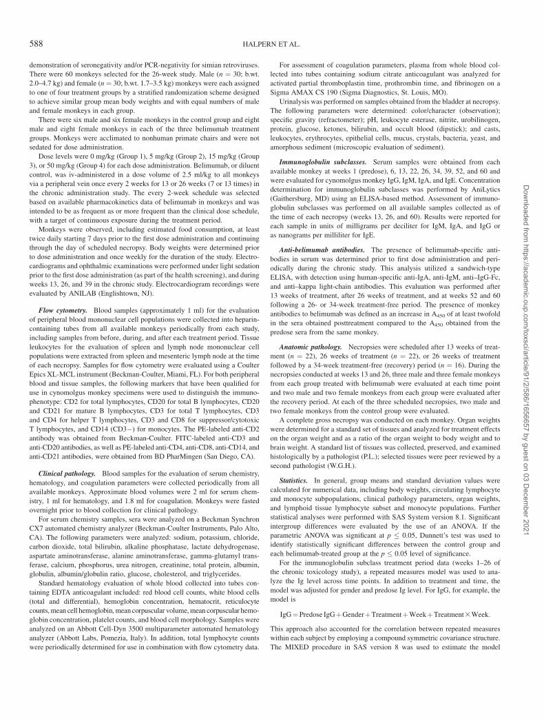
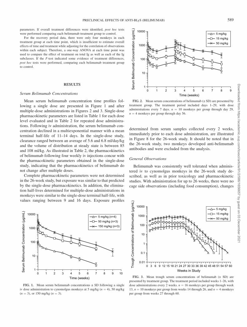
Mean serum belimumab concentration time profiles fol-lowing a single dose are presented in Figure 1 and aftermultiple-dose administrations in Figures 2 and 3. Single-dosepharmacokinetic parameters are listed in Table 1 for each doselevel evaluated and in Table 2 for repeated dose administra-tions. Following iv administration, the serum belimumab con-centration declined in a multiexponential manner with a meanterminal half-life of 11–14 days. In the single-dose study,clearance ranged between an average of 5.6 and 6.8 ml/day/kgand the volume of distribution at steady state is between 85and 108 ml/kg. As illustrated in Table 2, the pharmacokineticsof belimumab following four weekly iv injections concur withthe pharmacokinetic parameters obtained in the single-dosestudy, indicating that the pharmacokinetics of belimumab donot change after multiple doses.
Complete pharmacokinetic parameters were not determinedin the 26-week study, but exposure was similar to that predictedby the single-dose pharmacokinetics. In addition, the elimina-tion half-lives determined for multiple-dose administrations inmonkeys were similar to the single-dose terminal half-life, withvalues ranging between 9 and 16 days. Exposure profiles
determined from serum samples collected every 2 weeks,immediately prior to each dose administration, are illustratedin Figure 8 for the 26-week study. It should be noted that inthe 26-week study, two monkeys developed anti-belimumabantibodies and were excluded from the analysis.
General Observations
Belimumab was consistently well tolerated when adminis-tered iv to cynomolgus monkeys in the 26-week study de-scribed, as well as in prior toxicology and pharmacokineticstudies. With administration for up to 26 weeks, there were nocage side observations (including food consumption), changes
0 1 2 3 4 5 6 7 8 9 101
10
100
1000
100005 mg/kg (n=4)50 mg/kg (n=3)150 mg/kg (n=3)
Time (weeks)
Seru
m B
elim
umab
Con
cent
ratio
n (µ
g/m
L)
FIG. 1. Mean serum belimumab concentrations ± SD following a single
iv dose administration to cynomolgus monkeys at 5 mg/kg (n ¼ 4), 50 mg/kg
(n ¼ 3), or 150 mg/kg (n ¼ 3).
0 1 2 3 4 5 6 7 8 91
10
100
1000
100005 mg/kg15 mg/kg50 mg/kg
Time (weeks)
Seru
m B
elim
umab
Con
cent
ratio
n (µ
g/m
L)
FIG. 2. Mean serum concentrations of belimumab (± SD) are presented by
treatment group. The treatment period included days 1–29, with dose
administrations every 7 days. n ¼ 10 monkeys per group through day 29,
n ¼ 4 monkeys per group through day 56.
Weeks in Study
1
0 3 6 9 12 15 18 21 24 27 30 33 36 39 42 45 48 51 54 57 60
0.1
0.01
10
100
1000
100005 mg/kg15 mg/kg50 mg/kg
Trou
gh C
once
ntra
tions
of B
elim
umab
(µg/
mL)
FIG. 3. Mean trough serum concentrations of belimumab (± SD) are
presented by treatment group. The treatment period included weeks 1–26, with
dose administrations every 2 weeks. n ¼ 16 monkeys per group through week
13, n ¼ 10 monkeys per group from weeks 14 through 26, and n ¼ 4 monkeys
per group from weeks 27 through 60.
PRECLINICAL EFFECTS OF ANTI-BLyS (BELIMUMAB) 589
Dow
nloaded from https://academ
ic.oup.com/toxsci/article/91/2/586/1656657 by guest on 03 D
ecember 2021
in body weight, electrocardiographic findings, or ophthalmicfindings that were attributed to belimumab administration.Most cage side observations were considered typical of back-ground observations in monkeys undergoing study procedures,were distributed similarly across all treatment groups includingcontrols, and occurred both in multiple studies and, whereapplicable, in different rooms for the same study. During thechronic administration study, sneezing and red nasal dischargewere noted in 12 monkeys, including monkeys from both con-
trol and belimumab-treatment groups, between days 123 and133. Four additional (different) monkeys had similar symptomsfor 2–9 days between days 231 and 265, and all the affectedmonkeys had been housed in the same room. A bacterial in-fection (Branhamella catarrhalis) was considered the likelycause of this finding, and all affected monkeys responded totreatment with antibiotic (penicillin). Treatment with anti-biotics was limited to monkeys with overt clinical signs. Due tothe incidence in both control and treated monkeys, restrictionto one room, and response to antibiotic treatment, this findingwas not directly attributed to potential immunomodulatoryproperties of belimumab. However, a distinction between noeffect or mild effect of belimumab on susceptibility to thisinfection was not possible with the small numbers of monkeysaffected during each of the two ‘‘outbreaks’’ in this room.
Peripheral Blood Mononuclear Cells
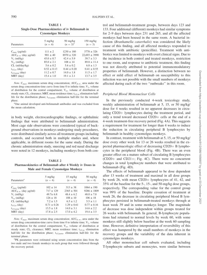
In the previously conducted 4-week toxicology study,weekly administration of belimumab at 5, 15, or 50 mg/kg/dose for 4 weeks resulted in no apparent decreases in circu-lating CD20þ lymphocytes during the treatment period, andonly a trend toward decreased CD20þ cells at the end of a4-week treatment-free recovery period (Fig. 4A). This suggestsa requirement for treatment for longer than 4 weeks to inducethe reduction in circulating peripheral B lymphocytes bybelimumab in healthy cynomolgus monkeys.
In contrast, treatment with belimumab at 5, 15, or 50 mg/kg/dose every other week for 13 or 26 weeks resulted in the ex-pected pharmacologic effect of decreasing CD20þ B lympho-cytes in the peripheral blood (Fig. 4B). There was an evengreater effect on a mature subset of circulating B lymphocytes(CD20þ and CD21þ; Fig. 4C). There were no concurrentchanges in total lymphocyte numbers that were attributed tobelimumab (Fig. 4D).
The effects of belimumab appeared to be dose dependentafter 13 weeks of treatment and maximal in all dose groupsby week 26, with mean CD20þ lymphocytes of 41, 42, and35% of the baseline for the 5-, 15-, and 50-mg/kg dose groups,respectively. The corresponding value for the control groupwas 99% of the baseline. Despite cessation of treatment atweek 26, the decrease in circulating peripheral blood B lym-phocytes persisted in belimumab-treated monkeys through atleast week 39 and in some monkeys longer. The magnitudeof decrease was dose independent within groups treated for26 weeks with belimumab. In general, B-lymphocyte popula-tions had returned to normal levels by week 60, with somemonkeys still slightly below baseline at the week 60 samplingtime. However, definitive interpretation of reversibility of thiseffect was hampered by the small numbers of monkeys in therecovery groups and the variability of the data inherent incynomolgus monkeys.
All other mononuclear cell subsets evaluated, includingT-lymphocyte subsets and monocytes, were similar between
TABLE 1
Single-Dose Pharmacokinetics of iv Belimumab in
Cynomolgus Monkeys
Parameters
5 mg/kg
(n ¼ 4)
50 mg/kg
(n ¼ 3)a150 mg/kg
(n ¼ 3)
Cmax (lg/ml) 111 ± 2 1230 ± 180 3778 ± 326
AUC0–N (day lg/ml) 901 ± 40 8970 ± 320 22,693 ± 3990
V1 (ml/kg) 45.0 ± 0.7 42.4 ± 5.9 39.7 ± 3.3
Vss (ml/kg) 85.0 ± 2.1 108 ± 14 89.8 ± 11.4
CL (ml/day/kg) 5.6 ± 0.2 5.6 ± 0.2 6.8 ± 1.3
t1/2,a (day) 0.52 ± 0.13 0.44 ± 0.20 0.41 ± 0.55
t1/2,term (day) 11.2 ± 0.7 14.0 ± 1.8 11.5 ± 1.7
MRT (day) 15.4 ± 1.0 19.1 ± 2.1 13.7 ± 3.5
Note. Cmax, maximum serum drug concentration; AUC0–N, area under the
serum drug concentration-time curve from time 0 to infinite time; V1, volume
of distribution for the central compartment; Vss, volume of distribution at
steady state; CL, clearance; MRT, mean residence time; t1/2,a, elimination half-
life for the distribution phase; t1/2,term, elimination half-life for the terminal
phase.aOne animal developed anti-belimumab antibodies and was excluded from
the mean calculation.
TABLE 2
Pharmacokinetics of Belimumab after 4 Weekly iv Doses in
Male and Female Cynomolgus Monkeys
Parametersa5 mg/kg
(n ¼ 4)
15 mg/kg
(n ¼ 4)
50 mg/kg
(n ¼ 4)
Cmax (lg/ml) 102 ± 14 313 ± 38 1064 ± 190
AUC0–N (day lg/ml) 717 ± 129 2365 ± 501 9286 ± 1809
V1 (ml/kg) 49.7± 6.8 48.4 ± 6.2 48.0 ± 7.8
Vss (ml/kg) 126 ± 18 108 ± 10 106 ± 18
CL (ml/day/kg) 7.2 ± 1.5 6.5 ± 1.2 5.5 ± 1.1
t1/2,a (day) 0.71 ± 0.26 1.29 ± 0.61 0.77 ± 0.16
t1/2,term (day) 13.5 ± 1.8 14.0 ± 3.1 14.6 ± 2.2
MRT (day) 17.8 ± 2.5 17.0 ± 4.2 19.4 ± 3.5
Note. Cmax, maximum serum drug concentration; AUC0–N, area under the
serum drug concentration-time curve from time 0 to infinite time; V1, volume
of distribution for the central compartment; Vss, volume of distribution at
steady state; CL, clearance; MRT, mean residence time; t1/2,a, elimination
half-life for the distribution phase; t1/2,term, elimination half-life for the
terminal phase.aPK parameters were estimated using serum concentration data from the
two male and two female monkeys in each group that were followed through
the recovery period.
590 HALPERN ET AL.
Dow
nloaded from https://academ
ic.oup.com/toxsci/article/91/2/586/1656657 by guest on 03 D
ecember 2021
belimumab-treated and control monkeys at each time point andcomparable to established baseline values for each parameter(data not shown).
Tissue Mononuclear Cells
The early effects of 4 weeks of treatment with belimumabon lymphoid tissue B lymphocytes in cynomolgus monkeyshave been previously reported (Baker et al., 2003). In thatstudy, statistically significant decreases in the relative percent-age of both CD20þ and CD20/CD21 dual-positive mono-nuclear cells were observed in the mesenteric lymph node and
spleen from all three belimumab-treated groups (as comparedto vehicle controls), but not until the end of the 4-week re-covery period. There were no statistically significant differ-ences between belimumab-treated groups for lymphoid tissuescollected at the end of the 4-week treatment period.
In the chronic study reported here, there were decreases inthe relative percentage of B lymphocytes in the spleen andmesenteric lymph node of monkeys in all belimumab-treatmentgroups compared to vehicle controls when evaluated at weeks13 and 26. There was no apparent dose dependency of effecton tissue B lymphocytes in monkeys treated with belimumabat 5, 15, or 50 mg/kg during the treatment period. The mean
Week 1312/16
Week 268/10
Week 334
Week 394
Week 454
Week 524
Week 604
0
25
50
75
100
125
150
175
200
225
2500 mg/kg/dose5 mg/kg/dose15 mg/kg/dose50 mg/kg/dose
0 mg/kg/dose5 mg/kg/dose15 mg/kg/dose50 mg/kg/dose
0 mg/kg/dose5 mg/kg/dose15 mg/kg/dose50 mg/kg/dose
0 mg/kg/dose5 mg/kg/dose15 mg/kg/dose50 mg/kg/dose
% B
asel
ine
CD
20+
Cel
ls (±
SEM
)
Week 1312/16
Week 268/10
Week 334
Week 394
Week 454
Week 524
Week 604
0255075
100125150175200225250275300325
Week 1312/16
Week 268/10
Week 334
Week 394
Week 454
Week 524
Week 604
0
25
50
75
100
125
150
175
200
225
250
% B
asel
ine
Tota
l Lym
phoc
ytes
(± S
EM)
A
C D
B
99%
86%30%
31%27%
41%42%
35%** *
**** * *
*
*** * * * *
*
* *
WeeksMonkeys Per Group (control/treated)
WeeksMonkeys Per Group (control/treated)
WeeksMonkeys Per Group (control/treated)
WeeksMonkeys Per Group
Week 210
Week 410
Week 84
0
25
50
75
100
125
150
175
% B
asel
ine
CD
20+
Cel
ls (±
SEM
)%
Bas
elin
e C
D20
+ /C
D21
+ C
ells
(± S
EM)
FIG. 4. Peripheral blood lymphocyte representation as a percentage of baseline levels according to belimumab dose level. Bars indicate mean percentage of
baseline ± SEM; the N for each time point is labeled for vehicle and belimumab-treated groups. For each study, baseline was the average of three measurements
taken prior to initiation of dosing. (A) Peripheral blood CD20þ cells as a percentage of baseline levels (± SEM) after 4 weeks of treatment (n¼ 10 per dose level),
and after 4 weeks of treatment followed by a 4-week treatment-free recovery period (n¼ 4 per dose level). (B–D) Belimumab, or vehicle control, was administered
iv every other week for 13 or 26 weeks, and peripheral blood lymphocytes were assessed by flow cytometric methods periodically over the treatment period and
a subsequent 34-week treatment-free recovery period. Asterisks indicate significant decreases in B lymphocytes as compared to baseline (p < 0.05). The actual
percentage of baseline is indicated for each treatment group at week 26. Assessment included CD20þ B lymphocytes (B), CD20þ/CD21þ mature B lymphocytes
(C), and total lymphocytes (D).
PRECLINICAL EFFECTS OF ANTI-BLyS (BELIMUMAB) 591
Dow
nloaded from https://academ
ic.oup.com/toxsci/article/91/2/586/1656657 by guest on 03 D
ecember 2021
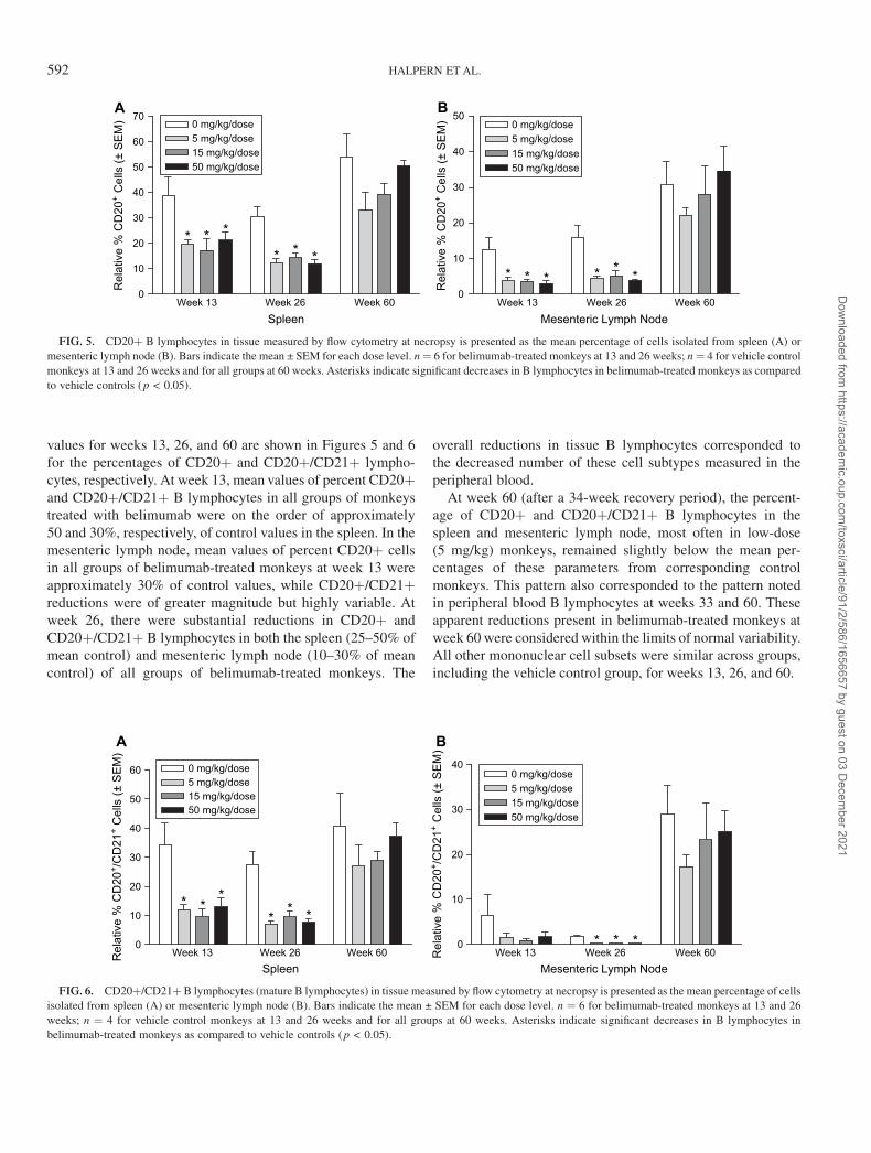
values for weeks 13, 26, and 60 are shown in Figures 5 and 6for the percentages of CD20þ and CD20þ/CD21þ lympho-cytes, respectively. At week 13, mean values of percent CD20þand CD20þ/CD21þ B lymphocytes in all groups of monkeystreated with belimumab were on the order of approximately50 and 30%, respectively, of control values in the spleen. In themesenteric lymph node, mean values of percent CD20þ cellsin all groups of belimumab-treated monkeys at week 13 wereapproximately 30% of control values, while CD20þ/CD21þreductions were of greater magnitude but highly variable. Atweek 26, there were substantial reductions in CD20þ andCD20þ/CD21þ B lymphocytes in both the spleen (25–50% ofmean control) and mesenteric lymph node (10–30% of meancontrol) of all groups of belimumab-treated monkeys. The
overall reductions in tissue B lymphocytes corresponded tothe decreased number of these cell subtypes measured in theperipheral blood.
At week 60 (after a 34-week recovery period), the percent-age of CD20þ and CD20þ/CD21þ B lymphocytes in thespleen and mesenteric lymph node, most often in low-dose(5 mg/kg) monkeys, remained slightly below the mean per-centages of these parameters from corresponding controlmonkeys. This pattern also corresponded to the pattern notedin peripheral blood B lymphocytes at weeks 33 and 60. Theseapparent reductions present in belimumab-treated monkeys atweek 60 were considered within the limits of normal variability.All other mononuclear cell subsets were similar across groups,including the vehicle control group, for weeks 13, 26, and 60.
A B
0
10
20
30
40
50
60 0 mg/kg/dose5 mg/kg/dose15 mg/kg/dose50 mg/kg/dose
Spleen
0
10
20
30
400 mg/kg/dose5 mg/kg/dose15 mg/kg/dose50 mg/kg/dose
Mesenteric Lymph Node
Rel
ativ
e %
CD
20+ /
CD
21+
Cel
ls (±
SEM
)
Rel
ativ
e %
CD
20+ /
CD
21+
Cel
ls (±
SEM
)
* * *
* **
* * *
Week 13 Week 26 Week 60 Week 13 Week 26 Week 60
FIG. 6. CD20þ/CD21þ B lymphocytes (mature B lymphocytes) in tissue measured by flow cytometry at necropsy is presented as the mean percentage of cells
isolated from spleen (A) or mesenteric lymph node (B). Bars indicate the mean ± SEM for each dose level. n ¼ 6 for belimumab-treated monkeys at 13 and 26
weeks; n ¼ 4 for vehicle control monkeys at 13 and 26 weeks and for all groups at 60 weeks. Asterisks indicate significant decreases in B lymphocytes in
belimumab-treated monkeys as compared to vehicle controls (p < 0.05).
Week 13 Week 26 Week 60 Week 13 Week 26 Week 600
10
20
30
40
50
60
700 mg/kg/dose5 mg/kg/dose15 mg/kg/dose50 mg/kg/dose
Spleen
Rel
ativ
e %
CD
20+
Cel
ls (±
SEM
)
0
10
20
30
40
500 mg/kg/dose5 mg/kg/dose15 mg/kg/dose50 mg/kg/dose
Mesenteric Lymph Node
Rel
ativ
e %
CD
20+
Cel
ls (±
SEM
)
* * *
* * ** * * * * *
A B
FIG. 5. CD20þ B lymphocytes in tissue measured by flow cytometry at necropsy is presented as the mean percentage of cells isolated from spleen (A) or
mesenteric lymph node (B). Bars indicate the mean ± SEM for each dose level. n¼ 6 for belimumab-treated monkeys at 13 and 26 weeks; n¼ 4 for vehicle control
monkeys at 13 and 26 weeks and for all groups at 60 weeks. Asterisks indicate significant decreases in B lymphocytes in belimumab-treated monkeys as compared
to vehicle controls (p < 0.05).
592 HALPERN ET AL.
Dow
nloaded from https://academ
ic.oup.com/toxsci/article/91/2/586/1656657 by guest on 03 D
ecember 2021
Gross Organ Weights
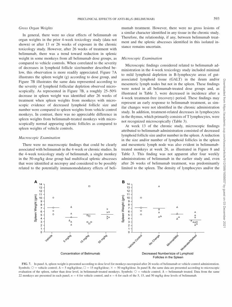
In general, there were no clear effects of belimumab onorgan weights in the prior 4-week toxicology study (data notshown) or after 13 or 26 weeks of exposure in the chronictoxicology study. However, after 26 weeks of treatment withbelimumab, there was a trend toward reduction in splenicweight in some monkeys from all belimumab dose groups, ascompared to vehicle controls. When correlated to the severityof decreases in lymphoid follicle size/number described be-low, this observation is more readily appreciated. Figure 7Aillustrates the spleen weight (g) according to dose group, andFigure 7B illustrates the same data represented according tothe severity of lymphoid follicular depletion observed micro-scopically. As represented in Figure 7B, a roughly 25–50%decrease in spleen weight was identified after 26 weeks oftreatment when spleen weights from monkeys with micro-scopic evidence of decreased lymphoid follicle size andnumber were compared to spleen weights from vehicle controlmonkeys. In contrast, there was no appreciable difference inspleen weights from belimumab-treated monkeys with micro-scopically normal appearing splenic follicles as compared tospleen weights of vehicle controls.
Macroscopic Examination
There were no macroscopic findings that could be clearlyassociated with belimumab in the 4-week or chronic studies. Inthe 4-week toxicology study of belimumab, a single monkeyin the 50-mg/kg dose group had multifocal splenic abscessesthat were identified at necropsy and considered to be possiblyrelated to the potentially immunomodulatory effects of beli-
mumab treatment. However, there were no gross lesions ofa similar character identified in any tissue in the chronic study.Therefore, the relationship, if any, between belimumab treat-ment and the splenic abscesses identified in this isolated in-stance remains uncertain.
Microscopic Examination
Microscopic findings considered related to belimumab ad-ministration in the 4-week toxicology study included minimalto mild lymphoid depletion in B-lymphocyte areas of gut-associated lymphoid tissue (GALT) in the ileum and/ormesenteric lymph nodes but not in the spleen. These findingswere noted in all belimumab-treated dose groups and, asillustrated in Table 3, were decreased in incidence after a4-week treatment-free (recovery) period. These findings mayrepresent an early response to belimumab treatment, as sim-ilar changes were not identified in the chronic administrationstudy. In addition, treatment-related decreases in lymphocytesin the thymus, which primarily consists of T lymphocytes, werenot recognized microscopically (Table 3).
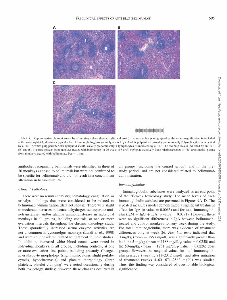
At week 13 of the chronic study, microscopic findingsattributed to belimumab administration consisted of decreasedlymphoid follicle size and/or number in the spleen. A reductionin the size and/or number of lymphoid follicles in the spleenand mesenteric lymph node was also evident in belimumab-treated monkeys at week 26, as illustrated in Figure 8 andTable 3. This finding was not apparent after four weeklyadministrations of belimumab in the earlier study and, evenafter 26 weeks of belimumab treatment, was predominantlylimited to the spleen. The density of lymphocytes and/or the
Vehicle
Con
trol
No Cha
nge
Minimal/
Mild
Modera
te/Mark
ed
A B
Concentration of Belimumab Decreased Number/size of Lymphoid Follicles in the Spleen
0123456789
1011121314
Sple
en W
eigh
t (gm
)
Vehicle
5 mg/k
g
15 m
g/kg
50 m
g/kg
0123456789
1011121314
Sple
en W
eigh
t (gm
)
FIG. 7. In panel A, spleen weight is presented according to dose level for monkeys necropsied after 26 weeks of belimumab or vehicle control administration.
Symbols: s ¼ vehicle control; D ¼ 5 mg/kg/dose; h ¼ 15 mg/kg/dose; 3 ¼ 50 mg/kg/dose. In panel B, the same data are presented according to microscopic
evaluation of the spleen, rather than dose level, in belimumab-treated monkeys. Symbols: s ¼ vehicle control; D ¼ belimumab treated. Data from the same
22 monkeys are presented in each panel; n ¼ 4 for vehicle control, and n ¼ 6 for each of the 5, 15, and 50 mg/kg dose levels of belimumab.
PRECLINICAL EFFECTS OF ANTI-BLyS (BELIMUMAB) 593
Dow
nloaded from https://academ
ic.oup.com/toxsci/article/91/2/586/1656657 by guest on 03 D
ecember 2021
follicular size/number in other lymphoid organs examined his-tologically (thymus, mesenteric and mandibular lymph nodes,GALT) morphologically normal in most belimumab-treatedmonkeys, despite the decreased B-lymphocyte representationwas determined by flow cytometry for mesenteric lymph nodeat week 26. The histomorphology of the spleen correlated withdecreases in B-lymphocyte representation in spleen as de-termined by flow cytometry after 13 or 26 weeks of treatmentwith belimumab and, as previously mentioned, was correlatedwith reduced spleen weights after 26 weeks of belimumabtreatment (Fig. 7B). Of monkeys treated with belimumab, theincidence of the splenic B-lymphocyte reduction was greatlyreduced in monkeys necropsied in week 60, after a 34-weektreatment-free recovery period; only one mid-dose (15 mg/kg/dose) monkey had a moderate decrease in lymphoid folliclesize/number in the spleen after the 34-week treatment-freeperiod. This isolated finding in this monkey may representnormal lymphoid tissue cycling, since other lymphoid tissuesappeared normal in this monkey, and the percent CD20þ andCD20þ/CD21þ lymphocytes had returned to baseline levels inperipheral blood and were similar to those of control monkeysin the spleen and mesenteric lymph node by flow cytometry(individual data not shown).
Serum Antibodies Specific for Belimumab
With chronic administration, antibodies specific for belimu-mab were detected in two of 60 monkeys during the 26-weektreatment period. Positive monkeys included a male from the5-mg/kg dose group and a female from the 50-mg/kg dosegroup. The appearance of anti-belimumab antibodies in thesetwo monkeys coincided with decreased belimumab detectedin serum, and a variable response to treatment. The low-dose(5 mg/kg) monkey’s B-lymphocyte counts were consideredessentially unaffected by belimumab, while the high-dose(50 mg/kg) monkey had a decrease in B lymphocytes similarto other high-dose monkeys treated with belimumab. All 16monkeys evaluated at the end of the recovery period in thisstudy were negative for belimumab-specific antibodies, includ-ing the two monkeys that were previously positive.
In addition, one monkey in a single-dose pharmacokineticstudy developed antibodies to belimumab after a single ivinjection at 50 mg/kg and had an altered pharmacokineticprofile. Therefore, three out of the 59 monkeys (5.1%) thatwere exposed to belimumab in these two studies developeda measurable antibody response that was specific for belimu-mab. None of the 12 control monkeys in these two studiesdeveloped antibodies to belimumab. In the 4-week study,
TABLE 3
Incidence of Minimal to Marked Decreases in Lymphoid Tissue Components in Cynomolgus Monkeys
According to Tissue, Dose and Schedule of Belimumab Treatment
Tissue Dose level
Treatment schedule
4-week toxicology studya 13/26-week toxicology study
Q7d 3 4
Q7d 3 4
þ 4 weeks
Q14d 3 7
(week 13)
Q14d 3 13
(week 26)
Q14d 3 13 þ 34 weeks
(week 60)
Spleen 0 mg/kg 0 of 6 0 of 4 0 of 4 0 of 4 0 of 4
5 mg/kg 0 of 6 0 of 4 2 of 6 6 of 6 0 of 4
15 mg/kg 0 of 6 0 of 4 3 of 6 4 of 6 1 of 4
50 mg/kg 0 of 6 0 of 4 1 of 6 3 of 6 0 of 4
Total 0 of 24 0 of 16 6 of 22 13 of 22 1 of 16
Mesenteric lymph node 0 mg/kg 0 of 6 0 of 4 0 of 4 0 of 4 0 of 4
5 mg/kg 1 of 6 1 of 4 0 of 6 1 of 6 0 of 4
15 mg/kg 2 of 6 0 of 4 0 of 6 2 of 6 0 of 4
50 mg/kg 5 of 6 1 of 4 0 of 6 2 of 6 0 of 4
Total 8 of 24 2 of 16 0 of 22 5 of 22 0 of 16
Ileum (GALT) 0 mg/kg 0 of 6 0 of 4 0 of 4 0 of 4 0 of 4
5 mg/kg 1 of 6 1 of 4 0 of 6 0 of 6 0 of 4
15 mg/kg 3 of 6 0 of 4 0 of 6 0 of 6 0 of 4
50 mg/kg 3 of 6 2 of 4 0 of 6 0 of 6 0 of 4
Total 7 of 24 3 of 16 0 of 22 0 of 22 0 of 16
Thymus 0 mg/kg 4 of 6 3 of 4 0 of 4 2 of 4 0 of 4
5 mg/kg 3 of 6 0 of 4 2 of 6 0 of 6 2 of 4
15 mg/kg 3 of 6 1 of 4 2 of 6 0 of 6 1 of 4
50 mg/kg 5 of 6 0 of 4 1 of 6 1 of 6 1 of 4
Total 15 of 24 4 of 16 5 of 22 3 of 22 5 of 16
aStudy design previously described (Baker et al., 2003).
594 HALPERN ET AL.
Dow
nloaded from https://academ
ic.oup.com/toxsci/article/91/2/586/1656657 by guest on 03 D
ecember 2021
antibodies recognizing belimumab were identified in three of30 monkeys exposed to belimumab but were not confirmed tobe specific for belimumab and did not result in a concomitantalteration in belimumab PK.
Clinical Pathology
There were no serum chemistry, hematology, coagulation, orurinalysis findings that were considered to be related tobelimumab administration (data not shown). There were slightto moderate increases in lactate dehydrogenase, aspartate ami-notransferase, and/or alanine aminotransferase in individualmonkeys in all groups, including controls, at one or moreevaluation intervals throughout the chronic toxicology study.These sporadically increased serum enzyme activities arenot uncommon in cynomolgus monkeys (Landi et al., 1990)and were not considered related to treatment in these studies.In addition, increased white blood counts were noted inindividual monkeys in all groups, including controls, at oneor more evaluation time points, including prestudy. Changesin erythrocyte morphology (slight anisocytosis, slight poikilo-cytosis, hypochromasia) and platelet morphology (largeplatelets, platelet clumping) were noted occasionally duringboth toxicology studies; however, these changes occurred in
all groups (including the control group), and in the pre-study period, and are not considered related to belimumabadministration.
Immunoglobulins
Immunoglobulin subclasses were analyzed as an end pointof the 26-week toxicology study. The mean levels of eachimmunoglobulin subclass are presented in Figures 9A–D. Therepeated measures model demonstrated a significant treatmenteffect for IgA (p value ¼ 0.0065) and for total immunoglob-ulin (IgM þ IgG þ IgA; p value ¼ 0.0391). However, therewere no significant differences in IgA between belimumab-treated and control monkeys for any week during the study.For total immunoglobulin, there was evidence of treatmentdifferences only at week 26. Post hoc tests indicated that0 mg/kg (mean ¼ 1553 mg/dl) was significantly greater thanboth the 5-mg/kg (mean ¼ 1188 mg/dl, p value ¼ 0.0250) andthe 50-mg/kg (mean ¼ 1231 mg/dl, p value ¼ 0.0226) dosegroups. However, the range of values for total immunoglob-ulin prestudy (week 1, 811–2312 mg/dl) and after initiationof treatment (weeks 4–60, 671–2562 mg/dl) was similar.Thus, this finding was considered of questionable biologicalsignificance.
FIG. 8. Representative photomicrographs of monkey spleen (hematoxylin and eosin); 1-mm size bar photographed at the same magnification is included
at the lower right. (A) illustrates typical spleen histomorphology in cynomolgus monkeys. A white pulp follicle, usually predominantly B lymphocytes, is indicated
by a ‘‘B.’’ A white pulp periarteriolar lymphoid sheath, usually predominantly T lymphocytes, is indicated by a ‘‘T.’’ The red pulp area is indicated by an ‘‘R.’’
(B) and (C) illustrate spleens from monkeys treated with belimumab for 26 weeks at 5 or 50 mg/kg, respectively. Note relative absence of ‘‘B’’ areas in the spleens
from monkeys treated with belimumab. Bar ¼ 1 mm.
PRECLINICAL EFFECTS OF ANTI-BLyS (BELIMUMAB) 595
Dow
nloaded from https://academ
ic.oup.com/toxsci/article/91/2/586/1656657 by guest on 03 D
ecember 2021
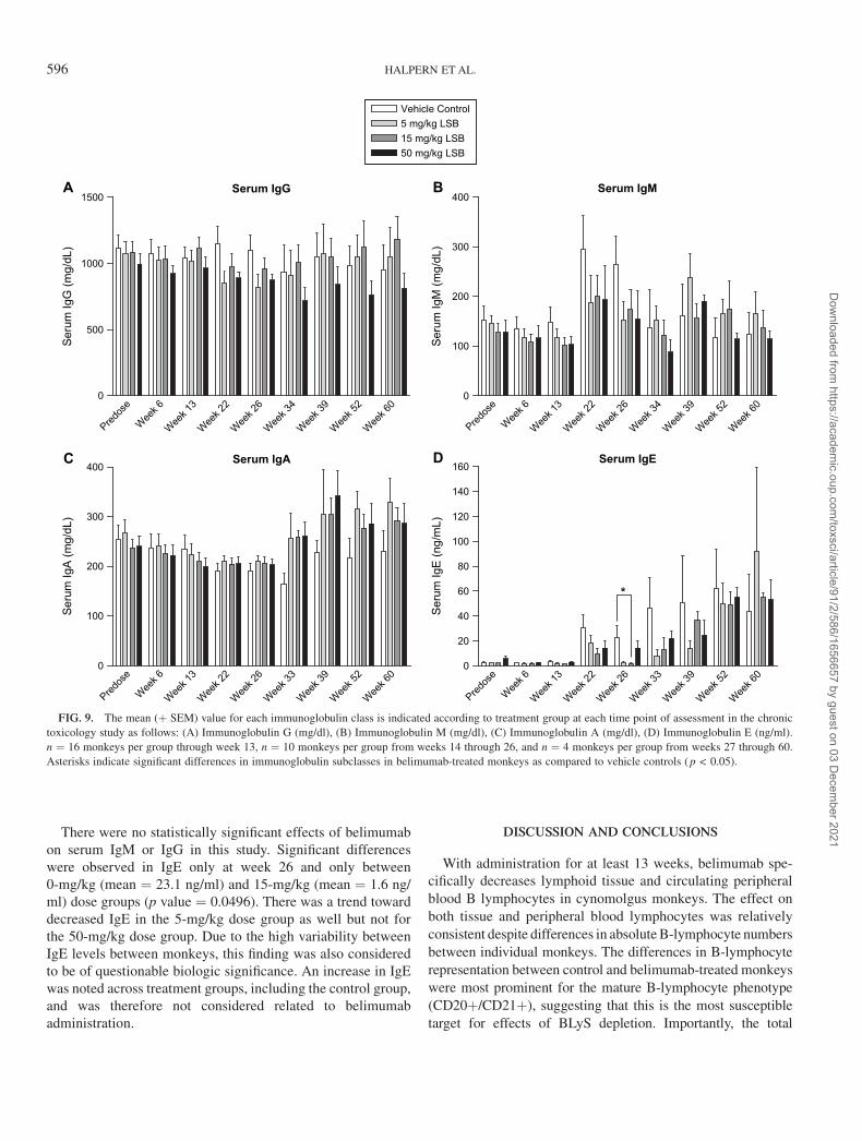
There were no statistically significant effects of belimumabon serum IgM or IgG in this study. Significant differenceswere observed in IgE only at week 26 and only between0-mg/kg (mean ¼ 23.1 ng/ml) and 15-mg/kg (mean ¼ 1.6 ng/ml) dose groups (p value ¼ 0.0496). There was a trend towarddecreased IgE in the 5-mg/kg dose group as well but not forthe 50-mg/kg dose group. Due to the high variability betweenIgE levels between monkeys, this finding was also consideredto be of questionable biologic significance. An increase in IgEwas noted across treatment groups, including the control group,and was therefore not considered related to belimumabadministration.
DISCUSSION AND CONCLUSIONS
With administration for at least 13 weeks, belimumab spe-cifically decreases lymphoid tissue and circulating peripheralblood B lymphocytes in cynomolgus monkeys. The effect onboth tissue and peripheral blood lymphocytes was relativelyconsistent despite differences in absolute B-lymphocyte numbersbetween individual monkeys. The differences in B-lymphocyterepresentation between control and belimumab-treated monkeyswere most prominent for the mature B-lymphocyte phenotype(CD20þ/CD21þ), suggesting that this is the most susceptibletarget for effects of BLyS depletion. Importantly, the total
Serum IgG
Predos
e
Week 6
Week 1
3
Week 2
2
Week 2
6
Week 3
4
Week 3
9
Week 5
2
Week 6
00
500
1000
1500
Vehicle Control5 mg/kg LSB15 mg/kg LSB50 mg/kg LSB
Seru
m Ig
G (m
g/dL
)
Serum IgM
Predos
e
Week 6
Week 1
3
Week 2
2
Week 2
6
Week 3
4
Week 3
9
Week 5
2
Week 6
00
100
200
300
400
Seru
m Ig
M (m
g/dL
)
Serum IgA
Predos
e
Week 6
Week 1
3
Week 2
2
Week 2
6
Week 3
3
Week 3
9
Week 5
2
Week 6
00
100
200
300
400
Seru
m Ig
A (m
g/dL
)
Serum IgE
A B
C D
Predos
e
Week 6
Week 1
3
Week 2
2
Week 2
6
Week 3
3
Week 3
9
Week 5
2
Week 6
00
20
40
60
80
100
120
140
160
Seru
m Ig
E (n
g/m
L)
*
FIG. 9. The mean (þ SEM) value for each immunoglobulin class is indicated according to treatment group at each time point of assessment in the chronic
toxicology study as follows: (A) Immunoglobulin G (mg/dl), (B) Immunoglobulin M (mg/dl), (C) Immunoglobulin A (mg/dl), (D) Immunoglobulin E (ng/ml).
n ¼ 16 monkeys per group through week 13, n ¼ 10 monkeys per group from weeks 14 through 26, and n ¼ 4 monkeys per group from weeks 27 through 60.
Asterisks indicate significant differences in immunoglobulin subclasses in belimumab-treated monkeys as compared to vehicle controls (p < 0.05).
596 HALPERN ET AL.
Dow
nloaded from https://academ
ic.oup.com/toxsci/article/91/2/586/1656657 by guest on 03 D
ecember 2021
lymphocyte count was not affected by belimumab, reflecting thelimited and targeted activity of this antibody. Finally, althoughthere were notable differences in belimumab effects betweenindividual monkeys, there was no consistent dose-related dif-ference in effect or in magnitude of the B-lymphocyte decreaseover the 5- to 50-mg/kg dose range explored. The lack ofbelimumab dose relationship to B-lymphocyte decrease mayindicate that these levels may be saturating for the target over thisrange and schedule in monkeys and also highlights the potentialfor variability in responses between individual monkeys at a givenexposure.
The timing of the response, with a delayed onset in mea-surable effects and a protracted recovery period, is consistentwith the hypothesis that BLyS antagonism indirectly decreasesB lymphocytes, possibly through decreased survival and/orproliferation. The previously reported 4-week study demon-strated decreased B-lymphocyte representation in spleen andmesenteric lymph node mononuclear cells that were mostapparent after 8 weeks (4 weeks of treatment followed by a4-week treatment-free period), but no significant difference inperipheral blood lymphocytes. However, in the chronic studydescribed here, both tissue and peripheral blood B lymphocyteswere decreased after 13 weeks of exposure, and this findingwas sustained through at least week 39, which was well into therecovery period.
The distribution of microscopic findings within the spleenafter 26 weeks of belimumab exposure further supports thehypothesis that belimumab activity is specific for B lympho-cytes. Interestingly, there was no apparent dose-response rela-tionship to the severity of the decreases in lymphoid folliclesize and number over the 5- to 50-mg/kg dose range evaluatedin the chronic study. In addition, when considered by treatmentgroup, any effect of belimumab on spleen weight was difficultto appreciate, in part due to the marked individual variabilityof this parameter in monkeys. However, when the spleen tobody weight ratio was evaluated against the severity of thedecreases in splenic follicular size and number rather than thedose level, an association of belimumab administration withdecreased spleen weight becomes apparent. This effect wasvariable between individual monkeys treated with belimumabfor 13 or 26 weeks and was not dose related over the 5- to50-mg/kg dose range in this study. With one exception, spleensfrom monkeys treated with LymphoStat-B for 26 weeks,followed by a 34-week treatment-free (recovery) period, werehistomorphologically similar to spleens from control monkeys.These data support the role of belimumab in the histomorpho-logic differences recognized at 26 weeks and suggest thatindividual animal sensitivity to belimumab may play a role inthe effects of belimumab observed in cynomolgus monkeys.
Microscopically, changes in lymphoid tissue morphologywere identified in GALT and mesenteric lymph node after fouriv administrations as noted in Table 2 but appeared to belimited to the spleen after chronic administration of belimu-mab. There were decreases in the number and size of lymphoid
follicles in the white pulp of the spleen in many, but not all, ofthe monkeys treated with belimumab for 13 or 26 weeks,without a clear dose-response relationship to incidence orseverity. However, there were no apparent histomorphologicdifferences across treatment groups in the other lymphoidtissues evaluated, such as lymph nodes, thymus, and GALT,at either 13 or 26 weeks. Thus, there may be differences inthe principle lymphoid organs affected (GALT vs. spleen)depending on the length of treatment with belimumab. Suchanatomic differences in B-lymphocyte sensitivity have alsobeen identified with anti-CD20 antibodies and may reflectdifferential dependence of these populations on survival fac-tors such as BLyS (Gong et al., 2005).
There were no clear effects of belimumab on basal immu-noglobulin levels in cynomolgus monkeys in either the 4-weekor the 26-week toxicology study. Serum immunoglobulinstended to fluctuate during the course of the chronic toxicologystudy. In general, there were no findings clearly attributable tobelimumab treatment, although there was a general trend forIgM and IgG to be decreased in belimumab-treated monkeys at22 and 26 weeks and for IgA to be increased in belimumab-treated monkeys during the recovery period. Isolated differ-ences for different groups versus control monkeys wereidentified, but without a temporal or dose-dependent patternindicating a specific effect of belimumab. The lack of effect ofbelimumab on basal immunoglobulin is consistent with datafrom other antibody therapeutics targeting B lymphocytes(Gong et al., 2005; Martin and Chan, 2004). Interestingly,IgE levels increased markedly over the 14-month period ofthe study, but this was noted for all treatment groups, includingcontrol monkeys. Therefore, this finding may reflect changes inenvironment, or possibly the immunologic maturational state,of the monkeys in this chronic study. Neither serum globulinnor the albumin:globulin ratio was affected by belimumabadministration (data not shown).
Although the presence of infections in even a small numberof monkeys in each study might suggest possible immunosup-pression, a clear relationship between infection and belimumabadministration could not be established. The multifocal ab-scesses identified in the spleen of one high-dose (50 mg/kg)monkey in the 4-week study were an isolated finding. Thismonkey had a mild peripheral neutrophilia for at least 2 weeksprior to belimumab administration and had consistentlyelevated serum IgG prior to and throughout the study, sug-gesting that the splenic abscessation may have been a preex-isting condition in this monkey. In the 26-week study, therewere cage side observations of red nasal discharge suggestiveof Branhamella infection across all treatment groups, includingcontrols. When antibiotic intervention was warranted for theaffected monkeys, all monkeys appeared to respond appropri-ately to that therapeutic intervention regardless of treatmentgroup. It should be noted that Branhamella has been caus-atively linked to the bloody nose syndrome cynomolgusmacaques (VandeWoude and Luzarraga, 1991), is reported
PRECLINICAL EFFECTS OF ANTI-BLyS (BELIMUMAB) 597
Dow
nloaded from https://academ
ic.oup.com/toxsci/article/91/2/586/1656657 by guest on 03 D
ecember 2021
sporadically in macaque colonies (reviewed in Olson andPalotay, 1983), and is occasionally observed at Charles RiverLaboratories Preclinical Services.
The pharmacokinetics profiles were linear across the 30-folddose range evaluated in the single-dose studies. The volume ofdistribution is relatively small but exceeds the plasma volume,indicating that belimumab distributes to tissues. Belimumabtotal body clearance is substantially lower than the glomerularfiltration rate for monkeys (2995 ml/day/kg), indicating littlerenal clearance of belimumab. The half-life is long, rangingbetween 9 and 16 days.
In the 26-week multiple-dose toxicology study detailedhere, all monkeys in belimumab-treated groups appear to havebeen exposed to appropriate levels of drug, with the exceptionof the two anti-belimumab antibody positive monkeys. Expo-sure of belimumab increased with dose and extended into therecovery period. The amount of drug accumulation at steadystate (approximately twofold) was consistent with the dosingregimen and expected half-life and PK appeared to be linearover the range of doses tested. The pharmacokinetic behaviorafter single- and multiple-dose administration is consistentwith large macromolecules such as other monoclonal anti-bodies (Gobburu et al., 1998).
In conclusion, belimumab was well tolerated by all mon-keys and demonstrated the expected pharmacologic activityof specifically decreasing B lymphocytes both in tissues andin peripheral blood. Toxicity of belimumab was not identifiedwith chronic administration. Exposures in these nonclinicalmonkey studies included levels 5- to 10-fold higher than ex-posures attained in phase 2 clinical studies. In addition, therewas minimal immunogenicity of belimumab detected in thischronic study. Finally, the treatment-related effects of belimu-mab were reversible after a treatment-free recovery period.
ACKNOWLEDGMENTS
The authors would like to thank Yuling Li and Melissa Perkins for the
production and formulation of the belimumab used in these studies. Todd
Riccobene provided the initial pharmacokinetic analyses for the repeat-dose
studies; Jean Recta provided the initial statistical analysis of immunoglobulin
subclass levels. Greg Beattie and Nancy Gillett contributed to study execution
and interpretation of findings in the toxicology studies. Youmei Wu performed
ELISA measurements of affinities with human and cynomolgus BLyS pro-
teins. The authors also thank Jim Fikes, Thi Sau Migone, Paul Moore, and
David Hilbert for helpful discussions in the development of the manuscript and
Terry Manspeaker for excellent assistance in preparing the figures.
REFERENCES
Baker, K. P., Edwards, B. M., Main, S. H., Choi, G. H., Wager, R. E., Halpern,
W. G., Lappin, P. B., Riccobene, T., Abramian, D., Sekut, L., et al. (2003).
Generation and characterization of LymphoStat-B, a human monoclonal
antibody that antagonizes the bioactivities of B lymphocyte stimulator.
Arthritis Rheum. 48, 3253–3265.
Do, R. K. G., Hatada, E., Lee, H., Tourigny, M. R., Hilbert, D., and Chen-
Kiang, S. (2000). Attenuation of apoptosis underlies B lymphocyte
stimulator enhancement of humoral immune response. J. Exp. Med. 192,
953–964.
Furie, R., Stohl, W., Ginzler, E., Becker, M., Mishra, N., Chatham, W., Merrill,
J. T., Weinstein, A., McCune, J., Zhong, J., et al. (2003). Safety,
pharmacokinetics and pharmacodynamic results of a phase 1 single and
double dose-escalation study of LymphoStat-B (human monoclonal antibody
to BLyS) in SLE patients. Arthritis Rheum. 48(Suppl.), 377 (Abstract).
Gobburu, J. V. S., Tenhoor, C., Rogge, M. C., Frazier, D. E., Thomas, D.,
Benjamin, C., Hess, D. M., and Jusko, W. J. (1998). Pharmacokinetics/
dynamics of 5c8, a monoclonal antibody to CD154 (CD40 ligand) suppression
of an immune response in monkeys. J. Pharmacol. Exp. Ther. 286, 925–930.
Gong, Q., Ou, Q., Ye, S., Lee, W. P., Cornelius, J., Diehl, L., Lin, W. Y., Hu, Z.,
Lu, Y., Chen, Y., et al. (2005). Importance of cellular microenvironment and
circulatory dynamics in B cell immunotherapy. J. Immunol. 174, 817–826.
Gross, J. A., Dillon, S. R., Mudri, S., Johnston, J., Littau, A., Roque, R.,
Rixon, M., Schou, O., Foley, K. P., Haugen, H., et al. (2001). TACI-Ig neu-
tralizes molecules critical for B cell development and autoimmune disease:
Impaired B cell maturation in mice lacking BLyS. Immunity 15, 289–302.
Gross, J. A., Johnston, J., Mudri, S., Enselman, R., Dillon, S. R., Madden, K.,
Xu, W., Parrish-Novak, J., Foster, D., Lofton-Day, C., et al. (2000). TACI and
BCMA are receptors for a TNF homologue implicated in B-cell autoimmune
disease. Nature 404, 995–999.
Khare, S. D., Sarosi, I., Xia, X.-Z., McCabe, S., Miner, K., Solovyev, I.,
Hawkins, N., Kelley, M., Chang, D., Van, G., et al. (2000). Severe B cell
hyperplasia and autoimmune disease in TALL-1 transgenic mice. Proc. Natl.
Acad. Sci. U.S.A. 97, 3370–3374.
Landi, M. S., Kissinger, J. T., Campbell, S. A., Kenney, C. A., and Jenkins, E. L.
(1990). The effects of four types of restraint on serum alanine aminotrans-
ferase and aspartate aminotransferase in the Macaca fascicularis. J. Am.
Coll. Toxicol. 9, 517–523.
Mackay, F., and Browning, J. L. (2002). BAFF: A fundamental survival factor
for B cells. Nat. Rev. Immunol. 2, 465–475.
Mackay, F., Woodcock, S. A., Lawton, P., Ambrose, C., Baetscher, M.,
Schneider, P., Tschopp, J., and Browning, J. L. (1999). Mice transgenic for
BAFF develop lymphocytic disorders along with autoimmune manifesta-
tions. J. Exp. Med. 190, 1697–1710.
Marsters, S. A., Yan, M., Pitti, R. M., Haas, P. E., Dixit, V. M., and
Ashkenazi, A. (2000). Interaction of the TNF homologues BLyS and APRIL
with the TNF receptor homologues BCMA and TACI.Curr. Biol.10,785–788.
Martin, F., and Chan, A. C. (2004). Pathogenic roles of B cells in human
autoimmunity: Insights from clinic. Immunity 20, 517–527.
Moore, P. A., Belvedere, O., Orr, A., Pieri, K., LaFleur, D. W., Feng, P.,
Soppet, D., Charters, M., Gentz, R., Parmelee, D., et al. (1999). BLyS:
Member of the tumor necrosis factor family and B lymphocyte stimulator.
Science 285, 260–263.
Mukhopadhyay, A., Ni, J., Zhai, Y., Yu, G.-L., and Aggarwal, B. B. (1999).
Identification and characterization of a novel cytokine, THANK, a TNF
homologue that activates apoptosis, nuclear factor-jB, and c-Jun NH2-
terminal kinase. J. Biol. Chem. 274, 15978–15981.
Nardelli, B., Belvedere, O., Roschke, V., Moore, P. A., Olsen, H. S., Migone, T. S.,
Sosnovtseva, S., Carrell, J. A., Feng, P., Giri, J. G., et al. (2001). Synthesis and
release of B lymphocyte stimulator from myeloid cells. Blood 97, 198–204.
Olson, L. C., and Palotay, J. L. (1983). Epistaxis and bullae in cynomolgus
macaques (Macaca fascicularis). Lab. Anim. Sci. 33, 377–379.
Parry, T. J., Riccobene, T. A., Strawn, S. J., Williams, R., Daoud, R., Carrell, J.,
Sosnovtseva, S., Micelli, R. C., Poortman, C. M., Sekut, L., et al. (2001).
Pharmacokinetics and immunological effects of exogenously administered
recombinant human B lymphocyte stimulator (BLyS) in mice. J. Pharmacol.
Exp. Ther. 296, 396–404.
Schneider, P., MacKay, F., Steiner, V., Hofmann, K., Bodmer, J. L., Holler, N.,
Ambrose, C., Lawton, P., Bixler, S., Acha-Orbea, H., et al. (1999). BAFF,
598 HALPERN ET AL.
Dow
nloaded from https://academ
ic.oup.com/toxsci/article/91/2/586/1656657 by guest on 03 D
ecember 2021
a novel ligand of the tumor necrosis factor family, stimulates B cell growth.
J. Exp. Med. 189, 1747–1756.
Shu, H.-B., Hu, W. H., and Johnson, H. (1999). TALL-1 is a novel member of
the TNF family that is downregulated by mitogens. J. Leukoc. Biol. 65,
680–683.
Shulga-Morskaya, S., Dobles, M., Walsh, M. E., Ng, L. G., MacKay, F., Rao,
S. P., Kalled, S. L., and Scott, M. (2004). B cell-activating factor be-
longing to the TNF family acts through separate receptors to support B cell
survival and T cell-independent antibody formation. J. Immunol. 173,
2331–2341.
Stohl, W. (2005). BLySfulness does not equal blissfulness in systemic lupus
erythematosus: A therapeutic role for BLyS antagonists. Curr. Dir. Auto-
immun. 8, 289–304.
Stohl, W., Metyas, S., Tan, S.-M., Cheema, G. S., Oamar, B., Xu, D., Roschke, V.,
Wu, Y., Baker, K. P., and Hilbert, D. M. (2003). B lymphocyte stimulator
overexpression in patients with systemic lupus erythematosus: Longitudinal
observations. Arthritis Rheum. 48, 3475–3486.
Thompson, J. S., Bixler, S. A., Qian, F., Vora, K., Scott, M. L., Cachero, T. G.,
Hession, C., Schneider, P., Sizing, I. D., Mullen, C., et al. (2001). BAFF-R,
a newly identified TNF receptor that specifically interacts with BAFF.
Science 293, 2108–2111.
Thompson, J. S., Schneider, P., Kalled, S. L., Wang, L. C., Lefevre, E. A.,
Cachero, T. G., Mackay, F., Bixler, S. A., Zafari, M., Liu, Z.-Y., et al. (2000).
BAFF binds to the tumor necrosis factor receptor-like molecule B cell
maturation antigen and is important for maintaining the peripheral B cell
population. J. Exp. Med. 192, 129–135.
VandeWoude, S. J., and Luzarraga, M. B. (1991). The role of Branhamella
catarrhalis in the ‘‘bloody-nose syndrome’’ of cynomolgus macaques. Lab.
Anim. Sci. 41, 401–406.
Wu, Y., Bressette, D., Carrell, J. A., Kaufman, T., Feng, P., Taylor, K.,
Gan, Y., Cho, Y.-H., Garcia, A. D., Gollatz, E., et al. (2000). Tumor necrosis
factor (TNF) receptor superfamily member TACI is a high affinity re-
ceptor for TNF family members APRIL and BLyS. J. Biol. Chem. 275,
35478–35485.
Yu, G., Boone, T., Delaney, J., Hawkins, N., Kelley, M., Ramakrishnan, M.,
McCabe, S., Qui, W.-R., Kornuc, M., Xia, X.-Z., et al. (2000). APRIL and
TALL-1 and receptors BCMA and TACI: System for regulating humoral
immunity. Nat. Immunol. 1, 252–256.
PRECLINICAL EFFECTS OF ANTI-BLyS (BELIMUMAB) 599
Dow
nloaded from https://academ
ic.oup.com/toxsci/article/91/2/586/1656657 by guest on 03 D
ecember 2021