Embed Size (px)
Citation preview
S Jang, et al
90 Ann Dermatol
Received July 8, 2015, Revised August 20, 2015, Accepted for publication September 15, 2015
Corresponding author: Sang Woong Youn, Department of Dermatology, Seoul National University Bundang Hospital, 82 Gumi-ro 173beon-gil, Bundang-gu, Seongnam 13620, Korea Tel: 82-31-787-7319, Fax: 82- 31-787-4058, E-mail: [email protected]
This is an Open Access article distributed under the terms of the Creative Commons Attribution Non-Commercial License (http:// creativecommons.org/licenses/by-nc/4.0) which permits unrestrictednon-commercial use, distribution, and reproduction in any medium, provided the original work is properly cited.
Ann Dermatol Vol. 28, No. 1, 2016 http://dx.doi.org/10.5021/ad.2016.28.1.90
CASE REPORT
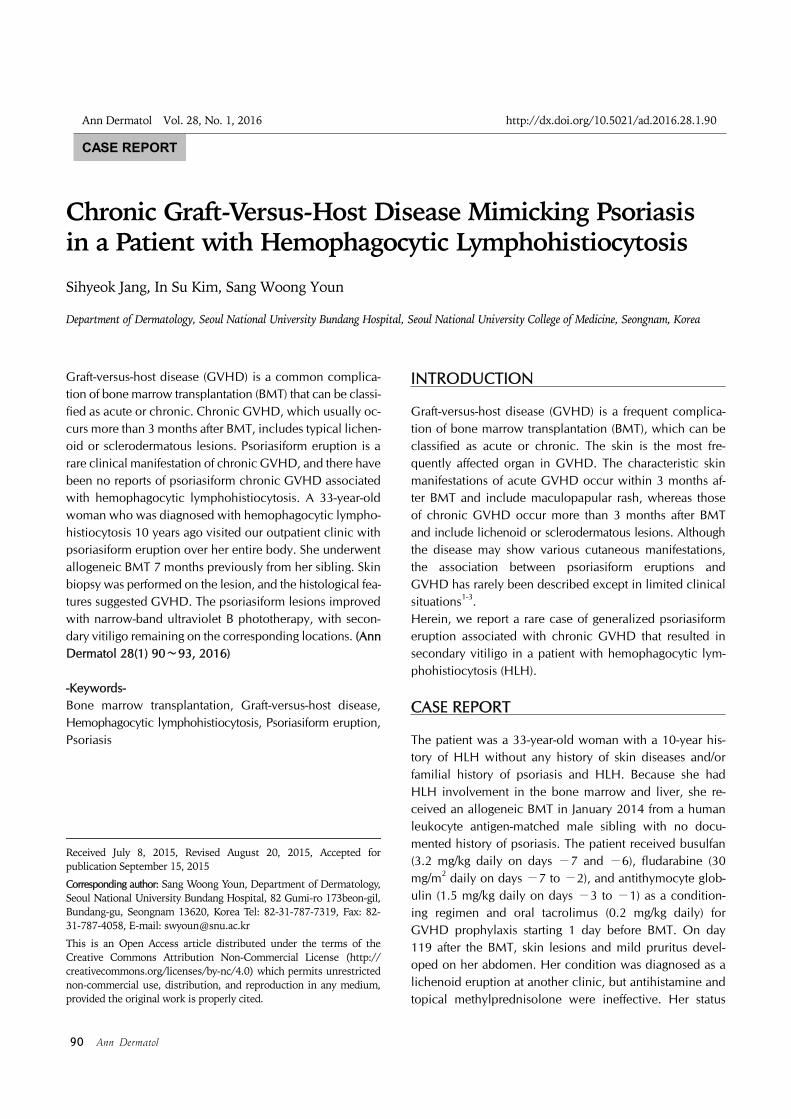
Chronic Graft-Versus-Host Disease Mimicking Psoriasis in a Patient with Hemophagocytic Lymphohistiocytosis
Sihyeok Jang, In Su Kim, Sang Woong Youn
Department of Dermatology, Seoul National University Bundang Hospital, Seoul National University College of Medicine, Seongnam, Korea
Graft-versus-host disease (GVHD) is a common complica-tion of bone marrow transplantation (BMT) that can be classi-fied as acute or chronic. Chronic GVHD, which usually oc-curs more than 3 months after BMT, includes typical lichen-oid or sclerodermatous lesions. Psoriasiform eruption is a rare clinical manifestation of chronic GVHD, and there have been no reports of psoriasiform chronic GVHD associated with hemophagocytic lymphohistiocytosis. A 33-year-old woman who was diagnosed with hemophagocytic lympho-histiocytosis 10 years ago visited our outpatient clinic with psoriasiform eruption over her entire body. She underwent allogeneic BMT 7 months previously from her sibling. Skin biopsy was performed on the lesion, and the histological fea-tures suggested GVHD. The psoriasiform lesions improved with narrow-band ultraviolet B phototherapy, with secon-dary vitiligo remaining on the corresponding locations. (Ann Dermatol 28(1) 90∼93, 2016)
-Keywords-Bone marrow transplantation, Graft-versus-host disease, Hemophagocytic lymphohistiocytosis, Psoriasiform eruption, Psoriasis
INTRODUCTION
Graft-versus-host disease (GVHD) is a frequent complica-tion of bone marrow transplantation (BMT), which can be classified as acute or chronic. The skin is the most fre-quently affected organ in GVHD. The characteristic skin manifestations of acute GVHD occur within 3 months af-ter BMT and include maculopapular rash, whereas those of chronic GVHD occur more than 3 months after BMT and include lichenoid or sclerodermatous lesions. Although the disease may show various cutaneous manifestations, the association between psoriasiform eruptions and GVHD has rarely been described except in limited clinical situations1-3.Herein, we report a rare case of generalized psoriasiform eruption associated with chronic GVHD that resulted in secondary vitiligo in a patient with hemophagocytic lym-phohistiocytosis (HLH).
CASE REPORT
The patient was a 33-year-old woman with a 10-year his-tory of HLH without any history of skin diseases and/or familial history of psoriasis and HLH. Because she had HLH involvement in the bone marrow and liver, she re-ceived an allogeneic BMT in January 2014 from a human leukocyte antigen-matched male sibling with no docu-mented history of psoriasis. The patient received busulfan (3.2 mg/kg daily on days −7 and −6), fludarabine (30 mg/m2 daily on days −7 to −2), and antithymocyte glob-ulin (1.5 mg/kg daily on days −3 to −1) as a condition-ing regimen and oral tacrolimus (0.2 mg/kg daily) for GVHD prophylaxis starting 1 day before BMT. On day 119 after the BMT, skin lesions and mild pruritus devel-oped on her abdomen. Her condition was diagnosed as a lichenoid eruption at another clinic, but antihistamine and topical methylprednisolone were ineffective. Her status

Chronic GVHD with Unusual Skin Lesion
Vol. 28, No. 1, 2016 91
Fig. 1. (A) Generalized erythema-tous papulosquamous lesions with whitish scales on the trunk (inlet) and erythematous papular lesion with whitish scales. (B) Improved skin lesions with remaining wide-spread hypopigmentation 8 weeks after the start of therapy. Some hyperpigmented spots correspon-ding to hair follicles were sus-pected to be due to repigmentation.
Fig. 2. Histopathology of skin lesions (H&E, ×100). Interface dermatitis with perivascular and periappendageal lymphocytic infiltration, apoptotic keratinocytes, exocytosis of lymphocytes, hyperkeratosis, and parakeratosis (inlet, ×400); dyskeratotic cells seen in the epidermis.
worsened and she visited our clinic on day 187. After 2 weeks of oral prednisolone (0.3 mg/kg daily) with topical desoxymethasone, the skin lesions improved, and we started tapering the medication. However, the eruptions abruptly recurred in the form of erythematous lesions across her whole body including the scalp, palms, and soles except the nails, without other general symptoms on day 201 just after the dosage was decreased. The erup-tions were composed of papulosquamous lesions with whitish scales, and the clinical features were similar to those of psoriasis (Fig. 1A).A laboratory examination including liver function testing found that all markers were within normal ranges. A histo-pathological examination of the arm indicated interface dermatitis with perivascular lymphocytic infiltration in the upper dermis, keratinocyte apoptosis, exocytosis of lympho-cytes, hyperkeratosis, and parakeratosis (Fig. 2). However, no typical histologic findings of psoriasis such as spongi-form pustules of Kogoj, Munro’s microabscesses, hypo-granulosis, or rete ridge elongation were observed.Overall, we found discrepancies between the clinical and histological findings. Although the clinical findings sug-gested psoriasiform lesions, the histological findings sug-gested GVHD, for example, dyskeratotic cells in the epidermis. Based on these findings, a diagnosis of mild chronic GVHD with a psoriasiform skin manifestation was made. Skin was the only involved organ in this case. Oral prednisolone (0.5 mg/kg daily) and narrow-band ultra-violet B phototherapy 3 times per week were started. These treatments were effective and the skin lesions were significantly improved, with widespread hypopigmented patches remaining (Fig. 1B).
DISCUSSION
The characteristic cutaneous manifestations of acute GVHD, which generally occur within 3 months after BMT, include maculopapular exanthema and perifollicular pap-ular lesions. In contrast, chronic GVHD, which usually oc-curs more than 3 months after BMT, includes typical li-chenoid or sclerodermatous lesions with poikilodermal elements.Although cutaneous involvement is common in GVHD, psoriasiform eruptions after BMT are rare. Several cases showing the development of psoriasis after BMT from do-nors with a history of psoriasis or resolution of psoriasis af-ter BMT for chronic myelogenous leukemia from a normal donor have been reported4,5, suggesting an adoptive trans-fer of susceptibility or disease-inducible immunity to psor-iasis from a donor with a genetic background of psoriasis.Three other reports of psoriasiform eruptions associated

S Jang, et al
92 Ann Dermatol
with GVHD have been reported in the literature6-8. The unusual psoriasiform skin presentation as described here could be a diagnostic challenge to clinicians. Even in HLH, the development of chronic GVHD is relatively rare (19%) compared to that of other conditions (30% from matched and 70% from mismatched donors)9. There are no other previous reports of psoriasiform chronic GVHD in a patient with HLH after BMT. Taking a detailed history was essential in considering GVHD, which was indicated by histopathological findings.GVHD and psoriasis share common immunological features. Both diseases are T-cell–mediated diseases dis-playing a T helper cell type 1 cytokine secretion profile, and both exhibit elevated human leukocyte antigen (HLA)-DR antigen expression in the lesional epidermal keratinocytes6. T-cell–directed immunosuppressants effec-tively control the disease activity of psoriasis, and psoriasis skin lesions are fully induced when T cells from patients with psoriasis are injected into symptomless psoriasis skin engrafted onto severe combined immunodeficiency mice10. Although it is unclear why this patient presented with psoriasiform eruption as a skin lesion of chronic GVHD, one possible explanation is that the interaction between donor lymphocytes and recipient keratinocytes during graft-versus-host reactions induced keratinocyte hyperpro-liferation.In addition, Langerhans cell (LC) counts have been re-ported to be decreased in the lesional skin of patients with GVHD and psoriasis6. Although the association of im-munological abnormalities relevant to local antigen pre-sentation and psoriasiform eruption remains to be fully un-derstood, it might be explained by the evidence of ex-acerbation of psoriasis in patients with acquired im-munodeficiency syndrome accompanied by a reduced number of epidermal LCs11 or the enhancement of in-flammatory skin reactions when epidermal LCs are de-pleted in the epidermis12. Thus, the decrease in LC counts also might play a role in the formation of psoriasiform eruption.Secondary vitiligo can be explained by autoimmune re-actions triggered by chronic GVHD, and the rather rapid repigmentation is probably due to the cessation of this process after the active inflammation is resolved. The asso-ciation of GVHD with various autoimmune conditions strengthens the probability of vitiligo as being a part of the autoimmune process on the basis of chronic GVHD13. GVHD appears to participate in the destruction of melano-cytes by skin-homing autoreactive melanocyte-specific cy-totoxic T lymphocytes. Furthermore, the influence of sex mismatch on the development of vitiligo, male donor to female recipient in our case, is consistent with previous
reports14,15.Herein, we present a case of a novel skin manifestation of chronic GVHD. We would like to call this cutaneous fea-ture “psoriasiform chronic cutaneous GVHD”. Further ac-cumulation of similar cases will be needed to enrich the understanding of the variety of immunological events and to clarify the mechanism associated with GVHD. We also emphasize the importance of maintaining a high index of suspicion of GVHD, even for uncommon lesions on the skin of patients with a history of BMT.
REFERENCES
1. Snowden JA, Heaton DC. Development of psoriasis after syngeneic bone marrow transplant from psoriatic donor: further evidence for adoptive autoimmunity. Br J Dermatol 1997;137:130-132.
2. Adkins DR, Abidi MH, Brown RA, Khoury H, Goodnough LT, Vij R, et al. Resolution of psoriasis after allogeneic bone marrow transplantation for chronic myelogenous leukemia: late complications of therapy. Bone Marrow Transplant 2000;26:1239-1241.
3. Ahn HS, Park HJ, Lee JY, Cho BK. A case of chronic cutaneous graft versus host disease with the clinical features of exfoliative dermatitis. Ann Dermatol 2009;21:319-322.
4. Masszi T, Farkas A, Remenyi P, Lueff S, Batai A, Krivan G, et al. Ten-year remission of psoriasis after allogeneic but not autologous bone marrow transplantation. Dermatology 2006;212:88-89.
5. Slavin S, Nagler A, Varadi G, Or R. Graft vs autoimmunity following allogeneic non-myeloablative blood stem cell transplantation in a patient with chronic myelogenous leukemia and severe systemic psoriasis and psoriatic poly-arthritis. Exp Hematol 2000;28:853-857.
6. Kawakami Y, Oyama N, Nakamura K, Kaneko F, Kikuta A, Suzuki H. Psoriasiform eruption associated with graft- versus-host disease. Acta Derm Venereol 2007;87:436-438.
7. Matsushita T, Hasegawa M, Shirasaki F, Fujimoto M, Yamazaki H, Sato S, et al. A case of acute cutaneous graft-versus-host disease mimicking psoriasis vulgaris. Dermatology 2008;216:64-67.
8. Taguchi S, Kawachi Y, Fujisawa Y, Nakamura Y, Furuta J, Otsuka F. Psoriasiform eruption associated with graft- versus-host disease. Cutis 2013;92:151-153.
9. Jordan MB, Filipovich AH. Hematopoietic cell transplan-tation for hemophagocytic lymphohistiocytosis: a journey of a thousand miles begins with a single (big) step. Bone Marrow Transplant 2008;42:433-437.
10. Nickoloff BJ, Wrone-Smith T. Injection of pre-psoriatic skin with CD4+ T cells induces psoriasis. Am J Pathol 1999; 155:145-158.
11. Zemelman V, Van Neer F, Roberts N, Patel P, Langtry J, Staughton RC. Epidermal Langerhans cells, HIV-1 infection and psoriasis. Br J Dermatol 1994;130:307-311.
12. Grabbe S, Steinbrink K, Steinert M, Luger TA, Schwarz T. Removal of the majority of epidermal Langerhans cells by

Chronic GVHD with Unusual Skin Lesion
Vol. 28, No. 1, 2016 93
topical or systemic steroid application enhances the effector phase of murine contact hypersensitivity. J Immunol 1995;155:4207-4217.
13. Ferrara JL, Deeg HJ. Graft-versus-host disease. N Engl J Med 1991;324:667-674.
14. Sanli H, Akay BN, Arat M, Koçyigit P, Akan H, Beksac M, et al. Vitiligo after hematopoietic cell transplantation: six cases
and review of the literature. Dermatology 2008;216:349- 354.
15. Zuo RC, Naik HB, Steinberg SM, Baird K, Mitchell SA, Kuzmina Z, et al. Risk factors and characterization of vitiligo and alopecia areata in patients with chronic graft- vs-host disease. JAMA Dermatol 2015;151:23-32.