Embed Size (px)
Citation preview
Circulation and Gas Exchange
Chapter 42
A.P. Biology
Mr. Knowles
Liberty Senior High
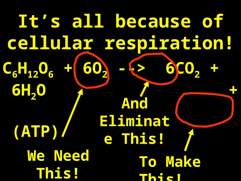
It’s all because of cellular respiration!
C6H12O6 + 6O2 --> 6CO2 + 6H2O +
(ATP)
We Need This! To Make This!
And Eliminate
This!
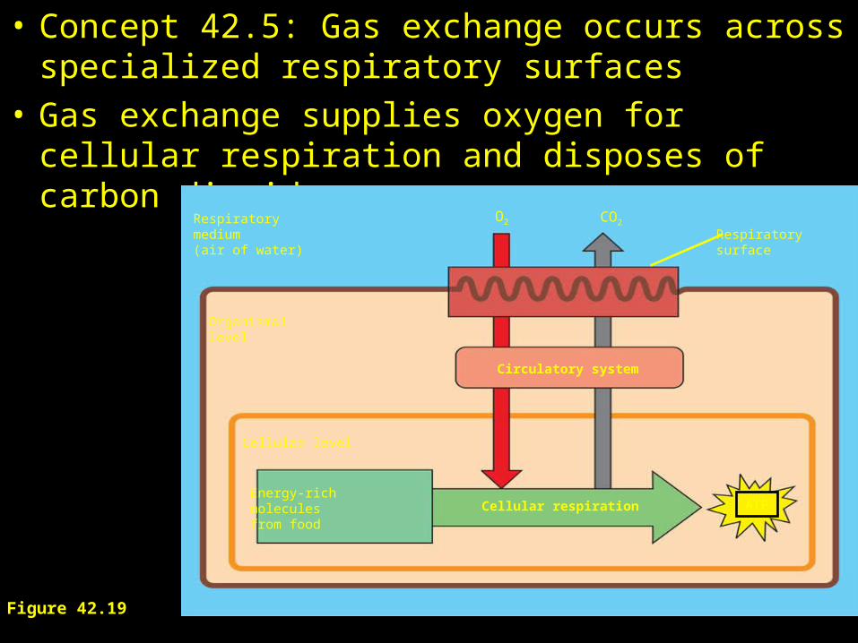
• Concept 42.5: Gas exchange occurs across specialized respiratory surfaces
• Gas exchange supplies oxygen for cellular respiration and disposes of carbon dioxide.
Figure 42.19
Organismal level
Cellular level
Circulatory system
Cellular respiration ATPEnergy-richmoleculesfrom food
Respiratorysurface
Respiratorymedium(air of water)
O2 CO2
• Animals require large, moist respiratory surfaces for the adequate diffusion of respiratory gases:
– Between their cells and the respiratory medium, either air or water.
• Overview: Trading with the Environment
• Every organism must exchange materials with its environment
–And this exchange ultimately occurs at the cellular level
• In unicellular organisms:
–These exchanges occur directly with the environment.
• For most of the cells making up multicellular organisms:
–Direct exchange with the environment is not possible.
• Concept 42.1: Circulatory systems reflect phylogeny
• Transport systems
– Functionally connect the organs of exchange with the body cells.
• Most complex animals have internal transport systems:
– That circulate fluid, providing a lifeline between the aqueous environment of living cells and the exchange organs, such as lungs, that exchange chemicals with the outside environment
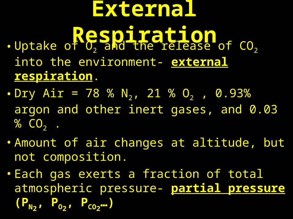
External Respiration• Uptake of O2 and the release of CO2 into the
environment- external respiration.
• Dry Air = 78 % N2, 21 % O2 , 0.93% argon and other inert gases, and 0.03 % CO2 .
• Amount of air changes at altitude, but not composition.
• Each gas exerts a fraction of total atmospheric pressure- partial pressure (PN2
, PO2, PCO2
…)
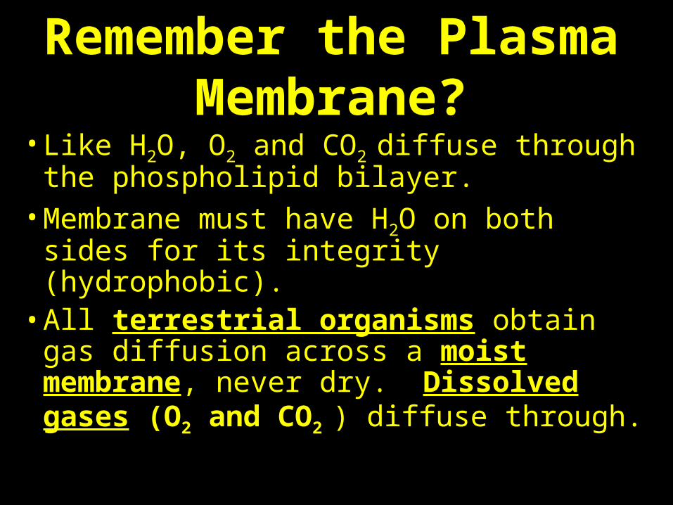
Remember the Plasma Membrane?
• Like H2O, O2 and CO2 diffuse through the phospholipid bilayer.
• Membrane must have H2O on both sides for its integrity (hydrophobic).
• All terrestrial organisms obtain gas diffusion across a moist membrane, never dry. Dissolved gases (O2 and CO2 ) diffuse through.
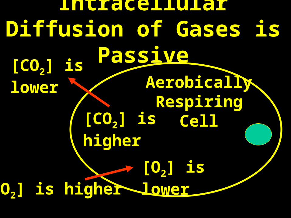
Intracellular Diffusion of Gases is Passive
Aerobically Respiring Cell
[O2] is lower
[CO2] is higher
[O2] is higher
[CO2] is lower
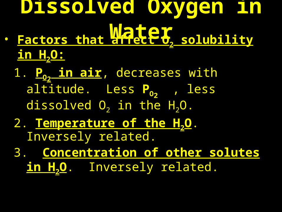
Dissolved Oxygen in Water• Factors that affect O2 solubility in H2O:
1. PO2 in air, decreases with altitude. Less
PO2 , less dissolved O2 in the H2O.
2. Temperature of the H2O. Inversely related.
3. Concentration of other solutes in H2O. Inversely related.
What happens to the oxygen level when tides
go out?
The Story of the Tarpon
Discovery: Blue Planet- Tidal Seas
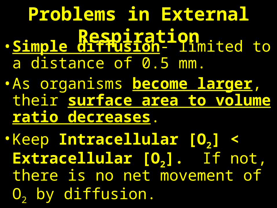
Problems in External Respiration• Simple diffusion- limited to a
distance of 0.5 mm. • As organisms become larger, their
surface area to volume ratio decreases.
• Keep Intracellular [O2] < Extracellular [O2]. If not, there is no net movement of O2 by diffusion.
Invertebrate Circulation
• The wide range of invertebrate body size and form:
–Is paralleled by a great diversity in circulatory systems
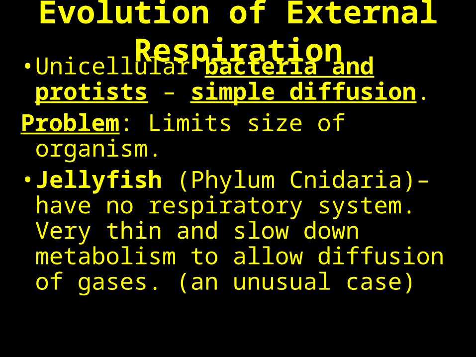
Evolution of External Respiration• Unicellular bacteria and protists –
simple diffusion. Problem: Limits size of organism. • Jellyfish (Phylum Cnidaria)– have
no respiratory system. Very thin and slow down metabolism to allow diffusion of gases. (an unusual case)
Gastrovascular Cavities
• Simple animals, such as cnidarians– Have a body wall only two cells thick that encloses a
gastrovascular cavity.
• The gastrovascular cavity– Functions in both digestion and distribution of
substances throughout the body.
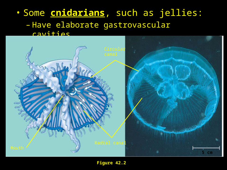
• Some cnidarians, such as jellies:– Have elaborate gastrovascular cavities
Figure 42.2
Circularcanal
Radial canal
5 cmMouth

The Jellyfish Life!
Discover: Blue Planet- Seasonal Seas
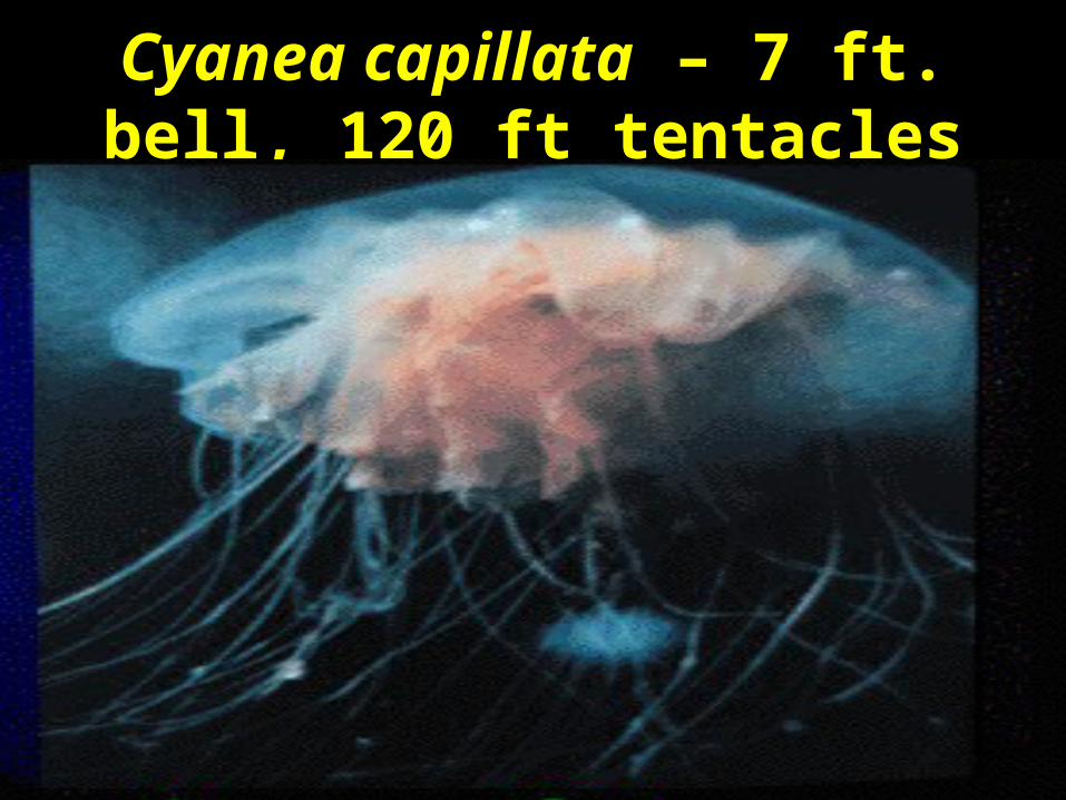
Cyanea capillata – 7 ft. bell, 120 ft tentacles
Creating a Water Current
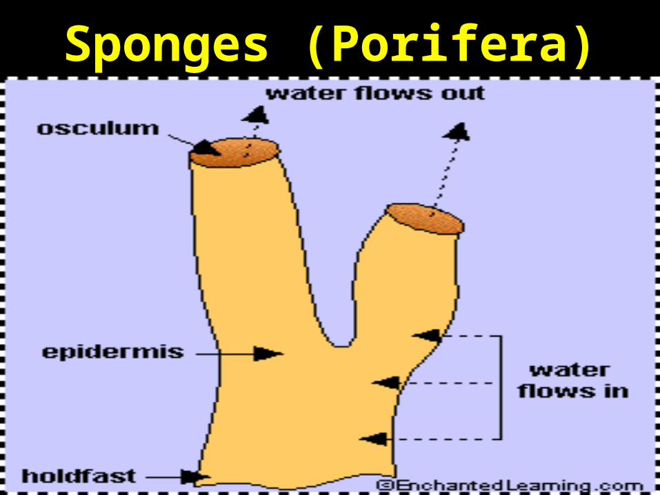
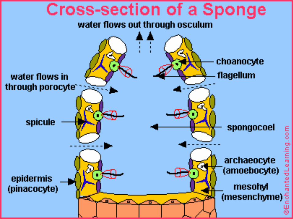
• Sponges (Phylum Porifera) – diffusion directly from surrounding water; set up a current using cilia. Beating cilia replace water over the diffusion surface.
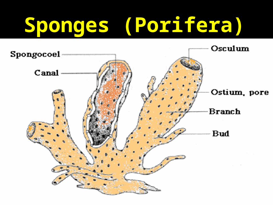
Sponges (Porifera)
Sponges (Porifera)

Sponges and Corals
Discovery: Blue Planet- Coral Seas
Creating a Water Current
• Problem: Limited to aquatic environments; not efficient for really large organisms.
But sponges are aquatic! What about terrestrial
organisms?
Enter Cutaneous Respiration!
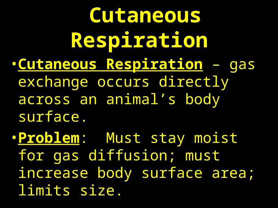
Cutaneous Respiration
• Cutaneous Respiration – gas exchange occurs directly across an animal’s body surface.
• Problem: Must stay moist for gas diffusion; must increase body surface area; limits size.
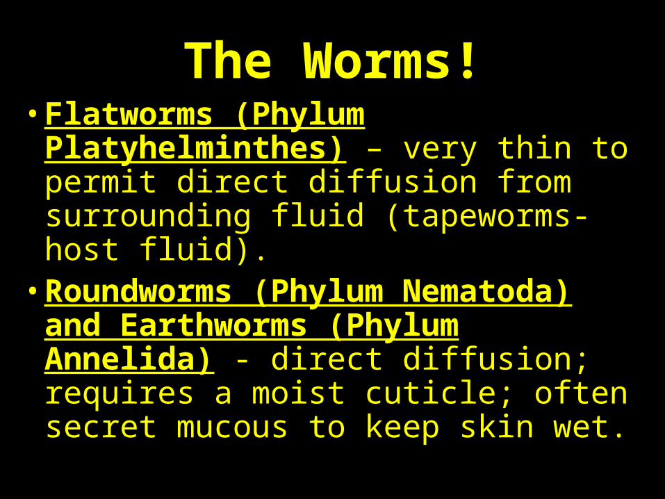
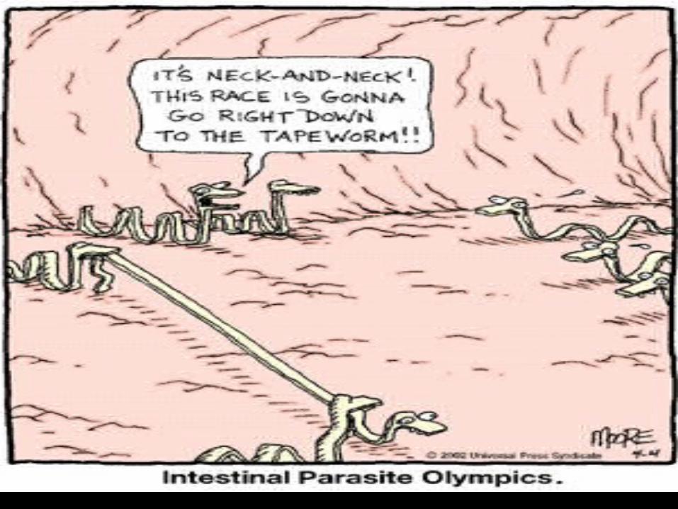
The Worms!• Flatworms (Phylum Platyhelminthes)
– very thin to permit direct diffusion from surrounding fluid (tapeworms-host fluid).
• Roundworms (Phylum Nematoda) and Earthworms (Phylum Annelida) - direct diffusion; requires a moist cuticle; often secret mucous to keep skin wet.
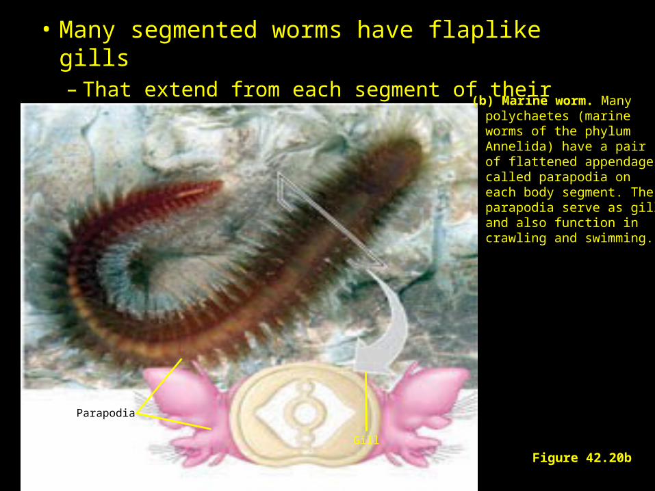
• Many segmented worms have flaplike gills– That extend from each segment of their body.
Figure 42.20b
(b) Marine worm. Many polychaetes (marine worms of the phylum Annelida) have a pair of flattened appendages called parapodia on each body segment. The parapodia serve as gillsand also function incrawling and swimming.
Gill
Parapodia
So why do earthworms die on your driveway
after a rain?
They dry out and, therefore, suffocate!
Mouth-to-skin, anyone?
What are the down sides to cutaneous respiration?
The World’s Largest Earthworm
Video: Nigel Marvin’s Giant Creepy Crawlies
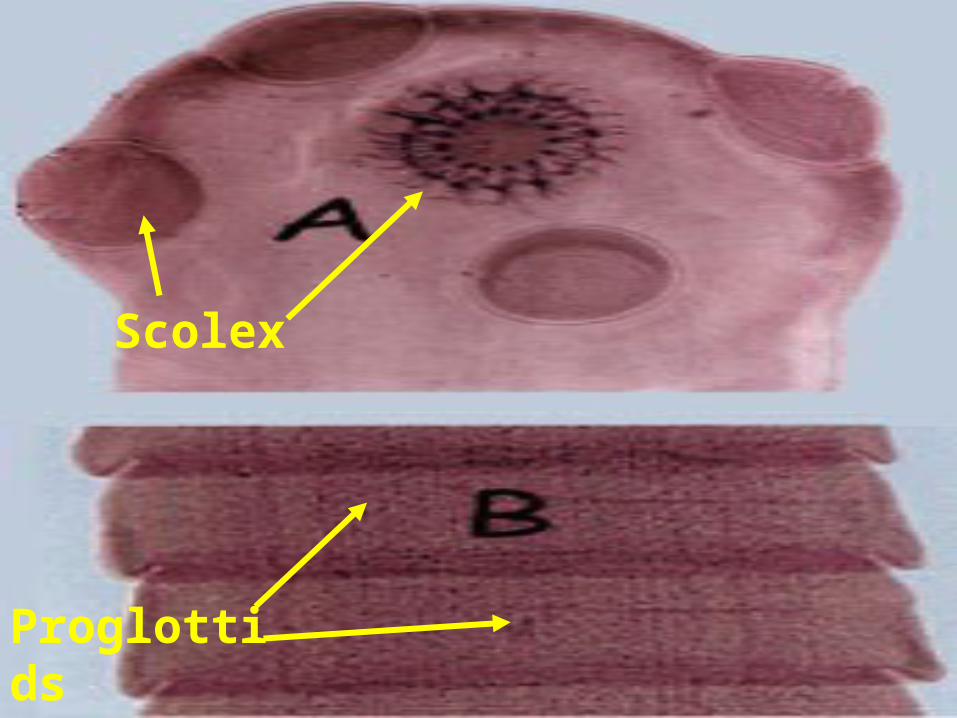
Scolex
Proglottids
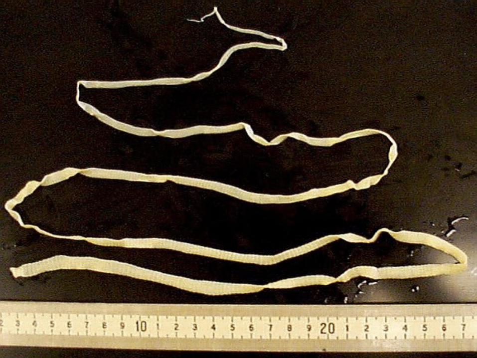
Cutaneous Respiration in a Tapeworm
Video: The Body Snatchers
Increasing the Diffusion Surface Area
• Advanced Invertebrates (Phylum Echinodermata, Mollusca, Arthropoda) – increase surface area and bring external fluid close to internal fluid.
• Use a primitive gill - increases diffusion surface area.
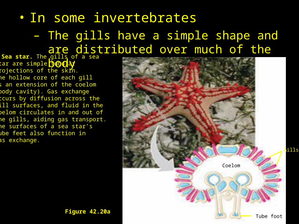
• In some invertebrates– The gills have a simple shape and are distributed
over much of the body(a) Sea star. The gills of a sea
star are simple tubular projections of the skin. The hollow core of each gillis an extension of the coelom(body cavity). Gas exchangeoccurs by diffusion across thegill surfaces, and fluid in thecoelom circulates in and out ofthe gills, aiding gas transport. The surfaces of a sea star’s tube feet also function in gas exchange.
Gills
Tube foot
Coelom
Figure 42.20a
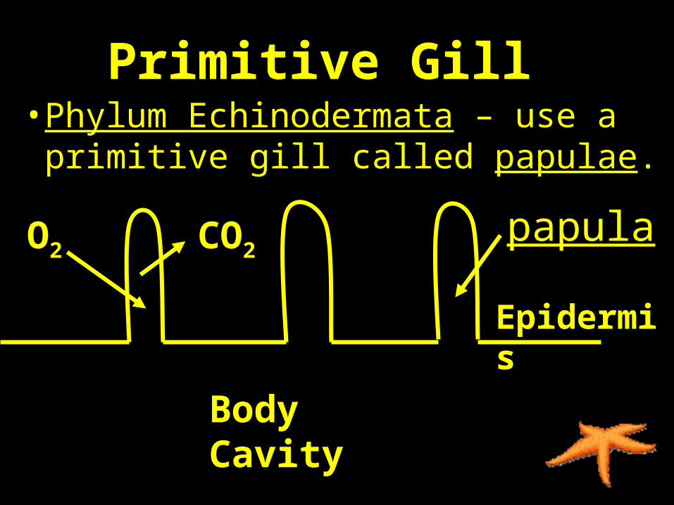
Primitive Gill • Phylum Echinodermata – use a
primitive gill called papulae.
Body Cavity
Epidermis
O2 CO2papula
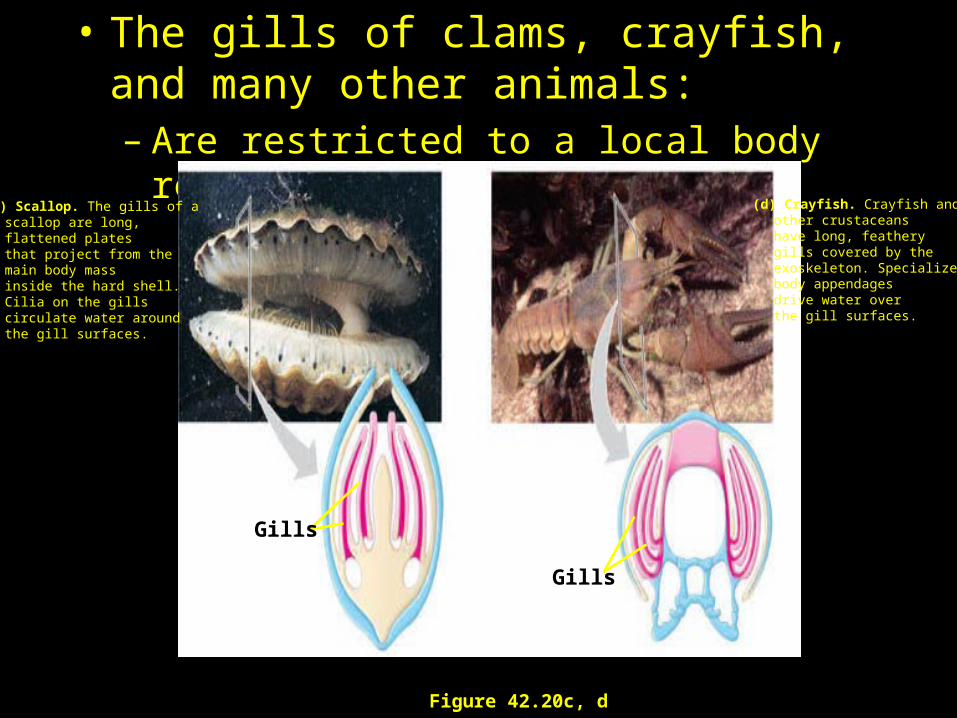
• The gills of clams, crayfish, and many other animals:– Are restricted to a local body region.
Figure 42.20c, d
(d) Crayfish. Crayfish and other crustaceanshave long, feathery gills covered by the exoskeleton. Specialized body appendagesdrive water over the gill surfaces.
(c) Scallop. The gills of a scallop are long, flattened plates that project from themain body mass inside the hard shell.Cilia on the gills circulate water around the gill surfaces.
Gills
Gills
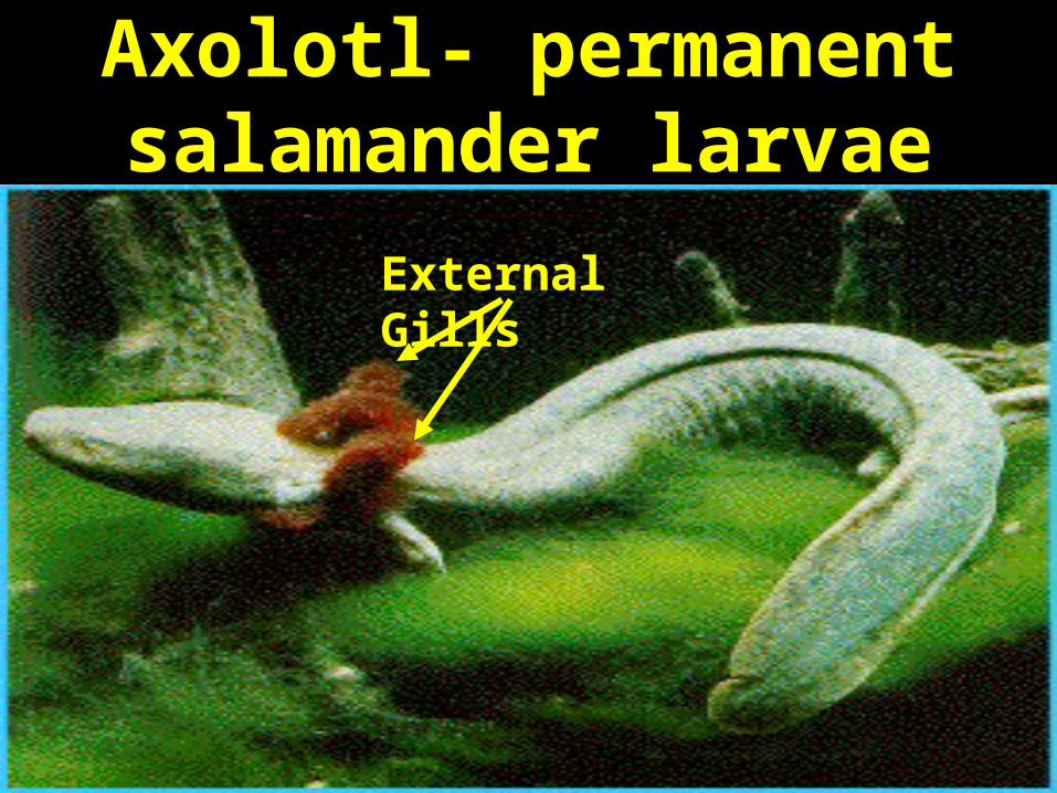
Axolotl- permanent salamander larvae
External Gills
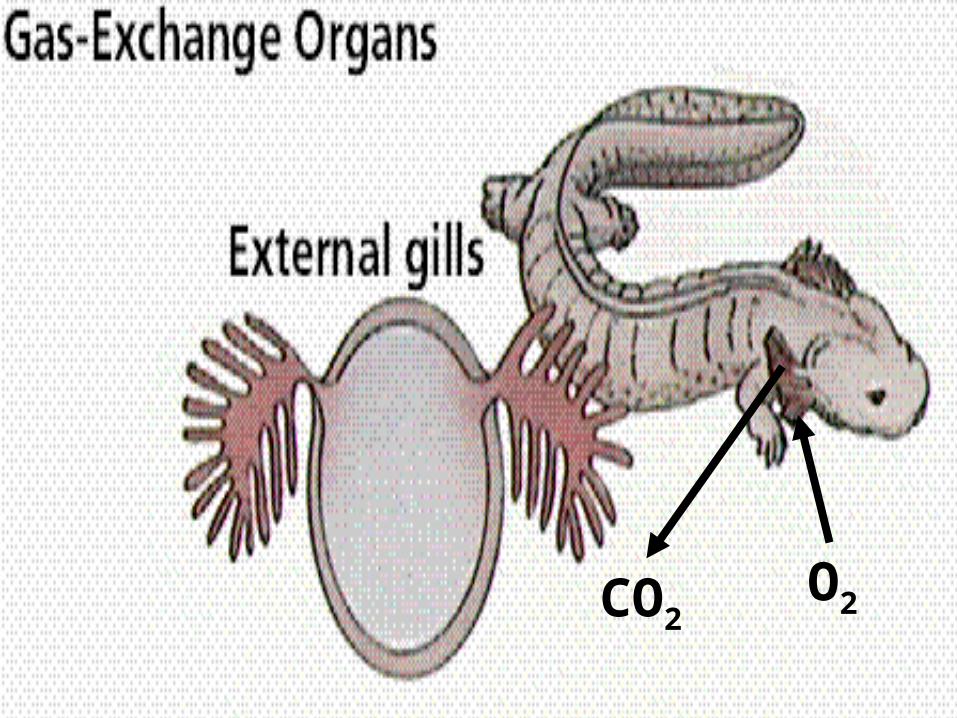
O2CO2
The External Gills• Some, like the axolotl (aquatic
salamander) physically moves its external gills through the water for improved gas exchange.
• A problem with external gills: Difficult to circulate water past surfaces constantly.
• Problem: external gills are fragile and offer resistance in water.
Brachial Chambers• Brachial chambers – a muscular,
internal pouch used to pump water over the gills.
• Phylum Mollusca – use an internal mantle cavity that pumps water over gills. Ex. Squid and octopi.
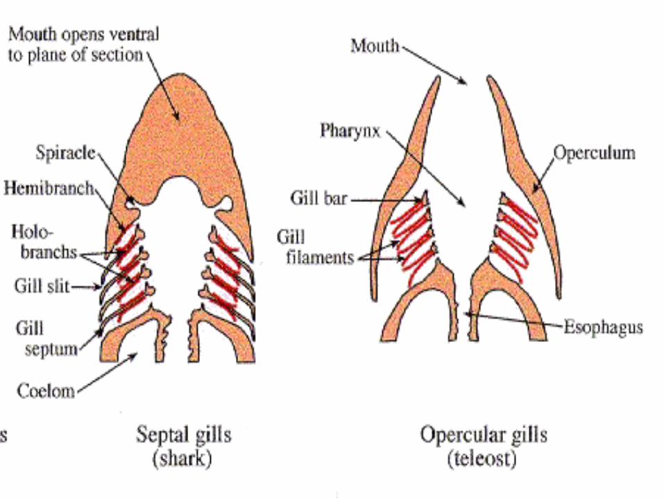
Internal Gills• Cartilaginous Fishes (Sharks and
Rays) – force water through mouth over internal gills by constant swimming. Water flows out gill slits.
• Swim with mouth open to force water over gills – ram ventilation.
• Problem: Must stay in motion or suffocate.
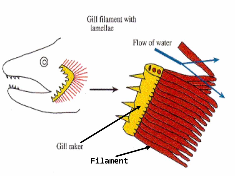
Filament
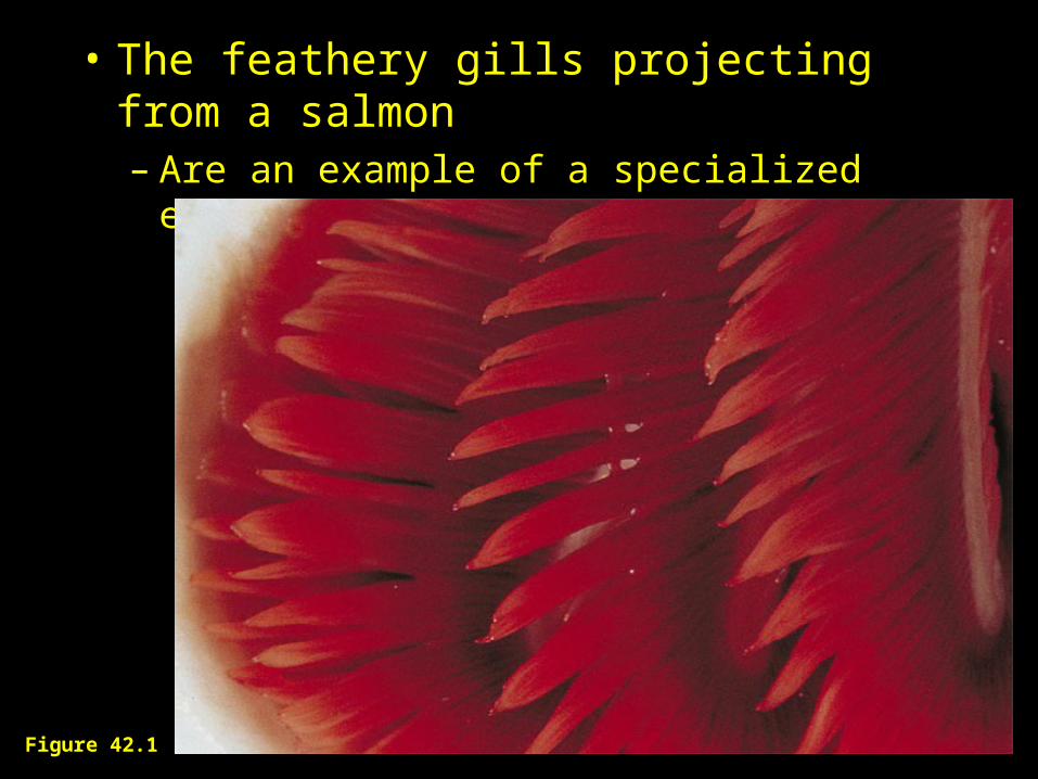
• The feathery gills projecting from a salmon– Are an example of a specialized exchange
system found in animals.
Figure 42.1
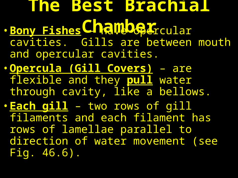
The Best Brachial Chamber• Bony Fishes – have opercular cavities. Gills
are between mouth and opercular cavities.• Opercula (Gill Covers) – are flexible and
they pull water through cavity, like a bellows.
• Each gill – two rows of gill filaments and each filament has rows of lamellae parallel to direction of water movement (see Fig. 46.6).
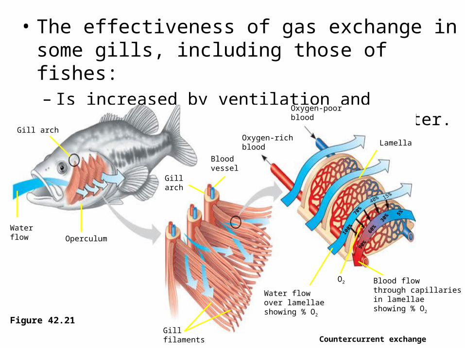
• The effectiveness of gas exchange in some gills, including those of fishes:– Is increased by ventilation and countercurrent flow of
blood and water.
Countercurrent exchange
Figure 42.21
Gill arch
Water flow Operculum
Gill arch
Blood vessel
Gillfilaments
Oxygen-poorblood
Oxygen-richblood
Water flowover lamellaeshowing % O2
Blood flowthrough capillariesin lamellaeshowing % O2
Lamella
100%
40%
70%
15%
90%
60%
30% 5%
O2
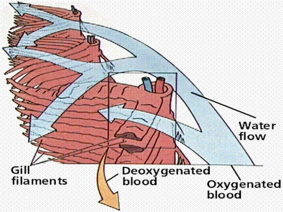
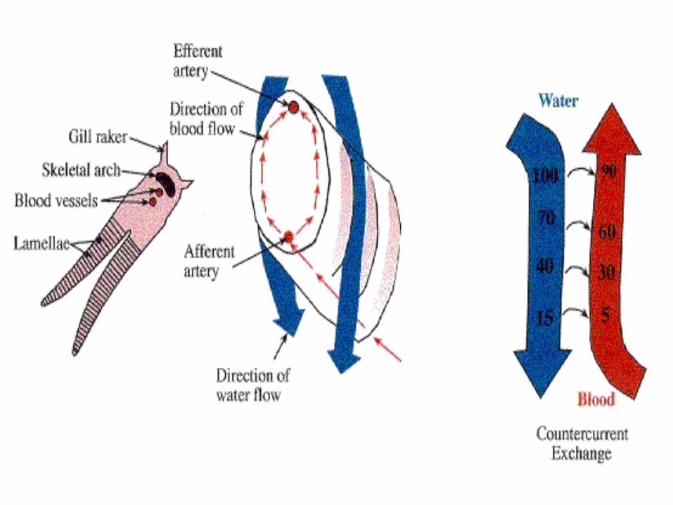
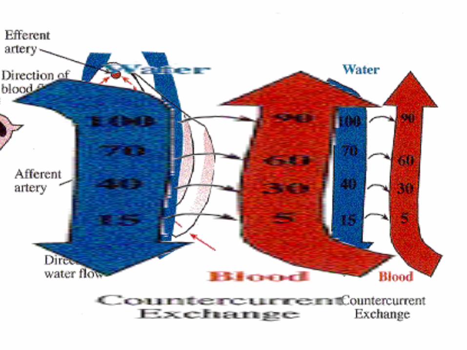
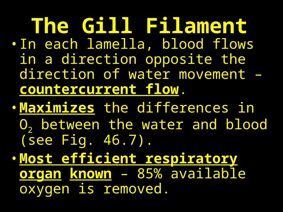
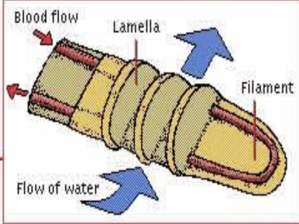
The Gill Filament• In each lamella, blood flows in a
direction opposite the direction of water movement – countercurrent flow.
• Maximizes the differences in O2 between the water and blood (see Fig. 46.7).
• Most efficient respiratory organ known – 85% available oxygen is removed.
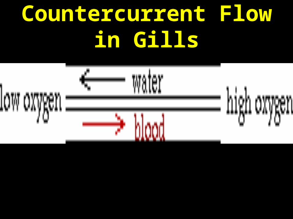
Countercurrent Flow in Gills

What if you’re not aquatic?
Why do fish die out of water?
They suffocate.
The Problem of Terrestrial Respiration
• Water – 5-10 ml of O2 per liter• Air – 210 ml O2 per liter (rich in O2)• Gills don’t work in air :
– Air is less buoyant than water, fragile lamellae collapse and reduce surface area and not enough gas diffusion.
– Water diffuses into air by evaporation. Gills provide too much surface area for water loss.
Terrestrial Organisms• Use two types of internal passage ways for
gas diffusion; sacrifice efficiency for reduced evaporation.
• Terrestrial Insects use tracheae – air-filled passages connecting the surface of the insect to all potions of its body. Diffusion directly with internal cells and no circulatory system.
• Use openings called spiracles along the abdomen that can be controlled. Effective for small animals.
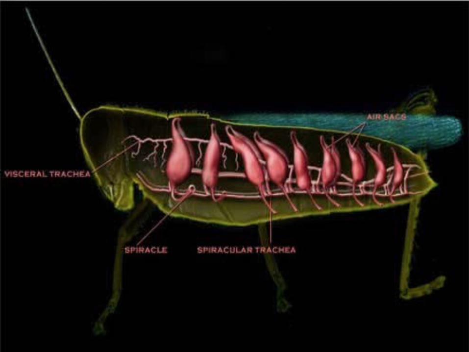
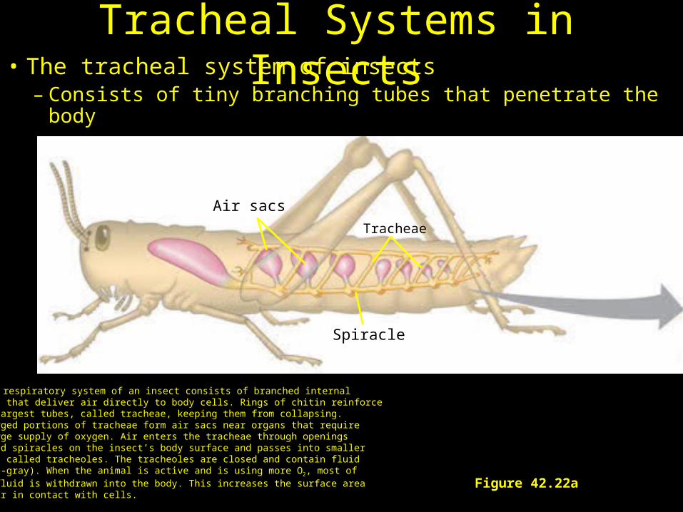
Figure 42.22a
Tracheae
Air sacs
Spiracle
(a) The respiratory system of an insect consists of branched internaltubes that deliver air directly to body cells. Rings of chitin reinforcethe largest tubes, called tracheae, keeping them from collapsing. Enlarged portions of tracheae form air sacs near organs that require a large supply of oxygen. Air enters the tracheae through openings called spiracles on the insect’s body surface and passes into smaller tubes called tracheoles. The tracheoles are closed and contain fluid(blue-gray). When the animal is active and is using more O2, most ofthe fluid is withdrawn into the body. This increases the surface area of air in contact with cells.
Tracheal Systems in Insects• The tracheal system of insects
– Consists of tiny branching tubes that penetrate the body
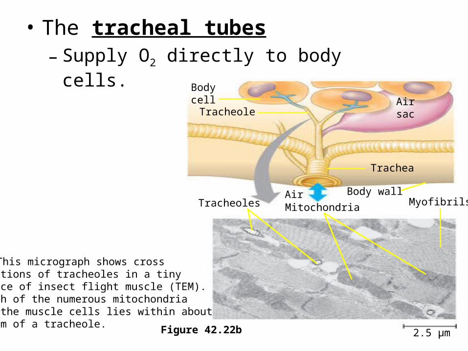
• The tracheal tubes– Supply O2 directly to body cells.
Airsac
Body cell
Trachea
Tracheole
Tracheoles Mitochondria MyofibrilsBody wall
(b) This micrograph shows crosssections of tracheoles in a tinypiece of insect flight muscle (TEM).Each of the numerous mitochondriain the muscle cells lies within about5 µm of a tracheole.
Figure 42.22b 2.5 µm
Air

How large can an insect become?
Video: Nigel Marvin’s Giant Creepy Crawlies
First Terrestrial Organism
• Problem: Tracheal breathing limits the size of the organism. Ventilation is by movement of organism.
Lungs
• Spiders, land snails, and most terrestrial vertebrates:
– Have internal lungs (simple sacs).
Other Terrestrial Organ• Lung – moves air through a moist,
internal, tubular passage and back out same passage.
• Benefit – minimizes evaporation.• Problem: lower efficiency than gill,
but O2 more abundant in air.• Four variations of the terrestrial,
vertebrate lung.

The First Terrestrial Animals?
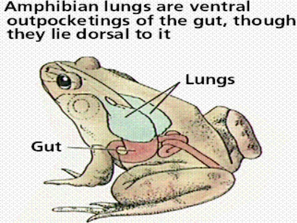
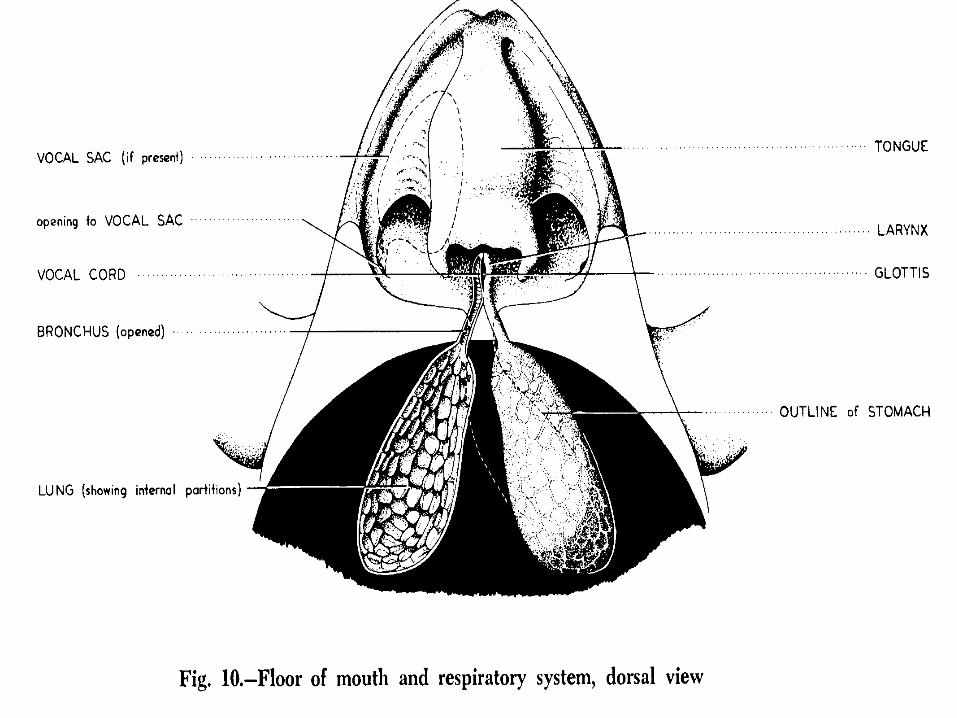
Class Amphibia• Amphibian Lung – simple sac with a
folded membrane; has trachea with a valve – glottis.
• Can breathe through nose and mouth.
• Perform positive pressure breathing – create a positive pressure outside and forces air into lungs (throat breathing in frogs).
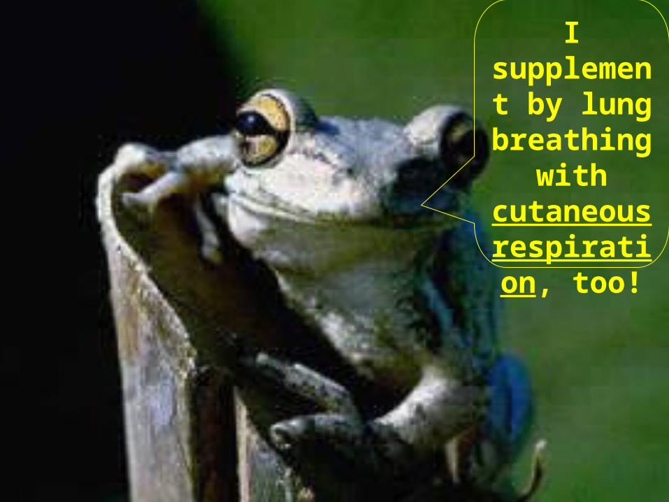
I supplement by lung
breathing with
cutaneous respiration,
too!
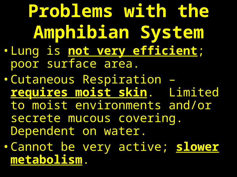
Problems with the Amphibian System
• Lung is not very efficient; poor surface area.
• Cutaneous Respiration – requires moist skin. Limited to moist environments and/or secrete mucous covering. Dependent on water.
• Cannot be very active; slower metabolism.
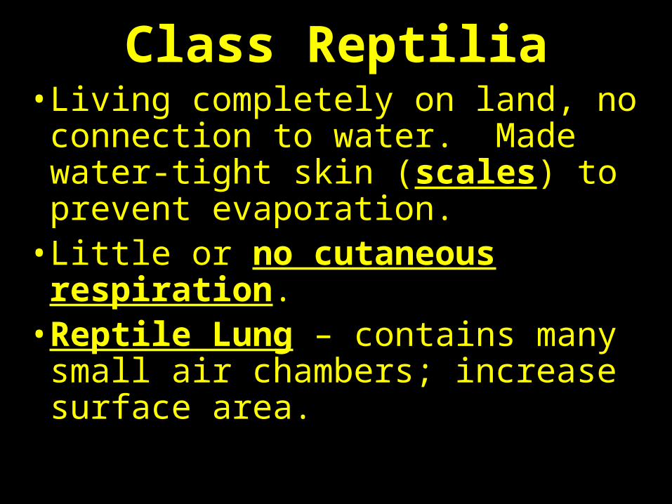
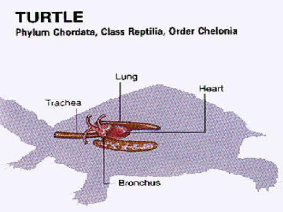
Class Reptilia• Living completely on land, no
connection to water. Made water-tight skin (scales) to prevent evaporation.
• Little or no cutaneous respiration.• Reptile Lung – contains many
small air chambers; increase surface area.
Class Reptilia• Reptiles use negative pressure
breathing – intercostal muscles and diaphragm to expand thoracic cavity and create a negative pressure in lungs.
• Air is pulled into lungs rather than pushed.
• Also called body cavity breathing or chest breathing.
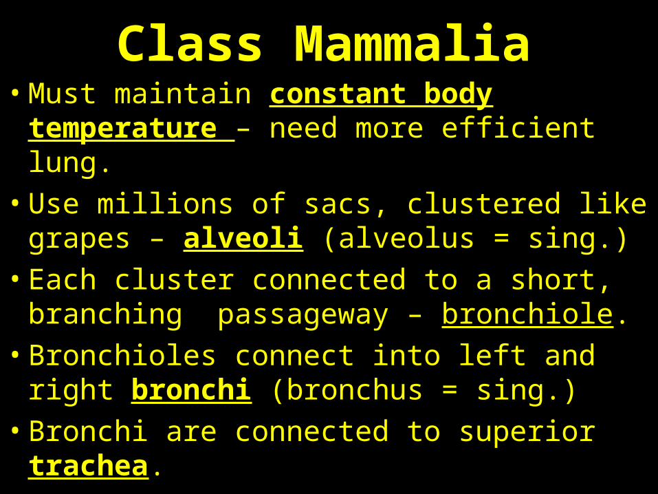
Class Mammalia• Must maintain constant body temperature –
need more efficient lung.
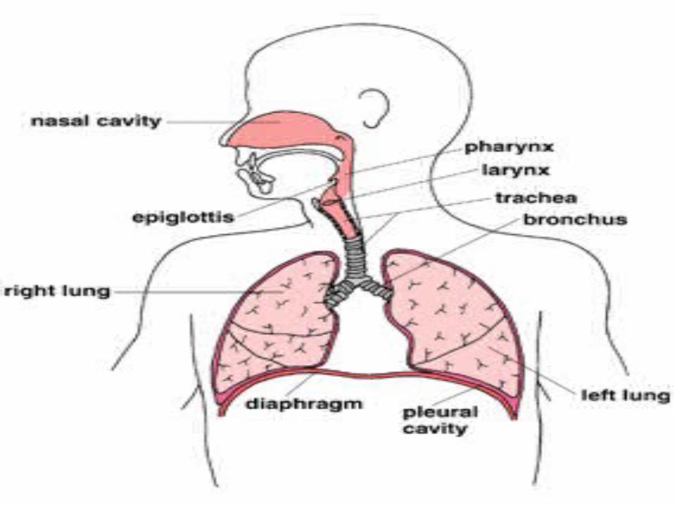
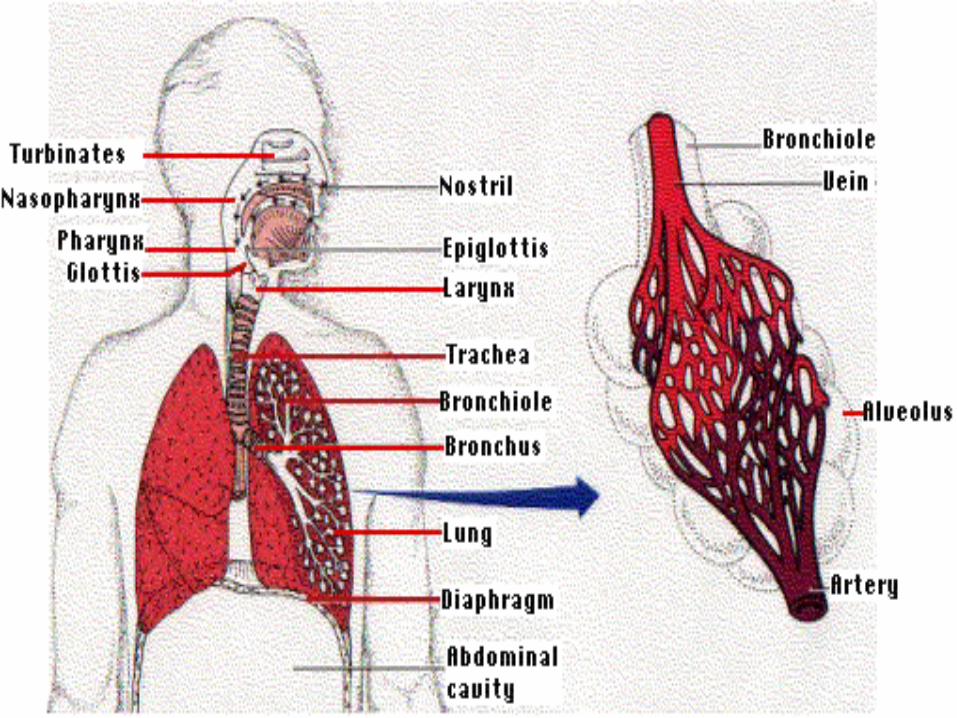
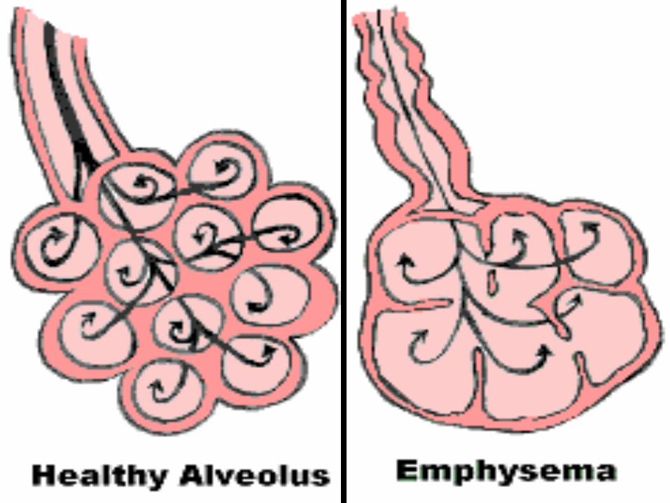
• Use millions of sacs, clustered like grapes – alveoli (alveolus = sing.)
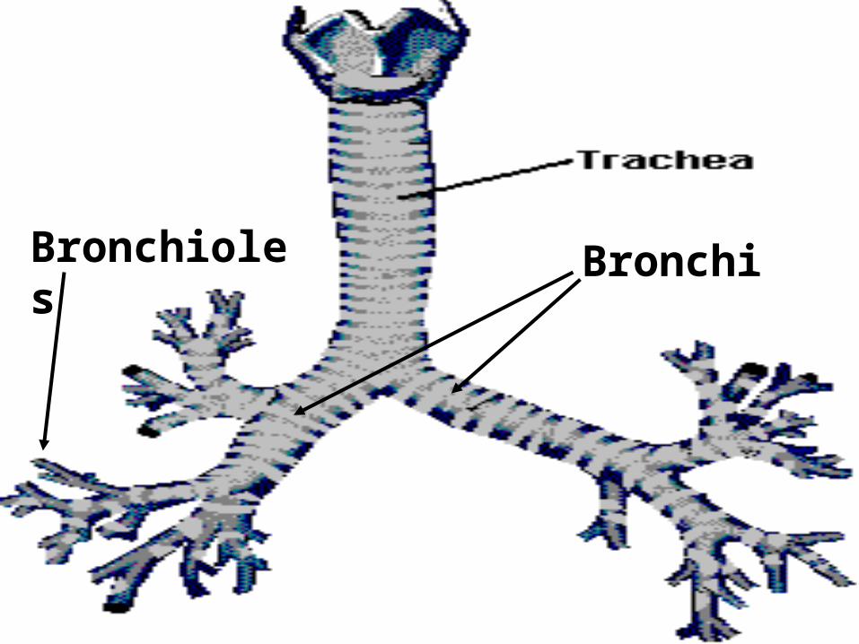
• Each cluster connected to a short, branching passageway – bronchiole.
• Bronchioles connect into left and right bronchi (bronchus = sing.)
• Bronchi are connected to superior trachea.
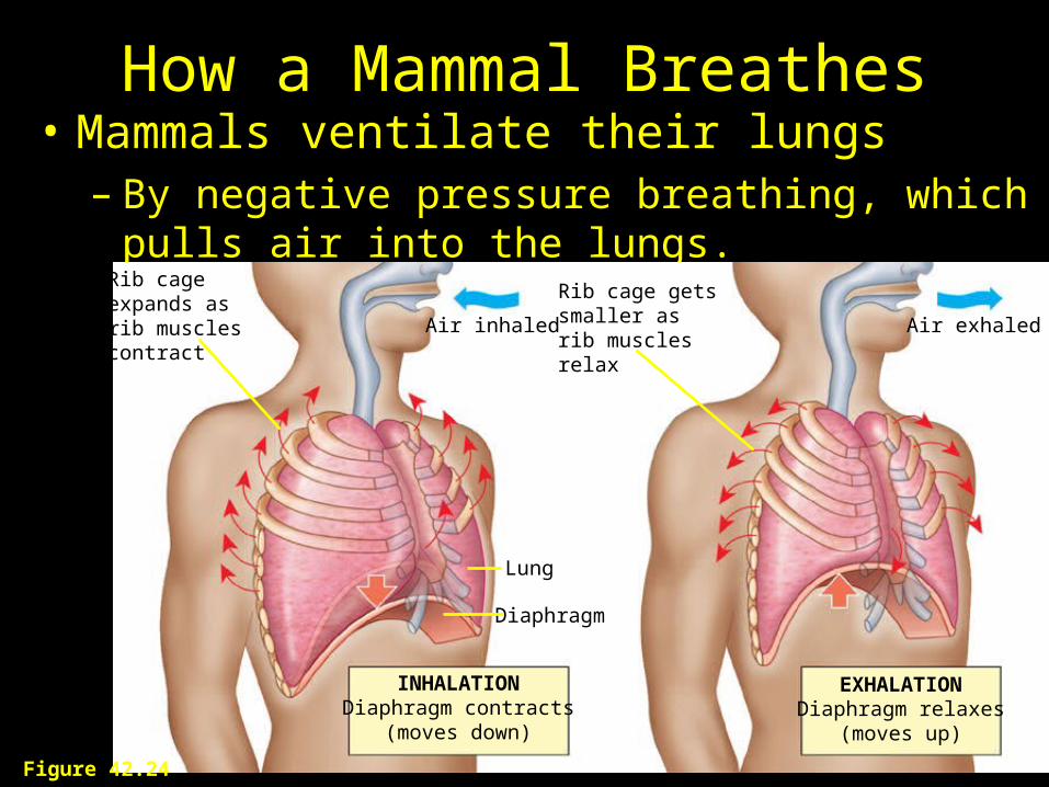
How a Mammal Breathes• Mammals ventilate their lungs
– By negative pressure breathing, which pulls air into the lungs.
Air inhaled Air exhaled
INHALATIONDiaphragm contracts
(moves down)
EXHALATIONDiaphragm relaxes
(moves up)
Diaphragm
Lung
Rib cage expands asrib muscles contract
Rib cage gets smaller asrib muscles relax
Figure 42.24
BronchiBronchioles
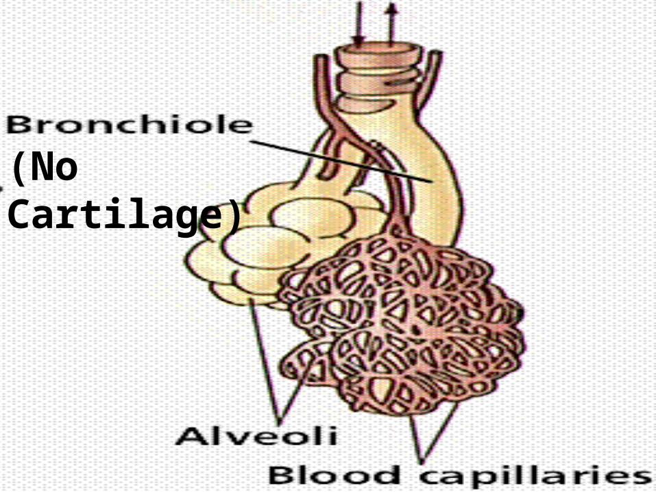
(No Cartilage)
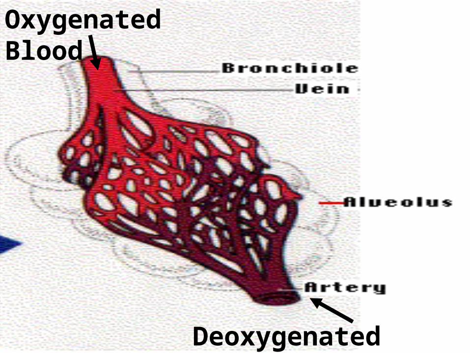
Deoxygenated Blood
Oxygenated Blood
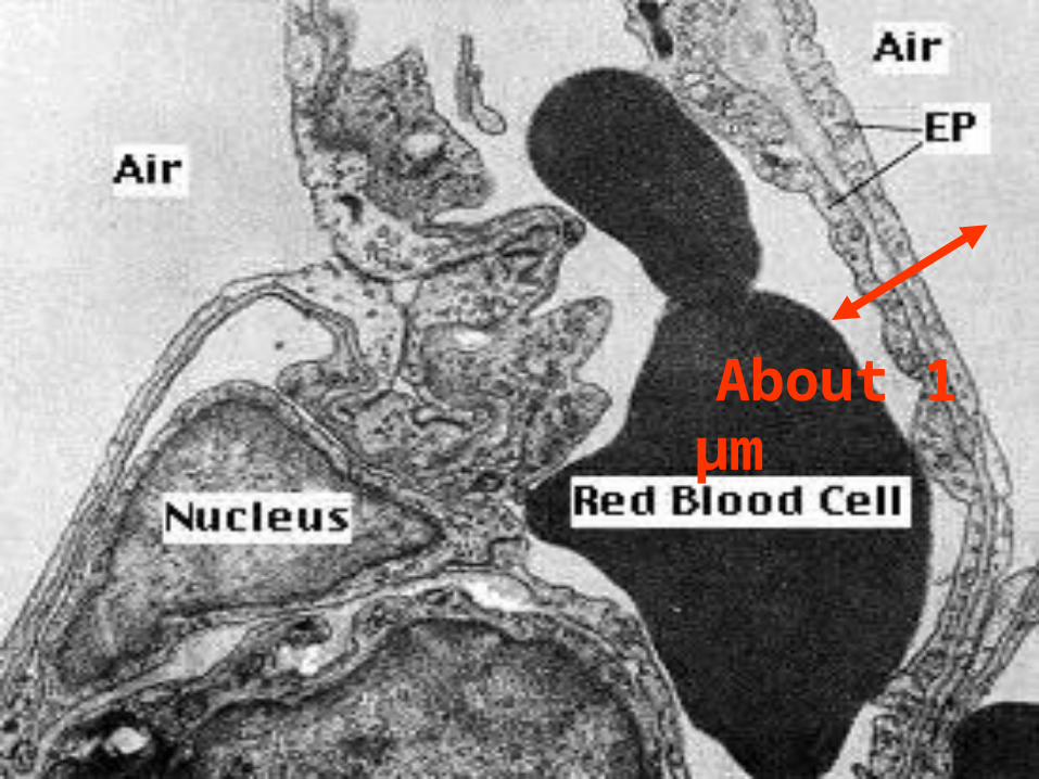
About 1 µm
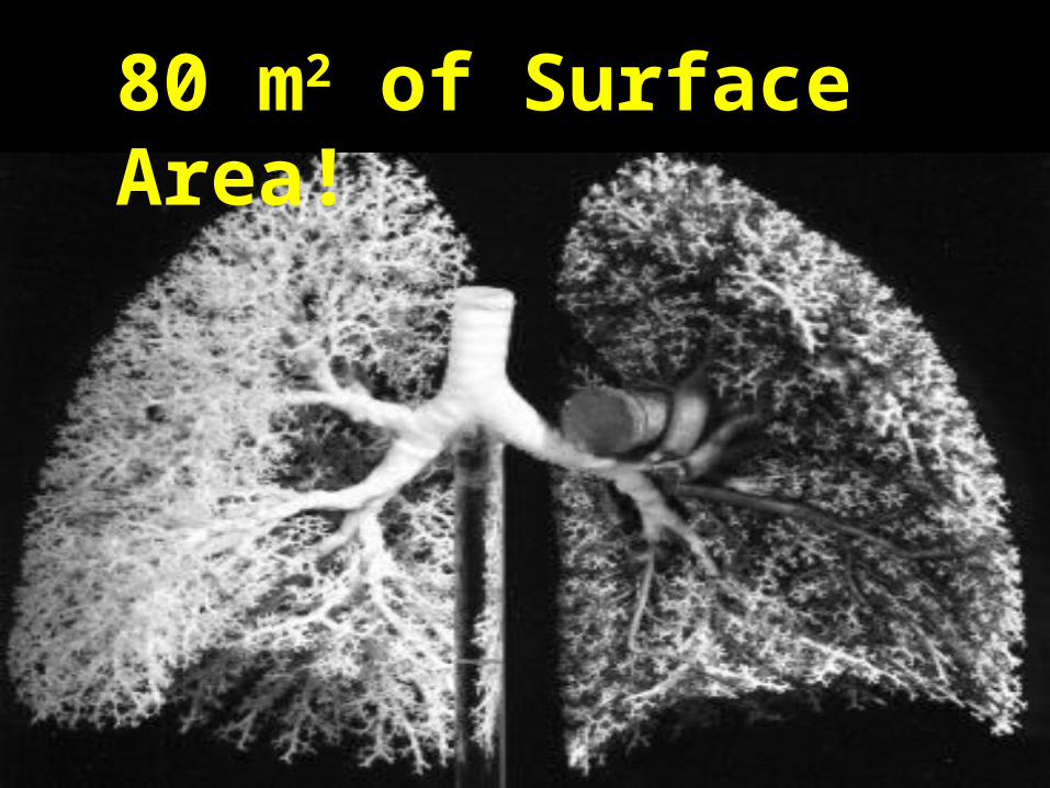
80 m2 of Surface Area!
Mechanics of Human Breathing• Trachea and Bronchi have hyaline cartilage,
but not bronchioles.• Bronchioles are surrounded by smooth
muscle.• Bronchoconstriction – nervous system or
hormones (histamine) signal smooth muscle to contract and narrow bronchioles (asthma).
• Bronchodilation - nervous system or hormones (epinephrine) signal smooth muscle to relax and open bronchioles.
Mechanics of Human Breathing
• Visceral Pleural Membrane – surrounds outside of lung.
• Parietal Pleural Membrane – lines thoracic cavity.
• Pleural Cavity – is fluid-filled space between; connects lung to wall of cavity.
Mechanics of Human Breathing• One-cycle pump.• Inspiration: intercostal muscles and
diaphragm contract = increase volume of thoracic cavity.
• Pleural membranes are coupled, lungs expand.
• Air pressure in lungs is decreased and air is pulled in – negative pressure breathing.
Mechanics of Human Breathing• One-cycle pump.
• Expiration: Intercostal muscles and diaphragm relax, elastic recoil of thoracic cavity = decrease volume of cavity and lungs.
• Air pressure in lungs is increased, forces air out.
Mechanics of Human Breathing• Tidal Volume = amount of air
moved into and out of lungs at rest (500 ml).
• Functional Residual Capacity = amount of air left in lungs after normal expiration at rest.
• Residual Volume = amount of air left after forceful, maximum expiration.
Mechanics of Human Breathing• Anatomical Dead Space = constant
amount of air trapped in trachea, bronchi, bronchioles (150 ml).
• Vital Capacity = max. amount of air exhaled after a forceful, maximum inhalation (VC = TV + IRV + ERV).
• Total Lung Capacity = TV + IRV + ERV + RV
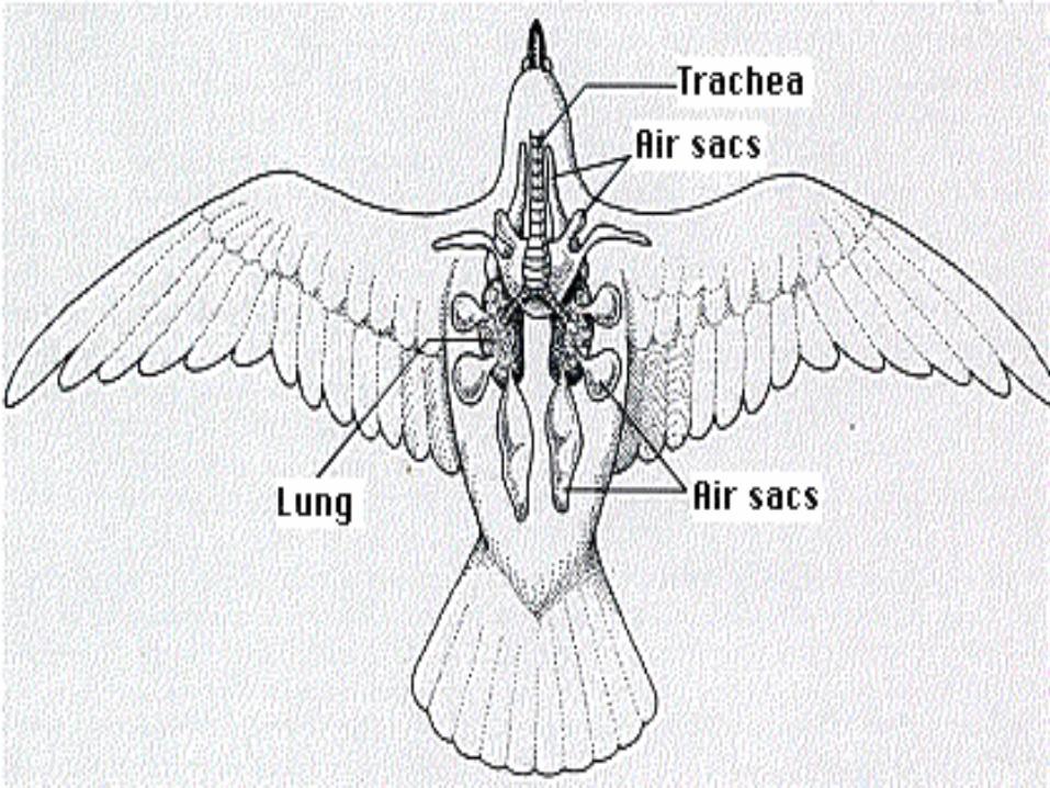
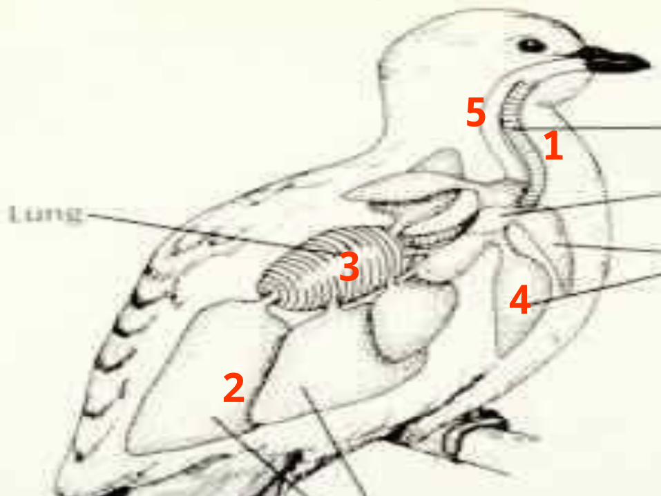
Class Aves
• Flight requires more ATP.
• Avian lung is a two-cycle pump (Fig. 46-9).
• Uses a system of anterior and posterior air sacs and a lung.
• Gas exchange occurs in lung only.
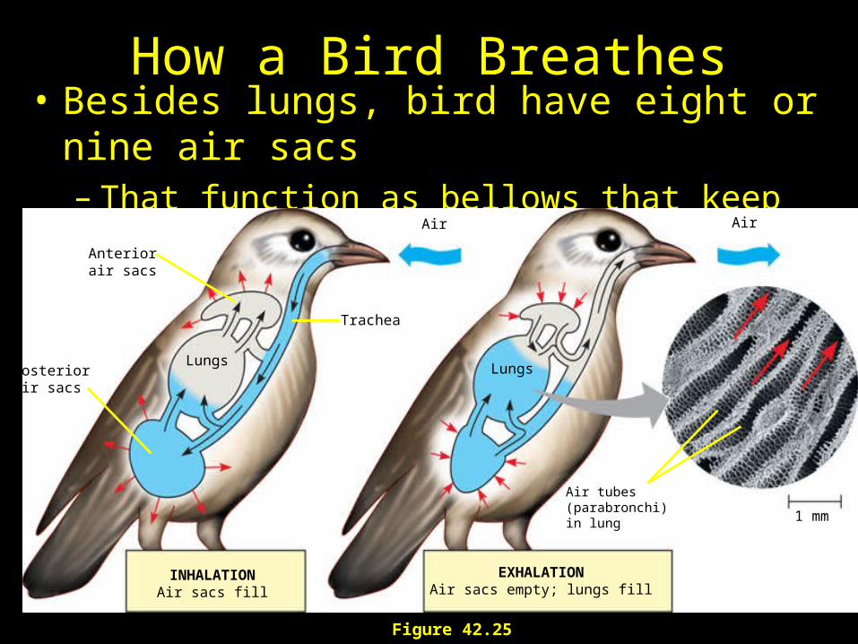
How a Bird Breathes• Besides lungs, bird have eight or nine air sacs
– That function as bellows that keep air flowing through the lungs.
INHALATIONAir sacs fill
EXHALATIONAir sacs empty; lungs fill
Anteriorair sacs
Trachea
LungsLungsPosterior
air sacs
Air Air
1 mm
Air tubes(parabronchi)in lung
Figure 42.25
1
2
34
5
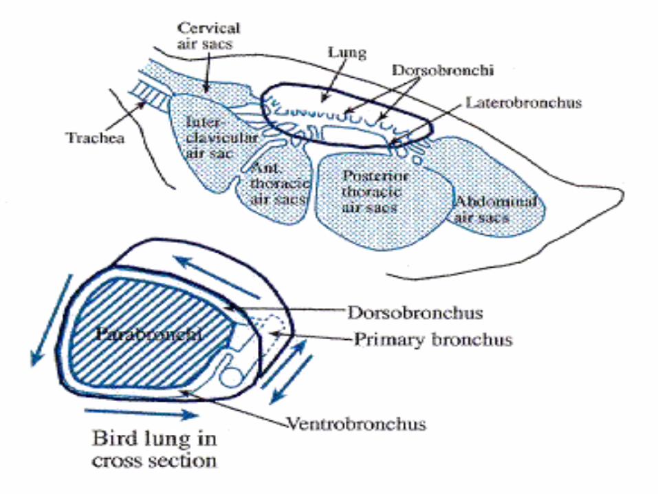
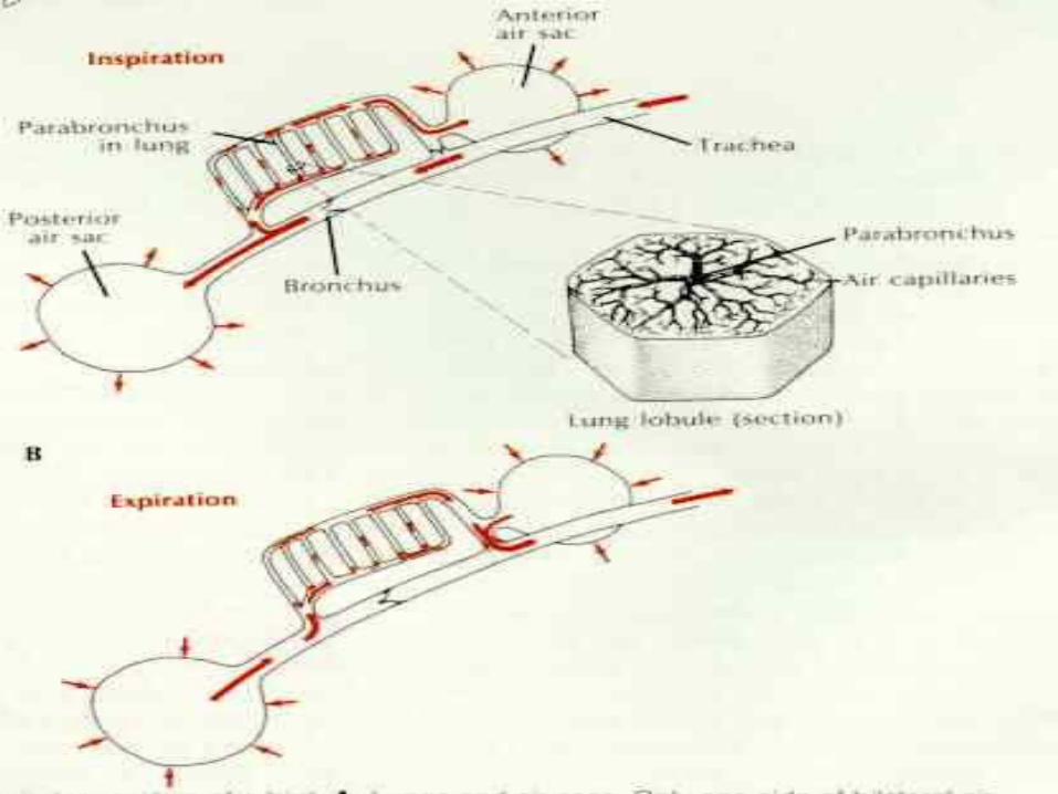
Two-Cycle Breathing• 1st Inspiration – air travels down
trachea to posterior air sacs.• 1st Expiration – air flows from sacs to
lung.• Lung – gas exchange.• 2nd Inspiration – air flows from lung to
anterior air sacs.• 2nd Exhalation – air flows from sacs out
through trachea.
Benefits to Avian Breathing• Unidirectional flow of air through lung
– no “dead volume” of air left in lung. Always fully oxygenated air.
• Flow of blood is perpendicular to air flow – cross-current flow.
• Very efficient at extracting oxygen from air.
• Most efficient terrestrial respiration.
Gas Transport and Exchange
• If transport were by simple diffusion, then O2 would require three years to travel from lung to toe.
• Use a circulatory system; but plasma could only carry 3 ml O2 per l.
• Use RBC with hemoglobin to carry 200 ml O2 per l.
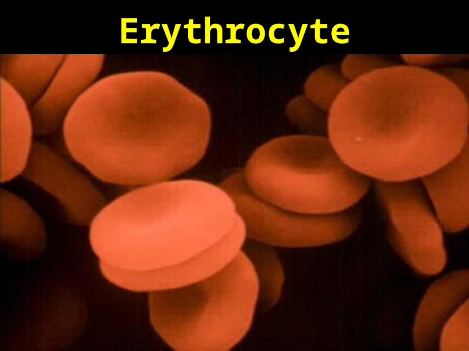
Erythrocyte
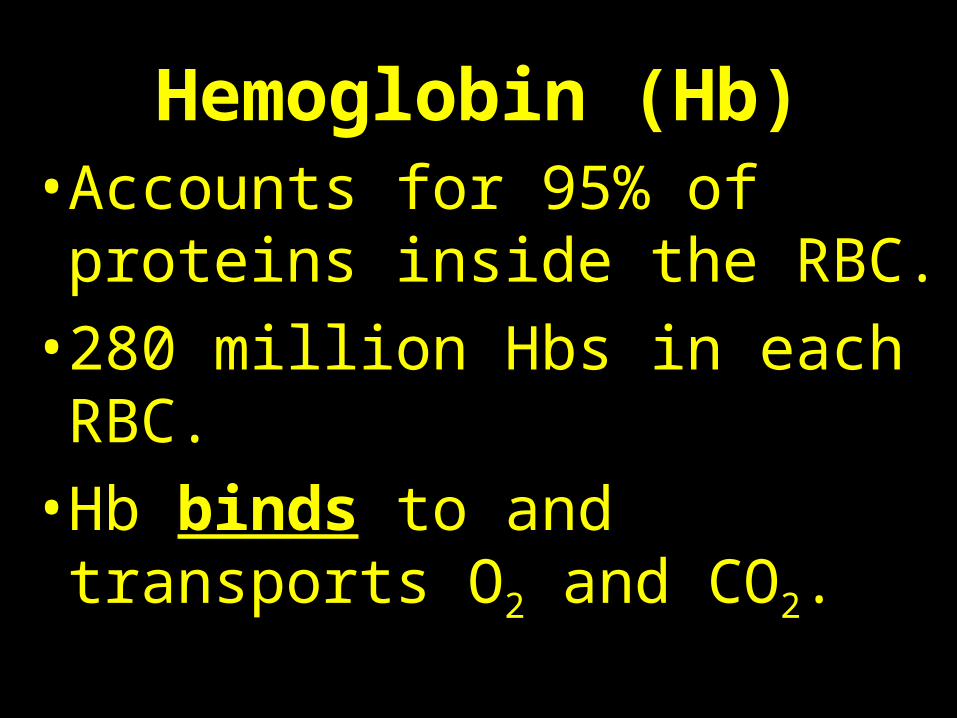
Hemoglobin (Hb)• Accounts for 95% of proteins
inside the RBC.
• 280 million Hbs in each RBC.
• Hb binds to and transports O2 and CO2.
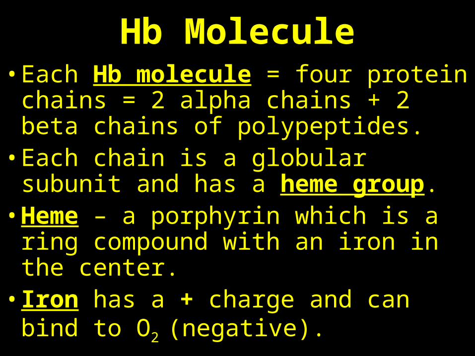
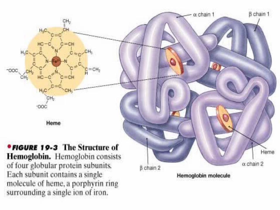
Hb Molecule• Each Hb molecule = four protein chains
= 2 alpha chains + 2 beta chains of polypeptides.
• Each chain is a globular subunit and has a heme group.
• Heme – a porphyrin which is a ring compound with an iron in the center.
• Iron has a + charge and can bind to O2
(negative).
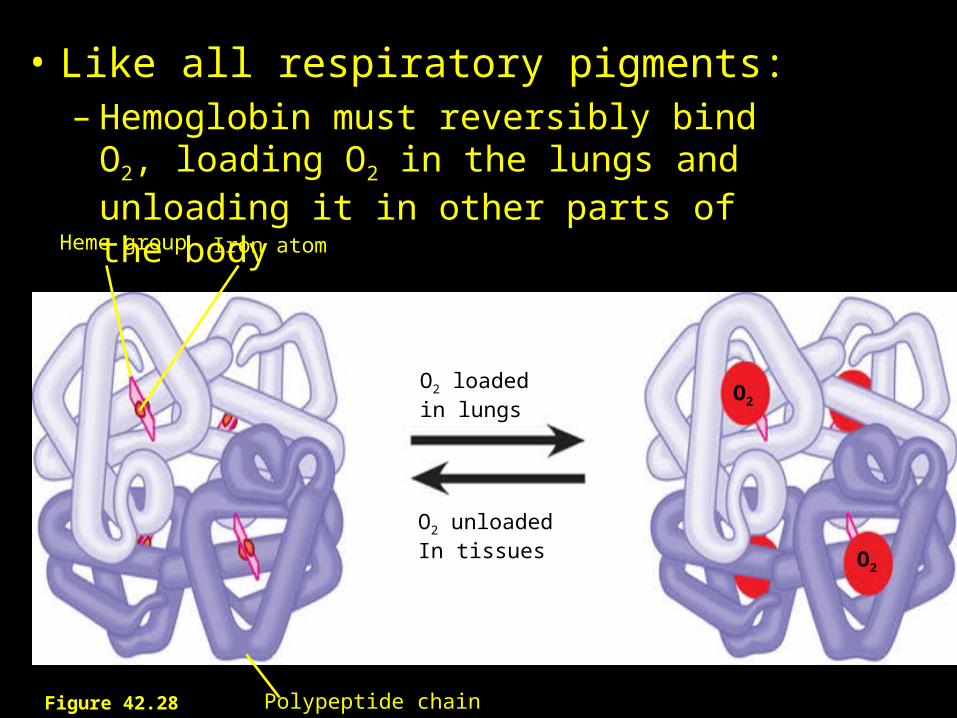
• Like all respiratory pigments:– Hemoglobin must reversibly bind O2, loading
O2 in the lungs and unloading it in other parts of the body
Heme group Iron atom
O2 loadedin lungs
O2 unloadedIn tissues
Polypeptide chain
O2
O2
Figure 42.28

Quaternary Structure of Hemoglobin
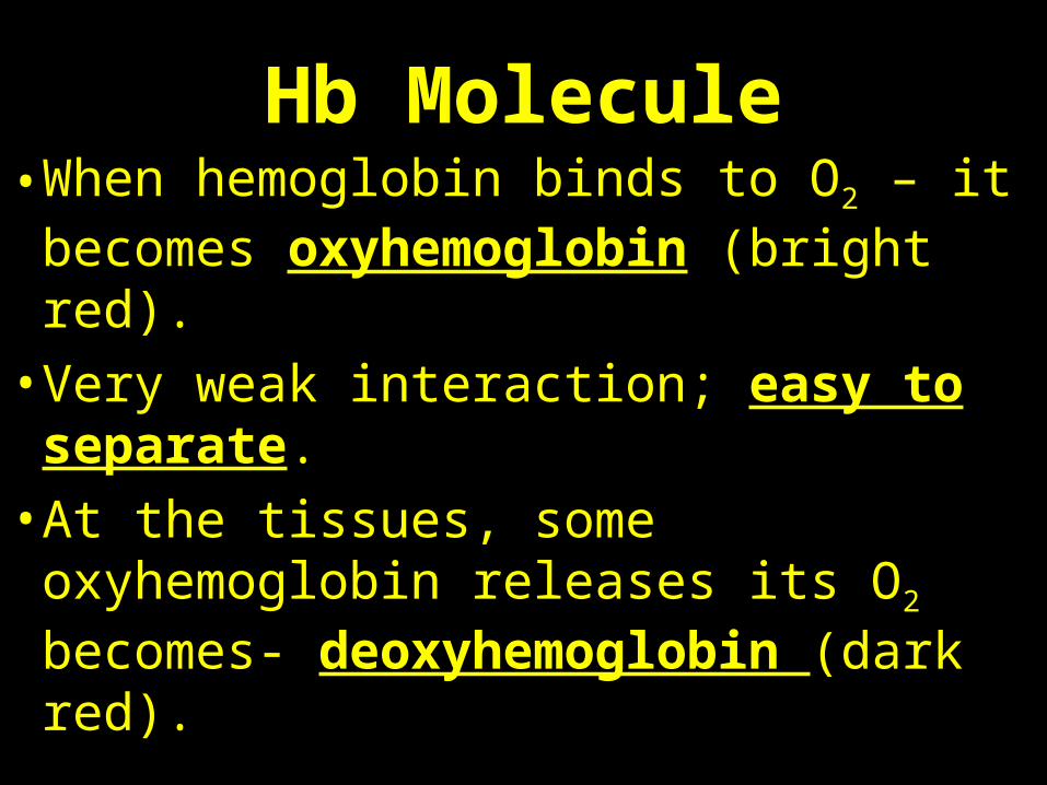
Hb Molecule• When hemoglobin binds to O2 – it
becomes oxyhemoglobin (bright red).
• Very weak interaction; easy to separate.
• At the tissues, some oxyhemoglobin releases its O2 becomes- deoxyhemoglobin (dark red).
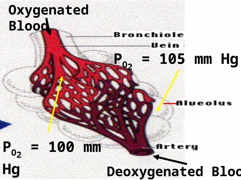
Deoxygenated Blood
Oxygenated Blood
PO2 = 105 mm Hg
PO2 = 100 mm Hg
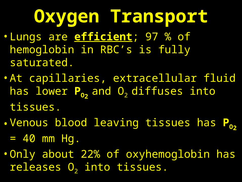
Oxygen Transport• Lungs are efficient; 97 % of hemoglobin
in RBC’s is fully saturated.
• At capillaries, extracellular fluid has lower PO2
and O2 diffuses into tissues.
• Venous blood leaving tissues has PO2 = 40
mm Hg.
• Only about 22% of oxyhemoglobin has releases O2 into tissues.
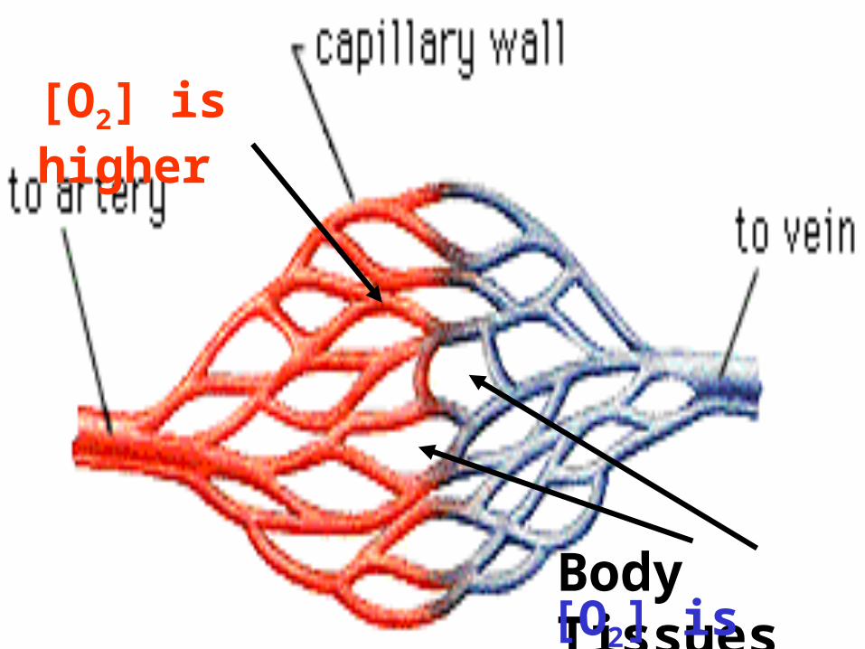
Body Tissues
[O2] is higher
[O2] is lower
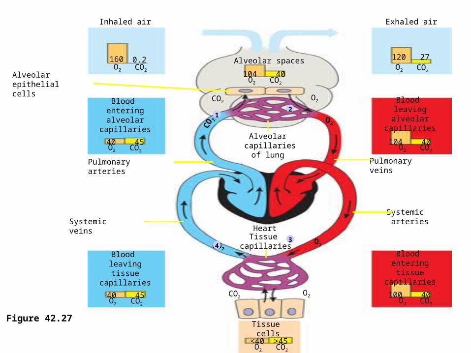
Inhaled air Exhaled air
160 0.2O2 CO2
O2 CO2
O2 CO2
O2 CO2 O2 CO2
O2 CO2 O2 CO2
O2 CO2
40 45
40 45
100 40
104 40
104 40
120 27
CO2O2
Alveolarepithelialcells
Pulmonaryarteries
Blood enteringalveolar
capillaries
Blood leavingtissue
capillaries
Blood enteringtissue
capillaries
Blood leaving
alveolar capillaries
CO2O2
Tissue capillaries
Heart
Alveolar capillaries
of lung
<40 >45
Tissue cells
Pulmonaryveins
Systemic arteriesSystemic
veinsO2CO2
O2
CO 2
Alveolar spaces
12
43
Figure 42.27
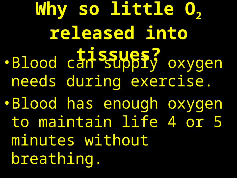
Why so little O2 released into tissues?
• Blood can supply oxygen needs during exercise.
• Blood has enough oxygen to maintain life 4 or 5 minutes without breathing.
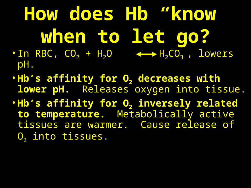
How does Hb “know” when to let go?
• In RBC, CO2 + H2O H2CO3 , lowers pH.• Hb’s affinity for O2 decreases with lower
pH. Releases oxygen into tissue.• Hb’s affinity for O2 inversely related to
temperature. Metabolically active tissues are warmer. Cause release of O2 into tissues.
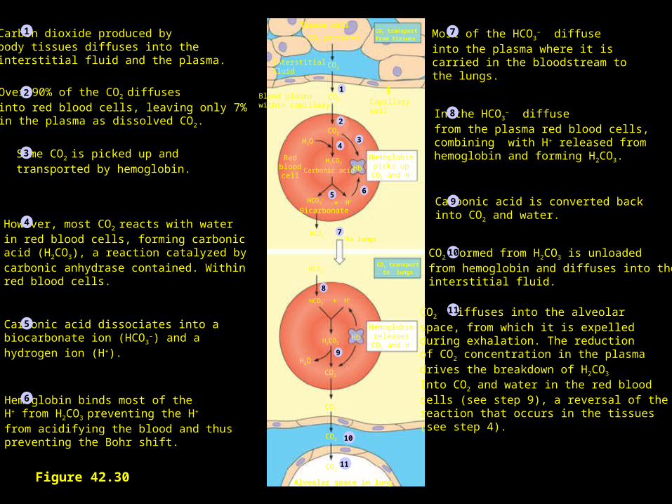
Figure 42.30
Tissue cell
CO2Interstitialfluid
CO2 producedCO2 transportfrom tissues
CO2
CO2
Blood plasmawithin capillary Capillary
wall
H2O
Redbloodcell
HbCarbonic acid
H2CO3
HCO3–
H++Bicarbonate
HCO3–
Hemoglobinpicks up
CO2 and H+
HCO3–
HCO3– H++
H2CO3Hb
Hemoglobinreleases
CO2 and H+
CO2 transportto lungs
H2O
CO2
CO2
CO2
CO2
Alveolar space in lung
2
1
34
56
7
8
9
10
11
To lungs
Carbon dioxide produced bybody tissues diffuses into the interstitial fluid and the plasma.
Over 90% of the CO2 diffuses into red blood cells, leaving only 7%in the plasma as dissolved CO2.
Some CO2 is picked up and transported by hemoglobin.
However, most CO2 reacts with water in red blood cells, forming carbonic acid (H2CO3), a reaction catalyzed bycarbonic anhydrase contained. Withinred blood cells.
Carbonic acid dissociates into a biocarbonate ion (HCO3
–) and a hydrogen ion (H+).
Hemoglobin binds most of the H+ from H2CO3 preventing the H+ from acidifying the blood and thuspreventing the Bohr shift.
CO2 diffuses into the alveolarspace, from which it is expelledduring exhalation. The reductionof CO2 concentration in the plasmadrives the breakdown of H2CO3 Into CO2 and water in the red bloodcells (see step 9), a reversal of the reaction that occurs in the tissues (see step 4).
Most of the HCO3– diffuse
into the plasma where it is carried in the bloodstream to the lungs.
In the HCO3– diffuse
from the plasma red blood cells, combining with H+ released from hemoglobin and forming H2CO3.
Carbonic acid is converted back into CO2 and water.
CO2 formed from H2CO3 is unloadedfrom hemoglobin and diffuses into the interstitial fluid.
1
2
3
4
5
6
7
8
9
10
11
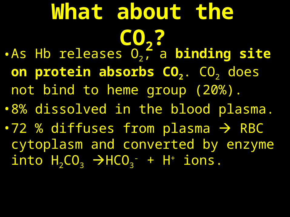
What about the CO2?
• As Hb releases O2, a binding site on protein absorbs CO2. CO2 does not bind to heme group (20%).
• 8% dissolved in the blood plasma. • 72 % diffuses from plasma RBC
cytoplasm and converted by enzyme into H2CO3 HCO3
- + H+ ions.
O2 unloaded fromhemoglobinduring normalmetabolism
O2 reserve that canbe unloaded fromhemoglobin totissues with highmetabolism
Tissues duringexercise
Tissuesat rest
100
80
60
40
20
0
100
80
60
40
20
0
100806040200
100806040200
Lungs
PO2 (mm Hg)
PO2 (mm Hg)
O2 s
atur
atio
n of
hem
oglo
bin
(%)
O2 s
atur
atio
n of
hem
oglo
bin
(%)
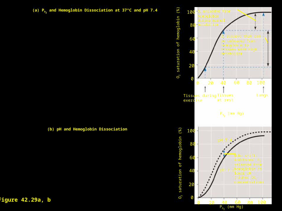
Bohr shift:Additional O2
released from hemoglobin at lower pH(higher CO2
concentration)
pH 7.4
pH 7.2
(a) PO2 and Hemoglobin Dissociation at 37°C and pH 7.4
(b) pH and Hemoglobin Dissociation
Figure 42.29a, b
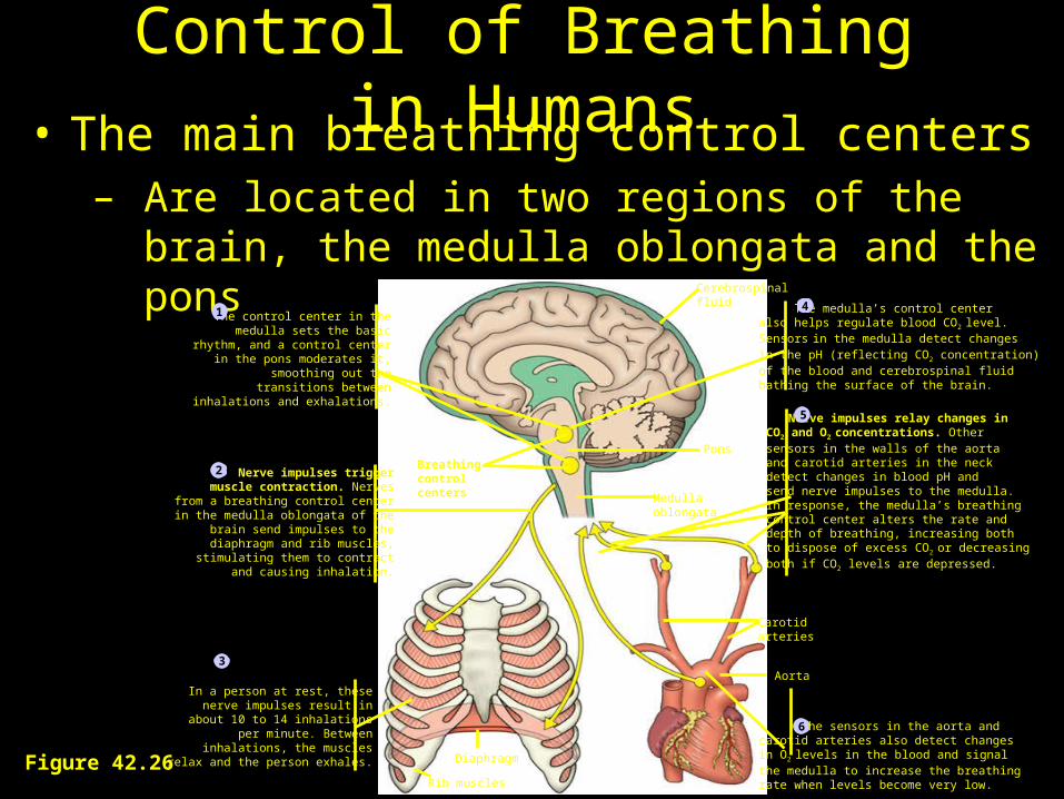
Control of Breathing in Humans• The main breathing control centers
– Are located in two regions of the brain, the medulla oblongata and the pons
Figure 42.26
PonsBreathing control centers Medulla
oblongata
Diaphragm
Carotidarteries
Aorta
Cerebrospinalfluid
Rib muscles
In a person at rest, these nerve impulses result in
about 10 to 14 inhalationsper minute. Between
inhalations, the musclesrelax and the person exhales.
The medulla’s control center also helps regulate blood CO2 level. Sensors in the medulla detect changes in the pH (reflecting CO2
concentration) of the blood and cerebrospinal fluid bathing the surface of the brain.
Nerve impulses relay changes in
CO2 and O2 concentrations. Other sensors in the walls of the aortaand carotid arteries in the neck detect changes in blood pH andsend nerve impulses to the medulla. In response, the medulla’s breathingcontrol center alters the rate anddepth of breathing, increasing bothto dispose of excess CO2 or decreasingboth if CO2 levels are depressed.
The control center in themedulla sets the basic
rhythm, and a control centerin the pons moderates it,
smoothing out thetransitions between
inhalations and exhalations.
1
Nerve impulses trigger muscle contraction. Nerves
from a breathing control centerin the medulla oblongata of the
brain send impulses to thediaphragm and rib muscles, stimulating them to contract
and causing inhalation.
2
The sensors in the aorta andcarotid arteries also detect changesin O2 levels in the blood and signal the medulla to increase the breathing rate when levels become very low.
6
5
3
4
Controlling Breathing• Respiratory Control Center – Medulla
Oblongata in brain.
• Impulses sent to diaphragm and intercostal muscles contraction and expand thoracic cavity (inhalation).
• No impulse, muscles relax and cavity becomes smaller (exhalation).
• Part of ANS but can be voluntary.
Controlling Breathing• If breathing stops, the PCO2 of plasma
rises.• Causes pH to drop (increase in [H+]).• Peripheral chemoreceptors in walls of
aorta and coratid arteries detect increase in [H+].
• Send signals to respiratory control center.• Initiates breathing.
What does exercise do?
• Working tissue causes ↑ PCO2 in
plasma and ↓in pH.
• As [H+] ↑, chemoreceptors cause an ↑ in respiratory rate.
• Can you indefinitely hyperventilate?
• Why can people hold their breath longer if they hyperventilate first?
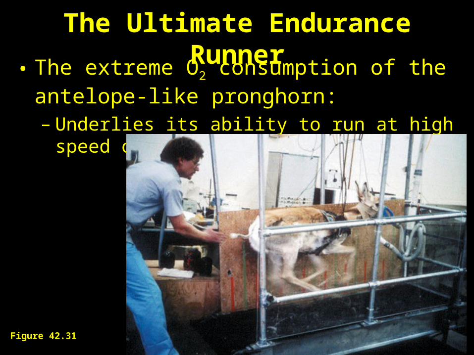
The Ultimate Endurance Runner• The extreme O2 consumption of the antelope-like
pronghorn:– Underlies its ability to run at high speed over long
distances
Figure 42.31