Embed Size (px)
Citation preview
JOURNAL OF BACTERIOLOGY, Nov. 1970, p. 826-833 Vol. 104, No. 2Copyright 0 1970 American Society for Microbiology Printed In U.S.A.
Citric Acid Cycle: Gene-Enzyme Relationshipsin Bacillus subtilis
BLANKA RUTBERGI AND JAMES A. HOCHDepartment ofMicrobiology, Scripps Clinic and Research Foundation, La Jolla, California 92037
Received for publication 6 July 1970
The genetic location of mutations affecting the citric acid cycle and the propertiesof mutants of Bacillus subtilis possessing these mutations have been examined. Genescoding for the component enzymes of the cycle were found to be unlinked to eachother and thus do not form an operon. The mutational defect in a mutant lackingfumarase mapped between thr-5 and cysB3. Mutations causing inability to produceisocitrate dehydrogenase and succinate dehydrogenase were found to map betweenargAIl and leu-1. The a-ketoglutarate dehydrogenase mutations were mapped at theterminal end of the B. subtilis chromosome through a weak linkage in phage PBS-1transduction of one class of these mutations to ilvA2 and metB4. A second class ofa-ketoglutarate dehydrogenase mutations mapped closer to ilvA2 and metB4 but stillterminal with respect to these markers. Aconitaseless mutants possessed mutationsthat could not be linked to any of the known transducing segments of the chromo-some. An effect of mutation conferring loss of one enzyme of the cycle on the specificactivity of the other enzymes in the cycle was observed.
The citric or tricarboxylic acid cycle plays acentral role in the metabolism of Bacillus speciesand of most cells. The specific activity of certainenzymes of the cycle has been shown to vary de-pending on the carbon source used for growth(13). Growth on glucose relegates the cycle to ananabolic role and, if glutamate is supplied, thespecific activity of both aconitase [EC 4.2.1.3,citrate (isocitrate) hydro-lyase] and citratesynthase (EC 4.1.3.7, citrate oxalacetate lyase)(7) may be shown to decline. The exhaustion ofglucose in the medium brings about an increasein the specific activity of the enzymes of the cycle,and the increase is coincident with the onset ofsporulation (14, 15). The coincidence of the twoevents led to the hypothesis that the increase inspecific activity was a required event for sporula-tion (14). The isolation of mutants devoid ofaconitase activity in which sporulation was de-creased pointed to a connection between citricacid cycle activity and sporulation (12). A similarinhibition of sporulation is obtained merely byinhibiting aconitase with chelating agents (8).Thus, the movement of substrates through thecycle is most important for successful sporula-tion.
Freese and Fortnagel (9, 10) examined a largenumber of sporulation-defective mutants for the
I Present address: Department of Bacteriology, KarolinskaInstitutet, S-10401, Stockholm 60, Sweden.
ability to incorporate uracil into ribonucleic acid(RNA) during sporulation in the presence ofvarious carbon sources. Among these sporula-tion-defective mutants were those blocked in thesynthesis of aconitase, isocitrate dehydrogenase[EC 1. 1. 1. 42, L-isocitrate: nicotinamide adeninedinucleotide phosphate (NADP) oxidoreduc-tase], a-ketoglutarate dehydrogenase (EC1.2.4.2, 2-oxoglutarate: lipoate oxidoreductase),and succinate dehydrogenase (EC 1. 3. 99. 1,succinate: oxidoreductase). Mutants blocked inthe citric acid cycle did not incorporate uracilwell even in the presence of ribose, suggestingthat the production of adenosine triphosphate(ATP) was limited by the mutational block.Thus, the poor sporulation of citric acid cyclemutants might be owing to a deficiency of ATPduring development. Direct ATP measurementson these mutants showed that the concentrationof ATP does decline after growth ceases and thatATP production may be restored by supplyingthe compound after the block (20). The restora-tion of sporulation is more difficult and has onlybeen observed in one mutant (29).Although much physiological data has ap-
peared on the properties of citric acid cyclemutants, nothing has been known of the geneticrelationships among these mutants. In an earlierreport (B. Rutberg and J. Hoch, 1969, Bacteriol.Proc., p. 54; reference 19), we indicated the
826
Dow
nloa
ded
from
http
s://j
ourn
als.
asm
.org
/jour
nal/j
b on
31
Janu
ary
2022
by
88.1
29.2
22.2
01.

CITRIC ACID CYCLE IN B. SUBTILIS
position on the chromosome map of some muta-tions affecting the citric acid cycle in relation tosporulation genes. The present study shows thegenetic location of genes for the citric acid cycleenzymes and some further properties of themutants.
MATERIALS AND METHODSBacteria. All bacterial strains used in this study were
derived from B. subtilis 168. The basic strains used inthe genetic experiments are listed in Table 1. The ma-jority of the cit- mutants were obtained by muta-genesis of strain 168 with N-methyl-N'-nitro-N'-nitro-soguanidine (Aldrich Chemical Co.) essentially by themethod of Adelberg et al. (1).
Media. Nutrient sporulation medium (NSMP) wasdescribed by Freese and Fortnagel (10) and minimalmedium by Spizizen (30). PAB is antibiotic mediumno. 3 (Difco). AK plates contain AK agar no. 2 (BBL)supplemented with 5 mm MgSO4. TBAB-Glu platesare made from Tryptose Blood Agar Base (Difco) plus1% glucose, 0.05 mm MnCI2, and 15 g of agar per liter.In minimal lactate plates, the glucose is replaced with0.5% sodium lactate.
Dissimilation of [14C] glutamate. A modification ofthe procedure of Fortnagel and Freese (9) was used.Bacteria from an overnight TBAB-Glu plate wereinoculated into PAB and grown to stationary phase byshaking for 4 hr at 37 C. Cells (10 ml) were centrifugedand resuspended in an equal volume of minimal me-dium containing 50 ,g of chloramphenicol per ml. Thecells were incubated with shaking at 37 C for 15 min,at which time 5 ,g of unlabeled glutamate per ml and1 ,uCi of [14C]glutamate (uniformly labeled, 167 Ci/mole; Schwarz BioResearch Inc.) per ml were added.After 5 min of further incubation, the cells were har-vested by centrifugation, washed in 5 ml of water, re-suspended in 0.4 ml of water, and boiled for 30 min.The cells were removed by centrifugation, and the ex-tracts were stored at -20 C. Extract (20 ,liters) wasapplied to cellulose thin-layer plates (MN-polygramcell 300, Brinkman Instruments) with a reference solu-tion (1 ,uliter) which was 0.1 M with respect to citrate,a-ketoglutarate, succinate, and malate. The plateswere chromatographed in a solvent system of ether-formic acid-water (7:2:1) (24). Autoradiographs ofthe chromatogram were made by exposure to Kodakmedical X-ray film for 5 days. The reference com-pounds were detected by spraying with 0.1% 2,6-di-chlorophenol-indophenol in ethanol (27).
Preparation of extracts. Bacteria from an overnightTBAB-Glu plate were inoculated into 50 to 100 ml ofNSMP in a 1-liter flask and grown with shaking at37 C for 4 hr (for a-ketoglutarate dehydrogenase) or6 hr (for all other enzymes). The cells were harvestedby centrifugation, washed with 0.05 M potassiumphosphate (pH 7.4), and suspended in 2 ml of thesame. Lysozyme (150,ug/ml) was added, and the sus-pension was incubated at 37 C until lysis (15 to 30min). The lysate was centrifuged at 10,000 X g for 15min. This supernatant was immediately assayed foraconitase and fumarase (EC 4.2.1.2, L-malate hydro-lyase) because of instability of these two enzymes. The
TABLE 1. Basic strains
Strain Genotype Origin
BR16 trpC2, lys-l B. ReillyGSY260 trpC2, ilvD2 C. Anagnosto-
poulosGSYIll trpC2, ilvAl C. Anagnosto-
poulosGSY384 argAII, leu-I C. Anagnosto-
poulosGSY184 ilvCJ, trpC2 C. Anagnosto-
poulosGSY1026 trpC2, metB4 C. Anagnosto-
poulosSB133 phe-J E. NesterBD92 hisAI, cysB3, trpC2 D. Dubnau
other activities were found to be relatively stable tostorage at -20 C. When succinate dehydrogenase wasto be assayed, 5 pAg of deoxyribonuclease was added tothe lysate, and the lysate was centrifuged at 35,000 Xg for 20 min. The pellet was suspended in 2 ml of phos-phate buffer and assayed.Enzyme assays. All assays were carried out at 25 C.
Activities were calculated as nanomoles of substrateconverted per minute per milligram of protein. Proteinwas determined by the method of Lowry et al. (22).The conversion of isocitrate to cis-aconitate was
followed spectrophotometrically at 240 nm by themethod of Racker (28). The molar extinction coeffi-cient for cis-aconitate was taken to be 3.3 X 10³ percm per mole (23).The isocitrate-dependent reduction of NADP to
reduced NADP (NADPH) was measured at 340 nm bythe method of Ochoa (25). The molar extinction coeffi-cient for NADPH or NADH was taken to be 6.22 X10³ per cm per mole.The a-ketoglutarate dehydrogenase reaction was
measured by following the reduction of nicotinamideadenine dinucleotide (NAD) at 340 nm by the methodof Bachmann, Allmann, and Green (4). Antimycin Awas excluded from the reaction mixture.The succinate-dependent reduction of indophenol
to leucoindophenol was followed spectrophotometri-callyat 600 nm by the method of Green et al. (11). Themolar extinction coefficient was taken to be 16.1 X10³ per cm per mole.The reduction of malate to fumarate was followed
spectrophotometrically at 240 nm by the method ofHanson and Cox (13). The molar extinction coefficientfor fumarate was taken to be 2.4 X 10³ per cm permole (6).
Malate dehydrogenase (EC 1.1.1. 37, L-malate:NAD oxidoreductase) was measured by following themalate-dependent reduction of NAD at 340 nm bythe method of Ochoa (25).
Genetic analysis. The methods for transformation(3) and transduction (18) were described. Selectionfor auxotrophic markers by using either transforma-tion or transduction was accomplished by plating onthe appropriately supplemented minimal medium.Selection for cit+ was done by plating on minimal-
VOL. 104, 1970 827
Dow
nloa
ded
from
http
s://j
ourn
als.
asm
.org
/jour
nal/j
b on
31
Janu
ary
2022
by
88.1
29.2
22.2
01.

RUTBERG AND HOCH
lactate medium. The frequency of recombination intwo-factor crosses is defined as 100 (1 - cotransfer)(reference 18).The recombination index method (5) was used to
show linkage of citF markers. Deoxyribonucleic acid(DNA) from citFIl trp+ and wild type was used totransform a citF8 trpC2 strain and a citF12 trpC2strain for cit+ and for trp+. The ratio cit+ to trp+ wascalculated for each donor, and the recombination in-dex was defined as the ratio cit- /trp+ (citFIl donor)divided by the ratio cit+/trp4 (wild donor).
RESULTSisolaion of cit- mutants During the isolation
of mutants of B. subtilis defective in sporulation,a large group of mutants appeared which had acharacteristic red pigmentation on AK sporula-tion agar. Some of these were found to requireglutamic acid, and the majority were unable toutilize lactic acid as sole carbon and energysource. This result suggested a block in citric acidcycle activity and led us to investigate the mutantsfurther. Genetic analysis was initiated by out-crossing the mutants to a reference strain tosegregatethe cit- defect from any other mutationsinduced by the mutagenesis procedure. DNAfrom the cir mutants was used at saturating con-centrations to transform BR16 to lysine inde-pendence. Among the lys+ transformants, cirtransformants were found as a result of the in-tegration of two independent DNA fragments.These strains were purified and used for allsubsequent experiments.
Preliminary lassification of cit mutants on thebasis of ['4C]glutamate disimilation. Fortnageland Freese (9) introduced a method for deter-mining enzymatic blocks in the citric acid cycleby scoring for the formation of radioactive inter-mediates from [t4C]glutamic acid. Each Cirmutant strain was subjected to this analysis byincubating a culture with [14C]glutamate in thepresence of chloramphenicol. After incubation,extracts were prepared, and distribution of label
TABLE 2. Dissimilation of [14C]glutamatein cit- mutants
Mutants Predominant labeledcompounds accumulated
cit-i, cit-3, cit-6, cit-10, Citrate or isocit-cit-15 rate, or both
cit-S, cit-7, cit-9, cit-14, cit- a-Ketoglutarate16, cit-18, cit-26, cit-28,cit-29, cit-33, cit-35
cit-2, cit-8, cit-li, cit-12 Succinate
cit-4 Fumarate and suc-cinate
among citric acid cycle intermediates was de-termined by thin-layer chromatography andautoradiography. The results of this analysis arepresented in Table 2.The glutamic acid-requiring mutants, cit-10,
cit-15, cit-i, cit-3, and cit-6, were found to ac-cumulate citrate or isocitrate, or both. Thesecompounds are not resolved in our solvent sys-tem. A small amount of labeled cis-aconitate wasfound for cit-i, cit-3, and cit-6 but not cit-10 andcit-15, suggesting an isocitrate dehydrogenasedefect in the former three. This suggestion wassupported by their growth response to isocitrateor a-ketoglutarate. Both isocitrate and a-keto-glutarate replaced glutamate for the growth ofcit-10 and cit-15, but only a-ketoglutarate waseffective in cit-i, cit-3, and cit-6. Since our mini-mal medium contains citrate, this result suggeststhat cit-10 and cit-15 cannot convert citrate toisocitrate and may be defective in aconitase.
Eleven mutants were found to accumulate a-ketoglutarate plus a small amount of succinate.A block in a-ketoglutarate dehydrogenase wouldbe expected to accumulate a-ketoglutarate; thepresence of succinate, however, was unexpected.Succinate might be formed from the conversionof a-ketoglutarate to isocitrate and subsequentcleavage to glyoxalate and succinate via isocitratelyase. Fortnagel and FRese (9) suggest, however,that this shunt is absent in B. subtilis since theirstrains do not use acetate as a carbon and energysource. Our strains, on the other hand, are ableto use acetate, albeit slowly, and J. Szulmajster(personal communication) has found substantiallevels of isocitrate lyase and malate synthase inextracts of B. subtilis. Alternate routes to succi-nate may be envisioned, however, such as viaglutamic decarboxylase or through the decarbox-ylation of a-ketoglutarate resulting from theextraction procedure. The accumulation of a-ketoglutarate in these 11 mutants is indicative ofa block in a-ketoglutarate dehydrogenase what-ever the route of succinate formation.Mutants which accumulate succinate pre-
dominantly, such as cit-2, cit-8, cit-il, and cit-12,were classified as lacking in succinate dehydro-genase. One mutant, cit-4, was found to accumu-late fumarate and succinate and was tentativelyclassified as a fumarase defect.Enzymatic assay of cit mutants. The tentative
assignment of enzyme blocks from accumulationstudies was found to hold in in vitro enzymeassays. Table 3 shows the results of these assays.In addition to confirming the tentative classifica-tion, some interesting effects of the mutant blockson the specific activity of other enzymes of thecycle are apparent. The aconitase mutants have atwo- to threefold increased level of isocitrate
828 J. BACrER1oL.
Dow
nloa
ded
from
http
s://j
ourn
als.
asm
.org
/jour
nal/j
b on
31
Janu
ary
2022
by
88.1
29.2
22.2
01.

CITRIC ACID CYCLE IN B. SUBTILIS
TABLE 3. Specific activity ofcitric acid cycle enzymes in B. subtilis 168 wild-type and cit mutants"
Mutant Isocitrate a-Ketoglu- SuccinateExtract Aconitase dehydro- tarate dehy- dehydro- Fumarase Malate dehydrogenasegenase drogenase genase
Wild type 241 351 13 40 1.6 X 10⁸ 5.2 X 10'cit-10 <0.5 790 10 21 1.0 X 10⁸ 10.3 X 10'cit-15 <0.5 689 14 49 1.2 X 10⁸ 9X4 X 10'cit-17 <0.5 856 10 68 1.1 X 10³ 8.6 X 10'cit-25 <0.5 556 13 47 1.9 X 10' 5.6 X 10'cit-i 966 <1 11 79 1.5 X 10' 9.7 X 10'cit-3 1,552 <1 5 47 1.2 X 10' 9.3 X 10'cit-6 1,352 <1 8 54 1.6 X 10' 8.5 X 10'cit-S 17 164 <0.2 33 1.1 X 10' 2.7 X 10'cit-7 19 267 <0.2 25 1.2 X 10' 3.3 X 10'cit-9 18 170 <0.2 37 1.2 X 10' 4.8 X 10'cit-14 23 224 <0.2 51 1.3 X 10' 2.7 X 10'cit-16 16 151 <0.2 58 1.1 X 10' 3.4 X 10'cit-18 13 147 <0.2 61 1.2 X 10³ 2.7 X 10³cit-26 14 73 <0.2 30 1.0 X 10³ 3.3 X 10'cit-28 11 89 <0.2 39 1.0 X 10³ 2.8 X 10'cit-29 19 57 <0.2 29 0.9 X 10' 4.4 X 10³cit-33 13 59 <0.2 20 1.1 X 10³ 3.0 X 10³cit-35 8 43 <0.2 20 0.7 X 10' 3.8 X 10'cit-2 28 390 16 <0.5 1.2 X 10³ 4.0 X 10'cit-8 33 110 18 <0.5 0.9 X 10' 3.6 X 10'cit-ll 29 299 21 <0.5 1.8 X 10' 2.5 X 10'cit-12 21 130 15 <0.5 1.0 X 10' 4.5 X 10'cit-4 132 148 11 40 <0.01 X 10' 6.6 X 10'
aSpecific activities are calculated as nanomoles of substrate converted per minute per milligram ofprotein.
dehydrogenase as compared to wild type. Iso-citrate dehydrogenase mutants have as much as asixfold elevated level of aconitase when comparedto wild type and a 100-fold increase when com-pared to a-ketoglutarate dehydrogenase mutants.Although the level of aconitase is low in all a-ketoglutarate dehydrogenase mutants, the mu-tants may be placed in two groups on the basisof their isocitrate dehydrogenase levels. In thefirst group, the specific activity of isocitrate de-hydrogenase is reduced about twofold, whereasthe level of this enzyme in the second group isreduced about fivefold. Studies reported belowwill show that the two groups are also geneticallyseparable. Succinate dehydrogenase mutants arelow in aconitase activity and somewhat reducedin isocitrate dehydrogenase. Finally, the fumarasemutant has about one-half the aconitase andisocitrate dehydrogenase levels of the wild type.The specific activities of a-ketoglutarate de-
hydrogenase, succinate dehydrogenase, fumarase,and malate dehydrogenase do not appear tofluctuate as widely as either aconitase or iso-citrate dehydrogenase in the mutant strains. Thespecific activity of a-ketoglutarate dehydrogenasehas been shown to increase with growth of theculture, and the enzyme is probably not constitu-tive (9). In Escherichia coli, on the other hand, a-
ketoglutarate dehydrogenase levels vary widelywith cultural conditions (2). Under the culturalconditions used here, the specific activities of a-ketoglutarate dehydrogenase, succinate dehydro-genase, fumarase, and malate dehydrogenase arerelatively constant and are unchanged by muta-tion in the cycle.
Genetic studies: fumarase (citG). Geneticanalysis of cir mutants proved somewhat frus-trating because of the variation in linkages ob-served in reciprocal crosses with some cir mu-tants. The data in Table 4 serve to illustrate thispoint. A transduction with citG4 as donor andthr-S as recipient gives a value of 61% cotransferfor the markers, whereas the reciprocal crossyields 21% cotransfer. When cysB3 was used asdonor, no linkage was found to citG4, whereas thereciprocal cross gave 50% cotransfer. A weaklinkage of citG4 to hisAl was also detected intwo-factor analysis. To determine the position ofcitG4, a three-factor analysis was undertaken(Table 5). The results of this analysis were con-sistent with the order thr-S-citG4-cysB3. Non-reciprocality of two-factor crosses has beenobserved before in other transduction systems(26). The present data suggest a disruption oflinkage to flanking markers due to the selectionfor cit+.
829VOL. 104, 1970
Dow
nloa
ded
from
http
s://j
ourn
als.
asm
.org
/jour
nal/j
b on
31
Janu
ary
2022
by
88.1
29.2
22.2
01.

RUTBERG AND HOCH
TABLE 4. Two-factor transduction croswith citG4
Donor
thr-5
citG4
cysB3
citG4
Recipient
citG4
thr-5
citG4
cysB3
TAT1 17 % Thrvoo
Classes
Cit+ Thr-Cit+ Thr
Thr+ Cit+Thr+ Cit
Cit+ Cys+Cit+ Cys-
Cys+ Cit+Cys+ Cit-
No.
30081
5079
1620
2323
I ALDL. J. i nrIC-JLctur crVu SVVJIu tu i
Donor Recipient Classes No. Implit
thr-5 cysB3 Cys+ Cit+ 94 thr-5citG4 Thr+ CYS
CyS+ Cit+ 0Thr-
CyS+ Cit7 9Thr+ 9
Cys+ Cit_ 55Thr-
The fumarase mutation, citG4, was the onlycit- mutant found to map in this region in two-factor transduction crosses with over 30 cit mu-tants.
Isocitrate dehydrogenase (citC) and succinatedehydrogenase (citF). Both citC- and citF muta-tions were found to be linked to phe-1 and ilv-J.Table 6 shows the results of two-factor transduc-tion crosses with these mutants. The cotransferof citF mutations with both phe-J and ilv-1 ishigher than that observed with citC mutations,suggesting the order citC-citF-ilv-1. Three-factor analysis (Table 7) with argAlI and leu-Jindicates the orders argAJl-citC6-leu-1 andargAII-citF12-Ieu-1. In two-factor transductioncrosses, we observed 50% cotransfer of citF12with citC3. These results, when combined withthose of previous studies (5), suggest the orderargAJI-citC3-citFJ2-leu-1-ilv-1-phe-I for thegenes in this transducing segment. Recently, Le-Hegarat and Anagnostopoulos (21) found thatin transformation experiments citCI is weaklylinked to argAlI and closely linked to the genesgoverning alkaline phosphatase synthesis.The results of Table 6 suggested that citFII
was closer to ilv-1 than the other citF mutationsand perhaps not in the same locus. To resolvethis question, a recombination index test by
ses transformation was performed. DNA from citFIland wild type was used to transform citF8 and
Recom- citF12 for trp+ and cit+. The recombination indexbination was determined as described in Materials and
Methods. The results of this analysis (Table 8)% show that citFIl is very tightly linked to citF879 and citF12 and very likely does not comprise a
second locus for succinate dehydrogenase.39 Aconitase (citB). No linkage of the four aco-
nitase mutants has ever been found to any of theknown transducing segments. A similar situation
100 exists for arol mutations (Hoch and Nester, inpreparation). Furthermore, citB and arol muta-tions are unlinked to each other in transduction.
50 It is not known whether these results are an arti-fact of the transduction system or whether largeareas of the chromosome are yet unaccounted for.
tG4 a-Ketoglutarate dehydrogenase (citK and citD).The division of a-ketoglutarate dehydrogenase
ed order mutants into two classes on the basis of enzyme
i-citG4-SB3 TABLE 6. Two-factor transduction crosses with
citC and citF
Donor phe-l Donor ilv-l
RecipientCit+ Recoin- Cit+Ilvw/Cit + Recoxn-
Phe-/Cit+ bination bination
citC3 56/157 64 80/156 49citC6 51/172 70 83/172 52citF2 29/60 52 44/60 27citF8 86/172 50 125/172 27citFIl 89/163 45 137/163 16citF12 43/112 62 71/112 37
TABLE 7. Three-factor transduction crosses withcitC and citF
Recipient Donor Classes No. Order implied
argAII citC6 Arg+ Cit+ 10 argAII-leu-l Leu+ citC6-
ArgLCite 80 leu-lLeu+
Arg+ Cit- 143Leu+
Arg+ Cit- 158Leu-
argAII citFJ2 Arg+ Cit+ 3 argAIl-leu-l Leu+ citF12-
ArglCite 146 leu-lleu-
Arg+ Cit- 42Leu+
ArgL Cit 17Leu-
830 J. BACTERIOL.
-fnrtfnr -rncc tn nrdor ri
Dow
nloa
ded
from
http
s://j
ourn
als.
asm
.org
/jour
nal/j
b on
31
Janu
ary
2022
by
88.1
29.2
22.2
01.

CITRIC ACID CYCLE IN B. SUBTILIS
TABLE 8. Recombination index analysis ofcitF mutants
DonorRecipient Recombination
citFil Wild type
citF8 0.09e 1.04 8.6citF12 0.06 1.12 5.3
a Ratio of cit+/trp+ transformants.
levels was confirmed by genetic studies. The mu-tations of the first group, citK, in which the levelof isocitrate dehydrogenase is only slightly re-duced, were found to map near the terminus ofthe chromosome. In two-factor transductioncrosses, all six members of this group were foundweakly linked to metB4 and ilvA2 (Table 9).Reciprocal crosses in which metB4 and ilvA2 wereused as donors confirmed this weak linkage. ThecitK mutations revert spontaneously, and thereversion rate is increased by N-methyl-N'-nitro-N-nitrosoguanidine, suggesting that theyare point mutations.The mutations of the second class of mutants
lacking a-ketoglutarate dehydrogenase, citD,were also found linked to metB4 and ilvA2, butthe results of crosses are difficult to interpret. ThecitD mutants are very poor recipients for trans-duction because they have a tendency to bepleomorphic in shape and nonmotile. After somedifficulty, however, we were able to preparetransducing lysates for use as donors. Two-factortransduction crosses with these lysates and metB4and ilvA2 recipients showed some variability inlinkage of the citD mutations to the recipientmarkers (Table 10). The citD markers appear tobe quite closely linked to ilvA2 and metB4 incontrast to the weak linkage of citK mutationsto these markers. The variability in linkages maybe due to selective loss of cir recombinants. Inreversion studies, we found that none of the citDmutations may be induced to revert by N-methyl-N'-nitro-N-nitrosoguanidine nor do they revertspontaneously. Thus, the possibility exists thatthey represent extensive deletions of geneticmaterial, and the deletions include the citKregion. This hypothesis is presently under in-vestigation.
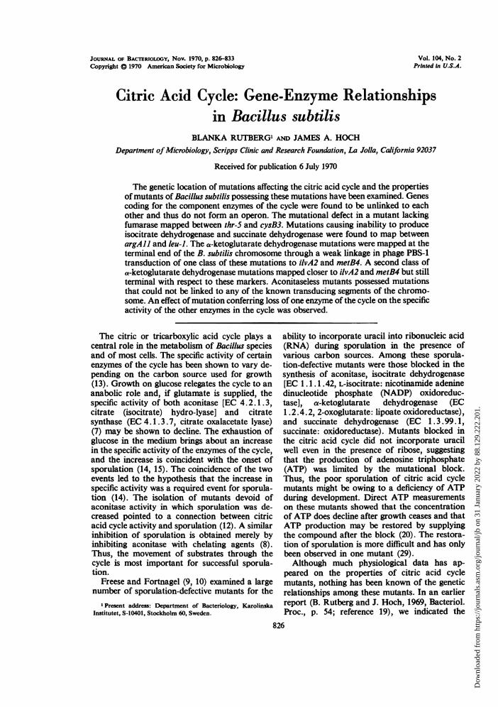
Genetic map of citric acid cycle mutations. Thelocations of the cit- mutants on their respectivetransducing fragments are presented in Fig. 1.The distances between genetic markers is ex-pressed in terms of per cent recombination inPBS-1 transduction, although the validity of thisnumber as a measure of genetic distance isquestionable in some cases. We were unable to
find linkage of citC4 to thr-S in transformation,but citC mutations are weakly linked to argAll(21) and citF mutations are linked to leu-l intransformation (unpublished data). The citD mu-
TABLE 9. Two-factor transduction crossesinvolving citK
Donor metB4 Donor ievA2Recipient
Cit+ Recom- Cit+Ilv/ Recom-Met/Cit+ bination Cit+ bination
citK5 7/104 93 5/104 95citK7 2/104 98 5/104 95citK9 5/104 95 6/104 94citKi4 2/104 98 3/104 97citK16 8/208 96 6/104 94citKi8 3/104 97 7/104 93
TABLE 10. Two-factor transduction crosseswith citD mutants
Recipient ilvAZ Recipient metB4Donor
Cit-/Ilv+ Recoi- Cit/Met+ Recomi-bination bination
citD26 97/104 7citD28 258/394 34citD29 192/200 4 656/794 17citD33 203/214 5 45/63 29citD35 144/208 31 54/117 54
thr-5 cit G4 cys 83 his Al
,,39,50 ,% 78699c F
.
arg All citC citF leu-I ilvCI phe-l
*1 I. *I
21 ,. S8 I- 26 252S51
met 84 iIvA2 cit D cit Ktlrt 1
It l,,I'm , ' J 95,. ~~~98
FIG. 1. Genetic maps of the transducing segmentsin which cit mutations are located. Numbers indicateper cent recombination in PBS-I transduction crosses.
831VoL. 104, 1970
Dow
nloa
ded
from
http
s://j
ourn
als.
asm
.org
/jour
nal/j
b on
31
Janu
ary
2022
by
88.1
29.2
22.2
01.

RUTBERG AND HOCH
tations are placed between ilvA2 and citK on thebasis of their recombination values with ilvA2.
DISCUSSIONThe genetic analysis of citric acid cycle mutants
has led to the discovery that the genes for thecomponent enzymes of the cycle do not form alinked group or operon but rather are scatteredon the B. subtilis chromosome. This finding isnot unexpected if one considers the diversecatabolic and anabolic roles the cycle mustfulfill. The biosynthetic role of the enzymes fromacetyl-coenzyme A (CoA) to a-ketoglutarate forthe synthesis of glutamate and amino acidsderived from glutamate is well known, and a-ketoglutarate was postulated to be the corepressorfor both citrate synthase and aconitase synthesis(7). This repression, however, may not be ac-complished in the absence of carbon sources, themetabolism of which does not depend on thecycle (13). Thus, the synthesis of these enzymesmay be under a more complex regulatory schemethan a simple biosynthetic pathway. The re-ductive branch of the cycle from oxalacetic acidto succinyl-CoA must also fulfill a biosyntheticrole. The succinyl-CoA formed is used in thebiosynthesis of both lysine and methionine. Itmight be predicted that these amino acids willinfluence the level of the reductive branch en-zymes directly or indirectly through succinyl-CoA. Although no direct data are available, itmay be relevant in this regard that the genefor fumarase is linked to genes involved inthreonine and threonine-methionine synthesis (C.Anagnostopoulos, personal communication), andsuccinate dehydrogenase is linked to isoleucine-valine and leucine genes.The properties of mutants blocked in a-keto-
glutarate dehydrvgenase are different from theproperties of similar mutants in E. coli (16, 17).In E. coli, a block in a-ketoglutarate dehydro-genase brings about a requirement for lysine andmethionine or succinate. This requirement stemsfrom the lack of succinyl-CoA since the reductivebranch stops at fumarate in aerobic cells. Underanaerobic conditions, this requirement disappearsbecause fumarate reductase is formed. Mutantsof B. subtilis blocked in a-ketoglutarate de-hydrogenase do not require succinate and mostlikely obtain this compound through a morefavorable equilibrium of succinate dehydrogenase.The genetic location of a-ketoglutarate mutantsplaces them as the most terminal markers foundon the B. subtilis chromosome. We determinedthat the citK gene replicates later than trp andthus is terminal in terms of DNA replication
(unpublished data). We did not find linkage inPBS-1 transduction of citK mutations to anyorigin marker, suggesting that a substantialgenetic distance exists between the terminus andorigin of the B. subtilis chromosome.The inability to link aconitase mutations to any
known transducing segment is probably a reflec-tion of the incomplete chromosome map.
Certain mutants of one enzyme of the cyclehave interesting effects on the levels of the otherenzymes. A block in aconitase increases thespecific activity of isocitrate dehydrogenase andvice versa. Neither block has much effect on thespecific activity of the other enzymes. The effectof the blocks is consistent with a derepression ofthe enzymes because of a lack of glutamate.Glutamate can be utilized by these mutants butnot replenished. The opposite is true for an a-ketoglutarate dehydrogenase mutant which cansynthesize glutamate but not oxidize it. In thesemutants, the specific activity of aconitase dropsdrastically but the level of isocitrate dehydrogen-ase is not as greatly affected. These results areconsistent with glutamate (or a-ketoglutarate) asthe corepressor of aconitase synthesis (13). Theresults also suggest that glutamate or a productderived from it regulates the synthesis of isocitratedehydrogenase, since the levels of this enzymerespond in the same general manner as aconitaseto the mutant blocks. The response, however, isnot as drastic as that of aconitase.The levels of a-ketoglutarate dehydrogenase,
succinate dehydrogenase, fumarase, and malatedehydrogenase are not greatly altered by mutantblocks elsewhere in the cycle. In contrast to E.coli in which a-ketoglutarate dehydrogenase is in-ducible (2), the specific activity of a-ketoglutaratedehydrogenase in B. subtilis does not seem tofluctuate much. It has been postulated that a-ketoglutarate is the inducer of a-ketoglutaratedehydrogenase in E. coli (2). If such were thecase in B. subtilis, we would expect low levels ofa-ketoglutarate dehydrogenase in aconitase andisocitrate dehydrogenase mutants and highlevels in fumarase or succinate dehydrogenasemutants. This expectation was not realized (Table3). In further studies (Hoch and Rutberg, unpub-lished data), we found the level of a-ketoglutaratedehydrogenase to vary at most 10-fold underdifferent cultural conditions. Thus, if the enzymeis inducible, the difference between induced andrepressed levels is much less than that seen inE. coli.These results provide the basis for a more
extensive study by genetic means of the regulationof the citric acid cycle.
832 J. BACrERIOL.
Dow
nloa
ded
from
http
s://j
ourn
als.
asm
.org
/jour
nal/j
b on
31
Janu
ary
2022
by
88.1
29.2
22.2
01.

CITRIC ACID CYCLE IN B. SUBTILIS
ACKNOWLEDGMENTS
These studies were supported by Public Health Service grantHD 02807 from the National Institute of Child Health and HumanDevelopment, grant GB 15602 from the National Science Founda-tion, and grants K70-16X-3038-01 and B71-16X-3038-02 from theSwedish Medical Research Council. James A. Hoch is a recipientofa Faculty Research Associate Award (PRA-66) from the Ameri-can Cancer Society.
Technical assistance was provided by Gayle Mildner andJudith Mathews.
LITERATURE CITED
1. Adelberg, E. A., M. Mandel, and Chein-Chin-Chen. 1965.Optimal conditions for mutalenesis by N-methyl-N'-nitro-N-nitrosoguanidine in Escherichia coli K12. Biochem. Bio-phys. Res. Commun. 18:788-795.
2. Amarasingham, C. R., and B. D. Davis. 1965. Regulation ofa-ketoglutarate dehydrogenase formation in Escherichiacolt. J. Biol. Chem. 240:3665-3668.
3. Anagnostopoulos, C., and J. Spizizen. 1962. Requirements fortransformation in Bacillus subtilis. J. Bacteriol. 81:741-746.
4. Bachmann, E., D. W. Allmann, and D. E. Green. 1966. Themembrane systems of the mitochondrion. I. The S fractionof the outer membrane of beef heart mitochondria. Arch.Biochem. Biophys. 115:153-164.
5. Barat, M., C. Anagnostopoulos, and A.-M. Schneider. 1965.Linkage relationships of genes controlling isoleucine, valine,and leucine biosynthesis in Bacillus subillis. J. Bacteriol.90:357-369.
6. Bock, R. M., and R. A. Alberty. 1953. Studies of the enzymefumarase. I. Kinetics and equilibrium. J. Amer. Chem. Soc.75:1921-1925.
7. Flechtner, V. R., and R. S. Hanson. 1969. Coarse and finecontrol of citrate synthase from Bacillus subtilis. Biochim.Biophys. Acta 184:252-262.
8. Fortnagel, P., and E. Freese. 1968. Inhibition of aconitase bychelation of transition metals causing inhibition of sporula-tion in Bacillus sublilis. J. Biol. Chem. 243:5289-5295.
9. Fortnagel, P., and E. Freese. 1968. Analysis of sporulationmutants. II. Mutants blocked in the citric acid cycle. J. Bac-teriol. 95:1431-1438.
10. Freese, E., and P. Fortnagel. 1967. Analysis of sporulationmutants. I. Response of uracil incorporation to carbonsources, and other mutant properties. J. Bacteriol. 94:1957-1969.
11. Green, D. E., S. Mii, and P. M. Kohout. 1955. Studies on theterminal electron transport system. J. Biol. Chem 217:551-567.
12. Hanson, R. S., J. Blicharska, and J. Szulmajster. 1964. Rela-tionship between the tricarboxylic acid cycle enzymes andsporulation ofB. subtilis. Biochem. Biophys. Res. Commun.17:1-7.
13. Hanson, R. S., and D. P. Cox. 1967. Effect of different nutri-tional conditions on the synthesis of tricarboxylic acid cycleenzymes. J. Bacteriol. 93: 1777-1787.
14. Hanson, R. S., V. R. Srinivasan, and H. 0. Halvorson. 1963.Biochemistry of sporulation. I. Metabolism of acetate byvegetative and sporulating cells. J. Bacteriol. 85:451-460.
15. Hanson, R. S., V. R. Srinivasan, and H. 0. Halvorson. 1963.Biochemistry of sporulation. II. Enzymatic changes duringsporulation of Bacillus cereus. J. Bacteriol. 86:45-50.
16. Herbert, A. A., and J. R. Guest. 1968. Biochemical andgenetic studies with lysine and methionine mutants ofEscherichia coll: lipoic acid and a-ketoglutarate dehy-drogenase-less mutants. J. Gen. Microbiol. 53:363-381.
17. Herbert, A. A., and J. R. Guest. 1969. Studies with a-keto-glutarate dehydrogenase mutants of Escherichia coli. Mol.Gen. Genet. 105:182-190.
18. Hoch, J. A., M. Barat, and C. Anagnostopoulos. 1967. Trans-formation and transduction in recombination-defectivemutants of Bacillus subtilis. J. Bacteriol. 93:1925-1937.
19. Hoch, J. A., and J. Spizizen. 1969. Genetic control of some
early events in sporulation of Bacillus subtilis 168, p. 112-120. In L. L. Campbell (ed.), Spores IV. American Societyfor Microbiology, Bethesda, Md.
20. Klofat, W., G. Picciolo, E. W. Chappelle, and E. Freese. 1969.Production of adenosine triphosphate in normal cells andsporulation mutants of Bacillus subtilis. J. Biol. Chem.244:3270-3276.
21. LeHegarat, J. -C., and C. Anagnostopoulos. 1969. Localisa-tion chromosomique d'un gene gouvernant la synthesed'une phosphatase alcaline chez Bacillus subtilis. C. R.Acad. Sci. (Paris) 269:2048-2050.
22. Lowry, 0. H., H. J. Rosebrough, A. L. Farr, and R. J. Ran-dall. 1951. Protein measurement with the Folin phenolreagent. J. Biol. Chem. 193:265-275.
23. Mahler, H. R., M. H. Wittenberger, and L. Brand. 1958.Biochemical studies of the developing avian embryo. II.
Enzymes of the citric acid cycle. J. Biol. Chem. 233:770-782.24. Myers, W. F., and K. -Y. Huang. 1966. Separation of inter-
mediates of the citric acid cycle and related compounds bythin-layer chromatography. Anal. Biochem. 17:210-213.
25. Ochoa, S. 1955. Isocitric dehydrogenase system (TPN) frompig heart, p. 699-700, and malic dehydrogenase frompig heart, p. 735-736. In S. P. Colowick and N. 0. Kaplan(ed.), Methods in enzymology, vol. 1. Academic Press Inc.,New York.
26. Ozeki, H. 1959. Chromosome fragments participating intransduction in Salmonella typhimurium. Genetics 44:457-470.
27. Passera, C., A. Pedrotti, and G. Ferrari. 1964. Thin-layerchromatography of carboxylic acids and keto acids ofbiological interest. J. Chromatogr. 14:289-291.
28. Racker, E. 1950. Spectrophotometric measurements of theenzymatic formation of fumaric and cis-aconitic acids.Biochim. Biophys. Acta 4:211-214.
29. Schmitt, R., and E. Freese. 1968. Curing of a sporulationmutant and antibiotic activity of Bacillus subtilis. J. Bac-teriol. 96:1255-1265.
30. Spizizen, J. 1958. Transformation of biochemically deficientstrains of Bacillus subtilis by deoxyribonucleate. Proc.Nat. Acad. Sci. U.S.A. 44:1072-1078.
VOL. 104, 1970 833
Dow
nloa
ded
from
http
s://j
ourn
als.
asm
.org
/jour
nal/j
b on
31
Janu
ary
2022
by
88.1
29.2
22.2
01.