Embed Size (px)
Citation preview
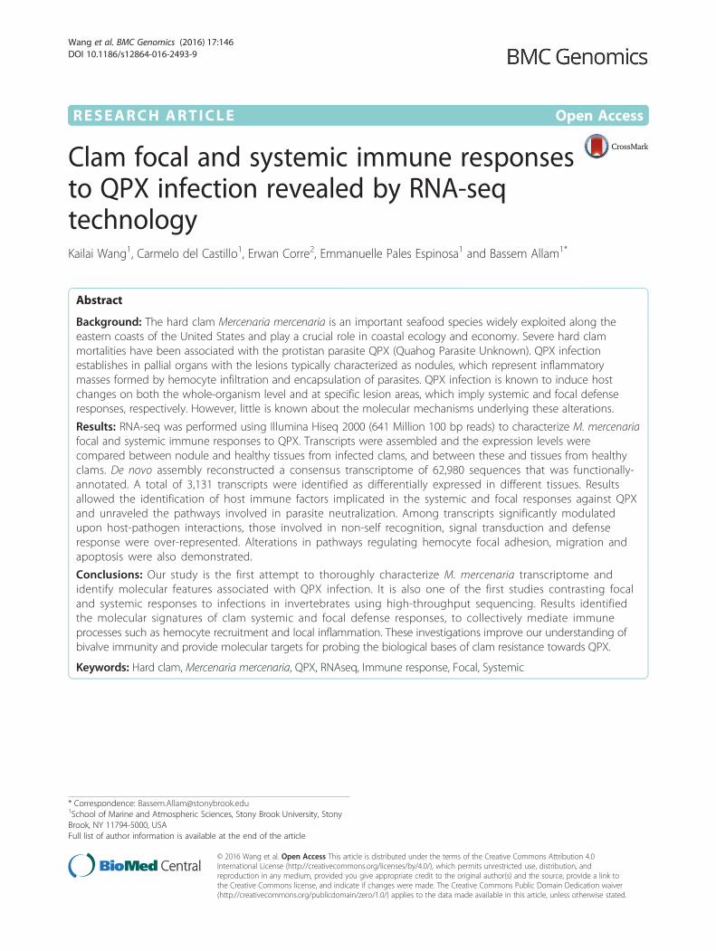
RESEARCH ARTICLE Open Access
Clam focal and systemic immune responsesto QPX infection revealed by RNA-seqtechnologyKailai Wang1, Carmelo del Castillo1, Erwan Corre2, Emmanuelle Pales Espinosa1 and Bassem Allam1*
Abstract
Background: The hard clam Mercenaria mercenaria is an important seafood species widely exploited along theeastern coasts of the United States and play a crucial role in coastal ecology and economy. Severe hard clammortalities have been associated with the protistan parasite QPX (Quahog Parasite Unknown). QPX infectionestablishes in pallial organs with the lesions typically characterized as nodules, which represent inflammatorymasses formed by hemocyte infiltration and encapsulation of parasites. QPX infection is known to induce hostchanges on both the whole-organism level and at specific lesion areas, which imply systemic and focal defenseresponses, respectively. However, little is known about the molecular mechanisms underlying these alterations.
Results: RNA-seq was performed using Illumina Hiseq 2000 (641 Million 100 bp reads) to characterize M. mercenariafocal and systemic immune responses to QPX. Transcripts were assembled and the expression levels werecompared between nodule and healthy tissues from infected clams, and between these and tissues from healthyclams. De novo assembly reconstructed a consensus transcriptome of 62,980 sequences that was functionally-annotated. A total of 3,131 transcripts were identified as differentially expressed in different tissues. Resultsallowed the identification of host immune factors implicated in the systemic and focal responses against QPXand unraveled the pathways involved in parasite neutralization. Among transcripts significantly modulatedupon host-pathogen interactions, those involved in non-self recognition, signal transduction and defenseresponse were over-represented. Alterations in pathways regulating hemocyte focal adhesion, migration andapoptosis were also demonstrated.
Conclusions: Our study is the first attempt to thoroughly characterize M. mercenaria transcriptome andidentify molecular features associated with QPX infection. It is also one of the first studies contrasting focaland systemic responses to infections in invertebrates using high-throughput sequencing. Results identifiedthe molecular signatures of clam systemic and focal defense responses, to collectively mediate immuneprocesses such as hemocyte recruitment and local inflammation. These investigations improve our understanding ofbivalve immunity and provide molecular targets for probing the biological bases of clam resistance towards QPX.
Keywords: Hard clam, Mercenaria mercenaria, QPX, RNAseq, Immune response, Focal, Systemic
* Correspondence: [email protected] of Marine and Atmospheric Sciences, Stony Brook University, StonyBrook, NY 11794-5000, USAFull list of author information is available at the end of the article
© 2016 Wang et al. Open Access This article is distributed under the terms of the Creative Commons Attribution 4.0International License (http://creativecommons.org/licenses/by/4.0/), which permits unrestricted use, distribution, andreproduction in any medium, provided you give appropriate credit to the original author(s) and the source, provide a link tothe Creative Commons license, and indicate if changes were made. The Creative Commons Public Domain Dedication waiver(http://creativecommons.org/publicdomain/zero/1.0/) applies to the data made available in this article, unless otherwise stated.
Wang et al. BMC Genomics (2016) 17:146 DOI 10.1186/s12864-016-2493-9
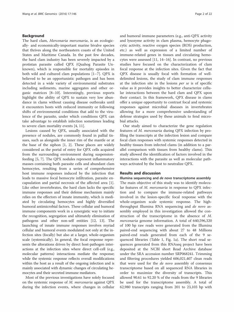
BackgroundThe hard clam, Mercenaria mercenaria, is an ecologic-ally- and economically-important marine bivalve speciesthat thrives along the northeastern coasts of the UnitedStates and Maritime Canada. In the past few decades,the hard clam industry has been severely impacted by aprotistan parasite called QPX (Quahog Parasite Un-known), which is responsible for mortality episodes inboth wild and cultured clam populations [1–7]. QPX isbelieved to be an opportunistic pathogen and has beendetected in a wide variety of environmental substratesincluding sediments, marine aggregates and other or-ganic matrices [8–10]. Interestingly, previous reportshighlight the ability of QPX to sustain very low abun-dance in clams without causing disease outbreaks untilit encounters hosts with reduced immunity or followingshifts of environmental conditions that add to the viru-lence of the parasite, under which conditions QPX cantake advantage to establish infection sometimes leadingto severe clam mortality events [4, 11].Lesions caused by QPX, usually associated with the
presence of nodules, are commonly found in pallial tis-sues, such as alongside the inner rim of the mantle or atthe base of the siphon [1, 2]. These places are widelyconsidered as the portal of entry for QPX cells acquiredfrom the surrounding environment during suspension-feeding [5, 7]. The QPX nodules represent inflammatorymasses containing both parasite cells and abundant clamhemocytes, resulting from a series of comprehensivehost immune responses induced by the infection thatleads to massive focal hemocyte infiltration, parasite en-capsulation and partial necrosis of the affected area [1].Like other invertebrates, the hard clam lacks the specificimmune responses and their defense mechanism mainlyrelies on the effectors of innate immunity, which is medi-ated by circulating hemocytes and highly diversifiedhumoral antimicrobial factors. These cellular and humoralimmune components work in a synergistic way to initiatethe recognition, segregation and ultimately elimination ofpathogens and other non-self entities [12, 13]. Thelaunching of innate immune responses involves myriadcellular and humoral events modulated not only at the in-fection sites (focally) but also at a larger, whole-organismscale (systemically). In general, the focal response repre-sents the alterations driven by direct host-pathogen inter-actions at the infection sites where direct cell-cell (e.g.,molecular patterns) interactions mediate the response;while the systemic response reflects overall modificationswithin the host as a result of the ongoing infection and ismainly associated with dynamic changes of circulating he-mocytes and their secreted immune mediators.Most of the previous investigations have solely focused
on the systemic response of M. mercenaria against QPXduring the infection events, where changes in cellular
and humoral immune parameters (e.g., anti-QPX activityand lysozyme activity in clam plasma, hemocyte phago-cytic activity, reactive oxygen species (ROS) production,etc.) as well as expression of a limited number ofimmune-related genes in tissues and circulating hemo-cytes were assessed [11, 14–16]. In contrast, no previousstudies have focused on the characterization of clamfocal response at the infection sites. Given the fact thatQPX disease is usually focal with formation of well-delimited lesions, the study of clam immune responsesat the infection site in the lesions per se is of specificvalue as it provides insights to better characterize cellu-lar interactions between the hard clam and QPX upontheir contact. In this framework, QPX disease in clamsoffer a unique opportunity to contrast focal and systemicresponses against microbial diseases in invertebratesallowing for a more comprehensive understanding ofdefense strategies used by these animals to fend micro-bial attacks.Our study aimed to characterize the gene regulation
features of M. mercenaria during QPX infection by pro-filing the transcripts at the infection lesion and comparefocal clam responses with systemic responses detected inhealthy tissues from infected clams (in addition to a par-allel comparison with tissues from healthy clams). Thisstudy allowed the identification of factors involved in theinteractions with the parasite as well as molecular path-ways activated by the host to neutralize QPX.
Results and discussionIllumina sequencing and de novo transcriptome assemblyThe main objective of this study was to identify molecu-lar features of M. mercenaria in response to QPX infec-tion and to compare the immune-related pathwaysinvolved in the lesion-specific focal response with thewhole-organism scale systemic response. The high-throughput Illumina RNA sequencing and de novo as-sembly employed in this investigation allowed the con-struction of the transcriptome in the absence of M.mercenaria genome information. A total of 640,596,320of 100 bp raw reads were generated from the Illuminapaired-end sequencing with about 27 to 48 Millionspaired-end reads generated from each of the 9 se-quenced libraries (Table 1, Fig. 1a). The short read se-quences generated from this RNAseq project have beendeposited at the NCBI short Read Archive databaseunder the SRA accession number SRP068241. Trimmingand filtering procedures yielded 606,021,407 clean readsthat were used for the de novo assembly of consensustranscriptome based on all sequenced RNA libraries inorder to maximize the diversity of transcripts. Thisallowed 90.61 to 92.20 % of the reads from the 9 librariesbe used for the transcriptome assembly. A total of62,980 transcripts ranging from 201 to 23,103 bp with
Wang et al. BMC Genomics (2016) 17:146 Page 2 of 22
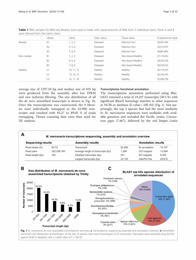
average size of 1297.59 bp and median size of 835 bpwere produced from the assembly after low FPKMand rare isoforms filtering. The size distribution of allthe de novo assembled transcripts is shown in Fig. 1b.Once the transcriptome was constructed, the 9 librar-ies were individually remapped to the 62,980 tran-scripts and resulted with 85.27 to 89.05 % of readsremapping. Theses counting data were then used forDE analysis.
Transcriptome functional annotationThe transcriptome annotation performed using Blas-t2GO returned a total of 19,107 transcripts (30.3 %) withsignificant BlastX homology matches to other sequencesin NCBI nr database (E-value < 10E-03) (Fig. 1). Not sur-prisingly, the top 3 species that had the most similarityto M. mercenaria sequences were mollusks with avail-able genomes and included the Pacific oyster, Crassos-trea gigas (7,467), followed by the owl limpet Lottia
Table 1 RNA samples for RNA-seq libraries. Each pool is made with equal amounts of RNA from 3 individual clams. Pools A and Bwere derived from the same clams
Library Clams Clam status Tissue status N paired-end reads
Nodule A1 1, 2, 3 Diseased Infection foci 30,491,569
A2 4, 5, 6 Diseased Infection foci 34,515,597
A3 7, 8, 9 Diseased Infection foci 46,861,893
Non-nodule B1 1, 2, 3 Diseased Non-lesion/Healthy 27,119,432
B2 4, 5, 6 Diseased Non-lesion/Healthy 28,254,720
B3 7, 8, 9 Diseased Non-lesion/Healthy 40,259,333
Healthy C1 10, 11, 12 Healthy Healthy 36,714,347
C2 13, 14, 15 Healthy Healthy 43,293,545
C3 16, 17, 18 Healthy Healthy 42,046,740
Fig. 1 M. mercenaria de novo assembled transcriptome summary. a Transcriptome sequencing, assembly and annotation overview. b Assembledtranscripts size distribution. c Distribution of the top 10 species with most homologues to M. mercenaria. Transcripts were searched using BLASTxagainst NCBI nr database with a cutoff value of E < 10E-03
Wang et al. BMC Genomics (2016) 17:146 Page 3 of 22
gigantea (3,539) and the California sea slug Aplysia cali-fornica (1,931) (Fig. 1c). KEGG Orthology (KO) termswere assigned to 6,425 sequences and reference path-ways were mapped to the KEGG database based on theassigned KO terms (Fig. 1a, Additional file 1). A total of29,815 sequences were identified to match to at leastone conserved protein domain in the InterPro database(Fig. 1a, Additional file 1).Gene ontology (GO) assignments were used to classify
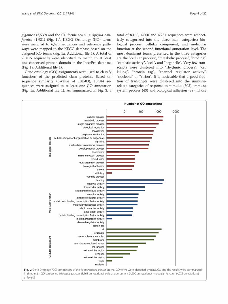
functions of the predicted clam proteins. Based onsequence similarity (E-value of 10E-03), 13,584 se-quences were assigned to at least one GO annotation(Fig. 1a, Additional file 1). As summarized in Fig. 2, a
total of 8,168, 4,600 and 4,231 sequences were respect-ively categorized into the three main categories: bio-logical process, cellular component, and molecularfunction at the second functional annotation level. Themost dominant terms presented in the three categoriesare the “cellular process”, “metabolic process”, “binding”,“catalytic activity”, “cell”, and “organelle”. Very few tran-scripts were clustered into “rhythmic process”, “cellkilling”, “protein tag”, “channel regulator activity”,“nucleoid” or “virion”. It is noticeable that a good frac-tion of transcripts were clustered into the immune-related categories of response to stimulus (503), immunesystem process (43) and biological adhesion (38). Those
Fig. 2 Gene Ontology (GO) annotations of the M. mercenaria transcriptome. GO terms were identified by Blast2GO and the results were summarizedin three main GO categories: biological process (8,168 annotations), cellular component (4,600 annotations), molecular function (4,231 annotations)at level-2
Wang et al. BMC Genomics (2016) 17:146 Page 4 of 22
transcripts were of special interest given that they mightbe involved in the M. mercenaria defense and resistancetoward QPX infection.A significant portion (69.7 %) of M. mercenaria tran-
scripts did not match any BlastX hit in NCBI nr data-base, in agreement with previous transcriptomic studiesin mollusks [17–20]. Most of the unannotated tran-scripts may represent transcripts spanning untranslatedmRNA regions, or transcripts containing only non-conserved protein domains [21, 22].
Identification of differentially expressed transcriptsThe generated transcriptome was used as a reference fordownstream investigations of global gene expression inthe three different tissues of interest (nodule, non-nodule and healthy) to identify genes associated with M.mercenaria’s focal and systemic immune responseagainst QPX. A gene-isoform relationship was estimatedusing RSEM over Trinity output isoforms. Resultsshowed that about 43 % (27,021) of all the transcriptshad 1 isoform, 19 % (12,307) had 2 isoforms and 38 %(23,652) had 3 isoforms, suggesting extensive isoform di-versity in M. mercenaria transcriptome. Transcript iso-form variation could affect mRNA stability, localizationand translation, as well as the production of protein vari-ants that differ in localization or function [23].By comparing the number of transcripts expressed in
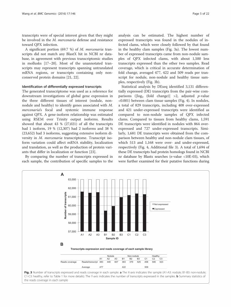
each sample, the contribution of specific samples to the
analysis can be estimated. The highest number ofexpressed transcripts was found in the nodules of in-fected clams, which were closely followed by that foundin the healthy clam samples (Fig. 3a). The lowest num-ber of expressed transcripts came from non-nodule sam-ples of QPX infected clams, with about 1,500 lesstranscripts expressed than the other two samples. Readcoverage, which is critical in accurate determination offold change, averaged 477, 422 and 509 reads per tran-script for nodule, non-nodule and healthy tissue sam-ples, respectively (Fig. 3b).Statistical analysis by DEseq identified 3,131 differen-
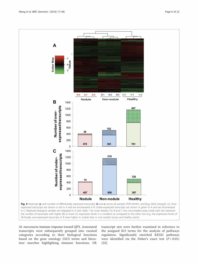
tially expressed (DE) transcripts from the pair-wise com-parisons (|log2 (fold change)| >2, adjusted p-value<0.001) between clam tissue samples (Fig. 4). In nodules,a total of 829 transcripts, including 408 over-expressedand 421 under-expressed transcripts were identified ascompared to non-nodule samples of QPX infectedclams. Compared to tissues from healthy clams, 1,591DE transcripts were identified in nodules with 864 over-expressed and 727 under-expressed transcripts. Simi-larly, 1,681 DE transcripts were obtained from the com-parison between healthy and non-nodule clam tissues, ofwhich 513 and 1,168 were over- and under-expressed,respectively (Fig. 4, Additional file 3). A total of 1,694 ofthese DE transcripts had protein homologs found in NCBInr database by Blastx searches (e-value <10E-03), whichwere further examined for their putative functions during
Fig. 3 Number of transcripts expressed and reads coverage in each sample. a The X-axis indicates the sample (A1-A3: nodule; B1-B3: non-nodule;C1-C3: healthy, refer to Table 1 for more details). The Y-axis indicates the number of transcripts expressed in the samples. b Summary statistics ofthe reads coverage in each sample
Wang et al. BMC Genomics (2016) 17:146 Page 5 of 22
M. mercenaria immune response toward QPX. Annotatedtranscripts were subsequently grouped into curatedcategories according to their biological functionsbased on the gene ontology (GO) terms and litera-ture searches highlighting immune functions. DE
transcript sets were further examined in reference tothe assigned KO terms for the analysis of pathwaysregulation. Significantly enriched KEGG pathwayswere identified via the Fisher’s exact test (P < 0.01)[24].
Fig. 4 Heatmap (a) and number of differentially expressed transcripts (b and c) across all samples (FDR ≤0.001, and |log2 (fold change)| >2). Over-expressed transcripts are shown in red in A and are enumerated in B. Under-expressed transcripts are shown in green in A and are enumeratedin C. Replicate biological samples are displayed in A (see Table 1 for more details). For B and C: the cross-shaded areas inside each bar representthe number of transcripts with higher (B) or lower (C) expression levels in a condition as compared to the other two (e.g., the expression levels of38 focally over-expressed transcripts in B were higher in nodule than in non-nodule tissues and healthy clams)
Wang et al. BMC Genomics (2016) 17:146 Page 6 of 22
Here we specifically focus on DE transcripts drawnfrom comparisons between “nodule vs. non-nodule” and“non-nodule vs. healthy” tissue samples, which were re-spectively considered to reflect the transcriptomicchanges caused by “focal” and “systemic” immune re-sponse of M. mercenaria toward QPX infection, respect-ively. The overview of DE transcripts drawn from thesetwo responses is presented in Fig. 4 and Additional files2 and 3.
Differential expression of immune related transcriptsFocal responseClam focal response reflected the alterations causedby direct clam-QPX interaction at the infection site(Additional file 2). QPX nodules are inflammatory massesresulting from massive hemocytes infiltration and encap-sulation of parasite cells [1, 2]. This process largely relieson the motility and adhesion properties of hemocytes,thus allowing these cells to migrate throughout the circu-latory system and recruit to the infection site. Hemocytescan sense stimuli in host tissues through an array of cellsurface receptors, and use these cues to adjust their be-havior accordingly [25]. The activation of hemocytes re-quires the binding of specific ligands to the cell surfacereceptors, which subsequently initiate the transduction ofextracellular signals into the cytoplasm via a variety of sig-naling pathways, thus inducing a series of hemocyte-mediated immune response such as phagocytosis, encap-sulation (prominent response against QPX in clams), ROSproduction, as well as secretion of immune effectors andcytokines [26–28]. A collection of DE transcripts involvedin these defense processes was identified during focal re-sponse, suggesting that strong and comprehensive host-pathogen interactions were taking places inside the QPXlesions (Table 2, Additional file 2). A large fraction of theDE transcripts of focal response was annotated as recep-tors or molecules with receptor activities, which putativelycontribute to the host defense against QPX as (1) cell sur-face receptors expressed on hemocytes that mediate therecognition and phagocytosis or encapsulation of foreignentities through microbe associated molecular patterns(MAMP); (2) signaling receptors activating intracellularsignaling cascades or (3) the soluble bridging moleculesmediating the linkage between MAMPs and hemocytes[29]. Among those receptors, most are identified as patho-gen pattern recognition receptors (PRRs), which includethe C-type lectins (CTLs), the scavenger receptors (SRs)and the toll-like receptors (TLRs).The C-type mannose receptor-2 (MRC2) identified during
the focal response (Table 2) is a member of the C-type lec-tins (CTLs) superfamily, a large group of Ca2+-dependentcarbohydrate-binding proteins that play crucial roles in in-nate immunity. CTLs recognize pathogens and facilitatetheir phagocytosis [30, 31] or encapsulation [32–34]. MRCs
are also key regulators of inflammatory responses and con-tribute to the removal of harmful inflammatory agents [35,36]. The 16-fold over-expression of MRC2 during the focalresponse suggested that active hemocyte encapsulation andlocal inflammation was induced by QPX at the infection le-sions, which is consistent with the results of histopatho-logical observations [1]. Another over-expressed CTLmember, the perlucin-like protein, has been previouslyshown to trigger immune response in Manila clams duringmicrobial infection [37].Scavenger receptors (SRs) were also among the strongly
over-expressed transcripts in infection foci (Table 2). Theseincluded somatomedin-b and thrombospondin type-1domain-containing (RPE spondin), insulin-related peptidereceptor, hemicentin-1, lysyl oxidase-like protein 2 andmam domain-containing glycosylphosphatidylinositol an-chor protein 1. SRs are structurally diverse PRRs that sharethe common function of recognizing oxidized or acetylatedlow-density lipoprotein (LDL) [38]. They contribute to in-nate immunity by recognizing MAMPs and mediatingnon-opsonic phagocytosis [29, 39]. They are extensivelyfound on immune cells and are able to interact with bothmodified-host components and exogenous ligands, whichmakes SRs a key component in host defense, apoptosis, in-flammation and lipoprotein homeostasis [40–42]. For ex-ample, scallop SRs bind not only to acetylated LDL butalso to MAMP including lipopolysaccharides (LPS), pepti-doglycans (PGN), mannan and zymosan particles [39]. Thesea urchin genome encodes approximately 150 genes con-sisting of one or more scavenger receptor cysteine-rich(SRCR) domains [43], and the members of this gene familyexhibit dynamic shifts in transcription after immune chal-lenge [40, 44].Our results also show an over-expression of TLR-1 and
Toll-8/tollo in nodules (Table 2), which is in agreementwith previous investigations showing up-regulation ofTLRs in M. mercenaria mantle following QPX challenge[15]. TLRs are among the most ancient and conservedPRRs. They are expressed by immune cells and interactwith a large variety of MAMPs. Bivalve TLRs have beencharacterized in the oyster C. gigas and the scallop C. far-reri where they exhibited significant response to LPSstimulation [45, 46]. Transcriptional modulation of TLRshas also been reported in Ruditapes philippinarum andMya arenaria following MAMPs stimulation and bacterialchallenge [47, 48]. Interestingly, Toll-8 (Tollo) has beenshown to participate in Drosophila epithelial immunitywhere it mediates host cells communication that subse-quently activates systemic immune responses [49]. Thissuggests that the Toll pathway could be one of the crucialpivoting links that allow coordination between focal andsystemic immune components during infection.The QPX nodules are formed as the result of granu-
lomatous inflammation, which is a chronic inflammatory
Wang et al. BMC Genomics (2016) 17:146 Page 7 of 22
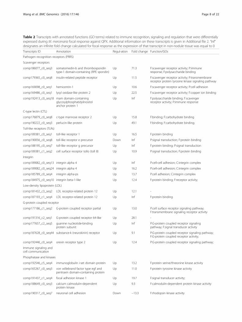
Table 2 Transcripts with annotated functions (GO terms) related to immune recognition, signaling and regulation that were differentiallyexpressed during M. mercenaria focal response against QPX. Additional information on these transcripts is given in Additional file 2. “Inf”designates an infinite fold change calculated for focal response as the expression of that transcript in non-nodule tissue was equal to 0
Transcripts ID Annotation Regul-ation Fold change Function/GOs
Pathogen recognition receptors (PRRS)
Scavenger receptors
comp186077_c0_seq3 somatomedin-b and thrombospondintype-1 domain-containing (RPE spondin)
Up 71.3 F:scavenger receptor activity; P:immuneresponse; F:polysaccharide binding
comp179365_c0_seq8 insulin-related peptide receptor Up 11.5 F:scavenger receptor activity; P:transmembranereceptor protein tyrosine kinase signaling pathway
comp169098_c0_seq1 hemicentin-1 Up 10.6 F:scavenger receptor activity; P:cell adhesion
comp169486_c0_seq1 lysyl oxidase-like protein 2 Up 22.5 F:scavenger receptor activity; F:copper ion binding
comp192413_c0_seq18 mam domain-containingglycosylphosphatidylinositolanchor protein 1
Up Inf F:polysaccharide binding; F:scavengerreceptor activity; P:immune response
C-type lectin (CTL)
comp176879_c0_seq8 c-type mannose receptor 2 Up 15.8 F:binding; F:carbohydrate binding
comp190222_c0_seq3 perlucin-like protein Up 49.1 F:binding; F:carbohydrate binding;
Toll-like receptors (TLRs)
comp189381_c0_seq1 toll-like receptor 1 Up 16.5 F:protein binding
comp190056_c0_seq8 toll-like receptor e precursor Down Inf P:signal transduction; F:protein binding
comp188195_c0_seq7 toll-like receptor g precursor Up Inf F:protein binding; P:signal transduction
comp189381_c1_seq2 cell surface receptor tollo (toll 8) Up 10.9 P:signal transduction; F:protein binding
Integrin
comp189082_c0_seq13 integrin alpha 4 Up Inf P:cell-cell adhesion; C:integrin complex
comp189082_c0_seq24 integrin alpha 4 Up 16.2 P:cell-cell adhesion; C:integrin complex
comp185789_c0_seq4 integrin alpha-ps Up 13.7 P:cell adhesion; C:integrin complex
comp184975_c0_seq10 integrin beta-1-like Up 12.4 F:protein binding; F:receptor activity;
Low-density lipoprotein (LDL)
comp181432_c3_seq2 LDL receptor-related protein 12 Up 12.1 -
comp187193_c1_seq4 LDL receptor-related protein 12 Up Inf F:protein binding
G-protein coupled receptor
comp177186_c1_seq2 G-protein coupled receptor partial Up 13.0 P:cell surface receptor signaling pathway;F:transmembrane signaling receptor activity
comp191316_c2_seq1 G-protein coupled receptor 64-like Up 28.1 -
comp177657_c2_seq5 guanine nucleotide-bindingprotein subunit
Up Inf P:G-protein coupled receptor signalingpathway; F:signal transducer activity
comp187628_c0_seq44 substance-k (neurokinin) receptor Up 9.1 P:G-protein coupled receptor signaling pathway;F:G-protein coupled receptor activity;
comp192446_c0_seq4 orexin receptor type 2 Up 12.4 P:G-protein coupled receptor signaling pathway;
Immune signaling andcell communication
Phosphatase and kinases
comp192546_c5_seq4 immunoglobulin i-set domain protein Up 13.2 F:protein serine/threonine kinase activity
comp165267_c0_seq3 von willebrand factor type egf andpentraxin domain-containing protein
Up 11.0 F:protein tyrosine kinase activity
comp191437_c1_seq4 focal adhesion kinase 1 Up 19.7 F:signal transducer activity;
comp188649_c0_seq3 calcium calmodulin-dependentprotein kinase
Up 9.3 F:calmodulin-dependent protein kinase activity
comp190317_c0_seq7 neuronal cell adhesion Down −13.3 F:rhodopsin kinase activity
Wang et al. BMC Genomics (2016) 17:146 Page 8 of 22
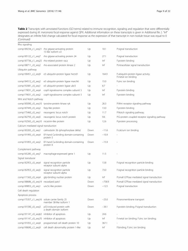
Table 2 Transcripts with annotated functions (GO terms) related to immune recognition, signaling and regulation that were differentiallyexpressed during M. mercenaria focal response against QPX. Additional information on these transcripts is given in Additional file 2. “Inf”designates an infinite fold change calculated for focal response as the expression of that transcript in non-nodule tissue was equal to 0(Continued)
Rho signaling
comp189234_c1_seq21 rho gtpase-activating protein15-like isoform x3
Up 18.1 P:signal transduction
comp185122_c1_seq7 rho gtpase-activating protein 24 Up 27.1 P:signal transduction
comp187736_c1_seq25 rho-related protein raca Up Inf F:protein binding
comp188711_c0_seq2 rho-associated protein kinase 2 Up Inf P:intracellular signal transduction
Ubiquitin pathway
comp190451_c2_seq9 e3 ubiquitin-protein ligase hectd1 Up 164.9 F:ubiquitin-protein ligase activity;F:metal ion binding
comp184512_c0_seq2 e3 ubiquitin-protein ligase march6 Up 15.0 F:zinc ion binding
comp192081_c0_seq1 e3 ubiquitin-protein ligase ubr3 Up 8.7 -
comp179031_c0_seq4 cop9 signalosome complex subunit 5 Up Inf F:protein binding
comp179031_c0_seq2 cop9 signalosome complex subunit 5 Up Inf F:protein binding
Wnt and Notch pathway
comp185090_c0_seq10 tyrosine-protein kinase ryk Up 26.3 P:Wnt receptor signaling pathway
comp187449_c0_seq2 fizzy-like protein Up 13.0 F:protein binding
comp175460_c0_seq1 neurogenic locus notch Up 11.7 P:Notch signaling pathway;
comp182793_c0_seq5 neurogenic locus notch protein Up 9.6 P:G-protein coupled receptor signaling pathway
comp192565_c0_seq10 nicastrin-like protein Up 12.6 P:protein processing
Calcium mediated signal transduction
comp183265_c0_seq1 calmodulin 3b (phosphorylase delta) Down −11.6 F:calcium ion binding
comp191993_c0_seq4 EF-hand Ca-binding domain-containingprotein 5
Down −10.9 -
comp191855_c0_seq2 EF-hand Ca-binding domain-containingprotein 6
Down −15.9 -
Complement pathway
comp165285_c0_seq7 macrophage-expressed gene 1 Up 11.5 -
Signal transducer
comp182953_c0_seq4 signal recognition particlereceptor subunit alpha
Up 13.8 F:signal recognition particle binding
comp182953_c0_seq5 signal recognition particlereceptor subunit alpha
Up 73.0 F:signal recognition particle binding
comp171563_c0_seq4 gtp-binding nuclear protein Up Inf P:small GTPase mediated signal transduction
comp188686_c0_seq19 neuralized pats1 Down −758.9 P:small GTPase mediated signal transduction
comp189853_c0_seq1 unc5c-like protein Down −12.5 P:signal transduction
Cell death regulation
Apoptosis process
comp175357_c1_seq16 solute carrier family 25member 38-like isoform 1
Down −25.0 P:transmembrane transport
comp191590_c0_seq3 p53-induced protein witha death domain isoform
Down −39.1 F:protein binding; P:signal transduction
comp191147_c0_seq62 inhibitor of apoptosis Up 24.6 -
comp191147_c0_seq70 inhibitor of apoptosis Up Inf F:metal ion binding; F:zinc ion binding;
comp191055_c2_seq4 programmed cell death protein 10 Up 367.1 -
comp190690_c2_seq9 cell death abnormality protein 1-like Up Inf F:binding; F:zinc ion binding
Wang et al. BMC Genomics (2016) 17:146 Page 9 of 22
reaction characterized by focal accumulation of activatedimmune cells to isolate the invading agent [50, 51]. Theformation of granuloma requires local recruitment ofhemocytes at the site of infection to execute extracellulardefense processes around the invaders [50]. An array oftranscripts associated with cell migration, adhesion andproliferation was regulated in nodules, including G-protein coupled receptors (GPCRs) and integrins fam-ilies (Table 2). GPCRs regulate inflammatory responsevia binding to chemokines and chemoattractants, thusactivating pathways mediating hemocyte migration andadhesion [52]. They also activate transcription factors inimmune cells, thus modulating the synthesis and secre-tion of certain pro- or anti-inflammatory substances[53]. On the other hand, integrins represent a majorgroup of cell adhesion mediators [54]. They not onlymodulate the cell-cell and cell-extracellular matrix adhe-sion, but also affect multiple signal transduction cas-cades regulating cell survival and proliferation [54].Overexpression of GPCRs and integrins in nodules sug-gests their role in hemocytes adhesion and aggregationassociated with the formation of granuloma [50, 55].Several enzymes regulating ROS production were also
over-expressed during focal response (Table 3). Theseincluded a dual oxidase, which is a key component me-diating host-microbe interactions in mucosa [56, 57].Dual oxidase regulates oxidative burst and ROS produc-tion in the gill muscosa of the shrimp Marsupenaeusjaponicus, favoring shrimp survivorship during viral in-fections [58]. Interestingly, transcripts of dual oxidasewere only expressed in nodules, suggesting this enzymewas induced upon direct clam-QPX interactions as apart of the mantle mucosa-related immune response.Other transcripts associated with oxidation-reductionprocesses also exhibited somewhat nodule-exclusive pat-tern, including the allene oxide synthase-lipoxygenase(AOSL), lysyl oxidase-like protein (LOXL), ww domain-containing oxidoreductase (WWOX), c-terminal bindingprotein (CtBP), isocitrate dehydrogenase (ICD) andmethylenetetrahydrofolate reductase (MTHFR). Thesemolecules are important for maintaining the redox
homeostasis of extracellular environment as they are keyregulators for oxi-reduction reactions. Over-expressionof these transcripts in nodules suggests the need for thehost to timely balance out excessive ROS and other toxicintermediates produced during interaction with QPX. Inaddition to redox-regulation, many of these moleculesalso take part in the immune modulation indirectly. Forexample, AOSL play a role in coral immunity by con-trolling the production of the inflammation regulatorarachidonic acid during apoptosis [59], and LOXL actsboth as a scavenger receptor and regulator for extracel-lular matrix remodeling that initiate hemocyte migrationand tissue regeneration [60], while WWOX was shownto promote proliferation of immune cells through inhib-ition of their apoptosis [61, 62]. In addition, ICD,MTHFR and cytochrome p450 are major detoxificationenzymes [63, 64]. In fact, immune cells and their se-creted effectors require the proper redox state in theextracellular environments to exert their immune func-tions, which makes the maintenance of redox homeosta-sis essential for persistent and effective host defense [65,66]. This is particularly true in the case of QPX diseasewhere the neutralization of parasites depends on extra-cellular killing pathways [26].Apoptosis is an essential host mechanism to effectively
remove damaged and infected cells without causing in-flammatory destructions to surrounding tissues [67, 68].Interestingly, apoptosis seems to be largely inhibitedduring M. mercenaria focal response, as shown by theunder-expression of pro-apoptosis transcripts (Table 2).For example, the tumor necrosis factor (TNF)-like pro-tein and 3-hydroxy-3-methylglutaryl-coenzyme A(HMG-CoA) reductase-like protein were significantlyunder-expressed in nodules. Similarly, the pro-apoptoticp53-induced protein and solute carrier family 25 mem-ber protein were also under-expressed in nodules. Mean-while, inhibitor of apoptosis protein (IAP) was over-expressed in nodules. IAPs regulate immune cell expan-sion and survival in highly inflammatory environmentsin mammals [69] and they may share similar function inclams by preventing hemocytes from death during
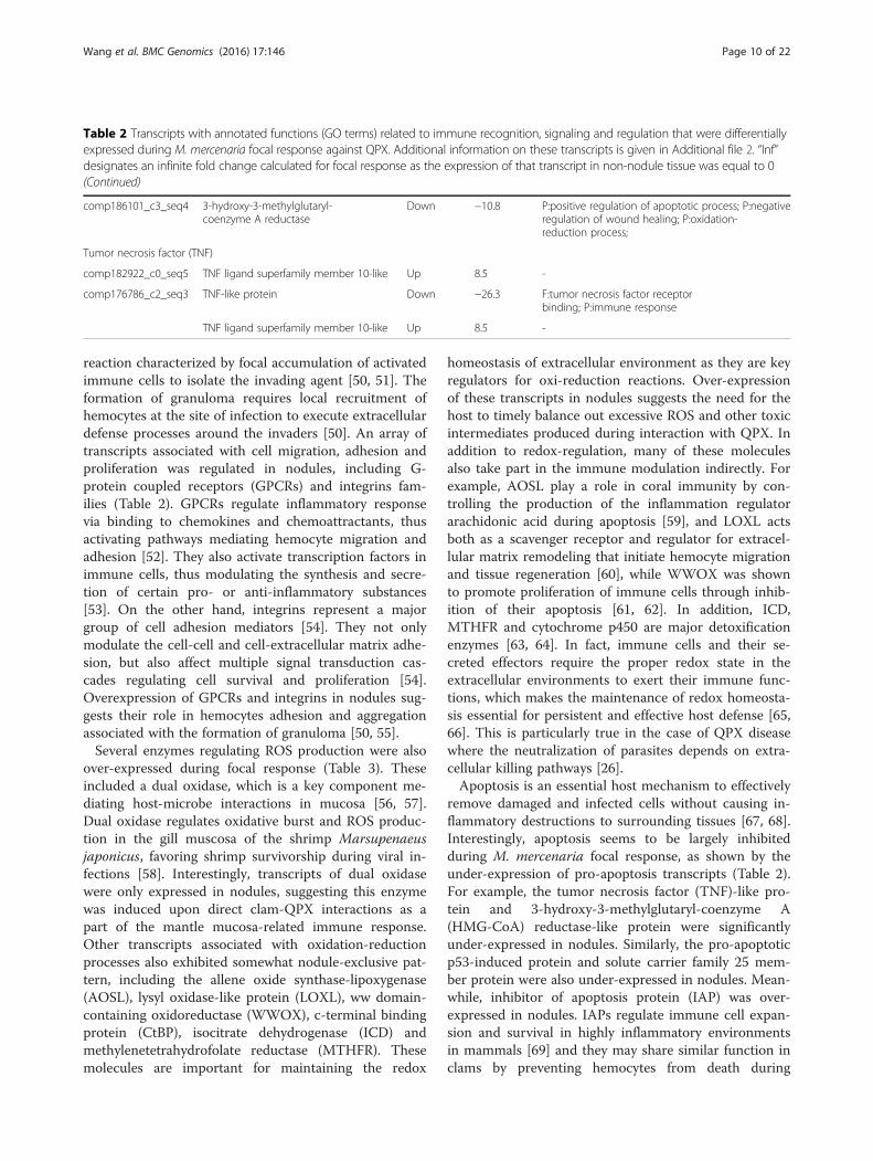
Table 2 Transcripts with annotated functions (GO terms) related to immune recognition, signaling and regulation that were differentiallyexpressed during M. mercenaria focal response against QPX. Additional information on these transcripts is given in Additional file 2. “Inf”designates an infinite fold change calculated for focal response as the expression of that transcript in non-nodule tissue was equal to 0(Continued)
comp186101_c3_seq4 3-hydroxy-3-methylglutaryl-coenzyme A reductase
Down −10.8 P:positive regulation of apoptotic process; P:negativeregulation of wound healing; P:oxidation-reduction process;
Tumor necrosis factor (TNF)
comp182922_c0_seq5 TNF ligand superfamily member 10-like Up 8.5 -
comp176786_c2_seq3 TNF-like protein Down −26.3 F:tumor necrosis factor receptorbinding; P:immune response
TNF ligand superfamily member 10-like Up 8.5 -
Wang et al. BMC Genomics (2016) 17:146 Page 10 of 22
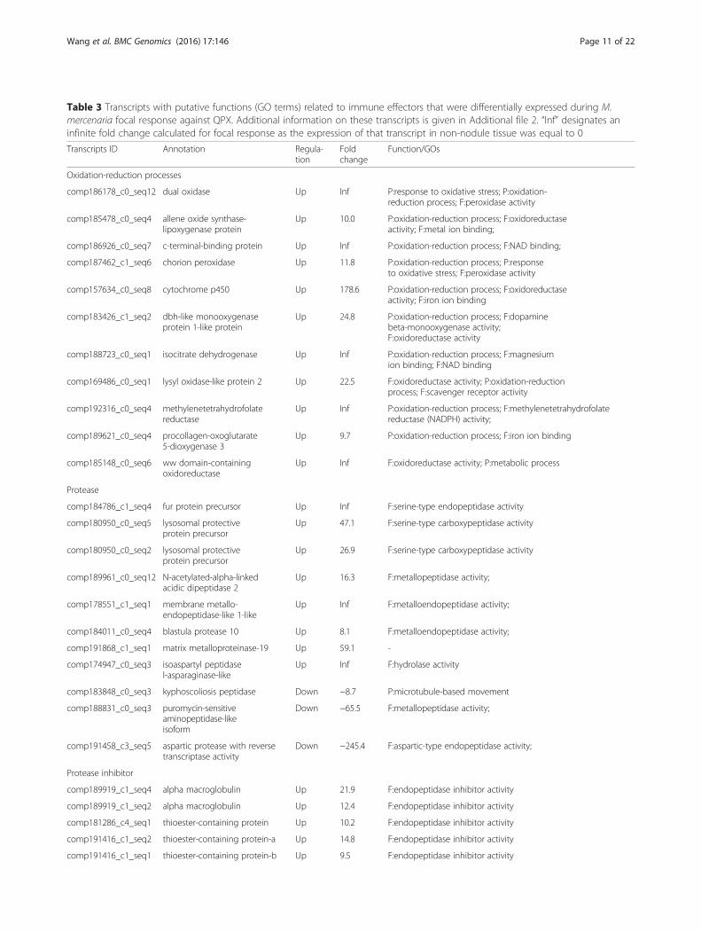
Table 3 Transcripts with putative functions (GO terms) related to immune effectors that were differentially expressed during M.mercenaria focal response against QPX. Additional information on these transcripts is given in Additional file 2. “Inf” designates aninfinite fold change calculated for focal response as the expression of that transcript in non-nodule tissue was equal to 0
Transcripts ID Annotation Regula-tion
Foldchange
Function/GOs
Oxidation-reduction processes
comp186178_c0_seq12 dual oxidase Up Inf P:response to oxidative stress; P:oxidation-reduction process; F:peroxidase activity
comp185478_c0_seq4 allene oxide synthase-lipoxygenase protein
Up 10.0 P:oxidation-reduction process; F:oxidoreductaseactivity; F:metal ion binding;
comp186926_c0_seq7 c-terminal-binding protein Up Inf P:oxidation-reduction process; F:NAD binding;
comp187462_c1_seq6 chorion peroxidase Up 11.8 P:oxidation-reduction process; P:responseto oxidative stress; F:peroxidase activity
comp157634_c0_seq8 cytochrome p450 Up 178.6 P:oxidation-reduction process; F:oxidoreductaseactivity; F:iron ion binding
comp183426_c1_seq2 dbh-like monooxygenaseprotein 1-like protein
Up 24.8 P:oxidation-reduction process; F:dopaminebeta-monooxygenase activity;F:oxidoreductase activity
comp188723_c0_seq1 isocitrate dehydrogenase Up Inf P:oxidation-reduction process; F:magnesiumion binding; F:NAD binding
comp169486_c0_seq1 lysyl oxidase-like protein 2 Up 22.5 F:oxidoreductase activity; P:oxidation-reductionprocess; F:scavenger receptor activity
comp192316_c0_seq4 methylenetetrahydrofolatereductase
Up Inf P:oxidation-reduction process; F:methylenetetrahydrofolatereductase (NADPH) activity;
comp189621_c0_seq4 procollagen-oxoglutarate5-dioxygenase 3
Up 9.7 P:oxidation-reduction process; F:iron ion binding
comp185148_c0_seq6 ww domain-containingoxidoreductase
Up Inf F:oxidoreductase activity; P:metabolic process
Protease
comp184786_c1_seq4 fur protein precursor Up Inf F:serine-type endopeptidase activity
comp180950_c0_seq5 lysosomal protectiveprotein precursor
Up 47.1 F:serine-type carboxypeptidase activity
comp180950_c0_seq2 lysosomal protectiveprotein precursor
Up 26.9 F:serine-type carboxypeptidase activity
comp189961_c0_seq12 N-acetylated-alpha-linkedacidic dipeptidase 2
Up 16.3 F:metallopeptidase activity;
comp178551_c1_seq1 membrane metallo-endopeptidase-like 1-like
Up Inf F:metalloendopeptidase activity;
comp184011_c0_seq4 blastula protease 10 Up 8.1 F:metalloendopeptidase activity;
comp191868_c1_seq1 matrix metalloproteinase-19 Up 59.1 -
comp174947_c0_seq3 isoaspartyl peptidasel-asparaginase-like
Up Inf F:hydrolase activity
comp183848_c0_seq3 kyphoscoliosis peptidase Down −8.7 P:microtubule-based movement
comp188831_c0_seq3 puromycin-sensitiveaminopeptidase-likeisoform
Down −65.5 F:metallopeptidase activity;
comp191458_c3_seq5 aspartic protease with reversetranscriptase activity
Down −245.4 F:aspartic-type endopeptidase activity;
Protease inhibitor
comp189919_c1_seq4 alpha macroglobulin Up 21.9 F:endopeptidase inhibitor activity
comp189919_c1_seq2 alpha macroglobulin Up 12.4 F:endopeptidase inhibitor activity
comp181286_c4_seq1 thioester-containing protein Up 10.2 F:endopeptidase inhibitor activity
comp191416_c1_seq2 thioester-containing protein-a Up 14.8 F:endopeptidase inhibitor activity
comp191416_c1_seq1 thioester-containing protein-b Up 9.5 F:endopeptidase inhibitor activity
Wang et al. BMC Genomics (2016) 17:146 Page 11 of 22
interaction with QPX. In fact, ROS production duringparasite killing may trigger apoptotic cell death in mol-luscs [67], and proper control of apoptosis mechanismsis required to maintain cellular homeostasis during im-mune response. This suspected inhibition of host apop-tosis is supported by the above-mentioned over-expression of integrins, as these were shown to protectcells from apoptosis and induce anti-apoptotic pathwaysduring cell adhesion and spreading in the snails Lym-naea stagnalis [70] and B. glabrata [71].Infection and tissue injury trigger host immune re-
sponses via immune signaling pathways [72], by activat-ing transcription factors and initiating the production ofimmune effectors and regulators. Immune signalingpathways identified in mollusks include Toll, MAPK/JNK and JAK/STAT signaling pathways [27, 47, 73, 74].During M. mercenaria focal response to QPX, a varietyof transcripts encoding kinases and phosphatases wereover-expressed (Table 2), suggesting the involvement ofMAPK and other kinase-mediated cascades in regulatingthe focal inflammatory response [75], whereas theunder-expression of EF-hand domain containing proteinand calmodulin may indicate the suppression of
calcium-regulated pathways [76]. Overexpression of E3ubiquitin-protein ligase and its upstream regulatorCOP9 signalosome suggests the activation of the damagesurveillance ubiquitin/proteasome pathway [77–79]. Inparallel, the over-expression of rho GTPase and rho kin-ase suggests the induction of anti-apoptotic Rho-mediated signaling pathway [80, 81] and reinforce theidea that apoptotic inhibition is extensively initiated byM. mercenaria to help fight QPX. However, a very lim-ited number of DE transcripts was detected in relationto those conventional signaling pathways of innate im-munity, such as the complement pathway and the Toll/TLR pathway. Only 1 transcript encoding macrophage-expressed gene protein 1 (MPEG1), a putative memberof the complement pathway [82, 83], was differentiallyexpressed in QPX nodules. As for the Toll/TLR pathway,only a few receptors were identified (Table 2) but noneof their downstream components.Interestingly, components of the Notch and the Wnt
signaling pathways were over-expressed during focal re-sponse (Table 2). These included two putative Notchfamily members, the neurogenic locus Notch proteinand the mediator protein nicastrin, and the tyrosin-
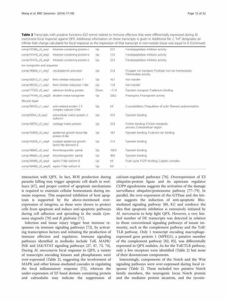
Table 3 Transcripts with putative functions (GO terms) related to immune effectors that were differentially expressed during M.mercenaria focal response against QPX. Additional information on these transcripts is given in Additional file 2. “Inf” designates aninfinite fold change calculated for focal response as the expression of that transcript in non-nodule tissue was equal to 0 (Continued)
comp192366_c0_seq1 thioester-containing protein-c Up 32.5 F:endopeptidase inhibitor activity
comp191416_c0_seq5 thioester-containing protein-e Up 12.9 F:endopeptidase inhibitor activity
comp191416_c0_seq4 thioester-containing protein-e Up 22.4 F:endopeptidase inhibitor activity
Ion transporter and sequester
comp190604_c1_seq1 ceruloplasmin precursor Up 21.8 P:copper ion transport; P:cellular iron ion homeostasis;F:ferroxidase activity
comp182612_c1_seq1 ferric-chelate reductase 1 Up 16.7 Iron transfer
comp180332_c1_seq1 ferric-chelate reductase 1-like Up 11.8 Iron transfer
comp177359_c0_seq1 selenium binding protein Down −11.0 P:protein transport; F:selenium binding
comp174164_c0_seq20 divalent metal transporter Up 328.2 P:transport; F:transporter activity
Wound repair
comp185425_c1_seq1 actin-related protein 2 3complex subunit 5-like
Up Inf C:cytoskeleton; P:regulation of actin filament polymerization
comp93954_c0_seq1 extracellular matrix protein 2isoform1
Up 25.9 F:protein binding
comp188753_c5_seq1 cartilage matrix protein Up 25.8 F:chitin binding; P:chitin metabolicprocess; C:extracellular region
comp142858_c0_seq1 epidermal growth factor-likeprotein 8-like
Up 18.5 F:protein binding; F:calcium ion binding
comp192650_c1_seq4 multiple epidermal growthfactor-like domains 6
Up 21.9 F:protein binding
comp186665_c0_seq1 thrombospondin- partial Up 106.9 F:protein binding
comp186665_c0_seq4 thrombospondin- partial Up 38.0 F:protein binding
comp184960_c0_seq4 septin-7-like isoform 8 Up Inf P:cell cycle; F:GTP binding; C:septin complex
comp184960_c0_seq42 septin-7-like isoform 6 Up 8.9 -
Wang et al. BMC Genomics (2016) 17:146 Page 12 of 22
protein kinase RYK which belongs to the Wnt pathway.Wnt signaling pathway regulates many cellular immuneprocesses and is evolutionarily conserved across taxa[84, 85]. Wnt signaling has been intensively exploited forits regulatory functions during wound healing and tissueregeneration [86, 87], so its over-expression may be re-lated to wound healing to repair damage resulting fromtissue digestion by the parasite or tissue necrosis. TheNotch signaling network was scarcely explored in bi-valves even though it has been reported to be associatedwith several aspects of immune response in mammals[88], especially in regulating granulomatous reactions toforeign bodies [89]. As an evolutionarily conserved path-way involved in modulating the intercellular signaling,the Notch pathway presumably shares an equally im-portant role in M. mercenaria by modulating the forma-tion of granuloma. Notch signaling triggers macrophageexpression of genes involved in pro-inflammatory re-sponses [90], but can suppress inflammation responsestriggered by canonical TLR cascade [91], in agreementwith our observations. These tightly regulated mecha-nisms ensure tailored immune responses against differ-ent pathogens and are crucial for the host to achievehigh immune efficiency while avoiding excessive im-mune activation and self-inflicted damages.Several proteases (A.K.A proteinases, peptidases) were
also differentially regulated during M. mercenaria focalresponse against QPX. These proteases mostly belong tothe serine and metallo protease families and were gener-ally over-expressed in QPX nodules (Table 3). Proteasesserve as key immune modulators partially through theirability to digest and remodel the extracellular matrixand tissues associated with hemocyte activation [92].Commonly associated with lysosomes and granules ofinflammatory cells, serine proteases participate in im-mune regulation either directly by degradation of patho-gens or indirectly through activation of cell surfacereceptors and signal molecules [92–94]. The function ofmetalloproteases in immune regulation is even more di-verse, acting as immune effectors, signal transducers,and mediators of immune cell development and migra-tion [95]. Metalloproteases are also known to be in-volved in many pro-inflammatory pathways, particularlyin the Notch pathway where they act as a type of down-stream element to Notch [96].At the same time, immune effectors with known uni-
versal protease inhibitor activities, such as alpha2-macroglobulin (α 2 M) and thioester-containing protein(TEP, a subfamily of α 2 M), were also collectively over-expressed in QPX nodules (Table 3). The α 2 M super-family inhibits peptidases of diverse origins [97, 98]. Thesimultaneous over-expression of proteases and proteaseinhibitors may reflect a finely adjusted defense responseof M. mercenaria to maintain homeostasis and regulate
self- and pathogen-derived proteases, as shown in otherhost-pathogen systems [92, 99], including bivalves [100].Proteases have been identified as major virulence factorsof QPX [101, 102], and are thought to degrade host pro-teinaceous immune effectors and hydrolyze host tissuesto fulfill nutritional requirements. Therefore, inhibitionof pathogen proteases contribute to host protection, andwas shown to represent a determinant factor for resist-ance against infectious diseases in bivalves [103–106]. Inparallel, TEPs have been extensively studied in molluskimmunity [99, 107], and beside their function as prote-ase inhibitors, they also play a role as PRRs or opsoninsto facilitate microbial phagocytosis and encapsulation.Consistent over-expression of TEPs was noted in thisstudy in agreement with findings following experimentalinfection with QPX [15]. These results support a criticalrole of TEPs in clam immune response against QPX ei-ther via the protease-inhibitor activity of these proteins,or by mediating parasite encapsulation, or both.In addition, several metal ion transporters were over-
expressed in nodules (Table 3), including the putativecopper ion binding protein ceruloplasmin precursor, thetransferrin enzyme ferric-chelate reductase and the diva-lent metal transporter (A.K.A natural resistance-associated macrophage protein; 328 fold increase). Thesemolecules contribute to host defense by controlling thesupply of essential micronutrients in the vicinity of in-fection sites thus reducing parasite survival [108, 109]and favoring the production of antimicrobial factors[110]. It is noteworthy to point out that some of the fo-cally over-expressed transcripts might be partly drivenby the dramatic increase of hemocyte proportion withintissues in the vicinity of infection foci as compared tothe surrounding host tissues [26, 111]. This would be es-pecially the case for transcripts known to be highlyexpressed in hemocytes, such as the cell surface PRRs,secreted humoral immune effectors, cell signal transduc-ers and enzymes associated with ROS production.
Systemic responseSignificant transcriptomic regulations were observedduring M. mercenaria systemic immune responseagainst QPX, with a total of 1,681 DE transcripts, whichis about two times the number of focal DE transcripts(829). However, only about one third of the DE tran-scripts (513) were over-expressed in response to QPXinfection, the larger remaining part (1,168) representedsignificantly under-expressed transcripts (Fig. 4, Add-itional file 3), possibly due to the chronic stress imposedby the infection. Transcriptome-wide depression hasbeen demonstrated in many marine invertebrates as theresult of pathogenic or environmental stress [18, 59,112–114]. A considerable number of systemically under-expressed transcripts was related to metabolism and
Wang et al. BMC Genomics (2016) 17:146 Page 13 of 22
biomineralization, which could be the result of host re-source allocation during on-going infection. The chronicinflammation induced by infection likely created extraenergy demands, which require resources being allocatedfrom other physiological processes, such as growth andreproduction, to immune processes which is critical forthe survivorship of the host. In fact, slow growth andlower tissue conditions are frequently observed in QPX-infected M. mercenaria [2], and similar energy trade-offsexisted between immune defense and other energy ex-penditure pathways [115–118]. Interestingly, a suite oftranscripts over-expressed during focal response was sig-nificantly under-expressed in non-nodule tissues as com-pared to healthy clams (Additional file 3), whichincluded several immune effectors and mediators associ-ated with nodule formation and focal inflammation (e.g.,integrins, notch proteins and peroxidases). In fact, main-taining high levels of these focally-induced moleculescould be costly and dangerous as some are toxic to boththe parasite and the host, so their production must berestrained within areas where they can directly exertdefense function, and reduced outside the infection foci tominimize risks of undesirable effects on the host [116, 117].On the other hand, systemically over-expressed tran-
scripts included stress proteins and other soluble immunefactors such as lysozyme (c-type lysozyme 2), lectins (C1qdomain containing protein, macrophage mannose recep-tor 1, low affinity immunoglobulin epsilon fc receptor),AMP (hemocyte defensin), proteases (cathepsin K, calpain11, isoaspartyl peptidase/L-asparaginase, ASRGL, count-ing factor associated protein d) and ferric-chelate reduc-tase (Table 4, Additional file 3). Over-expression of hoststress proteins, such as heat shock proteins (HSP 70, HSP90) and universal stress protein (USP) was also noted, inagreement with observations made during infection inother bivalve species [119–121]. Increased levels of stressproteins provide host cells with protection against in-correct protein folding caused by infection, inflamma-tion, oxidative stress and other destructive events[122, 123]. The systemic over-expression of solubleimmune effectors (e.g., humoral proteins) may helpmaintaining comparatively high immune capacity toprevent the spread of QPX (or secondary pathogens)throughout the host. In addition, transcripts of anti-apoptotic factors (IL17, deoxyguanosine, baculoviralmap repeat-containing proteins) were also over-expressed during the systemic response, indicatingthat anti-apoptotic processes noted during focal re-sponse are not limited to the infection foci.
Pathway alterations during M. mercenaria’s response to QPXTranscriptomic alterations during both focal and sys-temic response discussed above were also highlighted inthe pathway enrichment analysis of the DE transcripts.
This analysis aims at extracting an overview of pheno-typic changes on the underlying functional level, to re-duce the complexity of biological information given bythe long lists of DE genes/transcripts [124]. The KEGGpathways of focal adhesion (04510), ECM-receptor inter-action (04512), Notch signaling pathway (04330) andapoptosis (0421) were significantly over-represented dur-ing both focal and systemic response (Fig. 5), eventhough fold enrichment were generally higher during thefocal response. Other immune-related pathways particu-larly enriched during the focal response included regula-tion of actin cytoskeleton (04810), cell adhesionmolecules (CAMs, 04514), the leukocyte transendothe-lial migration (04670), complement and coagulation cas-cade (04610) and Wnt signaling pathway (04310). Thesepathways are critically involved in the immune cells acti-vation during migration, attachment and parasite encap-sulation, which serve as the underlying mechanisms fornodule formation and QPX killing. On the other hand,basic metabolic pathways such as the citrate cycle(TCA) and pyruvate metabolism were specially enrichedduring the systemic response. These alterations werelargely in accordance with the under-expression ofmetabolism-associated DE transcripts in infected tissuecompared to the healthy tissue (Fig. 4), possibly reflect-ing changes in the energy allocation strategy during in-fection as discussed above.Interdependence of KEGG pathways widely exist and
most of these are interrelated with each other via sharedcomponents, forming a signaling network to allow forpathway crosstalk. To investigate these interactions, weextracted the DE transcripts shared by multiple enrichedpathways and constructed a sketch of the hypotheticalpathways network that are significantly altered by QPXinfection (Fig. 6). In this framework, M. mercenaria re-sponse to QPX infection was initiated upon the sensingof danger signals via cell membrane receptors. The signalssubsequently transmitted down through the MAPK, Wntand Notch pathways and triggered the production of aseries of host defense factors as the end results. In parallel,activation of pathways regulating actin cytoskeleton andleukocyte transendothelial migration facilitated the re-cruitment of hemocytes to the infection area to build abarrier of cellular defense against the parasite. Recruitedhemocytes then attached to and encapsulated QPX cellsas suggested by the modulation of focal adhesion andECM receptor interaction pathways. These cellular activ-ities were performed under a tight regulation of the apop-tosis pathway to determine cell fates, resulting in eitherthe survival or death of M. mercenaria cells.
Distinctive transcriptomic pattern of healthy clamsA suite of transcripts (407, Fig. 4, Additional file 4 andTable 5) exhibited higher transcription levels in healthy
Wang et al. BMC Genomics (2016) 17:146 Page 14 of 22
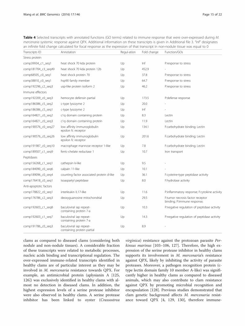
clams as compared to diseased clams (considering bothnodule and non-nodule tissues). A considerable fractionof these transcripts were related to metabolic processes,nucleic acids binding and transcriptional regulation. Theover-expressed immune-related transcripts identified inhealthy clams are of particular interest as they may beinvolved in M. mercenaria resistance towards QPX. Forexample, an antimicrobial protein (aplysianin A [125,126]) was exclusively identified in healthy clams with al-most no detection in diseased clams. In addition, thehighest expression levels of a serine protease inhibitorwere also observed in healthy clams. A serine proteaseinhibitor has been linked to oyster (Crassostrea
virginica) resistance against the protozoan parasite Per-kinsus marinus [103–106, 127]. Therefore, the high ex-pression of the serine protease inhibitor in healthy clamssupports its involvement in M. mercenaria’s resistanceagainst QPX, likely by inhibiting the activity of parasiteproteases. Moreover, a pathogen recognition protein (c-type lectin domain family 10 member A-like) was signifi-cantly higher in healthy clams as compared to diseasedanimals, which may also contribute to clam resistanceagainst QPX by promoting microbial recognition andencapsulation [128]. Previous studies demonstrated thatclam genetic background affects M. mercenaria resist-ance toward QPX [4, 129, 130], therefore immune-
Table 4 Selected transcripts with annotated functions (GO terms) related to immune response that were over-expressed during M.mercenaria systemic response against QPX. Additional information on these transcripts is given in Additional file 3. “Inf” designatesan infinite fold change calculated for focal response as the expression of that transcript in non-nodule tissue was equal to 0
Transcripts ID Annotation Regul-ation Fold change Function/GOs
Stress protein
comp39934_c1_seq1 heat shock 70 kda protein Up Inf P:response to stress
comp181704_c3_seq49 heat shock 70 kda protein 12b Up 452.9 -
comp68505_c0_seq1 heat shock protein 70 Up 37.8 P:response to stress
comp38810_c0_seq1 hsp90 family member Up 64.7 P:response to stress
comp192296_c2_seq3 usp-like protein isoform 2 Up 46.2 P:response to stress
Immune effectors
comp192209_c0_seq3 hemocyte defensin partial Up 173.5 P:defense response
comp186386_c5_seq2 c-type lysozyme 2 Up 20.0 -
comp186386_c5_seq1 c-type lysozyme 2 Up Inf -
comp164821_c0_seq2 c1q domain containing protein Up 8.3 Lectin
comp164821_c0_seq3 c1q domain containing protein Up 11.9 Lectin
comp190576_c0_seq27 low affinity immunoglobulinepsilon fc receptor
Up 134.1 F:carbohydrate binding; Lectin
comp190576_c0_seq26 low affinity immunoglobulinepsilon fc receptor
Up 201.6 F:carbohydrate binding; Lectin
comp191987_c0_seq10 macrophage mannose receptor 1-like Up 7.8 F:carbohydrate binding; Lectin
comp189507_c1_seq9 ferric-chelate reductase 1 Up 10.7 Iron transport
Peptidases
comp156268_c1_seq1 cathepsin k-like Up 9.5 -
comp184090_c0_seq6 calpain 11-like Up 10.1 -
comp189096_c0_seq4 counting factor associated protein d-like Up 36.1 F:cysteine-type peptidase activity
comp176418_c0_seq1 isoaspartyl peptidase Up 8.0 F:hydrolase activity
Anti-apoptotic factors
comp178822_c0_seq1 interleukin IL17-like Up 11.6 P:inflammatory response; F:cytokine activity
comp176786_c2_seq3 deoxyguanosine mitochondrial Up 29.5 F:tumor necrosis factor receptorbinding; P:immune response;
comp192603_c1_seq8 baculoviral iap repeat-containing protein 7-a
Up 10.3 P:negative regulation of peptidase activity
comp192603_c1_seq7 baculoviral iap repeat-containing protein 7-a
Up 14.3 P:negative regulation of peptidase activity
comp191786_c0_seq3 baculoviral iap repeat-containing protein partial
Up 8.9 -
Wang et al. BMC Genomics (2016) 17:146 Page 15 of 22
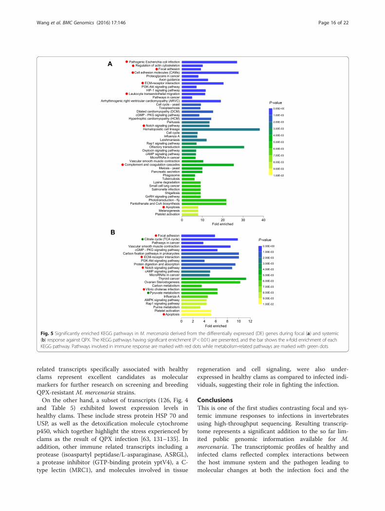
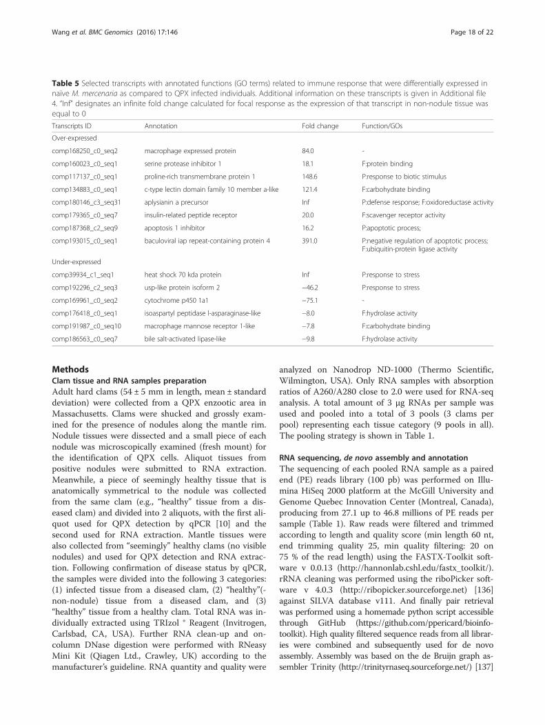
related transcripts specifically associated with healthyclams represent excellent candidates as molecularmarkers for further research on screening and breedingQPX-resistant M. mercenaria strains.On the other hand, a subset of transcripts (126, Fig. 4
and Table 5) exhibited lowest expression levels inhealthy clams. These include stress protein HSP 70 andUSP, as well as the detoxification molecule cytochromep450, which together highlight the stress experienced byclams as the result of QPX infection [63, 131–135]. Inaddition, other immune related transcripts including aprotease (isoaspartyl peptidase/L-asparaginase, ASRGL),a protease inhibitor (GTP-binding protein yptV4), a C-type lectin (MRC1), and molecules involved in tissue
regeneration and cell signaling, were also under-expressed in healthy clams as compared to infected indi-viduals, suggesting their role in fighting the infection.
ConclusionsThis is one of the first studies contrasting focal and sys-temic immune responses to infections in invertebratesusing high-throughput sequencing. Resulting transcrip-tome represents a significant addition to the so far lim-ited public genomic information available for M.mercenaria. The transcriptomic profiles of healthy andinfected clams reflected complex interactions betweenthe host immune system and the pathogen leading tomolecular changes at both the infection foci and the
Fig. 5 Significantly enriched KEGG pathways in M. mercenaria derived from the differentially expressed (DE) genes during focal (a) and systemic(b) response against QPX. The KEGG pathways having significant enrichment (P< 0.01) are presented, and the bar shows the x-fold enrichment of eachKEGG pathway. Pathways involved in immune response are marked with red dots while metabolism-related pathways are marked with green dots
Wang et al. BMC Genomics (2016) 17:146 Page 16 of 22
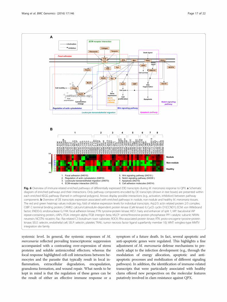
systemic level. In general, the systemic responses of M.mercenaria reflected prevailing transcriptomic suppressionaccompanied with a contrasting over-expression of stressproteins and soluble antimicrobial effectors; whereas thefocal response highlighted cell-cell interactions between he-mocytes and the parasite that typically result in local in-flammation, extracellular degradation, encapsulation,granuloma formation, and wound repair. What needs to bekept in mind is that the regulation of these genes can bethe result of either an effective immune response or a
symptom of a future death. In fact, several apoptotic andanti-apoptotic genes were regulated. This highlights a fineadjustment of M. mercenaria defense mechanisms to pre-cisely adapt to the infection development (e.g., through themodulation of energy allocation, apoptotic and anti-apoptotic processes and mobilization of different signalingpathways). In addition, the identification of immune-relatedtranscripts that were particularly associated with healthyclams offered new perspectives on the molecular featuresputatively involved in clam resistance against QPX.
Fig. 6 Overview of immune-related enriched pathways of differentially expressed (DE) transcripts during M. mercenaria response to QPX. a Schematicdiagram of enriched pathways and their interactions. Only pathway components encoded by DE transcripts (shown in text boxes) are presented withineach enriched KEGG pathway (framed in orthogonal polygons). Arrows display possible interactions (e.g., activation, inhibition) between pathwaycomponents. b Overview of DE transcripts expression associated with enriched pathways in nodule, non-nodule and healthy M. mercenaria tissues.The red and green heatmap values indicate log2 fold of relative expression levels for individual transcripts. Arp2/3: actin related protein 2/3 complex;CtBP: C-terminal binding protein; CAMK2: calcium/calmodulin-dependent protein kinase (CaM kinase) II; CycD: cyclin D1(CCND1); ECM: von Willebrandfactor; ENDO-G: endonuclease G; FAK: focal adhesion kinase; FYN: tyrosine-protein kinase; HES1: hairy and enhancer of split 1; IAP: baculoviral IAPrepeat-containing protein, cIAPs; ITGA: intergrin alpha; ITGB: intergrin beta; MLCP: serine/threonine-protein phosphatase PP1 catalytic subunit; NRXN:neurexin; NCSTN: nicastrin; Rac: Ras-related C3 botulinum toxin substrate; ROCK: Rho-associated protein kinase; RTK: proto-oncogene tyrosine-proteinkinase; SELE: selectin, endothelial cell; SELP: selectin, platelet; TRAIL: tumor necrosis factor ligand superfamily member 10); WNT: wingless-type MMTVintegration site family
Wang et al. BMC Genomics (2016) 17:146 Page 17 of 22
MethodsClam tissue and RNA samples preparationAdult hard clams (54 ± 5 mm in length, mean ± standarddeviation) were collected from a QPX enzootic area inMassachusetts. Clams were shucked and grossly exam-ined for the presence of nodules along the mantle rim.Nodule tissues were dissected and a small piece of eachnodule was microscopically examined (fresh mount) forthe identification of QPX cells. Aliquot tissues frompositive nodules were submitted to RNA extraction.Meanwhile, a piece of seemingly healthy tissue that isanatomically symmetrical to the nodule was collectedfrom the same clam (e.g., “healthy” tissue from a dis-eased clam) and divided into 2 aliquots, with the first ali-quot used for QPX detection by qPCR [10] and thesecond used for RNA extraction. Mantle tissues werealso collected from “seemingly” healthy clams (no visiblenodules) and used for QPX detection and RNA extrac-tion. Following confirmation of disease status by qPCR,the samples were divided into the following 3 categories:(1) infected tissue from a diseased clam, (2) “healthy”(-non-nodule) tissue from a diseased clam, and (3)“healthy” tissue from a healthy clam. Total RNA was in-dividually extracted using TRIzol ® Reagent (Invitrogen,Carlsbad, CA, USA). Further RNA clean-up and on-column DNase digestion were performed with RNeasyMini Kit (Qiagen Ltd., Crawley, UK) according to themanufacturer’s guideline. RNA quantity and quality were
analyzed on Nanodrop ND-1000 (Thermo Scientific,Wilmington, USA). Only RNA samples with absorptionratios of A260/A280 close to 2.0 were used for RNA-seqanalysis. A total amount of 3 μg RNAs per sample wasused and pooled into a total of 3 pools (3 clams perpool) representing each tissue category (9 pools in all).The pooling strategy is shown in Table 1.
RNA sequencing, de novo assembly and annotationThe sequencing of each pooled RNA sample as a pairedend (PE) reads library (100 pb) was performed on Illu-mina HiSeq 2000 platform at the McGill University andGenome Quebec Innovation Center (Montreal, Canada),producing from 27.1 up to 46.8 millions of PE reads persample (Table 1). Raw reads were filtered and trimmedaccording to length and quality score (min length 60 nt,end trimming quality 25, min quality filtering: 20 on75 % of the read length) using the FASTX-Toolkit soft-ware v 0.0.13 (http://hannonlab.cshl.edu/fastx_toolkit/).rRNA cleaning was performed using the riboPicker soft-ware v 4.0.3 (http://ribopicker.sourceforge.net) [136]against SILVA database v111. And finally pair retrievalwas performed using a homemade python script accessiblethrough GitHub (https://github.com/ppericard/bioinfo-toolkit). High quality filtered sequence reads from all librar-ies were combined and subsequently used for de novoassembly. Assembly was based on the de Bruijn graph as-sembler Trinity (http://trinityrnaseq.sourceforge.net/) [137]
Table 5 Selected transcripts with annotated functions (GO terms) related to immune response that were differentially expressed innaïve M. mercenaria as compared to QPX infected individuals. Additional information on these transcripts is given in Additional file4. “Inf” designates an infinite fold change calculated for focal response as the expression of that transcript in non-nodule tissue wasequal to 0
Transcripts ID Annotation Fold change Function/GOs
Over-expressed
comp168250_c0_seq2 macrophage expressed protein 84.0 -
comp160023_c0_seq1 serine protease inhibitor 1 18.1 F:protein binding
comp117137_c0_seq1 proline-rich transmembrane protein 1 148.6 P:response to biotic stimulus
comp134883_c0_seq1 c-type lectin domain family 10 member a-like 121.4 F:carbohydrate binding
comp180146_c3_seq31 aplysianin a precursor Inf P:defense response; F:oxidoreductase activity
comp179365_c0_seq7 insulin-related peptide receptor 20.0 F:scavenger receptor activity
comp187368_c2_seq9 apoptosis 1 inhibitor 16.2 P:apoptotic process;
comp193015_c0_seq1 baculoviral iap repeat-containing protein 4 391.0 P:negative regulation of apoptotic process;F:ubiquitin-protein ligase activity
Under-expressed
comp39934_c1_seq1 heat shock 70 kda protein Inf P:response to stress
comp192296_c2_seq3 usp-like protein isoform 2 −46.2 P:response to stress
comp169961_c0_seq2 cytochrome p450 1a1 −75.1 -
comp176418_c0_seq1 isoaspartyl peptidase l-asparaginase-like −8.0 F:hydrolase activity
comp191987_c0_seq10 macrophage mannose receptor 1-like −7.8 F:carbohydrate binding
comp186563_c0_seq7 bile salt-activated lipase-like −9.8 F:hydrolase activity
Wang et al. BMC Genomics (2016) 17:146 Page 18 of 22
using the default parameters. Assembly quality was con-trolled by re-mapping raw reads back to the transcriptsusing bowtie 2 and RSEM within Trinity package scripts.The assembled transcripts with sequence length longerthan 200 bp, re-mapping FPKM (fragments per kilobase oftranscript per million mapped reads) greater than 1 andisoform discovery level greater than 1 % were then consid-ered for the following annotation and transcript abundancequantitation. Annotation of this de novo assembled tran-scriptome was performed using Blast2GO (http://www.blast2go.com) with a semi-automated functional an-notation based on sequence homology search. Putativegene identities was obtained by Blastx search against Na-tional Center for Biotechnology Information (NCBI) non-redundant sequences (nr) database with the E-value thresh-old setting at 1E-03. Putative gene functions were predictedby sequence similarity search against Gene Ontology (GO,http://www.geneontology.org/) database and assigning GOannotation terms to each mapped transcript. Protein do-main search and enzyme annotation were also performedusing InterPro scan and the Kyoto Encyclopedia of Genesand Genomes (KEGG). KEGG Orthology (KO) terms andKEGG pathways were also assigned to the assembled se-quences using the online KEGG Automatic AnnotationServer (KAAS, http://www.genome.jp/kegg/kaas/) using bi-directional best-hit methods [138]. This server provides KOannotation and pathway mapping.
Differential gene expression analysisThe consensus transcriptome generated in the previoussteps was used as a reference for transcript abundanceanalysis. Reads from each single library were separatelymapped to this reference transcriptome using the “RNA-Seq by Expectation-Maximization (RSEM)” method thatis bundled within the Trinity package. The expressionlevel of each transcript was determined as the totalmapped reads count. The differences in gene expressionbetween clam tissue samples (nodule, non-nodule andhealthy tissues) were estimated using the DESeq Biocon-ductor package (https://github.com/Bioconductor-mir-ror/SESeq/tree/release-3.2) [139] in R statistical software(R Development Core Team, 2010; http://www.R-projec-t.org). The threshold for defining significant differentiallyexpressed (DE) transcripts between two different condi-tions (3 replications in each condition) was set as ad-justed p-value smaller than 0.001 and absolute log2 (foldchange) values greater than 2. Expression patterns of DEtranscripts were also analyzed by a K-means clusteringmethod using Euclidean distance based on expressionlevels over all input samples. For further analysis, onlythose DE transcripts with annotation were considered ascandidates of interest and were subsequently dividedinto over- and under-expressed groups.
Availability of supporting dataThe data sets supporting the results of this study areavailable at the NCBI short Read Archive database underthe SRA accession number SRP068241.
Additional files
Additional file 1: Sequence summary of M. mercenaria de novoassembled transcriptome. This is a Microsoft Excel worksheet thatcontains descriptions and annotation information of individual transcriptsequences. (XLSX 6600 kb)
Additional file 2: Differentially expressed (DE) sequences associatedwith M. mercenaria focal response against QPX. This file is a MicrosoftExcel worksheet that contains separate lists of over- and under-expressedtranscripts with their expression levels and fold change values given.(XLSX 143 kb)
Additional file 3: Differentially expressed (DE) sequences associatedwith M. mercenaria systemic response against QPX. This file is aMicrosoft Excel worksheet that contains separate lists of over- andunder-expressed transcripts with their expression levels and foldchange values given. (XLSX 327 kb)
Additional file 4: Transcripts that were differentially expressed in naïveM. mercenaria as compared to QPX infected individuals. This file is aMicrosoft Excel worksheet that contains separate lists of over- and under-expressed transcripts with their expression levels and fold change valuesgiven. (XLSX 128 kb)
Competing interestsThe authors declare that they have no competing interests.
Authors’ contributionsBA and EPE designed the study, coordinated experiments and superviseddata analysis and manuscript drafting. KW carried out the experiments,analyzed the data and drafted the manuscript. EC participated in dataanalysis and helped in drafting the methodology section of the manuscript.CDC helped with data analysis. All authors read and approved the finalmanuscript.
AcknowledgmentsThis research was supported by projects R/XG-19 and R/FBM-34 (to BA andEPE), funded under award NA07OAR4170010 from the National Sea GrantCollege Program of NOAA to the Research Foundation of State University ofNew York on behalf of New York Sea Grant. The study was also partiallysupported by project IOS 1050596 (to BA and EPE) supported by theNational Science Foundation. The statements, findings, conclusions, viewsand recommendations are those of the authors and do not necessarilyreflect the views of any of those organizations.
Author details1School of Marine and Atmospheric Sciences, Stony Brook University, StonyBrook, NY 11794-5000, USA. 2Analyses and Bioinformatics for Marine Science,Station Biologique de Roscoff, 29688 Roscoff Cedex, France.
Received: 6 October 2015 Accepted: 17 February 2016
References1. Whyte SK, Cawthorn RJ, McGladdery SE. QPX (Quahaug Parasite X), a pathogen
of northern quahaug Mercenaria mercenaria from the gulf of St-Lawrence,Canada. Dis Aquat Organ. 1994;19(2):129–36.
2. Smolowitz R, Leavitt D, Perkins F. Observations of a protistan disease similarto QPX in Mercenaria mercenaria (hard clams) from the coast ofMassachusetts. J Invertebr Pathol. 1998;71(1):9–25.
3. Anderson RS, Kraus BS, McGladdery SE, Reece KS, Stokes NA. A thraustochytridprotist isolated from Mercenaria mercenaria: molecular characterization andhost defense responses. Fish Shellfish Immun. 2003;15(3):183–94.
Wang et al. BMC Genomics (2016) 17:146 Page 19 of 22
4. Ford SE, Kraeuter JN, Barber RD, Mathis G. Aquaculture-associated factors inQPX disease of hard clams: density and seed source. Aquaculture. 2002;208(1–2):23–38.
5. Ragone Calvo LM, Walker JG, Burreson EM. Prevalence and distribution ofQPX, Quahog Parasite Unknown, in hard clams Mercenaria mercenaria inVirginia, USA. Dis Aquat Organ. 1998;33(3):209–19.
6. MacCallum GS, McGladdery SE. Quahog Parasite Unknown (QPX) in thenorthern quahog Mercenaria mercenaria (Linnaeus, 1758) and M.mercenaria var. notata from Atlantic Canada, survey results from threemaritime provinces. J Shellfish Res. 2000;19(1):43–50.
7. Dove ADM, Bowser PR, Cerrato RM. Histological Analysis of an Outbreak ofQPX Disease in Wild Hard Clams Mercenaria mercenaria in New York. JAquat Anim Health. 2004;16(4):246–50.
8. Lyons MM, Ward JE, Smolowitz R, Uhlinger KR, Gast RJ. Lethal marine snow:Pathogen of bivalve mollusc concealed in marine aggregates. LimnolOceanogr. 2005;50(6):1983–8.
9. Gast RJ, Moran DM, Audemard C, Lyons MM, DeFavari J, Reece KS, et al.Environmental distribution and persistence of Quahog Parasite Unknown(QPX). Dis Aquat Organ. 2008;81(3):219–29.
10. Liu QQ, Allam B, Collier JL. Quantitative Real-Time PCR Assay for QPX(Thraustochytriidae), a Parasite of the Hard Clam (Mercenaria mercenaria).Appl Environ Microb. 2009;75(14):4913–8.
11. Perrigault M, Dahl SF, Espinosa EP, Gambino L, Allam B. Effects of temperatureon hard clam (Mercenaria mercenaria) immunity and QPX (Quahog ParasiteUnknown) disease development: II. Defense parameters. J Invertebr Pathol.2011;106(2):322–32.
12. Bayne CJ. Phagocytosis and non-self recognition in invertebrates.Bioscience. 1990;40(10):723–31.
13. Canesi L, Gallo G, Gavioli M, Pruzzo C. Bacteria-hemocyte interactions andphagocytosis in marine bivalves. Microsc Res Tech. 2002;57(6):469–76.
14. Perrigault M, Dahl SF, Espinosa EP, Allam B. Effects of salinity on hard clam(Mercenaria mercenaria) defense parameters and QPX disease dynamics.J Invertebr Pathol. 2012;110(1):73–82.
15. Perrigault M, Tanguy A, Allam B. Identification and expression of differentiallyexpressed genes in the hard clam, Mercenaria mercenaria, in response toquahog parasite unknown (QPX). Bmc Genomics. 2009;10:377.
16. Perrigault M, Allam B. Differential immune response in the hard clam(Mercenaria mercenaria) against bacteria and the protistan pathogenQPX (quahog parasite unknown). Fish Shellfish Immun. 2012;32(6):1124–34.
17. Leite RB, Milan M, Coppe A, Bortoluzzi S, dos Anjos A, Reinhardt R, et al.mRNA-Seq and microarray development for the Grooved carpet shell clam,Ruditapes decussatus: a functional approach to unravel host -parasiteinteraction. Bmc Genomics. 2013;14(1):741.
18. Zhao X, Yu H, Kong L, Li Q. Transcriptomic Responses to Salinity Stress inthe Pacific Oyster Crassostrea gigas. PLoS ONE. 2012;7(9):e46244.
19. Teaniniuraitemoana V, Huvet A, Levy P, Klopp C, Lhuillier E, Gaertner-MazouniN, et al. Gonad transcriptome analysis of pearl oyster Pinctada margaritifera:identification of potential sex differentiation and sex determining genes.Bmc Genomics. 2014;15(1):491.
20. Adema CM, Hanington PC, Lun C-M, Rosenberg GH, Aragon AD, Stout BA, et al.Differential transcriptomic responses of Biomphalaria glabrata (Gastropoda,Mollusca) to bacteria and metazoan parasites, Schistosoma mansoni andEchinostoma paraensei (Digenea, Platyhelminthes). Mol Immunol.2010;47(4):849–60.
21. Wang JP, Lindsay BG, Leebens-Mack J, Cui L, Wall K, Miller WC, et al. ESTclustering error evaluation and correction. Bioinformatics.2004;20(17):2973–84.
22. Mittapalli O, Bai X, Mamidala P, Rajarapu SP, Bonello P, Herms DA. Tissue-SpecificTranscriptomics of the Exotic Invasive Insect Pest Emerald Ash Borer (Agrilusplanipennis). PLoS ONE. 2010;5(10):e13708.
23. Pelechano V, Wei W, Steinmetz LM. Extensive transcriptional heterogeneityrevealed by isoform profiling. Nature. 2013;497(7447):127–31.
24. Luo W, Friedman MS, Shedden K, Hankenson KD, Woolf PJ. GAGE: generallyapplicable gene set enrichment for pathway analysis. BMC Bioinformatics.2009;10(1):161.
25. Humphries JE, Yoshino TP. Cellular receptors and signal transduction inmolluscan hemocytes: connections with the innate immune system ofvertebrates. Integr Comp Biol. 2003;43(2):305–12.
26. Soudant P, E Chu FL, Volety A. Host-parasite interactions: Marine bivalvemolluscs and protozoan parasites, Perkinsus species. J Invertebr Pathol.2013;114(2):196–216.
27. Canesi L, Betti M, Ciacci C, Lorusso LC, Pruzzo C, Gallo G. Cell signalling inthe immune response of mussel hemocytes. Invertebrate Survival Journal:ISJ. 2006;3:40–9.
28. Hatanaka R, Sekine Y, Hayakawa T, Takeda K, Ichijo H. Signaling pathways ininvertebrate immune and stress response. Invertebrate Survival Journal:ISJ. 2009;6:32–43.
29. Jeannin P, Jaillon S, Delneste Y. Pattern recognition receptors in the immuneresponse against dying cells. Curr Opin Immunol. 2008;20(5):530–7.
30. Wang L, Wang L, Zhang D, Li F, Wang M, Huang M, et al. A novel C-typelectin from crab Eriocheir sinensis functions as pattern recognition receptorenhancing cellular encapsulation. Fish Shellfish Immun. 2013;34(3):832–42.
31. Vazquez-Mendoza A, Carrero JC, Rodriguez-Sosa M. Parasitic infections: arole for C-type lectins receptors. Biomed Res Int. 2013;2013:456352.
32. Yang J, Qiu L, Wei X, Wang L, Wang L, Zhou Z, et al. An ancient C-typelectin in Chlamys farreri (CfLec-2) that mediate pathogen recognition andcellular adhesion. Dev Comp Immunol. 2010;34(12):1274–82.
33. Wang L, Wang L, Yang J, Zhang H, Huang M, Kong P, et al. A multi-CRD C-type lectin with broad recognition spectrum and cellular adhesion fromArgopecten irradians. Dev Comp Immunol. 2012;36(3):591–601.
34. Ao J, Ling E, Yu XQ. Drosophila C-type lectins enhance cellularencapsulation. Mol Immunol. 2007;44(10):2541–8.
35. Lasky LA. Selectin-Carbohydrate Interactions and the Initiation of theInflammatory Response. Annu Rev Biochem. 1995;64(1):113–40.
36. Gazi U, Martinez-Pomares L. Influence of the mannose receptor in hostimmune responses. Immunobiology. 2009;214(7):554–61.
37. Moreira R, Milan M, Balseiro P, Romero A, Babbucci M, Figueras A, et al.Gene expression profile analysis of Manila clam (Ruditapes philippinarum)hemocytes after a Vibrio alginolyticus challenge using an immune-enrichedoligo-microarray. Bmc Genomics. 2014;15(1):267.
38. Gordon S. Pattern recognition receptors: doubling up for the innateimmune response. Cell. 2002;111(7):927–30.
39. Liu L, Yang J, Qiu L, Wang L, Zhang H, Wang M, et al. A novel scavengerreceptor-cysteine-rich (SRCR) domain containing scavenger receptoridentified from mollusk mediated PAMP recognition and binding.Dev Comp Immunol. 2011;35(2):227–39.
40. Mukhopadhyay S, Gordon S. The role of scavenger receptors in pathogenrecognition and innate immunity. Immunobiology. 2004;209(1–2):39–49.
41. Blumbach B, Pancer Z, Diehl-Seifert B, Steffen R, Munkner J, Muller I, et al.The putative sponge aggregation receptor. Isolation and characterization ofa molecule composed of scavenger receptor cysteine-rich domains andshort consensus repeats. J Cell Sci. 1998;111(Pt 17):2635–44.
42. van der Laan LJ, Dopp EA, Haworth R, Pikkarainen T, Kangas M, Elomaa O, et al.Regulation and functional involvement of macrophage scavenger receptorMARCO in clearance of bacteria in vivo. J Immunol. 1999;162(2):939–47.
43. Bowdish DM, Gordon S. Conserved domains of the class A scavengerreceptors: evolution and function. Immunol Rev. 2009;227(1):19–31.
44. Rast JP, Smith LC, Loza-Coll M, Hibino T, Litman GW. Genomic insights intothe immune system of the sea urchin. Science. 2006;314(5801):952–6.
45. Zhang Y, He X, Yu F, Xiang Z, Li J, Thorpe KL, et al. Characteristic and functionalanalysis of toll-like receptors (TLRs) in the lophotrocozoan, Crassostrea gigas, revealsancient origin of TLR-mediated innate immunity. PLoS One. 2013;8(10):e76464.
46. Qiu L, Song L, Xu W, Ni D, Yu Y. Molecular cloning and expression of a Tollreceptor gene homologue from Zhikong Scallop, Chlamys farreri. FishShellfish Immunol. 2007;22(5):451–66.
47. Moreira R, Balseiro P, Planas JV, Fuste B, Beltran S, Novoa B, et al. Transcriptomicsof in vitro immune-stimulated hemocytes from the Manila clam Ruditapesphilippinarum using high-throughput sequencing. PLoS One. 2012;7(4):e35009.
48. Mateo DR, Greenwood SJ, Araya MT, Berthe FC, Johnson GR, Siah A.Differential gene expression of gamma-actin, Toll-like receptor 2 (TLR-2) andinterleukin-1 receptor-associated kinase 4 (IRAK-4) in Mya arenariahaemocytes induced by in vivo infections with two Vibrio splendidusstrains. Dev Comp Immunol. 2010;34(7):710–4.
49. Akhouayri I, Turc C, Royet J, Charroux B. Toll-8/Tollo Negatively RegulatesAntimicrobial Response in the Drosophila Respiratory Epithelium. PlosPathog. 2011;7(10):e1002319.
50. Williams GT, Williams WJ. Granulomatous inflammation–a review. J ClinPathol. 1983;36(7):723–33.
51. de Brito T, Franco MF. Granulomatous inflammation. Revista do Instituto deMedicina Tropical de Sao Paulo. 1994;36(2):185–92.
52. Sun L, Ye RD. Role of G protein-coupled receptors in inflammation. Actapharmacologica Sinica. 2012;33(3):342–50.
Wang et al. BMC Genomics (2016) 17:146 Page 20 of 22
53. Cho H, Kehrl JH. Regulation of immune function by G protein-coupled receptors,trimeric G proteins, and RGS proteins. Prog Mol Biol Transl Sci. 2009;86:249–98.
54. Zhang Y, Wang H. Integrin signalling and function in immune cells.Immunology. 2012;135(4):268–75.
55. Stavitsky AB. Regulation of granulomatous inflammation in experimentalmodels of schistosomiasis. Infect Immun. 2004;72(1):1–12.
56. Ha EM, Oh CT, Bae YS, Lee WJ. A direct role for dual oxidase in Drosophilagut immunity. Science. 2005;310(5749):847–50.
57. Bae YS, Choi MK, Lee WJ. Dual oxidase in mucosal immunity and host-microbe homeostasis. Trends Immunol. 2010;31(7):278–87.
58. Inada M, Kihara K, Kono T, Sudhakaran R, Mekata T, Sakai M, et al. Decipheringof the Dual oxidase (Nox family) gene from kuruma shrimp, Marsupenaeusjaponicus: Full-length cDNA cloning and characterization. Fish Shellfish Immun.2013;34(2):471–85.
59. Libro S, Kaluziak ST, Vollmer SV. RNA-seq Profiles of Immune Related Genesin the Staghorn Coral Acropora cervicornis Infected with White BandDisease. PLoS ONE. 2013;8(11):e81821.
60. Bignon M, Pichol-Thievend C, Hardouin J, Malbouyres M, Brechot N, Nasciutti L,et al. Lysyl oxidase-like protein-2 regulates sprouting angiogenesis and type IVcollagen assembly in the endothelial basement membrane. Blood. 2011;118(14):3979–89.
61. Chang NS, Hsu LJ, Lin YS, Lai FJ, Sheu HM. WW domain-containingoxidoreductase: a candidate tumor suppressor. Trends Mol Med. 2007;13(1):12–22.
62. Yang Z, Zhao T, Liu Y. Upregulation of tumor suppressor WWOX promotesimmune response in glioma. Cell Immunol. 2013;285(1–2):1–5.
63. Behrendt L, Jonsson ME, Goldstone JV, Stegeman JJ. Induction ofcytochrome P450 1 genes and stress response genes in developingzebrafish exposed to ultraviolet radiation. Aquat Toxicol. 2010;98(1):74–82.
64. Pan L, Liu N, Xu C, Miao J. Identification of a novel P450 gene belonging tothe CYP4 family in the clam Ruditapes philippinarum, and analysis of basal-and benzo(a)pyrene-induced mRNA expression levels in selected tissues.Environ Toxicol Pharmacol. 2011;32(3):390–8.
65. Lotze MT, Zeh HJ, Rubartelli A, Sparvero LJ, Amoscato AA, Washburn NR, et al.The grateful dead: damage-associated molecular pattern molecules andreduction/oxidation regulate immunity. Immunol Rev. 2007;220:60–81.
66. Mone Y, Monnin D, Kremer N. The oxidative environment: a mediator ofinterspecies communication that drives symbiosis evolution. Proc Biol Sci/The Royal Society. 2014;281(1785):20133112.
67. Terahara K, Takahashi KG. Mechanisms and immunological roles ofapoptosis in molluscs. Curr Pharm Des. 2008;14(2):131–7.
68. Sokolova IM. Apoptosis in molluscan immune defense. Invertebrate SurvivalJournal: ISJ. 2009;6:49–58.
69. Gentle IE, Moelter I, Lechler N, Bambach S, Vucikuja S, Hacker G, et al.Inhibitors of apoptosis proteins (IAPs) are required for effective T-cellexpansion/survival during antiviral immunity in mice. Blood.2014;123(5):659–68.
70. Plows LD, Cook RT, Davies AJ, Walker AJ. Phagocytosis by Lymnaea stagnalishaemocytes: A potential role for phosphatidylinositol 3-kinase but notprotein kinase A. J Invertebr Pathol. 2006;91(1):74–7.
71. Yoshino TP, Laursen JR. Production of Schistosoma mansoni daughtersporocysts from mother sporocysts maintained in synxenic culture withBiomphalaria glabrata embryonic (Bge) cells. J Parasitol. 1995;81(5):714–22.
72. Newton K, Dixit VM. Signaling in innate immunity and inflammation. ColdSpring Harb Perspect Biol. 2012;4(3). doi: 10.1101/cshperspect.a006049.
73. Toubiana M, Rosani U, Giambelluca S, Cammarata M, Gerdol M, Pallavicini A, et al.Toll signal transduction pathway in bivalves: complete cds of intermediateelements and related gene transcription levels in hemocytes ofimmune stimulated Mytilus galloprovincialis. Dev Comp Immunol.2014;45(2):300–12.
74. Philipp EER, Kraemer L, Melzner F, Poustka AJ, Thieme S, Findeisen U, et al.Massively Parallel RNA Sequencing Identifies a Complex Immune GeneRepertoire in the lophotrochozoan Mytilus edulis. PLoS ONE. 2012;7(3):e33091.
75. Theodosiou A, Ashworth A. MAP kinase phosphatases. Genome Biol. 2002;3(7):REVIEWS3009.
76. Grabarek Z. Insights into modulation of calcium signaling by magnesium incalmodulin, troponin C and related EF-hand proteins. Biochim Biophys Acta(BBA) - Molecular Cell Research. 2011;1813(5):913–21.
77. Liu Y, Schiff M, Serino G, Deng XW, Dinesh-Kumar SP. Role of SCF ubiquitin-ligase and the COP9 signalosome in the N gene-mediated resistanceresponse to Tobacco mosaic virus. The Plant cell. 2002;14(7):1483–96.
78. Bech-Otschir D, Seeger M, Dubiel W. The COP9 signalosome: at theinterface between signal transduction and ubiquitin-dependent proteolysis.J Cell Sci. 2002;115(Pt 3):467–73.
79. Nezames CD, Deng XW. The COP9 signalosome: its regulation of cullin-based E3 ubiquitin ligases and role in photomorphogenesis. Plant Physiol.2012;160(1):38–46.
80. Tosello-Trampont AC, Nakada-Tsukui K, Ravichandran KS. Engulfment ofapoptotic cells is negatively regulated by Rho-mediated signaling. J BiolChem. 2003;278(50):49911–9.
81. Krijnen PA, Sipkens JA, Molling JW, Rauwerda JA, Stehouwer CD, Muller A,et al. Inhibition of Rho-ROCK signaling induces apoptotic and non-apoptotic PS exposure in cardiomyocytes via inhibition of flippase. J MolCell Cardiol. 2010;49(5):781–90.
82. He X, Zhang Y, Yu Z. An Mpeg (macrophage expressed gene) from thePacific oyster Crassostrea gigas: molecular characterization and geneexpression. Fish Shellfish Immunol. 2011;30(3):870–6.
83. D’Angelo ME, Dunstone MA, Whisstock JC, Trapani JA, Bird PI. Perforinevolved from a gene duplication of MPEG1, followed by a complex patternof gene gain and loss within Euteleostomi. Bmc Evol Biol. 2012;12(1):59.
84. Staal FJ, Luis TC, Tiemessen MM. WNT signalling in the immune system:WNT is spreading its wings. Nat Rev Immunol. 2008;8(8):581–93.
85. Holstein TW. The evolution of the Wnt pathway. Cold Spring Harb PerspectBiol. 2012;4(7):a007922.
86. Zhang Z, Deb A, Zhang Z, Pachori A, He W, Guo J, et al. Secreted frizzledrelated protein 2 protects cells from apoptosis by blocking the effect ofcanonical Wnt3a. J Mol Cell Cardiol. 2009;46(3):370–7.
87. Fathke C, Wilson L, Shah K, Kim B, Hocking A, Moon R, et al. Wnt signaling inducesepithelial differentiation during cutaneous wound healing. Bmc Cell Biol. 2006;7(1):4.
88. Yuan JS, Kousis PC, Suliman S, Visan I, Guidos CJ. Functions of notchsignaling in the immune system: consensus and controversies. Annu RevImmunol. 2010;28:343–65.
89. Rangel JR, Chung Y, Rosenbach M, Ziober AF, Zhang PJ, Schaffer A. Expressionof Notch signaling components in cutaneous foreign body and sarcoidalgranulomas and fusing macrophages. Am J Dermatopathol. 2014;36(5):409–13.
90. Palaga T, Buranaruk C, Rengpipat S, Fauq AH, Golde TE, Kaufmann SH, et al.Notch signaling is activated by TLR stimulation and regulates macrophagefunctions. Eur J Immunol. 2008;38(1):174–83.
91. Zhang Q, Wang C, Liu Z, Liu X, Han C, Cao X, et al. Notch signal suppressesToll-like receptor-triggered inflammatory responses in macrophages byinhibiting extracellular signal-regulated kinase 1/2-mediated nuclear factorkappaB activation. J Biol Chem. 2012;287(9):6208–17.
92. Manoury B, Roghanian A, Sallenave JM. Serine and cysteine proteases and theirinhibitors as antimicrobial agents and immune modulators. In: Vergnolle N,Chignard M, editors. Proteases and their receptors in inflammation. Progress inInflammation Research. Basel: Springer; 2011. p. 27–50.
93. Shpacovitch V, Feld M, Hollenberg MD, Luger TA, Steinhoff M. Role ofprotease-activated receptors in inflammatory responses, innate andadaptive immunity. J Leukoc Biol. 2008;83(6):1309–22.
94. Meyer-Hoffert U, Wiedow O. Neutrophil serine proteases: mediators ofinnate immune responses. Curr Opin Hematol. 2011;18(1):19–24.
95. Khokha R, Murthy A, Weiss A. Metalloproteinases and their natural inhibitorsin inflammation and immunity. Nat Rev Immunol. 2013;13(9):649–65.
96. Murthy A, Shao YW, Narala SR, Molyneux SD, Zuniga-Pflucker JC, Khokha R.Notch activation by the metalloproteinase ADAM17 regulatesmyeloproliferation and atopic barrier immunity by suppressing epithelialcytokine synthesis. Immunity. 2012;36(1):105–19.
97. Sottrup-Jensen L. Alpha-macroglobulins: structure, shape, and mechanismof proteinase complex formation. J Biol Chem. 1989;264(20):11539–42.
98. Borth W. Alpha 2-macroglobulin, a multifunctional binding protein withtargeting characteristics. Faseb J. 1992;6(15):3345–53.
99. Armstrong PB. Proteases and protease inhibitors: a balance of activities inhost–pathogen interaction. Immunobiology. 2006;211(4):263–81.
100. Allam B, Pales Espinosa E, Tanguy A, Jeffroy F, Le Bris C, Paillard C.Transcriptional changes in Manila clam (Ruditapes philippinarum) inresponse to Brown Ring Disease. Fish Shellfish Immunol. 2014;41(1):2–11.
101. Anderson RS, Kraus BS, McGladdery S, Smolowitz R. QPX, a pathogen ofquahogs (hard clams), employs mucoid secretions to resist hostantimicrobial agents. J Shellfish Res. 2003;22(1):205–8.
102. Rubin E, Tanguy A, Perrigault M, Pales Espinosa E, Allam B. Characterization of thetranscriptome and temperature-induced differential gene expression in QPX, thethraustochytrid parasite of hard clams. Bmc Genomics. 2014;15(1):245.
Wang et al. BMC Genomics (2016) 17:146 Page 21 of 22
103. He Y, Yu H, Bao Z, Zhang Q, Guo X. Mutation in promoter region of a serineprotease inhibitor confers Perkinsus marinus resistance in the eastern oyster(Crassostrea virginica). Fish Shellfish Immunol. 2012;33(2):411–7.
104. La Peyre JF, Xue QG, Itoh N, Li Y, Cooper RK. Serine protease inhibitor cvSI-1potential role in the eastern oyster host defense against the protozoanparasite Perkinsus marinus. Dev Comp Immunol. 2010;34(1):84–92.
105. Yu H, He Y, Wang X, Zhang Q, Bao Z, Guo X. Polymorphism in a serineprotease inhibitor gene and its association with disease resistance in theeastern oyster (Crassostrea virginica Gmelin). Fish Shellfish Immunol.2011;30(3):757–62.
106. Xue QG, Waldrop GL, Schey KL, Itoh N, Ogawa M, Cooper RK, et al. A novelslow-tight binding serine protease inhibitor from eastern oyster (Crassostreavirginica) plasma inhibits perkinsin, the major extracellular protease of theoyster protozoan parasite Perkinsus marinus. Comp Biochem Physiol BBiochem Mol Biol. 2006;145(1):16–26.
107. Moné Y, Gourbal B, Duval D, Du Pasquier L, Kieffer-Jaquinod S, Mitta G. Alarge repertoire of parasite epitopes matched by a large repertoire of hostimmune receptors in an invertebrate host/parasite model. PLoS Negl TropDis. 2010;4(9):e813.
108. Ong ST, Shan Ho JZ, Ho B, Ding JL. Iron-withholding strategy in innateimmunity. Immunobiology. 2006;211(4):295–314.
109. Stafford SL, Bokil NJ, Achard ME, Kapetanovic R, Schembri MA, McEwan AG,Sweet MJ. Metal ions in macrophage antimicrobial pathways: emergingroles for zinc and copper. Biosci Rep. 2013;33(4). doi: 10.1042/BSR20130014.
110. Sun Y, Zhang Y, Fu X, Zhang R, Zou J, Wang S, et al. Identification of twosecreted ferritin subunits involved in immune defense of Yesso scallopPatinopecten yessoensis. Fish Shellfish Immunol. 2014;37(1):53–9.
111. Torreilles J, Guerin MC, Roch P. Reactive oxygen species and defensemechanisms in marine bivalves. Cr Acad Sci Iii-Vie. 1996;319(3):209–18.
112. Sun Y, Li F, Sun Z, Zhang X, Li S, Zhang C, et al. Transcriptome Analysis ofthe Initial Stage of Acute WSSV Infection Caused by Temperature Change.PLoS One. 2014;9(3):e90732.
113. Pauletto M, Milan M, Moreira R, Novoa B, Figueras A, Babbucci M, et al. Deeptranscriptome sequencing of Pecten maximus hemocytes: a genomic resourcefor bivalve immunology. Fish Shellfish Immunol. 2014;37(1):154–65.
114. Moya A, Huisman L, Ball EE, Hayward DC, Grasso LC, Chua CM, et al. Wholetranscriptome analysis of the coral Acropora millepora reveals complexresponses to CO(2)-driven acidification during the initiation of calcification.Mol Ecol. 2012;21(10):2440–54.
115. Lourenco AP, Martins JR, Bitondi MM, Simoes ZL. Trade-off between immunestimulation and expression of storage protein genes. Arch Insect BiochemPhysiol. 2009;71(2):70–87.
116. Rauw WM. Immune response from a resource allocation perspective.Front Genet. 2012;3:267.
117. Hegemann A, Matson KD, Versteegh MA, Tieleman BI. Wild SkylarksSeasonally Modulate Energy Budgets but Maintain Energetically CostlyInflammatory Immune Responses throughout the Annual Cycle. PLoS ONE.2012;7(5):e36358.
118. Simmons LW. Resource allocation trade-off between sperm quality andimmunity in the field cricket, Teleogryllus oceanicus. Behav Ecol.2011;23(1):168–73.
119. Fabbri E, Valbonesi P, Franzellitti S. HSP expression in bivalves. InvertebrateSurvival Journal: ISJ. 2008;5:135–61.
120. Moreira R, Balseiro P, Romero A, Dios S, Posada D, Novoa B, et al. Geneexpression analysis of clams Ruditapes philippinarum and Ruditapesdecussatus following bacterial infection yields molecular insights intopathogen resistance and immunity. Dev Comp Immunol. 2012;36(1):140–9.
121. Genard B, Miner P, Nicolas J-L, Moraga D, Boudry P, Pernet F, et al.Integrative Study of Physiological Changes Associated with BacterialInfection in Pacific Oyster Larvae. PLoS ONE. 2013;8(5):e64534.
122. Zugel U, Kaufmann SH. Role of heat shock proteins in protection from andpathogenesis of infectious diseases. Clin Microbiol Rev. 1999;12(1):19–39.
123. Osterloh A, Breloer M. Heat shock proteins: linking danger and pathogenrecognition. Med Microbiol Immunol. 2008;197(1):1–8.
124. Ouzounis CA, Khatri P, Sirota M, Butte AJ. Ten Years of Pathway Analysis:Current Approaches and Outstanding Challenges. Plos Comput Biol. 2012;8(2):e1002375.
125. Kamiya H, Muramoto K, Yamazaki M. Aplysianin-A, an antibacterial andantineoplastic glycoprotein in the albumen gland of a sea hare, Aplysiakurodai. Experientia. 1986;42(9):1065–7.
126. Iijima R, Kisugi J, Yamazaki M. Antifungal activity of Aplysianin E, a cytotoxicprotein of sea hare (Aplysia kurodai) eggs. Dev Comp Immunol. 1995;19(1):13–9.
127. Oliver JL, Gaffney PM, Allen SK, Faisal M, Kaattari SL. Protease InhibitoryActivity in Selectively Bred Families of Eastern Oysters. J Aquat Anim Health.2000;12(2):136–45.
128. Chernikov OV, Molchanova VI, Chikalovets IV, Kondrashina AS, Li W,Lukyanov PA. Lectins of marine hydrobionts. Biochemistry (Moscow).2013;78(7):760–70.
129. Smolowitz R, Defaveri J, Walton W, Murphy D, Leavitt D. The interaction oftemperature and hard clam (Mercenaria mercenaria) strain on the occurrenceof QPX disease in the laboratory and in the field. J Shellfish Res.2008;27(4):1052–3.
130. Dahl SF, Perrigault M, Allam B. Laboratory transmission studies of QPXdisease in the hard clam: Interactions between different host strains andpathogen isolates. Aquaculture. 2008;280(1–4):64–70.
131. Monari M, Foschi J, Rosmini R, Marin MG, Serrazanetti GP. Heat shockprotein 70 response to physical and chemical stress in Chameleagallina. J Exp Mar Biol Ecol. 2011;397(2):71–8.
132. Metzger DC, Pratt P, Roberts SB. Characterizing the Effects of Heavy Metaland Vibrio Exposure on Hsp70 Expression in Crassostrea Gigas Gill Tissue.J Shellfish Res. 2012;31(3):627–30.
133. Roberts RJ, Agius C, Saliba C, Bossier P, Sung YY. Heat shock proteins(chaperones) in fish and shellfish and their potential role in relation to fishhealth: a review. J Fish Dis. 2010;33(10):789–801.
134. Narusaka Y, Narusaka M, Seki M, Umezawa T, Ishida J, Nakajima M, et al.Crosstalk in the responses to abiotic and biotic stresses in Arabidopsis:Analysis of gene expression in cytochrome P450 gene superfamily by cDNAmicroarray. Plant Mol Biol. 2004;55(3):327–42.
135. Cardona P-J, Seifart Gomes C, Izar B, Pazan F, Mohamed W, Mraheil MA, et al.Universal Stress Proteins Are Important for Oxidative and Acid Stress Resistanceand Growth of Listeria monocytogenes EGD-e In Vitro and In Vivo. PLoS ONE.2011;6(9):e24965.
136. Schmieder R, Lim YW, Edwards R. Identification and removal of ribosomalRNA sequences from metatranscriptomes. Bioinformatics. 2012;28(3):433–5.
137. Grabherr MG, Haas BJ, Yassour M, Levin JZ, Thompson DA, Amit I, et al.Full-length transcriptome assembly from RNA-Seq data without a referencegenome. Nat Biotechnol. 2011;29(7):644–52.
138. Moriya Y, Itoh M, Okuda S, Yoshizawa AC, Kanehisa M. KAAS: an automaticgenome annotation and pathway reconstruction server. Nucleic Acids Res.2007;35(Web Server issue):W182–5.
139. Anders S, Huber W. Differential expression analysis for sequence count data.Genome Biol. 2010;11(10):R106.
• We accept pre-submission inquiries
• Our selector tool helps you to find the most relevant journal
• We provide round the clock customer support
• Convenient online submission
• Thorough peer review
• Inclusion in PubMed and all major indexing services
• Maximum visibility for your research
Submit your manuscript atwww.biomedcentral.com/submit
Submit your next manuscript to BioMed Central and we will help you at every step:
Wang et al. BMC Genomics (2016) 17:146 Page 22 of 22


![[PPT]Clam Dissection · Web viewDissection of the Clam Venus mercenaria * copyright cmassengale * * * * * * copyright cmassengale Resource * * * * * * * * * * * * * * * * * * * * *](https://img.pdfslide.net/doc/110x75/5aa6232f7f8b9a7c1a8e5555/pptclam-dissection-viewdissection-of-the-clam-venus-mercenaria-copyright-cmassengale.jpg)