Embed Size (px)
Citation preview
*Corresponding Author Address: Dr Lavina Loungani.Email: [email protected]
International Journal of Dental and Health Sciences
Volume 03, Issue 05
Case Report
CLEAR CELL ODONTOGENIC CARCINOMA OF MAXILLA:
CASE REPORT OF A RARE ENTITY
Lavina Loungani1,Surekha Bhalekar2, Prakash Roplekar3, Snigdha Mukharji4
1.Post Graduate Resident,Department of Pathology,D. Y. Patil University, School of Medicine, Nerul, Navi Mumbai 2.Associate Professor, Department of Pathology, D. Y. Patil University, School of Medicine, Nerul, Navi Mumbai 3.Professor and Head, Department of Pathology, D. Y. Patil University, School of Medicine, Nerul, Navi Mumbai 4.Assistant Professor, Department of Pathology, D. Y. Patil University, School of Medicine, Nerul, Navi Mumbai
ABSTRACT:
Clear Cell Odontogenic Carcinoma (CCOC) is a rare odontogenic tumor and is most commonly seen in the posterior mandible. It mainly affects females in their fifth to seventh decades of life. CCOC is an aggressive tumor with both local invasive and distant metastatic properties. This tumor is also known to recur. CCOC of maxilla is rarer and only few cases have been reported so far. We, therefore, report a case of 55-year-old male patient complaining of a hard swelling on the right cheek of 2 ½ months duration. Fine needle Aspiration of the same was performed followed by histopathological evaluation of the resected maxillectomy specimen. Due to its aggressive and recurring nature, this entity demands a nodal dissection along with adjuvant radiotherapy. Keywords: Clear cell, Clear cell odontogenic carcinoma, CCOC, Maxilla INTRODUCTION
Hanset et al.[1] and Waldron et al.[2] were
first to describe Clear Cell Odontogenic
Carcinoma (CCOC) in 1985 [3,4,5]. It is a rare
aggressive intraosseous jaw tumor
predominantly seen in females of 40-60
years of age [3,6,5]. Mandible, mainly the
posterior part, is the most commonly
affected site [3,7].
Formerly, known as Clear Cell
Odontogenic Tumor or Clear Cell
Ameloblastoma, it was classified as a
benign entity under World Health
Organization (WHO) classification (1992) [3,5,7]. Due to its locally invasive nature and
much more aggressive behaviour as
compared to Ameloblastoma,
classification of odontogenic tumors was
revised in 2005 by WHO. Since then CCOC
is considered a malignant tumor with an
increased tendency of recurrence,
metastasis to regional lymph nodes and
other distant sites [3-6]. Distant pulmonary
metastasis was reported by Bang et al.[8]
whereas Kumar et al. [9] reported a case
metastasizing to 5th lumbar vertebrae and
to the hip 3 years after initial diagnosis.
We, hereby, report a case of Clear Cell
Odontogenic Carcinoma, a rare entity in a
rarer site, i.e., maxilla in a 55-year-old
male patient.
CASE DETAIL
55-year-old male patient came to ENT OPD with complaints of rapidly increasing
Loungani L. et al., Int J Dent Health Sci 2016; 3(5): 1018-1023
1019
painless right sided cheek swelling after a tooth extraction 2 ½ months back. He also complained of nasal discharge from the right nostril after applying pressure on it. On physical examination, swelling measured 3x2 cm, was firm in consistency and was immobile (Figure 1). Patient was a chronic tobacco smoker and alcoholic because of which clinicians thought it to be squamous cell carcinoma. However, examination of the oral cavity did not show any mucosal lesion. CT PNS was performed which showed an abnormal soft tissue mass in the right maxillary, ethmoid, sphenoid and frontal sinus with hyperdense areas within along with smooth expansion of the sinuses. Cortical breach and extension into the pre-maxillary region, right masticator space and extra cortical portion of right orbit was noted. Right osteomeatal unit was widened along with blockage and rarefaction of the ethmoidal trabeculae. Both radiologists and the clinicians thought it to be right maxillary sinus abscess along with allergic fungal sinusitis. However, malignancy was not ruled out. Followed by which Fine Needle Aspiration (FNA) of the swelling was performed which revealed highly cellular smears. The cell clusters were arranged in papillary configuration with a fibrovascular septae surrounded by mild inflammatory cells along with plenty of cyst macrophages. Individual cells were round to elongated and showed cellular as well as nuclear pleomorphism, hyperchromatic nuclei and scant to moderate amount of cytoplasm (Figure 2 & 3). FNA report was given out as High Grade Epithelial Malignancy most probably Adenocarcinoma. Patient underwent right hemimaxillectomy procedure and subsequently the resected specimen was
sent for histopathological evaluation. We received a right hemimaxillectomy specimen which included the right maxilla, the frontal process, part of hard palate and 3 attached teeth. A firm to hard, grey-white well circumscribed tumor mass was seen sitting on the hard palate measuring 5x5x4 cm. Specimen also consisted of friable, soft to firm grey-brown tissue bits (Figure 4). Adequate sections were taken from the tumor mass and revealed nests of clear cells with thin delicate cell membrane, hyperchromatic nuclei and cytoplasmic clearing. Cells also showed peripheral nuclear palisading or basaloid appearance giving an ameloblastomatous picture. These nests were separated by fibrocollagenous stroma. (Figure 5 & 6) Impression: Clear cell Odontogenic Carcinoma of Maxilla
DISCUSSION
Clear cells may result from several reasons
like fixation artifacts, intracellular storage
of substances like glycogen, mucin or lipid,
and can be seen in many other tumors. In
maxillofacial area, clear cell tumors are
either salivary or odontogenic in origin or
occasionally metastatic from a primary
elsewhere [10]. Odontogenic neoplasms
showing predominantly clear cells are rare
and include odontogenic cysts, clear cell
variants of calcifying epithelial
odontogenic tumor (CEOT),
Ameloblastoma and clear cell odontogenic
carcinoma (CCOC) [4].
Clear cell odontogenic carcinoma
(CCOC) is a rare and unusual tumor and
most of these cases are seen in the
anterior region of the mandible followed
by maxilla with mandible: maxilla ratio of
Loungani L. et al., Int J Dent Health Sci 2016; 3(5): 1018-1023
1020
3:1 [6]. Females are predominantly
affected as compared to males with a
ratio of 1.5-2:1, usually in the 5th to 7th
decade of life [7,10]. The presenting case is
unusual as this was diagnosed in a 55-
year-old male patient and the site
affected was the right maxilla. Although
few cases have been recently reported by
Siraj et al. [4], Keswani et al.[3], Dhariwal et
al. [5] and Surej Kumar LK et al. [9].
Clinically, these patients present
with a painless swelling of the mandible or
maxilla [3,5]. Sometimes, they also present
with loosening of teeth or gingival
swelling [4]. Our patient complained of an
enlarging painless swelling after
extraction of tooth.
Radiologically, these lesions are
radiolucent and ill-defined with irregular
margins often associated with root
resorption [3,4,10]. Kwon et al.[6] has
mentioned that the radiolucent picture of
such lesions can be misdiagnosed as an
infected cyst. Similar thing was noticed in
the present case as the radiologists and
clinicians thought it to be an abscess along
with fungal sinusitis.
Histologically, CCOC show 3
different patterns –
Common of all is a biphasic
pattern which shows nests of clear
cells admixed with polygonal cells
and eosinophilic cytoplasm.
A monophasic pattern that is
composed purely of clear cells.
The least common pattern shows
nests of clear cells with an
ameloblastomatous pattern [3,4,7].
The case presented here shows the third
pattern histologically.
These tumors show a strong PAS positivity
and is also immunoreactive to
cytokeratins mainly CK8, 13, 18 & 19 due
to presence of glycogen in these cells [3,4,5]. Occasional EMA, S-100 and
Antiameloblastoma Antigen are present [3,10].
Since CCOC is difficult to diagnose
histopathologically, the differential
diagnosis of jaw tumors with cytoplasmic
clearing should always be kept in mind
and ruled out with adequate sampling of
the tumor and use of appropriate special
stains and IHCs [6].
Table 1 below provides a list of differential
diagnosis of malignant clear cell tumors of
oral cavity [4].
Treatment options for CCOC depend on
the size of the lesion, its location, soft
tissue involvement and lymph node
metastasis [4]. Due to its aggressive
behaviour and increased affinity of
recurrence, treatment of this tumor
includes complete excision, curettage or
enucleation or surgical resection with
atleast 1 cm of tumor-free margins with or
without lymph node dissection [3,6,7].
It has been mentioned that an en bloc
resection of the bone and any other soft
tissue involvement reduces the risk of
recurrence [4]. It is also advised that
ultrasonographic evaluation of the liver,
Loungani L. et al., Int J Dent Health Sci 2016; 3(5): 1018-1023
1021
kidneys and spleen should also be carried
out to rule out metastasis [9].
Adjuvant radiotherapy has also proved to
be beneficial especially in patients with
extensive soft tissue and perineural
invasion and in which tumor-free margins
are not possible and with positive nodes [5,7]. Our patient underwent a
hemimaxillectomy procedure and was put
on radiotherapy.
CONCLUSION
Diagnosis and treatment of CCOC are
challenging for surgeons due to the rarity
of this tumor in the maxilla. A thorough
and careful analysis is needed to reach a
correct diagnosis. Clear cell lesions of jaw
are many and CCOC should always be
considered as a differential diagnosis. Due
to the aggressive nature and increased
rate of recurrence of this entity, a wide
local excision of the tumor mass along
with lymph node dissection and adjuvant
radiotherapy is required. A regular and
close follow up is mandatory for patients
diagnosed with CCOC.
REFERENCES
1. Hansen LS, Eversole LR, Green TL,
Powell NB. Clear cell odontogenic
tumor – A new histologic variant with
aggressive potential. Head Neck Surg
1985;8:115-23.
2. Waldron CA, Small IA, Silverman H.
Clear cell ameloblastoma: an
odontogenic carcinoma. J Oral
Maxillofac Surg. 1985;43:701-717.
3. TL Yogesh, Hema Keswani, Akshay
Shetty, Hemavathy S., “Maxillary Clear
Cell Odontogenic Carcinoma - A Rare
Entity” British Journal of Medicine &
Medical Research (2016) 18(3),1-5
4. Siraj, Fouzia, Manveen Kaur, and Usha
Agrawal. "Clear cell odontogenic
carcinoma of maxilla: A diagnostic
challenge." Clinical Cancer
Investigation Journal 5.3 (2016): 256.
5. Swain N, Dhariwal R, Ray JG. Clear cell
odontogenic carcinoma of maxilla: A
case report and mini review. Journal
of Oral and Maxillofacial Pathology.
2013 Jan 1;17(1):89.
6. Kwon IJ, Kim SM, Amponsah EK,
Myoung H, Lee JH, Lee SK. Mandibular
clear cell odontogenic carcinoma.
World journal of surgical oncology.
2015 Sep 24;13(1):1.
7. Adebiyi K, Ugboko V. Clear cell
odontogenic carcinoma of the maxilla:
A case report. Internet J Dent Sci.
2005;2.
8. Bang G, Koppang H. Clear cell
odontogenic carcinoma. In: Barnes L,
Eveson JW, Reichart P, Sidransky D,
editors. WHO Classification of Tumors.
Pathology and Genetics of Head and
Neck Tumors. Lyon: IARC Press; 2005.
p. 294.
9. Surej Kumar, L. K., Sherin A. Khalam,
and Nikhil Mathew Kurien. "Clear cell
odontogenic carcinoma of maxilla: A
rare case report." The Southeast Asian
Journal of Case Report and Review 2.3
(2013): 159-163.
10. Maiorano E, Altini M, Viale G, Piattelli
A, Favia G. Clear cell odontogenic
Loungani L. et al., Int J Dent Health Sci 2016; 3(5): 1018-1023
1022
carcinoma. American journal of clinical
pathology. 2001 Jul 1;116(1):107-14.
TABLE:
Table 1: Differential Diagnosis with their histopathological features and IHC markers
FIGURES:
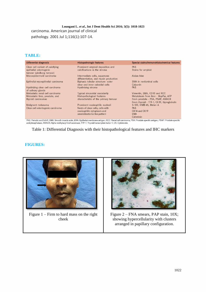
Figure 1 – Firm to hard mass on the right
cheek
Figure 2 – FNA smears, PAP stain, 10X;
showing hypercellularity with clusters
arranged in papillary configuration.
Loungani L. et al., Int J Dent Health Sci 2016; 3(5): 1018-1023
1023
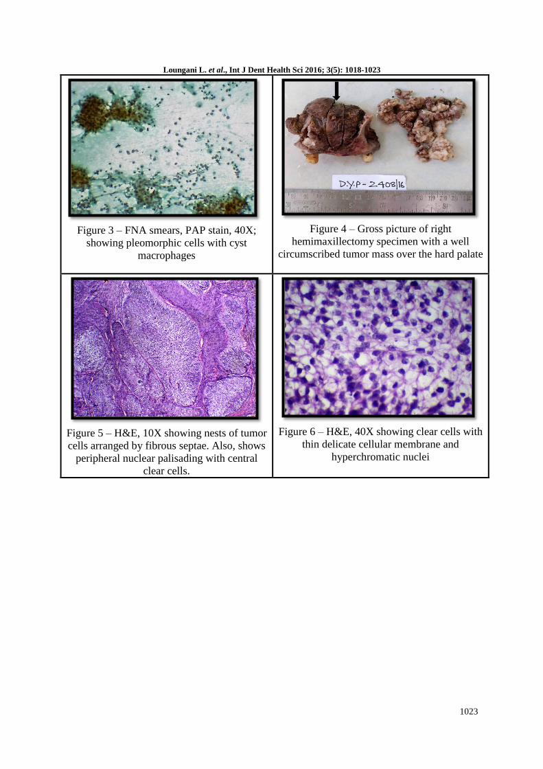
Figure 3 – FNA smears, PAP stain, 40X;
showing pleomorphic cells with cyst
macrophages
Figure 4 – Gross picture of right
hemimaxillectomy specimen with a well
circumscribed tumor mass over the hard palate
Figure 5 – H&E, 10X showing nests of tumor
cells arranged by fibrous septae. Also, shows
peripheral nuclear palisading with central
clear cells.
Figure 6 – H&E, 40X showing clear cells with
thin delicate cellular membrane and
hyperchromatic nuclei


![Fibroma of the Maxilla Trabecular Variant Juvenile … · contains cementicles [2], and while it is of odontogenic origin, it predominantly occurs in the second and third decades](https://img.pdfslide.net/doc/110x75/5b810d1f7f8b9a2b6f8b7676/fibroma-of-the-maxilla-trabecular-variant-juvenile-contains-cementicles-2.jpg)