Embed Size (px)
Citation preview

How to Diagnose the Most Common Odontogenic Lesions
Robert A. Robinson, M.D., Ph.D.
Department of Pathology Carver College of Medicine
University of Iowa

No Conflicts of Interest to Report
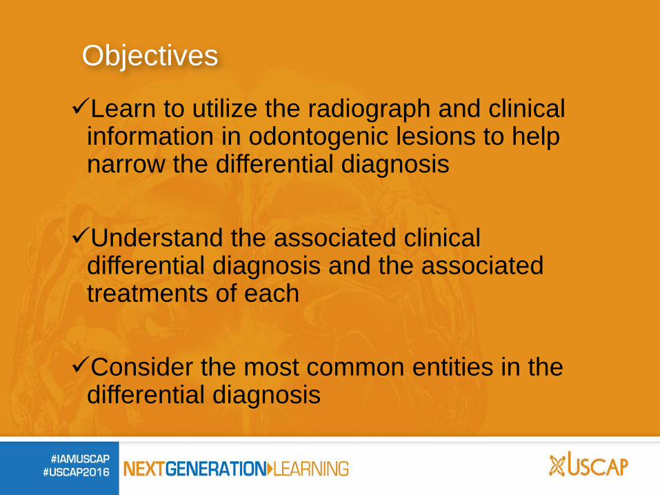
Objectives
Learn to utilize the radiograph and clinical information in odontogenic lesions to help narrow the differential diagnosis Understand the associated clinical
differential diagnosis and the associated treatments of each
Consider the most common entities in the
differential diagnosis
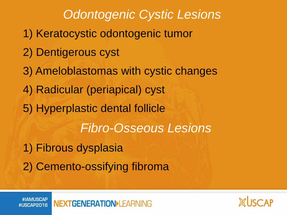
Odontogenic Cystic Lesions 1) Keratocystic odontogenic tumor
2) Dentigerous cyst
3) Ameloblastomas with cystic changes
4) Radicular (periapical) cyst
5) Hyperplastic dental follicle
Fibro-Osseous Lesions 1) Fibrous dysplasia
2) Cemento-ossifying fibroma
Odontogenic Cystic Lesions
A. Cyst from mandibular area #32
Clinical history from a pathology requisition
1) What is the oral surgeon actually asking? 2) What do they really want to know?
I’ve already enucleated this cyst that I have sent to the laboratory. Is there anything else I need to do or worry about?
What the oral surgeon really means:
A. Cyst from mandibular area #32
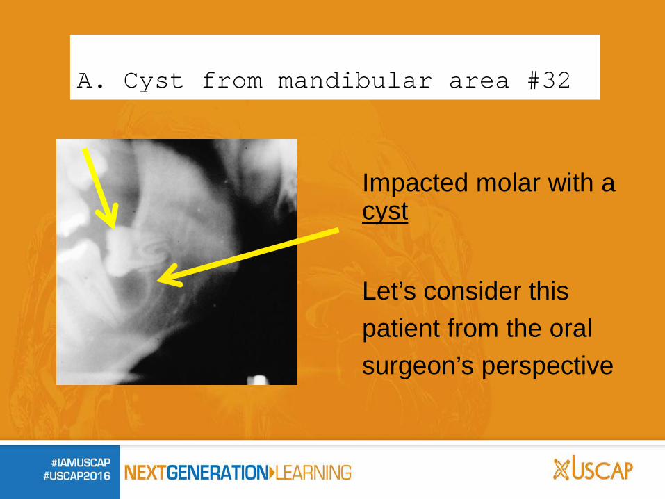
Impacted molar with a cyst
Let’s consider this patient from the oral surgeon’s perspective
A. Cyst from mandibular area #32
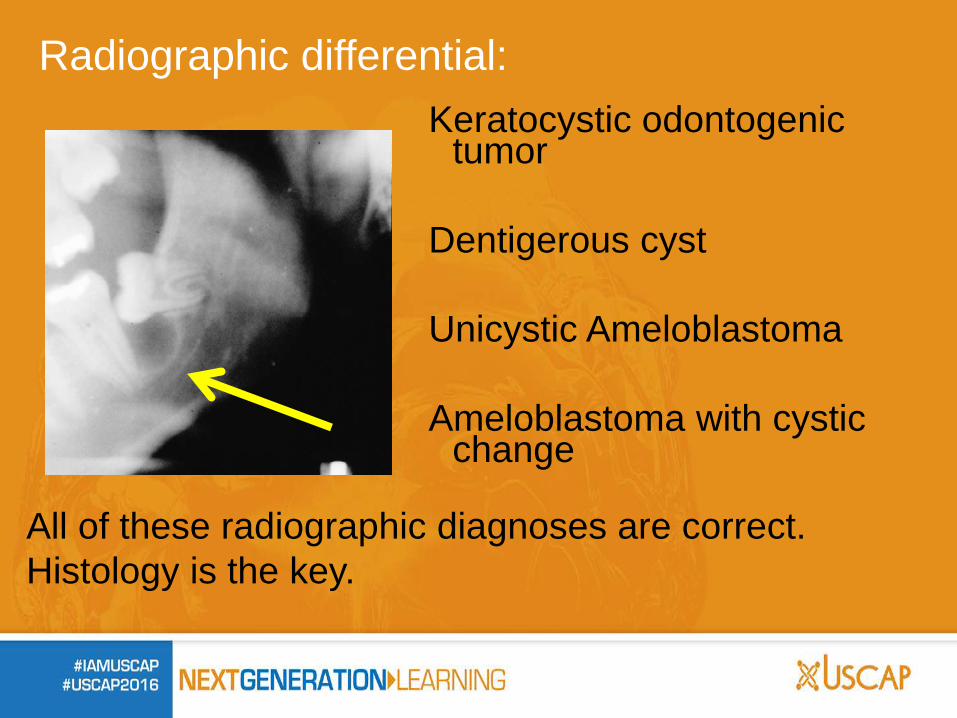
Radiographic differential: Keratocystic odontogenic
tumor Dentigerous cyst
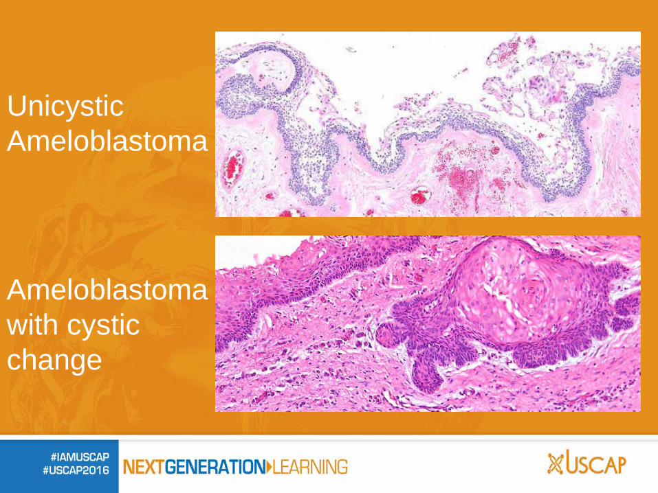
Unicystic Ameloblastoma Ameloblastoma with cystic
change
All of these radiographic diagnoses are correct. Histology is the key.
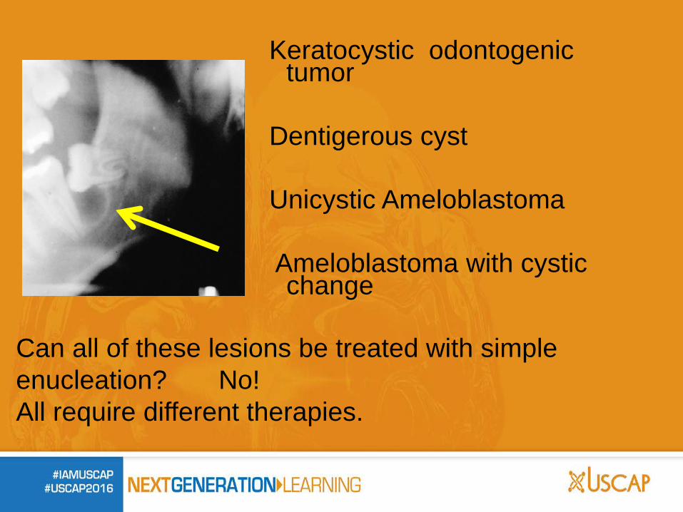
Keratocystic odontogenic tumor
Dentigerous cyst Unicystic Ameloblastoma Ameloblastoma with cystic
change
Can all of these lesions be treated with simple enucleation? No! All require different therapies.
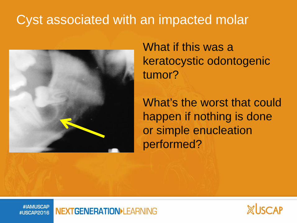
Cyst associated with an impacted molar
What if this was a keratocystic odontogenic tumor? What’s the worst that could happen if nothing is done or simple enucleation performed?
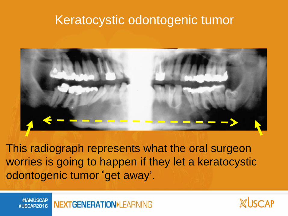
Keratocystic odontogenic tumor
This radiograph represents what the oral surgeon worries is going to happen if they let a keratocystic odontogenic tumor‘get away’.
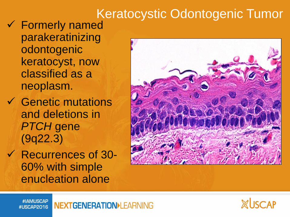
Formerly named parakeratinizing odontogenic keratocyst, now classified as a neoplasm.
Genetic mutations and deletions in PTCH gene (9q22.3)
Recurrences of 30-60% with simple enucleation alone
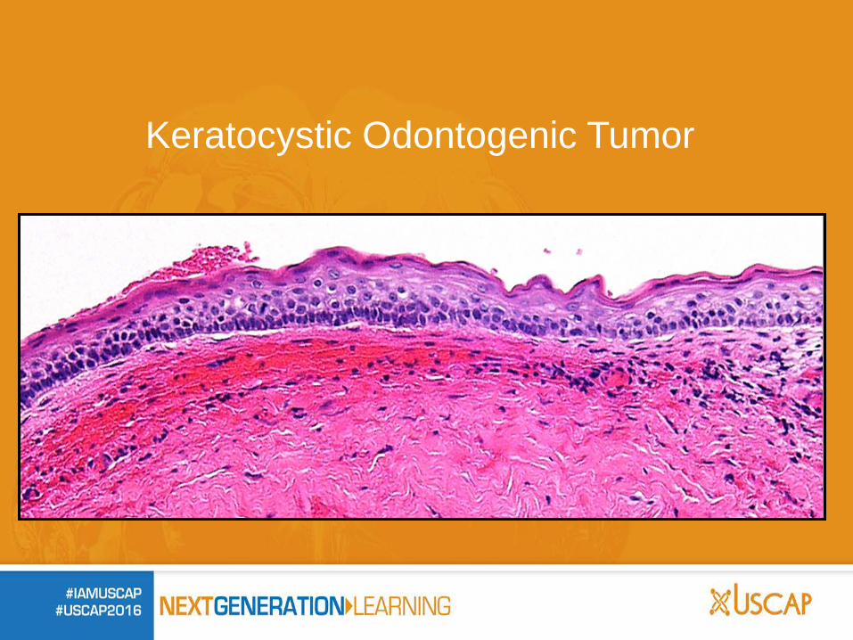
Keratocystic Odontogenic Tumor
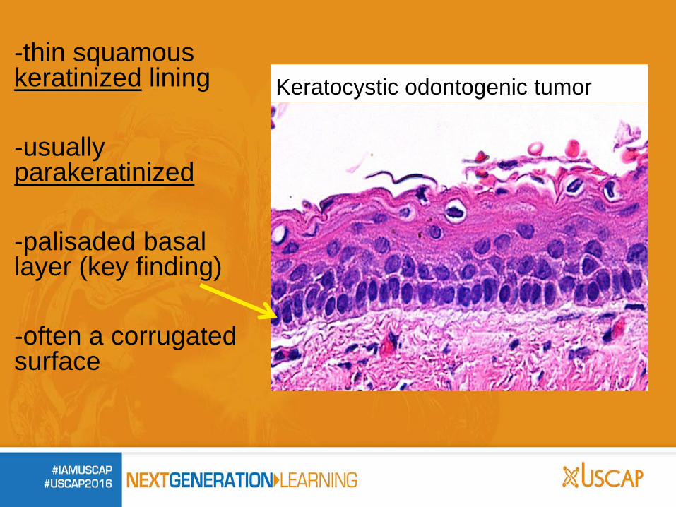
-thin squamous keratinized lining -usually parakeratinized -palisaded basal layer (key finding) -often a corrugated surface
Keratocystic odontogenic tumor
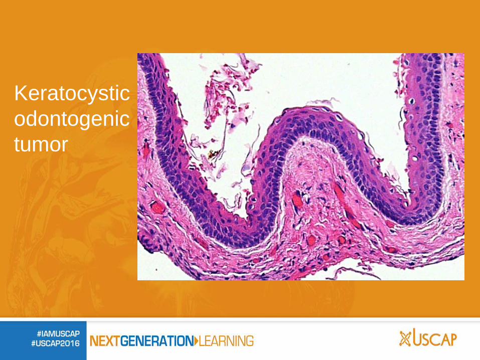
Keratocystic odontogenic tumor
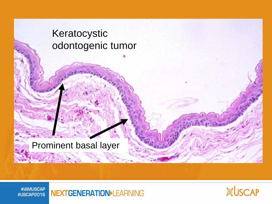
Keratocystic odontogenic tumor
Prominent basal layer
Keratocystic odontogenic tumor When a patient has more than one
keratocystic odontogenic tumor, this very likely represents
Basal Cell Nevus syndrome (Basal Cell Carcinoma Nevus Syndrome, commonly known as Gorlin or Gorlin-Goltz
Syndrome) Often presents in the first and second decade
of life
Treatment: Keratocystic Odontogenic Tumor
Requires additional surgical procedure with complete curettage and chemical cautery of the area of the cyst with Carnoy’s solution and in some cases, removal of some bone (perpheral ostectomy). Long term (10 yr) followup with radiograph every year for 5 years and then every other year thereafter.
Dentigerous cyst This is also a squamous lined cyst on histology. But, we are looking for what is NOT present: no parakeratosis no palisaded basal layer no surface corrugation
We need to make sure this is not a keratocystic odontogenic tumor.
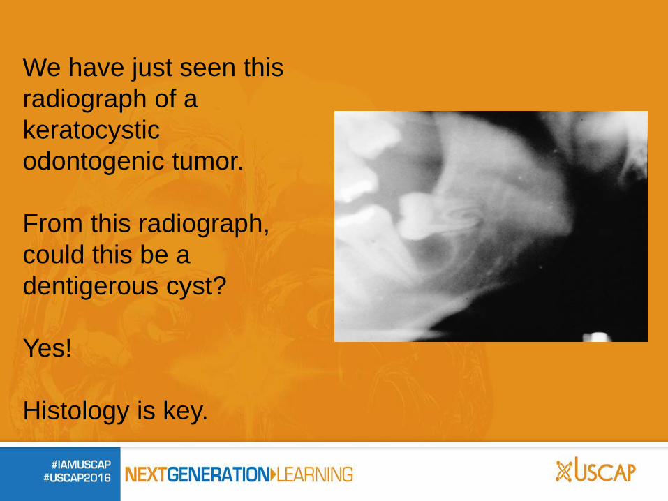
We have just seen this radiograph of a keratocystic odontogenic tumor. From this radiograph, could this be a dentigerous cyst? Yes! Histology is key.
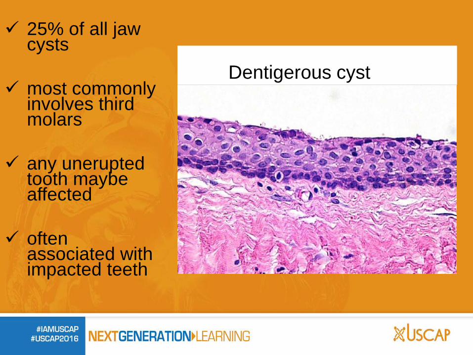
25% of all jaw cysts
most commonly involves third molars
any unerupted tooth maybe affected
often associated with impacted teeth
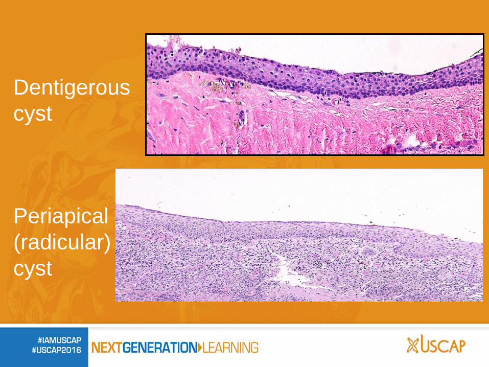
Dentigerous cyst
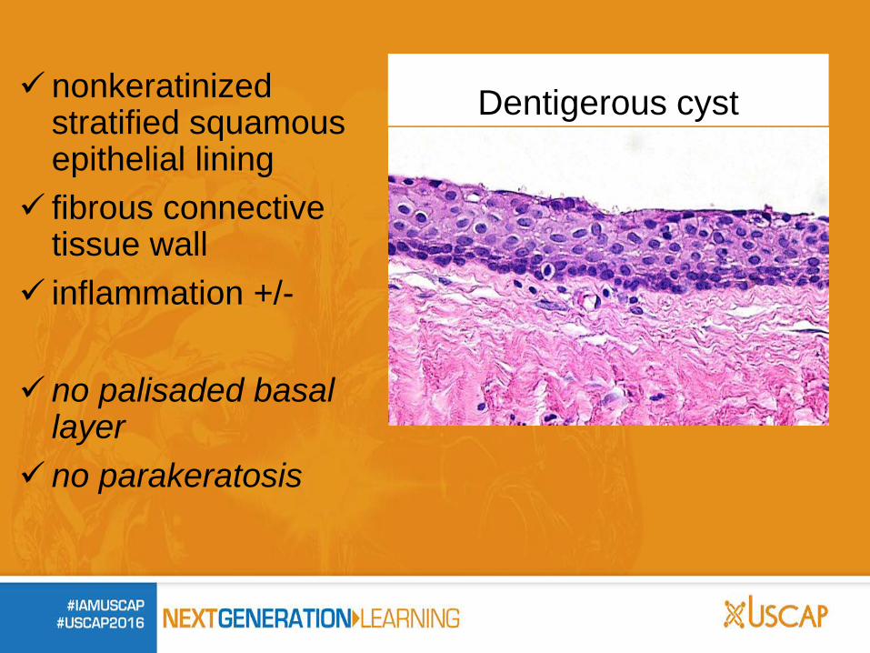
nonkeratinized stratified squamous epithelial lining
fibrous connective tissue wall
inflammation +/-
no palisaded basal layer
no parakeratosis
Dentigerous cyst
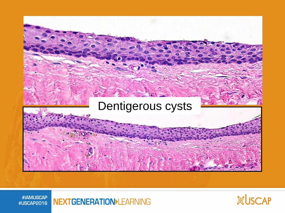
Dentigerous cysts
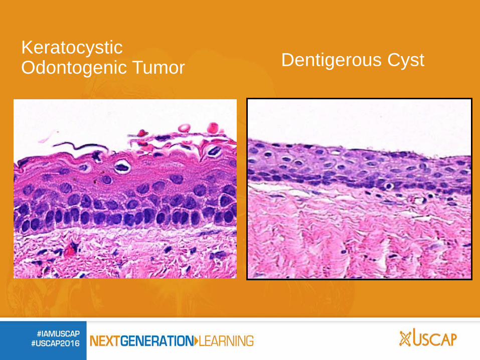
Keratocystic Odontogenic Tumor Dentigerous Cyst
Treatment: Dentigerous cyst
Enucleation of the cyst with the tooth. No further treatment required.
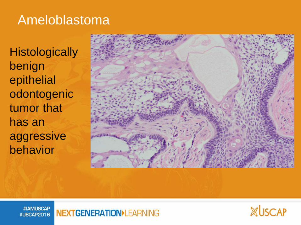
Ameloblastoma
Histologically benign epithelial odontogenic tumor that has an aggressive behavior
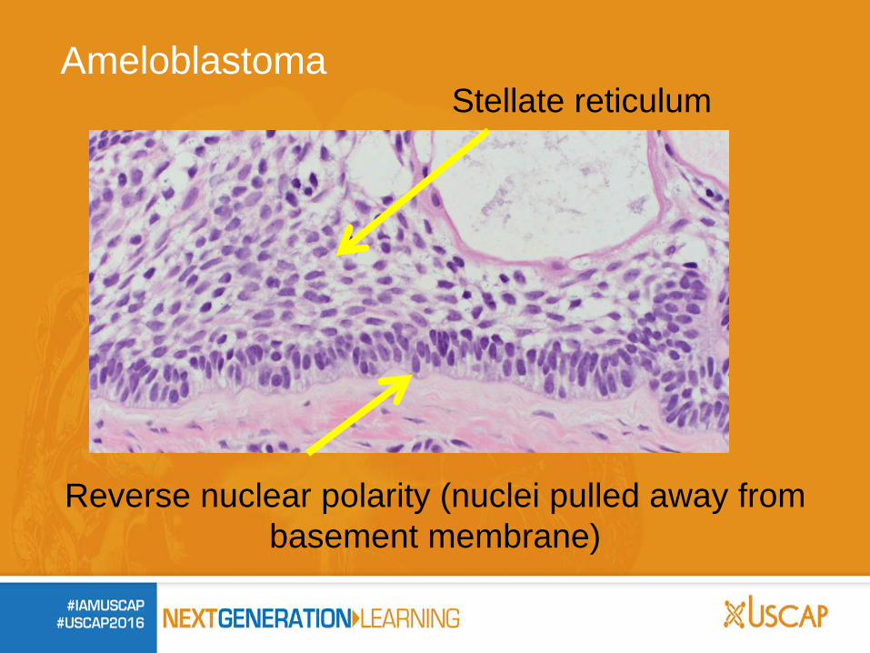
Reverse nuclear polarity (nuclei pulled away from basement membrane)
Ameloblastoma Stellate reticulum
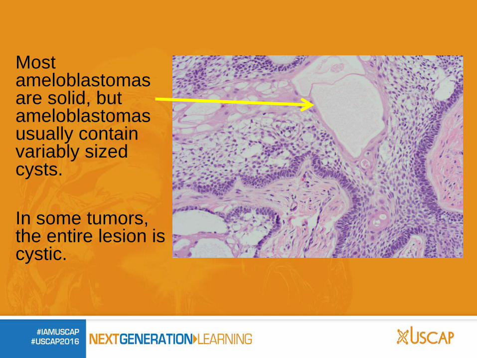
Most ameloblastomas are solid, but ameloblastomas usually contain variably sized cysts. In some tumors, the entire lesion is cystic.
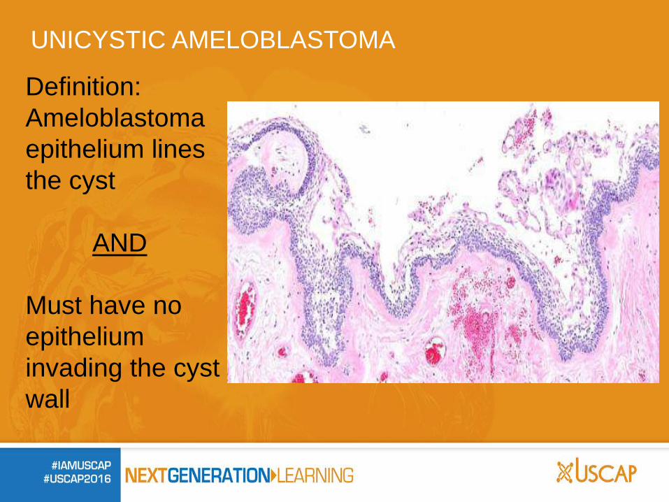
UNICYSTIC AMELOBLASTOMA Definition: Ameloblastoma epithelium lines the cyst AND Must have no epithelium invading the cyst wall
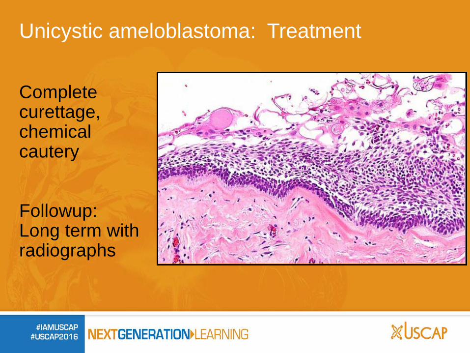
Unicystic ameloblastoma: Treatment
Complete curettage, chemical cautery Followup: Long term with radiographs
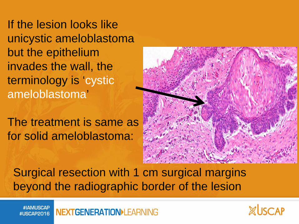
If the lesion looks like unicystic ameloblastoma but the epithelium invades the wall, the terminology is ‘cystic ameloblastoma’ The treatment is same as for solid ameloblastoma:
Surgical resection with 1 cm surgical margins beyond the radiographic border of the lesion
In the cases presented so far, we have noted that the radiograph has limited value as to establishing a specific diagnosis. But that is not the case for all cystic lesions. Radicular (periapical) cysts can’t be diagnosed without the radiograph
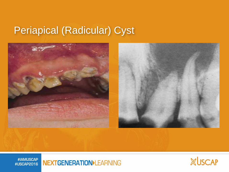
Periapical (Radicular) Cyst
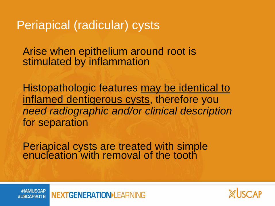
Arise when epithelium around root is stimulated by inflammation
Histopathologic features may be identical to
inflamed dentigerous cysts, therefore you need radiographic and/or clinical description for separation
Periapical cysts are treated with simple
enucleation with removal of the tooth
Periapical (radicular) cysts
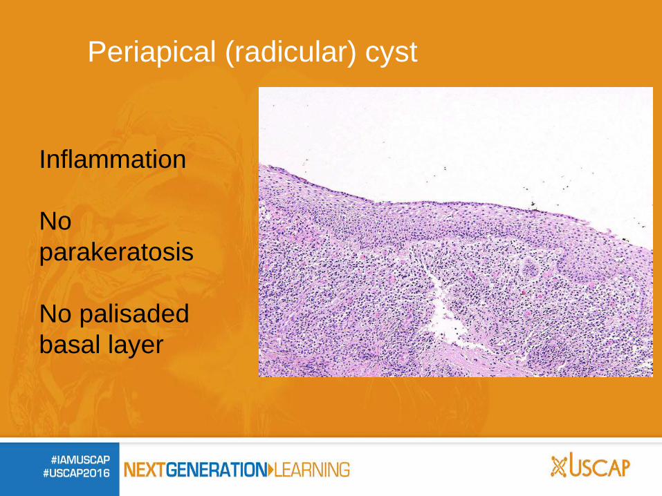
Inflammation No parakeratosis No palisaded basal layer
Periapical (radicular) cyst
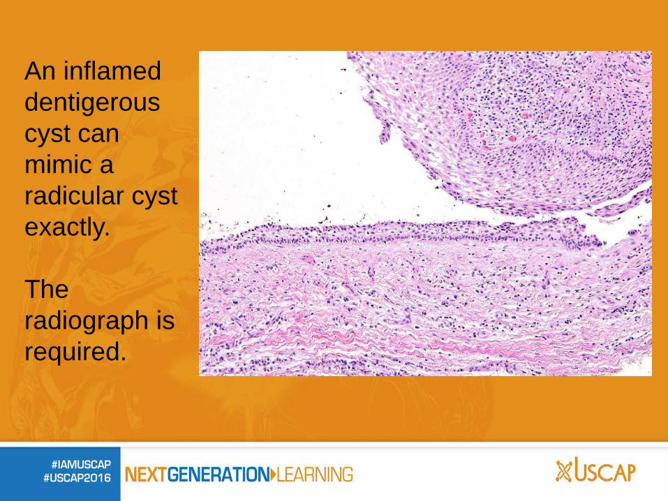
An inflamed dentigerous cyst can mimic a radicular cyst exactly. The radiograph is required.
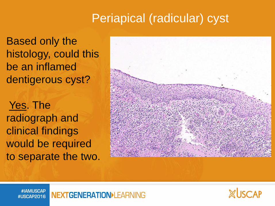
Periapical (radicular) cyst
Based only the histology, could this be an inflamed dentigerous cyst? Yes. The radiograph and clinical findings would be required to separate the two.
Is it clinically imperative to separate dentigerous cyst
from periapical cyst? No One would always like to be correct with matching
clinical and histologic findings but essentially the treatment has been performed and no other therapy is required.
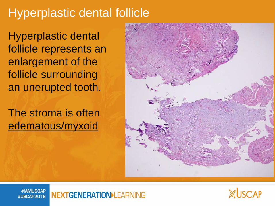
Hyperplastic dental follicle
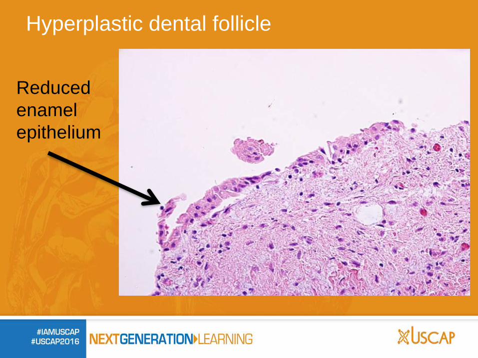
Hyperplastic dental follicle represents an enlargement of the follicle surrounding an unerupted tooth. The stroma is often edematous/myxoid
Hyperplastic dental follicle
The specimen will be sent to the lab as ‘odontogenic cyst’ or ‘dental follicle’
‘Radiographic distinction between a small
dentigerous cyst and an enlarged dental follicle is difficult and may be an academic exercise’
General rule: If the cyst is > 1.0 cm, designated dentigerous cyst
Hyperplastic dental follicle
Reduced enamel epithelium
Hyperplastic dental follicle
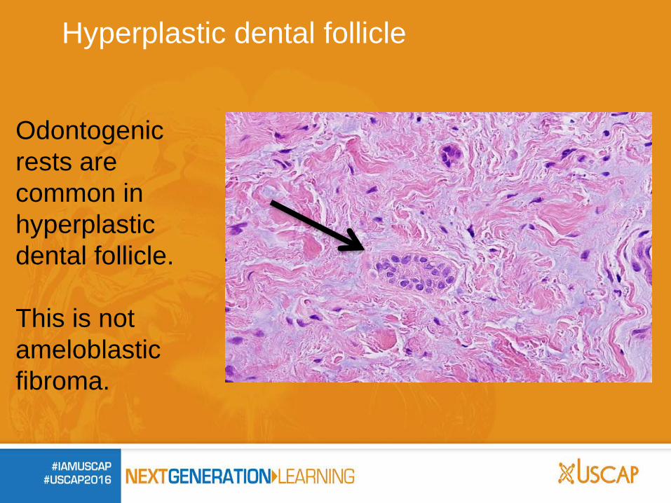
Odontogenic rests are common in hyperplastic dental follicle. This is not ameloblastic fibroma.
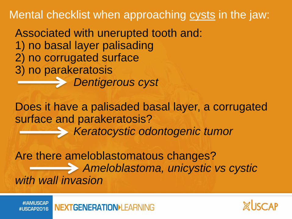
Associated with unerupted tooth and: 1) no basal layer palisading 2) no corrugated surface 3) no parakeratosis Dentigerous cyst Does it have a palisaded basal layer, a corrugated surface and parakeratosis? Keratocystic odontogenic tumor Are there ameloblastomatous changes? Ameloblastoma, unicystic vs cystic with wall invasion
Mental checklist when approaching cysts in the jaw:
What do you do if you have no history or radiographic information yet see what appears to be a dentigerous cyst?
The diagnostic line should be:
“Squamous lined cyst” with a comment that there are no features of
keratocystic odontogenic tumor or ameloblastoma.
In every pathology report, even where I have the history or radiograph and the diagnosis is one of these three:
-dentigerous cyst
-hyperplastic dental follicle
-radicular cyst
I still always note somewhere in the report that there are no features of keratocystic odontogenic tumor or ameloblastoma.
Dentigerous cyst
Periapical (radicular) cyst
Keratocystic Odontogenic Tumor
Unicystic Ameloblastoma
Ameloblastoma with cystic change
Fibro-osseous Lesions of the Jaws
Fibrous dysplasia Cemento-ossifying fibroma Fibro-osseous dysplasia
Fibro-osseous Lesions of the Jaws
Fibro-osseous lesions of the jaws can appear very similar histologically. Although there can be some histologic hints, true separation is most safely carried out with the addition of clinical and radiographic findings.
Fibrous dysplasia
Cemento-ossifying fibroma
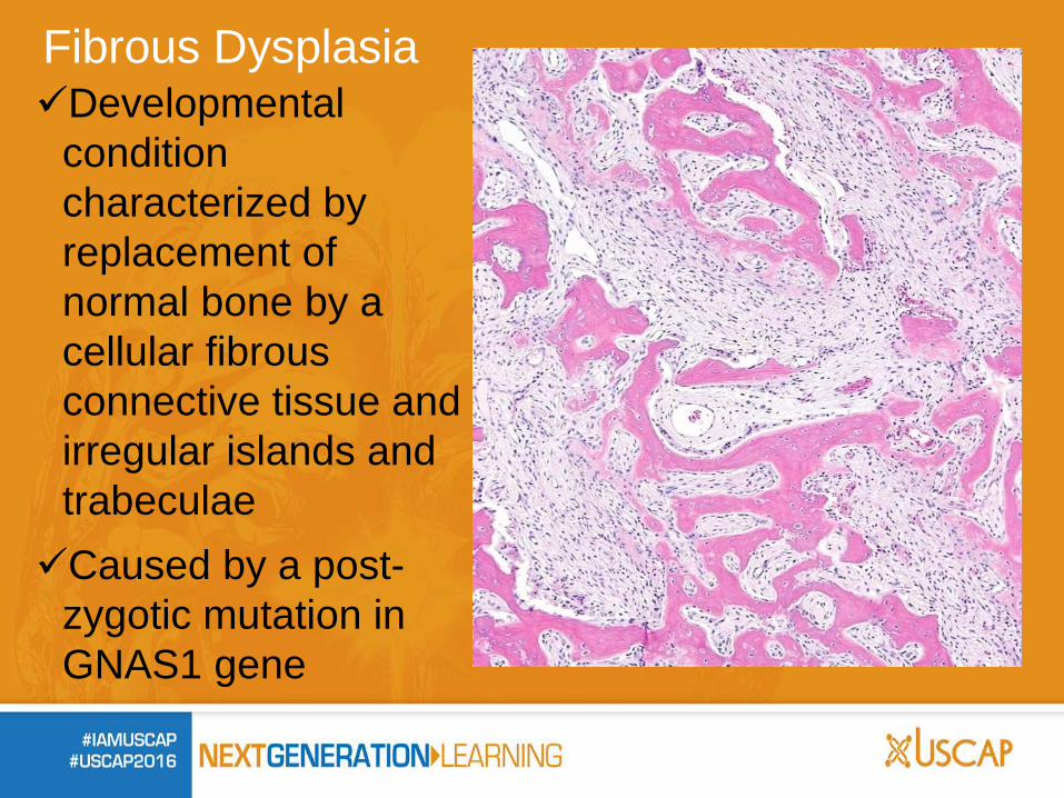
Fibrous Dysplasia
Developmental condition characterized by replacement of normal bone by a cellular fibrous connective tissue and irregular islands and trabeculae Caused by a post-
zygotic mutation in GNAS1 gene
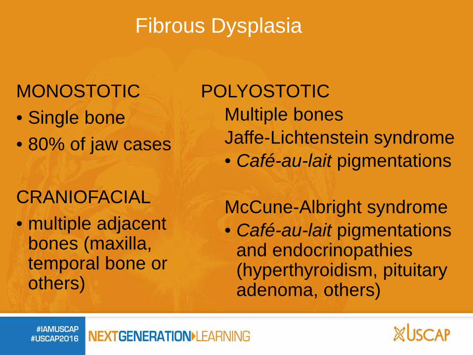
Fibrous Dysplasia
MONOSTOTIC • Single bone • 80% of jaw cases CRANIOFACIAL • multiple adjacent
bones (maxilla, temporal bone or others)
POLYOSTOTIC Multiple bones Jaffe-Lichtenstein syndrome • Café-au-lait pigmentations
McCune-Albright syndrome • Café-au-lait pigmentations
and endocrinopathies (hyperthyroidism, pituitary adenoma, others)
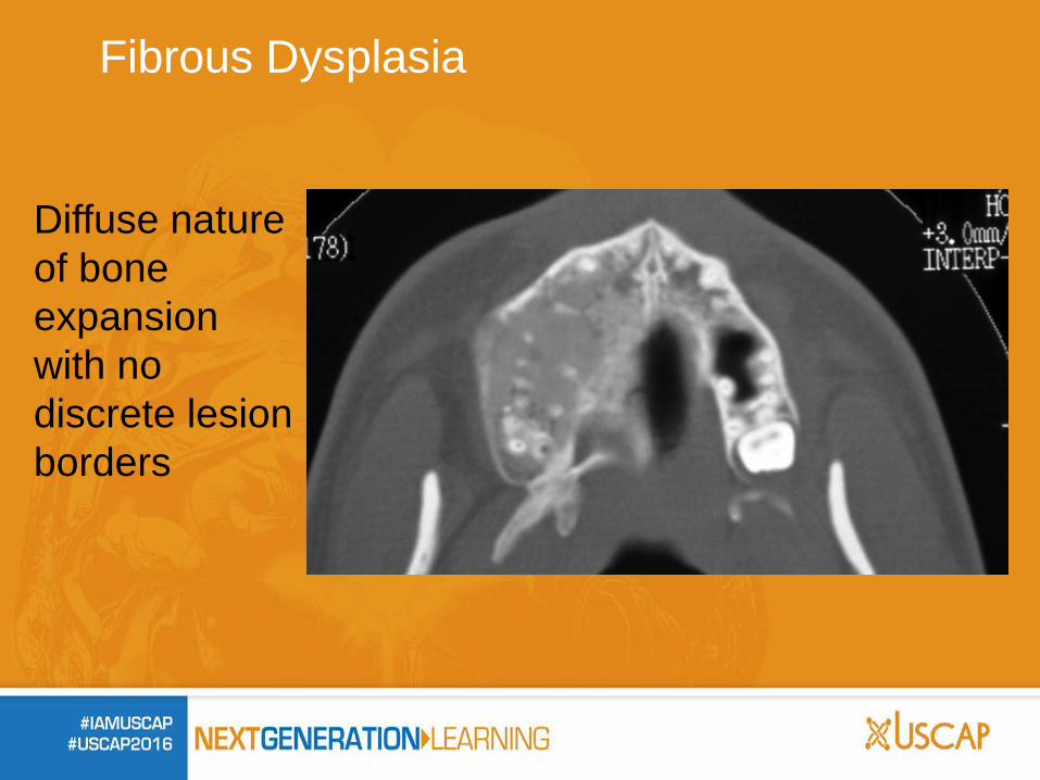
Fibrous Dysplasia
Diffuse nature of bone expansion with no discrete lesion borders
Fibrous Dysplasia
Irregular trabeculae of metaplastic woven bone Fibrous connective tissue with varying cellularity Blends with normal bone at lesion periphery (no capsule) Bone spherules Few osteoblasts
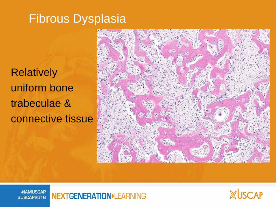
Fibrous Dysplasia
Relatively uniform bone trabeculae & connective tissue
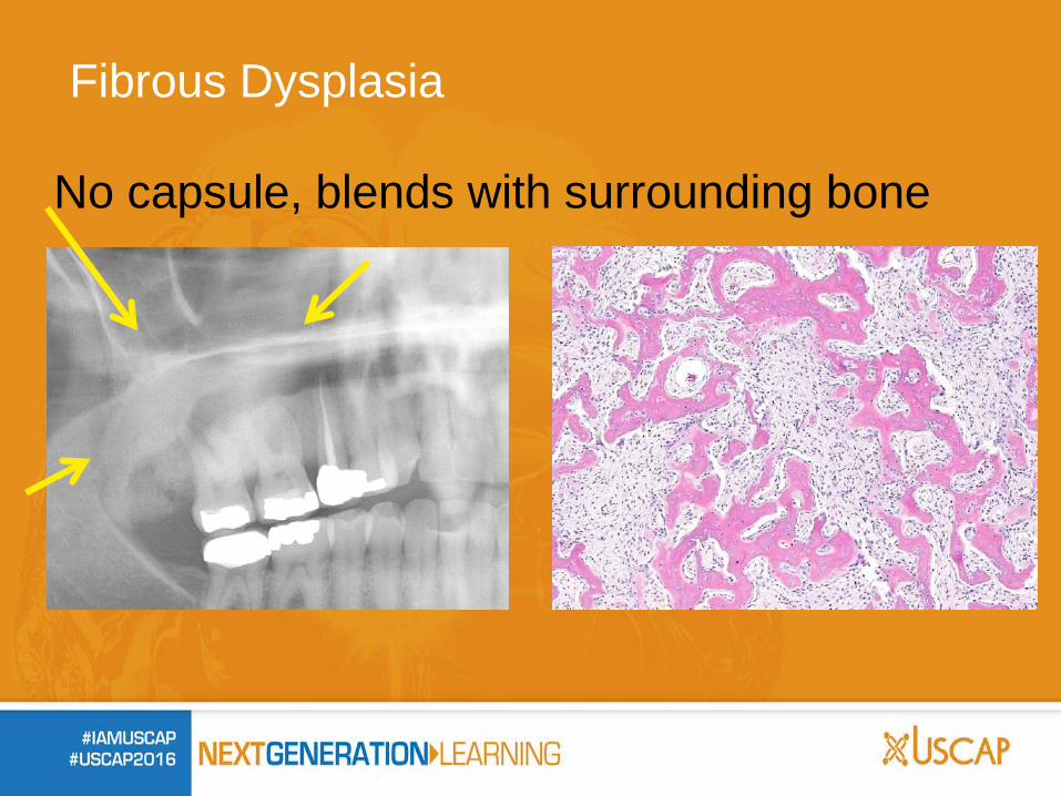
No capsule, blends with surrounding bone
Fibrous Dysplasia
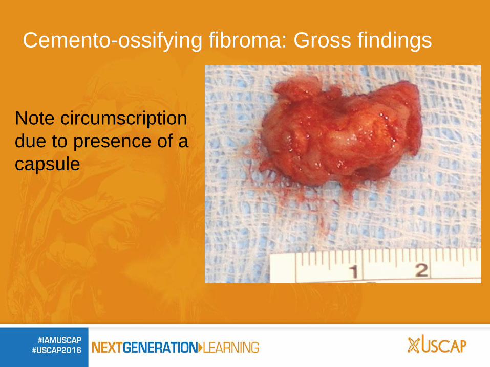
Cemento-ossifying fibroma: Gross findings
Note circumscription due to presence of a capsule
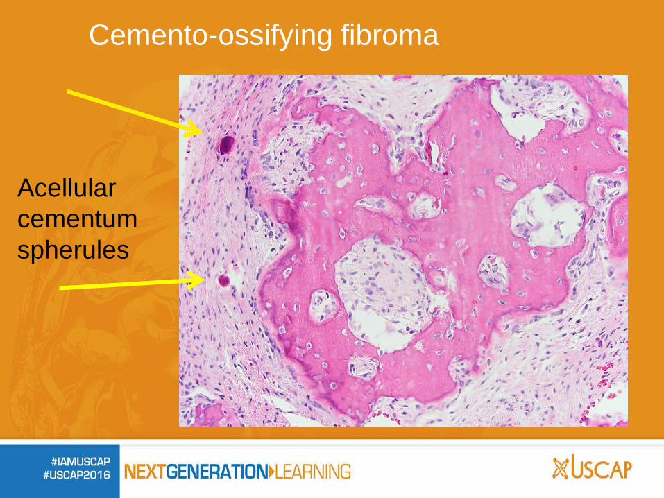
Bone trabeculae & basophilic cellular spherules which resemble cementum Spherules of cementum like material often demonstrate peripheral brush borders with blending into the adjacent connective tissue Trabeculae are variable in size and show a mixture of woven and lamellar patterns Peripheral osteoblastic rimming is usually present
Cemento-ossifying fibroma
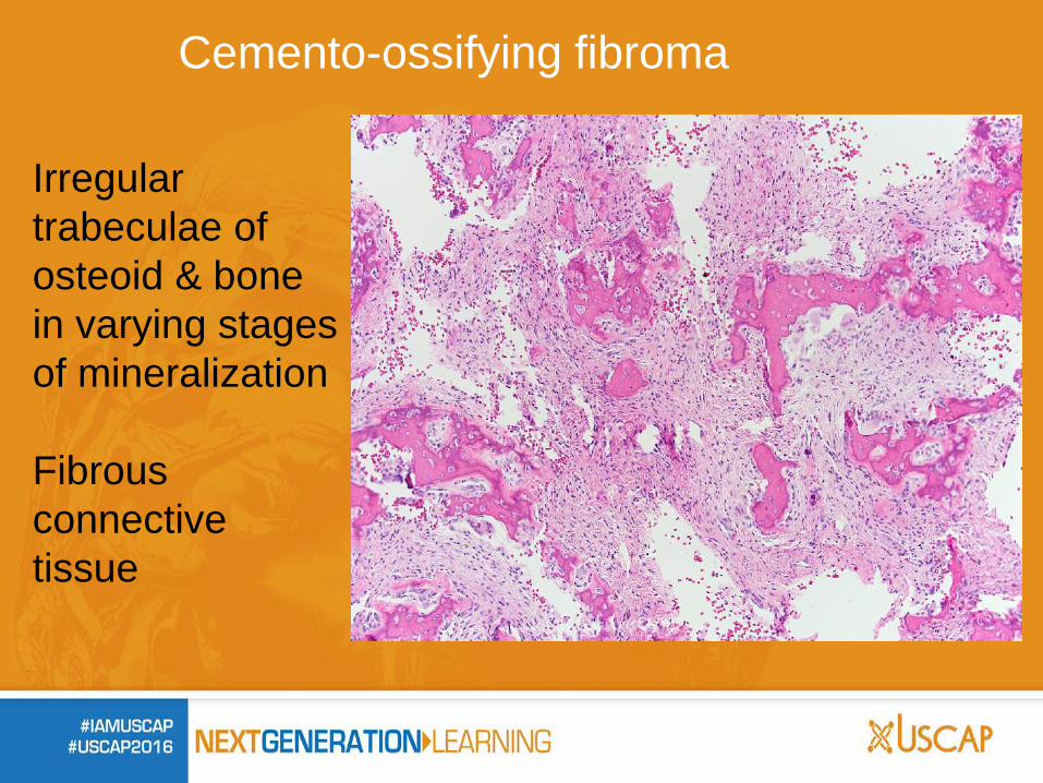
Cemento-ossifying fibroma
Irregular trabeculae of osteoid & bone in varying stages of mineralization Fibrous connective tissue
Cemento-ossifying fibroma
Acellular cementum spherules
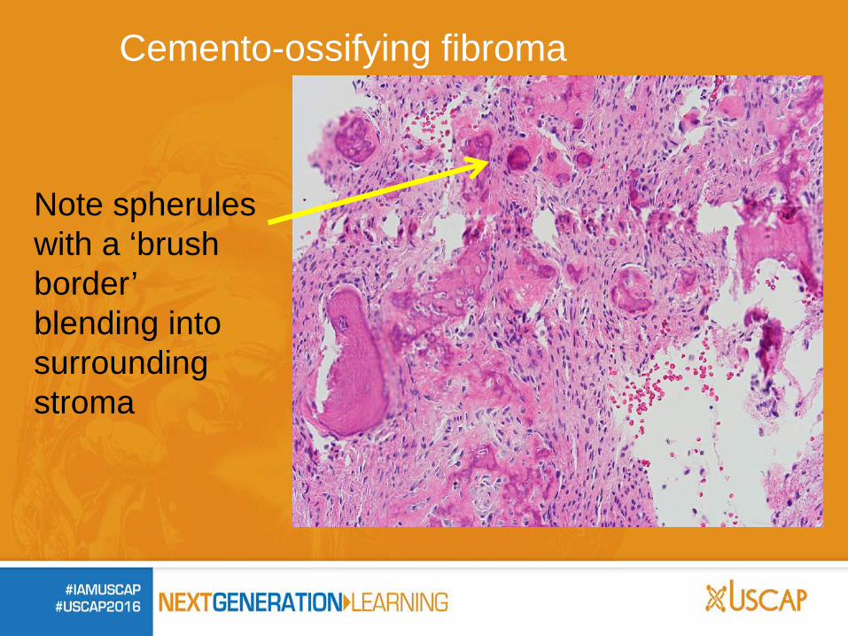
Cemento-ossifying fibroma
Note spherules with a ‘brush border’ blending into surrounding stroma
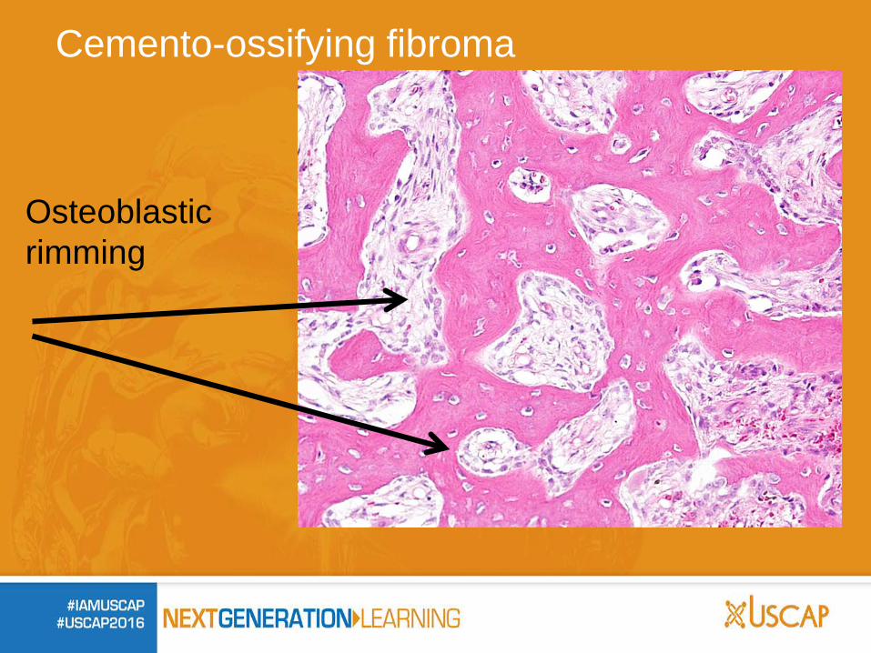
Cemento-ossifying fibroma
Osteoblastic rimming
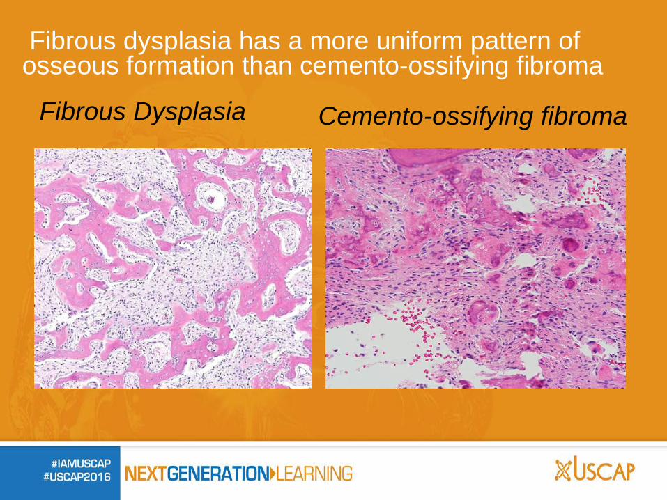
Fibrous dysplasia has a more uniform pattern of osseous formation than cemento-ossifying fibroma
Fibrous Dysplasia Cemento-ossifying fibroma
A mix of mineralization patterns resembling bone or cementum along with abundant fibrous tissue is much more suggestive of cemento-ossifying fibroma over fibrous dysplasia. Differentiating these lesions still usually requires clinical and radiographic correlation. No distinct cortical border is seen in dysplasias.
Fibrous dysplasia or Cemento-ossifying fibroma?
Treatment of Fibro-osseous Lesions
Fibrous Dysplasia: Various modalities; therapy not always required Recontouring can be performed for asymmetry Cemento-ossifying fibroma: Conservative excision

![Non-syndromic multiple keratocystic ... - Case report · syndrome (NBCCS) /Gorlin–Goltz syndrome/ Bifid rib syndrome) usually in younger patients [3, 4]. Occurrence of multiple](https://img.pdfslide.net/doc/110x75/60460eb40960e6162151c0eb/non-syndromic-multiple-keratocystic-case-syndrome-nbccs-gorlinagoltz.jpg)