Embed Size (px)
Citation preview
University of Dundee
DOCTOR OF MEDICINE
Airway challenges in different clinical phenotypes and their relationship to markers ofdisease and treatment
Clearie, Karine Leila
Award date:2010
Link to publication
General rightsCopyright and moral rights for the publications made accessible in the public portal are retained by the authors and/or other copyright ownersand it is a condition of accessing publications that users recognise and abide by the legal requirements associated with these rights.
• Users may download and print one copy of any publication from the public portal for the purpose of private study or research. • You may not further distribute the material or use it for any profit-making activity or commercial gain • You may freely distribute the URL identifying the publication in the public portal
Take down policyIf you believe that this document breaches copyright please contact us providing details, and we will remove access to the work immediatelyand investigate your claim.
Download date: 25. Mar. 2022
DOCTOR OF MEDICINE
Airway challenges in different clinicalphenotypes and their relationship tomarkers of disease and treatment
Karine Leila Clearie
2010
University of Dundee
Conditions for Use and DuplicationCopyright of this work belongs to the author unless otherwise identified in the body of the thesis. It is permittedto use and duplicate this work only for personal and non-commercial research, study or criticism/review. Youmust obtain prior written consent from the author for any other use. Any quotation from this thesis must beacknowledged using the normal academic conventions. It is not permitted to supply the whole or part of thisthesis to any other person or to post the same on any website or other online location without the prior writtenconsent of the author. Contact the Discovery team ([email protected]) with any queries about the useor acknowledgement of this work.
1
Airway challenge in different clinical phenotypes and their
relationship to markers of disease and treatment
Dr Karine Leila Clearie
Doctor of Medicine (MD) University of Dundee
October 2010
2
Table of contents List of abbreviations 5 Declaration 7 Summary statement 8
Chapter 1: Introduction and literature review 10 Burden of disease (incidence and prevalence of asthma) 10 National guidelines and the treatment of asthma 11
Strategy 1: Inflammation 15 Induced sputum 18 Peripheral blood eosinophil counts and ECP 20 Exhaled tidal nitric oxide 21 Airway hyper-responsiveness and bronchial challenge 25 Direct challenge 27
Indirect challenge 30 Surrogate markers of inflammation and research 32 Strategy 2: Phenotype 33 Smokers 33 Elite swimmers 35
Chapter 2: Methods 41 Airway measurements 42 Lung function 42 Mannitol challenge 42 Methacholine challenge 43 Nitric oxide measurement 44 Impulse oscillometry 44 Peak expiratory flow rate 45 Quality of life/ symptom measures 45 Juniper mini AQLQ/ ACQ 45 Symptom scores 46 Skin prick testing 46 Blood and urine collection 46 Overnight urinary cortisol/ creatinine 46 Blood eosinophil count and eosinophil cationic protein 47 Quality control 48
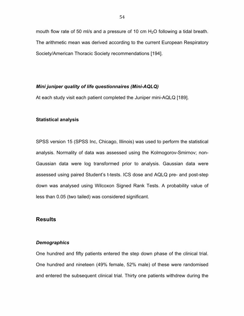
Chapter 3: Establishing the role of surrogate markers of inflammation in clinical and research settings 49 Part a: Supervised step-down of inhaled steroids in a community setting
Introduction 50 Methods 51 Results 54 Discussion 64 Conclusion 70 Critique 71
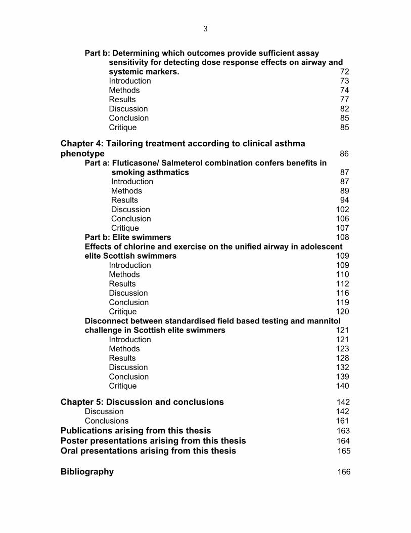
3
Part b: Determining which outcomes provide sufficient assay sensitivity for detecting dose response effects on airway and systemic markers. 72
Introduction 73 Methods 74 Results 77 Discussion 82 Conclusion 85 Critique 85
Chapter 4: Tailoring treatment according to clinical asthma phenotype 86
Part a: Fluticasone/ Salmeterol combination confers benefits in smoking asthmatics 87 Introduction 87
Methods 89 Results 94 Discussion 102 Conclusion 106 Critique 107
Part b: Elite swimmers 108 Effects of chlorine and exercise on the unified airway in adolescent elite Scottish swimmers 109
Introduction 109 Methods 110 Results 112 Discussion 116 Conclusion 119 Critique 120
Disconnect between standardised field based testing and mannitol challenge in Scottish elite swimmers 121
Introduction 121 Methods 123 Results 128 Discussion 132 Conclusion 139 Critique 140
Chapter 5: Discussion and conclusions 142 Discussion 142 Conclusions 161 Publications arising from this thesis 163 Poster presentations arising from this thesis 164 Oral presentations arising from this thesis 165 Bibliography 166
4
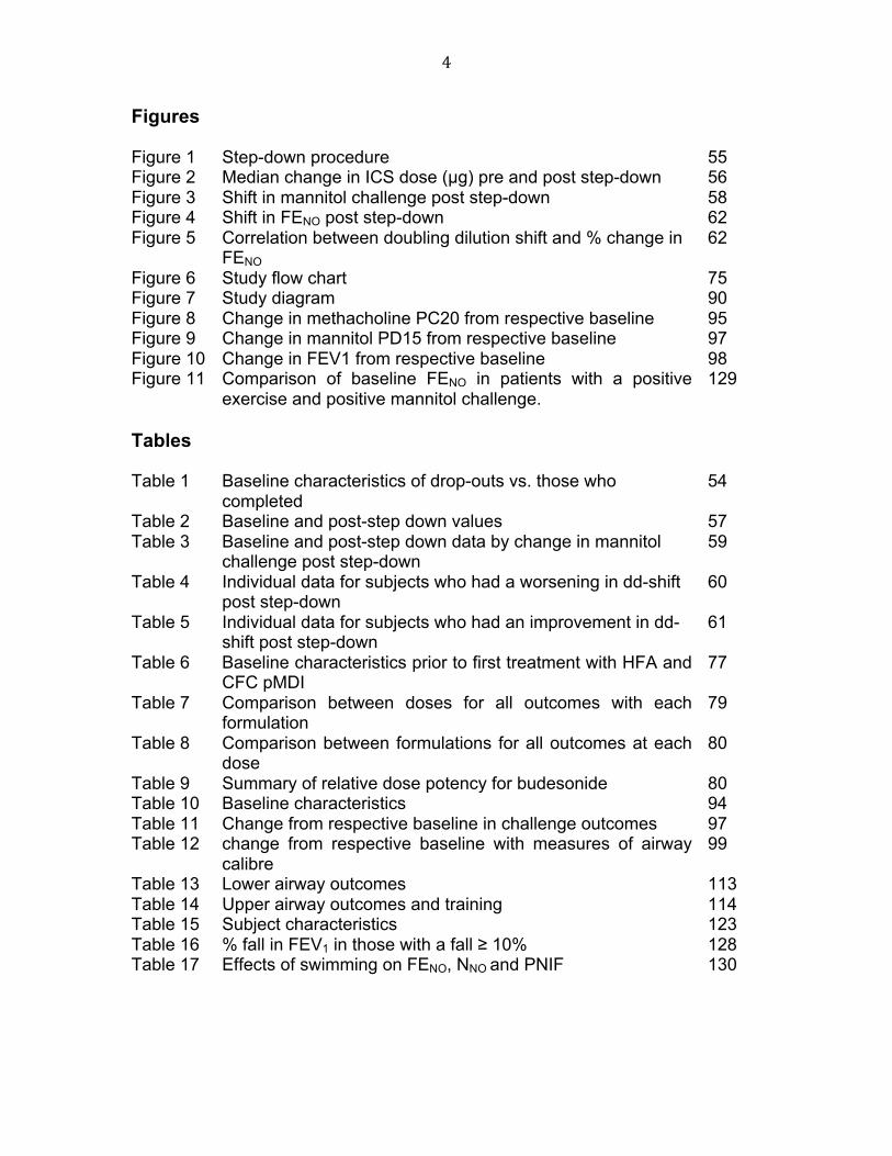
Figures Figure 1 Step-down procedure 55 Figure 2 Median change in ICS dose (µg) pre and post step-down 56 Figure 3 Shift in mannitol challenge post step-down 58 Figure 4 Shift in FENO post step-down 62 Figure 5 Correlation between doubling dilution shift and % change in
FENO
62
Figure 6 Study flow chart 75 Figure 7 Study diagram 90 Figure 8 Change in methacholine PC20 from respective baseline 95 Figure 9 Change in mannitol PD15 from respective baseline 97 Figure 10 Change in FEV1 from respective baseline 98 Figure 11 Comparison of baseline FENO in patients with a positive
exercise and positive mannitol challenge. 129
Tables Table 1 Baseline characteristics of drop-outs vs. those who
completed 54
Table 2 Baseline and post-step down values 57 Table 3 Baseline and post-step down data by change in mannitol
challenge post step-down 59
Table 4 Individual data for subjects who had a worsening in dd-shift post step-down
60
Table 5 Individual data for subjects who had an improvement in dd-shift post step-down
61
Table 6 Baseline characteristics prior to first treatment with HFA and CFC pMDI
77
Table 7 Comparison between doses for all outcomes with each formulation
79
Table 8 Comparison between formulations for all outcomes at each dose
80
Table 9 Summary of relative dose potency for budesonide 80 Table 10 Baseline characteristics 94 Table 11 Change from respective baseline in challenge outcomes 97 Table 12 change from respective baseline with measures of airway
calibre 99
Table 13 Lower airway outcomes 113 Table 14 Upper airway outcomes and training 114 Table 15 Subject characteristics 123 Table 16 % fall in FEV1 in those with a fall ≥ 10% 128 Table 17 Effects of swimming on FENO, NNO and PNIF 130
5
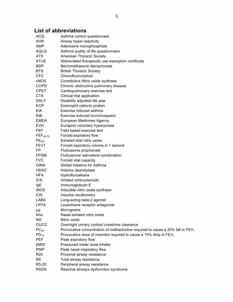
List of abbreviations ACQ Asthma control questionnare AHR Airway hyper-reactivity AMP Adenosine monophosphate AQLQ Asthma quality of life questionnaire ATS American Thoracic Society ATUE Abbreviated therapeutic use exemption certificate BDP Beclomethasone diproprionate BTS British Thoracic Society CFC Chlorofluorocarbon cNOS Constitutive Nitric oxide synthase COPD Chronic obstructive pulmonary disease CPET Cardiopulmonary exercise test CTA Clinical trial application DALY Disability adjusted life year ECP Eosinophil cationic protein EIA Exercise induced asthma EIB Exercise induced bronchospasm EMEA European Medicines Agency EVH Eucapnic voluntary hyperpnoea FBT Field based exercise test FEF25-75 Forced expiratory flow FENO Exhaled tidal nitric oxide FEV1 Forced expiratory volume in 1 second FP Fluticasone proprionate FPSM Fluticasone/ salmeterol combination FVC Forced vital capacity GINA Global Initiative for Asthma HDAC Histone deacetylase HFA Hydrofluroalkane ICS Inhaled corticosteroids IgE Immunoglobulin E iNOS Inducible nitric oxide synthase IOS Impulse oscillometry LABA Long-acting beta-2 agonist LRTA Leukotriene receptor antagonist µg Micrograms Nno Nasal exhaled nitric oxide NO Nitric oxide OUCC Overnight urinary cortisol creatinine clearance PC20 Provocative concentration of methacholine required to cause a 20% fall in FEV1 PD15 Provocative dose of mannitol required to cause a 15% drop in FEV1 PEF Peak expiratory flow pMDI Pressured meter dose inhaler PNIF Peak nasal inspiratory flow R20 Proximal airway resistance R5 Total airway resistance R5-20 Peripheral airway resistance RADS Reactive airways dysfunction syndrome

6
RDR Response dose rate SEM Standard error of the mean WADA World anti-doping agency X5 Peripheral reactance
7
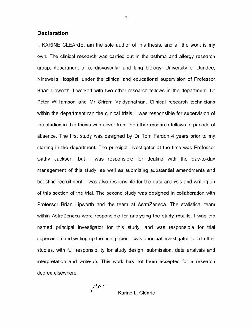
Declaration I, KARINE CLEARIE, am the sole author of this thesis, and all the work is my
own. The clinical research was carried out in the asthma and allergy research
group, department of cardiovascular and lung biology, University of Dundee,
Ninewells Hospital, under the clinical and educational supervision of Professor
Brian Lipworth. I worked with two other research fellows in the department. Dr
Peter Williamson and Mr Sriram Vaidyanathan. Clinical research technicians
within the department ran the clinical trials. I was responsible for supervision of
the studies in this thesis with cover from the other research fellows in periods of
absence. The first study was designed by Dr Tom Fardon 4 years prior to my
starting in the department. The principal investigator at the time was Professor
Cathy Jackson, but I was responsible for dealing with the day-to-day
management of this study, as well as submitting substantial amendments and
boosting recruitment. I was also responsible for the data analysis and writing-up
of this section of the trial. The second study was designed in collaboration with
Professor Brian Lipworth and the team at AstraZeneca. The statistical team
within AstraZeneca were responsible for analysing the study results. I was the
named principal investigator for this study, and was responsible for trial
supervision and writing up the final paper. I was principal investigator for all other
studies, with full responsibility for study design, submission, data analysis and
interpretation and write-up. This work has not been accepted for a research
degree elsewhere.
Karine L. Clearie
8
Summary Statement
Asthma is a chronic disease, which affects over 300 million people worldwide.
Despite the introduction of both national and international guidelines, asthma
control remains poor. The aim of this thesis is therefore to explore possible new
strategies for improving the management of asthma. The first strategy to be
explored is ‘titrating asthma treatment to suppress underlying airway
inflammation’. The benefits of such a strategy have already been demonstrated,
however, the lack of an adequate ‘inflammometer’ have limited its application to
the research/ hospital setting. Mannitol challenge appears to be the most
promising candidate, as it is portable and relatively cheap. The aim of the first
study in this thesis is therefore to trial the use of mannitol challenge in a
community setting. The selection of an appropriate ‘inflammometer’ is not limited
to the clinical setting, it is equally important in research. This is particularly true
when determining therapeutic equivalence between inhaled steroids. The aim of
the second study in this thesis is therefore to determine which inflammatory
outcomes demonstrate sufficient assay sensitivity, as part of a cross-over trial, to
detect dose response effects on airway and systemic markers.
The second strategy to be examined in this thesis is tailoring asthma therapy
according to asthmatic phenotype. Two groups of asthmatics that differ
significantly from traditional ‘inflammatory asthma’ have been selected. Asthmatic
smokers are known to develop relative resistance to the beneficial effects of
inhaled steroids. A recent post hoc analysis of the GOAL trial has suggested that
9
smokers may gain greater benefit from the addition of a long-acting beta-2
agonist vs. doubling the dose of inhaled steroid. The third study in this thesis
therefore aims to examine this in a prospective fashion. Another group of
individuals in whom the traditional approach to asthma management and
diagnosis may not be appropriate is elite athletes. In has been well documented
that the mechanism of bronchospasm in athletes involves the drying/ cooling of
airways. However, even within this category there are athletes to which this
mechanism of action is unlikely to apply. Elite swimmers, for example, exercise in
a warm, humidified environment. It therefore seems unlikely that tests designed
to reproduce hyperosmolar shifts will have the same diagnostic sensitivity as they
do in cold weather or track athletes. The aim of the fourth and fifth studies
included in this thesis is therefore to compare various diagnostic tests in
swimmers to determine which are the most sensitive.
10
Introduction
Burden of disease (incidence and prevalence of asthma)
Asthma is a chronic disorder of the respiratory tract which is thought to affect
over 300 million people worldwide [1]. It has been estimated that this figure will
increase exponentially over the coming years as communities adopt western
lifestyles and become increasingly urbanised [2-4]. Urbanisation of the world’s
population is projected to increase from 45% to 59% in 2052, potentially leading
to an additional 100 million new diagnoses of asthma worldwide [1].
The projected increase in prevalence of asthma is of concern due to the
significant burden which asthma places on society [5]. In 1996 Peter Barnes
published a review article examining this burden, which he sub-divided into
direct, indirect and intangible costs [5]. He determined that the direct costs of
asthma associated with medical care in the UK were between £322- £686 million
[5]. 22% of this was attributed to physician care (75% to GP consultations, 25%
to specialists), 37% to drug costs, and 25% to hospital costs (70-80% to in-patent
care, and 14-18% to emergency care) [5]. ‘Indirect’ costs were more difficult to
accurately quantify as they only occurred when asthma became sufficiently
intrusive to interfere with lifestyle. These were associated with loss of productive
work, premature retirement, time spent by others caring for sick relatives and
premature death [5]. The GINA (Global Initiative for Asthma) Committee
quantified this by ranking common disorders in terms of disability-adjusted life
11
years (DALYs), which combines information about mortality and morbidity in
numbers of years lost. The number of DALYs lost due to asthma was estimated
at about 15 million per year [1], ranking asthma as the 25th leading cause of
DALYs lost worldwide. This is comparable with Diabetes Mellitus (23rd) cirrhosis
of the liver (24th) and dementia (28th) [1]. ‘Intangible’ costs were associated with
the impact of asthma on a patient’s quality of life [5]. This was examined as part
of the UK Action Asthma Survey, which collected qualitative data from 61234
asthmatic patients, through the provision of questionnaires in GP surgeries,
pharmacies and outpatient clinics. They determined that 62% of patients felt that
asthma had a moderate impact on their lives and 40% felt that it imposed a
moderate restriction on their daily activities [6]. Jones et al examined the impact
of asthma on an employed population using the St George’s Hospital Respiratory
questionnaire and found that up to 95% of respondents had an impaired quality
of life compared to age matched controls [7].
National guidelines and the treatment of asthma
In 1994 the British Thoracic Society implemented the first national guidelines on
the management of asthma [8]. Their aim was to decrease the mortality and
morbidity associated with asthma by standardising its management. They set
about doing this by reviewing the most up to date evidence based medicine and
issuing recommendations. Current guidelines outline a stepwise management
plan, beginning with short acting beta-2 agonists for the treatment of mild
intermittent asthma at step 1 (BTS guideline), and culminating with the use of
12
oral corticosteroids and immunosuppressants at step 5. The aims of
pharmacological management are the control of symptoms (including nocturnal
symptoms and exercise induced symptoms), prevention of exacerbations and the
achievement of best pulmonary function with minimal side-effects [8].
There is little doubt that inhaled corticosteroids (ICS) remain the treatment of
choice for all severities of asthma. They are potent anti-inflammatory agents,
which act in a relatively non-specific manner, inhibiting a wide variety of
inflammatory cells, cytokines, and transcription factors [9]. These effects have
been confirmed clinically in bronchial biopsy studies [10]. Evidence also suggests
that early intervention with inhaled corticosteroid drugs may prevent any long
term decline in lung function resulting from airway remodelling caused by
untreated chronic inflammation [9]. The benefits of early treatment with ICS were
confirmed by the START trial in which 7241 steroid naïve persistent asthmatic
patients were randomised to receive either daily budesonide or placebo for 3
years. At the end of the study period, participants who had received budesonide
had fewer severe exacerbations, fewer courses of oral corticosteroids, a longer
time until their first exacerbation, and a greater number of symptom free days
[11]. An earlier trial also found that mild-persistent asthmatics who received low-
dose budesonide had fewer exacerbations and better symptom control, than
patients receiving placebo [12]. These trials have firmly established the role of
ICS in treating persistent asthma, however there is increasing concern over the
side effect profile of ICS. For most adults with mild to moderate asthma, the
13
steep part of the dose-response curve for anti-asthma effects generally occurs at
doses below 800 µg/day BDP (beclomethasone dipropionate) or equivalent [13].
In addition, the curve for systemic adverse effects becomes much steeper at
doses above 800 µg/day, resulting in an inverted U shaped curve for the benefit
to risk ratio [13]. This has been shown to be true, even in severe asthmatics. The
main side effects associated with inhaled steroids include: adrenal suppression,
osteoporosis, growth suppression, skin bruising, cataracts and ocular
hypertension [13]. Current guidelines therefore recommend starting treatment
with a relatively high dose for four to eight weeks in order to gain rapid optimal
control, then gradually tapering the dose to determine the lowest effective
maintenance dose [8].
Despite the publication of these guidelines and improvements in asthma
treatment options, asthma remains a significant cause of mortality and morbidity
for all age groups throughout the UK. This was echoed in the Asthma Insights
and Reality Europe (AIRE) survey, which questioned asthmatic adults (n= 2083)
from seven European countries about their symptom severity, healthcare use,
exacerbation frequency, and perceived control [14]. Only 5.3% of all patients met
all the criteria required for ‘asthma control’ as defined by the GINA guidelines
[15]. 30.5% of adults complained of asthma-related sleep disturbance at least
once a week, 27.9% had required an unscheduled urgent care visit over the last
12 months, and 57.2% of adults reported symptoms such as shortness of breath,
14
cough, chest tightness and wheeze at least once a month [14].
Mortality associated with asthma also remains a significant problem, and it has
been estimated that approximately 1 in 250 deaths worldwide are due to asthma
[1]. Mathers et al predicted, using mathematical modeling, that the global
mortality rate from asthma will rise from roughly 240 000 in 2002 to between 320
000 - 402 000 by 2030 [16]. The most concerning aspect of this prediction is that
mortality is mostly limited to those aged 15-59 [16]. Whilst new developments in
pharmacological treatments do appear promising, none has made a radical
impact on asthma mortality or morbidity. It is therefore clear that changes need to
be made in the way we approach the management of asthma, in order to
maximise the efficacy of treatments which are currently available to us. The aim
of this thesis was to investigate this further:
Strategy 1) Titrating anti-inflammatory treatment to suppress surrogate markers
of inflammation. In particular to identify which outcomes are most
suitable in different clinical and research situations.
Strategy 2) Looking at different phenotypes of asthma rather than treating
asthma as a homogenous disease.
15
Strategy 1: Inflammation
Airway inflammation is a salient feature of asthma. The link between asthma and
inflammatory cells in the airways was first established during early case series of
fatal asthma [17]. These described profound cellular infiltration of the respiratory
mucosa associated with widespread mucus plugging [17]. More recently, Carroll
et al compared the histological appearances of patients who died from asthma
with those of non-asthmatic controls, and found a greater percentage of
degranulated mast cells and increased numbers of neutrophils in the submucosal
glands [18]. De Magalhaes Simoes et al described eosinophilic infiltration of the
respiratory mucosa, extending from the nasal mucosa to the distal lung [19].
These results have been substantiated by a number of other studies, confirming
that a diverse population of inflammatory cells including eosinophils, mast cells,
neutrophils, and a variety of lymphocyte sub-sets are involved in the asthmatic
process [20-22]. Interestingly, Carroll et al demonstrated that the distribution of
inflammation within the lung varied with clinical severity [23]. Mild to moderate
asthmatics were found to have predominantly central (i.e. large airways)
infiltration, whereas patients with severe asthma had more distal disease [23].
Sputum eosinophil counts have been shown to increase in severe or poorly
controlled asthma [24, 25] and after allergen challenge [26]. High sputum
eosinophil counts have been shown to predict the development of asthma
exacerbations [25, 27] and to predict failure of steroid reduction in clinically stable
16
asthmatics [26]. Green et al demonstrated that airway eosinophil counts
decrease following treatment with inhaled corticosteroids and that this was linked
with an improvement in symptoms and pulmonary function tests [28]. Reductions
have also been noted following treatment with other anti-inflammatory agents
such as theophyllines [29] and anti-leukotrienes [30].
Airway inflammation is known to correlate very poorly with subjective symptoms
and measures of airflow limitation [21]. This is particularly true in mild to
moderate asthmatics, in which unsuppressed airway inflammation may occur
despite clinically stable disease, potentially leaving many patients with
unchecked indolent inflammation [31]. This could result in frequent exacerbations
and eventually irreversible damage due to airway remodeling.
Many studies have documented that asthmatic subjects have extensive structural
changes in their airway [17, 32, 33]. These changes are present throughout the
airway wall and the whole length of the bronchial tree and include basement
membrane thickening, smooth muscle hypertrophy, abnormal deposition of
matrix components, angiogenesis, proliferation of airway nerves and hypertrophy
of glands [17]. Endobronchial biopsy studies have demonstrated that these
changes, in particular reticular basement membrane thickness, are proportional
to the number of inflammatory cells in the airways [34]. Autopsy studies have
confirmed that extensive remodelling is present in both fatal and non-fatal
asthma [35]. Laprise et al followed up 10 asymptomatic asthmatics and 10
17
control subjects with bronchial biopsy over a 2 year period. At baseline, groups
had similar eosinophil counts, IgE levels and percentage predicted FEV1. At 2
years, four had developed asthma and bronchial biopsies in these subjects
revealed an increase in sub-epithelial fibrosis [31]. In a landmark trial Haahtela et
al randomized 37 asthmatic patients to receive either 400µg budesonide or
placebo in a double-blind manner for two years. A third group who had received
terbutaline only for two years were crossed over in a open-label manner to
treatment with 1200µg budesonide daily for the third year. They found significant
differences between the placebo and budesonide groups in terms of FEV1 and
histamine PC20. The condition of patients in the third group, who had initially
been treated with terbutaline, improved, however, the degree of improvement
appeared to be less than those who were treated with budesonide at the
beginning of the three-year study. This suggests that early intervention with
inhaled steroids is crucial when avoiding fixed airway obstruction [36]. These
results were confirmed in the more recent Steroid Treatment as Regular Therapy
(START) study, which involved 7,000 individuals (adults and children) with mild
persistent asthma. The beneficial effects of ICS on accelerated decline of airway
function over a 3 year period were established, as the rate of decline in post-
bronchodilator FEV1 was reduced by 22% in children and 42% in adults [11].
The benefits of a treatment strategy based on targeting inflammation were first
demonstrated in 1999 by Sont and colleagues who carried out a parallel group
study involving 75 mild to moderate asthmatics [37]. Subjects were randomised
18
to a treatment strategy aimed at reducing airway hyper-responsiveness (a
surrogate of inflammation) or standard BTS management. Patients in the AHR
group had a 1.8 fold lower rate of exacerbations and a greater improvement in
their FEV1 compared to the BTS group [37]. Bronchial biopsy specimens
revealed that subjects in the AHR group also had a greater reduction in the
thickness of their sub-epithelial reticular layer [37]. These findings were
substantiated by Green and colleagues [38] who compared standard BTS
management to titrating therapy with the aim of normalising induced sputum
eosinophil counts. They found that eosinophil counts were 62% lower (p=0.002)
in the treatment group, and that these patients suffered significantly fewer
exacerbations and admissions compared with the BTS group [38]. The average
steroid dose was the same in both groups [38]. These two studies have clearly
demonstrated the benefits of a treatment strategy aimed at targeting surrogate
markers of inflammation.
In order to accurately target inflammation a number of non-invasive surrogate
markers have been developed. The pros and cons of each are outlined below:
Induced sputum
Induced sputum is probably the most obvious surrogate marker of inflammation.
Sputum from asthmatic subjects is known to contain increased numbers of
inflammatory cells such as eosinophils, which are strongly implicated in the
pathogenesis of asthma [39]. Raised sputum eosinophil counts have been shown
19
to predict impending loss of asthma control, and to be a robust predictor of
improvement in FEV1 following a course of oral corticosteroids [25, 40]. Leuppi et
al reported that, at a cut-off point of 6.3%, sputum eosinophils had a sensitivity of
90% and a specificity of 63% for loss of control after stepwise reduction of
inhaled steroid dose [41]. Using a cut-off point of 4%, Jones et al [42] reported a
sensitivity of 59% and a specificity of 60% for loss of control after complete
steroid withdrawal. In a landmark trial Green et al used these observations to
titrate corticosteroid treatment against sputum eosinophil count [38]. Subjects in
the eosinophil group had their treatment up-titrated if their counts were >3%. This
led to a five-fold reduction in exacerbation rates compared to subjects titrated
according to symptoms and measures of airflow limitation alone. Similar
reductions were noted in other surrogate markers of inflammation such as FENO,
and methacholine PC20 [38]. However, no differences were seen in symptoms or
any spirometric measure, lending further weight to the argument that these
methods are inefficient at monitoring disease activity [38]. Interestingly, no
significant difference was seen in inhaled steroid dose between the two groups,
indicating that this novel treatment strategy does not owe it’s success to
increased steroid dose [38]. The main drawback of induced sputum is that it is
technically difficult, requiring expertise in sputum induction, sputum handling, cell
counting and interpretation. Whilst the Leicester group have obtained
consistently good results, with high sputum yields, other centres, such as ours,
have time and again failed to do so. In addition, the technical expertise required
to collect and process these samples make it an inaccessible and impractical test
20
for use in the community, limiting its use to specialist centres.
Peripheral blood eosinophil counts and ECP
An alternative method of quantifying eosinophil counts is to measure them in
peripheral blood. It has been suggested that peripheral eosinophil count is
representative the overall ‘systemic burden’ of the disease [43, 44]. High blood
eosinophil counts have been shown to predict failure of corticosteroid withdrawl
[45]. Eosinophil counts have also been shown to reduce following treatment with
inhaled corticosteroids [44]. However, it has been clearly established that
eosinophils exhibit greater sensitivity for assessing the anti-inflammatory
response in sputum than they do in blood [44]. This suggests that activated
eosinophils in the target tissue are more important than the total number in the
blood stream, making measurement of blood eosinophil counts less applicable to
use in routine clinical practice [44].
Eosinophil cationic protein (ECP) is a potent cytotoxic molecule that is released
through the degranulation of eosinophils, and is thereby felt to indicate levels of
eosinophil activation. It is therefore felt to be a more robust marker of asthmatic
inflammation than total blood eosinophil count [46]. ECP levels in serum have
been shown to correlate well with those in bronchiolar lavage fluid and sputum
[47]. Studies looking at serum ECP in asthma indicate that levels are related to
the severity of asthmatic disease, as measured by lung function and
methacholine challenge [48]. ECP has been shown to rise following late phase
allergen challenge and natural allergen exposure [49], and to decrease following
21
treatment with inhaled steroids [50]. However, ECP in sputum has been shown to
be more sensitive than ECP in peripheral blood [44, 51]. Wever et al suggested
that high ECP levels in serum can predict acute exacerbations, in spite of
apparently satisfactory anti-inflammatory treatment [52]. However, these results
conflict with those of Ferguson et al, who determined that ECP was a poor
indicator of disease activity in chronic asthma, as it could not differentiate
between bronchial and nasal inflammation [53]. Only one study has evaluated the
efficacy of a treatment strategy based on ECP. In 2002, Lowhagen et al
compared steroid titration according to serum ECP vs. pre-bronchodilator
morning peak flow, and found no significant difference in exacerbation rates,
symptom scores or FEV1 between the groups [54]. These results suggest that
although ECP is a useful clinical tool, it is best used in combination with other
markers of disease activity.
Exhaled tidal nitric oxide
Nitric oxide (NO) is a molecule which is synthesised throughout the pulmonary
epithelium and vascular tree by nitric oxide synthase (NOS) enzymes [55]. Two
isoforms of NOS exist: the constitutive form (cNOS) which appears to protect the
airways from excessive bronchoconstriction, and the inducible form (iNOS) which
has a modulatory role in inflammatory disorders [56]. cNOS is produced in
platelets, neuronal, epithelial and endothelial cells. It releases tiny quantities of
NO (fM or pM) when airway receptors are stimulated by agonists such as
acetylcholine and bradykinin [56]. iNOS is produced by neutrophils,
22
macrophages, epithelial, mesangial and vascular smooth muscle cells [56][58]. It
releases large quantities (nM) of pro-inflammatory NO in response to up-
regulation by immune cytokines, which may persist for hours or days [56]. NO
has been detected in the exhaled breath of animals and humans by
chemiluminescence [57]. Numerous authors have reported that the fraction of
exhaled nitric oxide (FENO) measured in the expired breath of asthmatics is
elevated compared to healthy controls. This has been linked to increased
expression of iNOS [58], and is thought to reflect airway inflammation [59, 60].
FENO has been shown to increase during exacerbations and following exposure
to allergen and viral infection [61, 62]. It correlates well with other markers of
inflammation including sputum and peripheral eosinophil counts, ECP and airway
hyper-responsiveness to methacholine [60, 63-67]. FENO has therefore been
suggested as an aid in the diagnosis of asthma: A study by Berkman et al found
it to have a similar diagnostic sensitivity to methacholine challenge, with a
positive predictive value of more than 80%, however this finding was limited to
steroid naïve patients [68]. Oral and inhaled corticosteroids have been shown to
result in a rapid (6hrs after a single dose), dose dependent reduction in FENO [69-
71]. Non-compliance or cessation of treatment with ICS will return FENO levels
rapidly (3 to 5 days) to the pretreatment level [69]. This decrease is thought to
occur due to a reduction in airway inflammation and an inhibitory effect on iNOS
expression [55]. FENO has therefore been suggested as a marker of treatment
response [72]. It has also been used to predict exacerbations, both spontaneous
[61] and those induced by steroid reduction [25, 73], as high levels in treated
23
asthmatics have been found to herald a loss of clinical control [42]. Jones et al
reported that a 60% increase in FENO between two visits provided a positive
predictive value of 83% for loss of control after stepwise reduction of inhaled
steroid dose [42]. More recently, Michils et al reported that changes in FENO in
relation to asthma control (as measured by the Asthma Control Questionnaire)
were prognostically helpful, as a single FENO measurement of >45 ppb excluded
well-controlled asthma with a negative predictive value of 89% [74]. On the basis
of repeated measurements, a 40% decrease in FENO was found to have a high
positive predictive value (83%) and similar negative predictive value (79%) for a
clinically relevant improvement in asthma control. A FENO reading consistently
<30 ppb was associated with a low likelihood of exacerbation within 3 months.
Van Veen et al reported that persistently high FENO readings are a predictor of
accelerated decline in lung function in ‘‘difficult asthma’’ [75]. This suggests that
serial monitoring could be used to predict and thereby prevent exacerbations.
However, studies in which steroid dose has been titrated to suppress FENO have
proved disappointing [76-79]. Smith et al randomized 97 asthmatic subjects to
titration of their treatment based on either FENO or symptoms for one year. They
failed to demonstrate a significant difference in exacerbation rates between the
two groups, however there was a trend towards decreased exacerbations in the
FENO group. In addition, the FENO cohort required significantly less inhaled
steroids at the end of the study [78]. These results were confirmed by those of
Shaw et al who carried out a similar study involving 118 asthmatic patients [77].
FENO is known to have a relatively shallow dose response curve, with a plateau
24
at approximately 400µg of BDP or equivalent, which may explain it’s limited use
in upwards steroid titration [80, 81]. iNOS is also suppressed by cigarette smoke,
limiting the application of FENO to a wider patient population [82]. FENO should
also be used with caution as a diagnostic tool, as increased levels have been
noted in other conditions such as bronchiectasis [83], rhinitis [84], and atopy [85].
Nevertheless, when interpreted appropriately FENO remains a useful tool in the
management of asthma. Measurement of FENO is quick, painless, and non-
invasive with a high degree of reproducibility and tolerability amongst subjects. It
is easier to perform in a clinic environment than bronchial challenge, and the
development of hand held analysers means that its use can be extended into a
community setting.
25
Airway hyper-responsiveness and bronchial challenge
Airway hyper-responsiveness is a characteristic feature of asthma. It represents
the tendency of the airways to constrict and narrow in response to irritant stimuli
[86]. AHR has been shown to correlate well with airway inflammation on
bronchial biopsy [34, 37], and with other surrogate markers of inflammation
including FENO, sputum and blood eosinophil count, and ECP [60, 66]. AHR is
known to vary over time, increasing during exacerbations and decreasing
following treatment with anti-inflammatory medications. Sont and colleagues
demonstrated the efficacy of a treatment programme in which ICS dose was
titrated in order to suppress AHR [37]. They were able to achieve a 1.8 fold
reduction in mild exacerbations compared to conventional steroid titration using
symptoms and spirometry alone (0.23 and 0.43 exacerbations per year per
patient respectively). The clinical improvement in the AHR group was
accompanied by a significant reduction in sub-epithelial reticular basement
thickness at the end of the two-year follow-up period [37].
AHR can be measured using either ‘direct’ or ‘indirect’ stimuli. ‘Direct’ stimuli
include aerosols such as methacholine or histamine which act by stimulating
specific receptors on the bronchial smooth muscle to cause bronchoconstriction
[87]. ‘Indirect’ stimuli include adnosine monophosphate (AMP), mannitol,
hypertonic saline, exercise and EVH. They provoke airway narrowing through the
release of mediators from inflammatory cells and sensory nerves [88, 89]. These
mediators then act on bronchial smooth muscle, causing it to contract and the
26
airways to narrow. The response to indirect stimuli is therefore dependent on the
presence of inflammatory cells, and is thereby felt to be more reflective of
airways inflammation than direct challenge. This theory was substantiated in a
study by Van den Berge et al who demonstrated a stronger association between
sputum eosinophil concentration and AHR to indirect stimuli, than direct stimuli
[90].
The major advantage of indirect challenges is their capacity to act on many
different cells causing the release of a wide variety of inflammatory substances
(e.g. histamine, leukotrienes, prostaglandins, neuropeptides) [88, 91-94]. As ICS
are known to cause a reduction in the number of inflammatory cells in the airway,
it has been suggested that indirect stimuli, which act via these cells, may better
reflect inflammatory status following treatment [95]. For example, taking
budesonide (400–1000 µg) daily for 4–8 weeks has been shown to markedly
inhibit responses to exercise [96-99], hypertonic saline [100-102], mannitol [103],
and AMP [95, 104, 105]. A negative response to an indirect challenge suggests
that inflammatory cells are not present in sufficient numbers to cause airway
narrowing. In other words, the subject either does not have asthma or their
asthma is currently under control with treatment. By contrast, it is highly likely
that the same subject treated for the same time and with the same dose of
steroid will remain hyper-responsive to inhaled histamine or methacholine [38,
101, 106, 107]. This suggests that direct stimuli may be more representative of
airway ‘smooth muscle stability’ than inflammation per se.
27
Direct bronchial challenge (methacholine and histamine)
Direct bronchial challenges act by stimulating histaminic or cholinergic receptors
on airway smooth muscle to cause bronchoconstriction. They are highly sensitive
for clinically current symptomatic asthma and particularly useful to exclude
current asthma, as they have a high negative predictive value. AHR, as assessed
by direct challenge, increases following allergen exposure, both in the laboratory
[108] and following natural [109] exposure. AHR has in turn been shown to be an
important determinant of the airway response to allergen. The early asthmatic
response to allergen is dependent on the degree of allergen sensitisation and the
level of airway smooth muscle hyper-responsiveness [110-112]. Indeed, studies
have shown that methacholine PC20 can be used to predict the allergen PC20 to
within 3 doubling concentrations in 94% of cases [113].
Short-term within-subject repeatability studies (l-8 weeks) have shown that the
95% confidence intervals for repeat determinations of methacholine PC20 lie
within 1 doubling dilution [114]. Unlike other tests, methacholine challenge has a
minimal significant difference equal to the variability of the test (i.e. 1 doubling
dilution). The clinical significance of a change of this magnitude was clearly
established by Sont et al who demonstrated that a difference of 0.64 doubling
dilutions equated to a 1.8 fold reduction in exacerbation rates [37]. Responses to
histamine have been shown to correlate closely with responsiveness to
methacholine (r2 =0 85) [115]. Ward et al demonstrated significant
improvements in methacholine PC20 following treatment with regular inhaled
28
corticosteroids. This was accompanied by improvements in inflammatory cell
count on bronchoalveolar lavage at 3 months, and a decrease in reticular
basement membrane thickness at 12 months, compared with placebo [34].
Methacholine challenge has also been shown to demonstrate dose response
following treatment with high and low dose corticosteroids [116].
Direct challenges have been found to have a sensitivity of only 57% for
identifying people with asthma in a random population [117]. Their use is limited
by their poor positive predictive value. A positive response can be seen in
healthy people with no symptoms, smokers, congestive cardiac failure, and those
with other diseases of the lung such as COPD, cystic fibrosis and bronchitis [118-
121]. About 30% of patients without asthma but with allergic rhinitis have a PC20
in the borderline AHR range [122]. The clinical significance of a positive
methacholine challenge in the absence of symptoms has yet to be fully
determined. Several possible explanations have been suggested [123]:
1. The patient has mild intermittent asthma but is a “poor perceiver’ of their
asthma symptoms
2. The patient never exercises or is never exposed to environmental triggers.
3. The mild AHR is due to a cause other than asthma (e.g. post-viral upper
respiratory tract infection or cigarette smoking)
4. The patient has very mild or sub-clinical asthma that will become
symptomatic in the future [36, 124]. Studies have shown that between 1.5
29
and 45% of asymptomatic patients with AHR develop asthma during 2-3
years of follow- up.
The pre-test clinical probability of asthma is therefore of vital importance in the
assessment of results. Studies have also shown that the degree of AHR cannot
be used to assess the clinical severity of asthma, as the correlation between the
two is weak [125-127].
Methacholine and histamine act on specific receptors on the bronchial smooth
muscle causing it to contract. Thus an important limitation in using one or other of
these agents is that AHR to only a single mediator is tested. The involvement of
many cells in the airway inflammation of asthma means that there are many
substances to which the smooth muscle could respond. Furthermore, it is known
that some inflammatory mediators (e.g., leukotrienes and prostaglandins) are
more potent than either histamine or methacholine in causing airways to narrow
[128, 129]. Thus failure to find AHR to one mediator would not exclude a positive
response to another. It should be noted that a diagnosis of EIB is not excluded
on the basis of a negative test to challenge with histamine or methacholine [129-
131]. The most likely reason for this is that leukotrienes and prostaglandins [88,
93] are involved in the response to exercise.
30
Indirect challenge (AMP, mannitol, hyperosmolar saline, exercise and EVH)
Bronchial provocation tests (BPTs) have been used to assess patients with
suspected AHR in clinical practice since the 1960s. Initially only direct aerosols,
such as methacholine or histamine were used. However, in the 1970s, exercise
as a BPT was developed for use in children [132]. It was recognised that most
individuals with asthma experience symptoms when they exercise and that this is
often one of the last symptoms to disappear following treatment with anti-
inflammatory medications [133]. Studies investigating the pathophysiology
behind the effects of exercise determined that water and heat loss from the
airways were the major factors leading to bronchoconstriction [134]. Thus, the
water content of the air inspired and ventilation rate were identified as the factors
to be controlled to provoke exercise induced asthma (EIA). This led to
development of eucapnic hyperventilation with dry air as a surrogate of exercise.
Eucapnic hyperventilation was standardized by members of the U.S. Army and
used to assess recruits for EIA, making testing fast, simple, and less expensive
to perform than exercise [135]. The complex machinery involved and
requirements for highly trained staff limited the applicability of this test outside the
armed forces. Hypertonic saline was therefore suggested as an alternative, as it
mimicked the osmotic shift that occurs following water loss from the airways.
Later, an alternative osmotic bronchial challenge using a dry powder of mannitol
was introduced [136]. The use of adenosine monophosphate (AMP) as a
bronchial challenge has been developed over the last 15 years, however the
31
machinery and trained staff required limit its use to the research setting [137].
The airway response to AMP has many features in common with hyperpnoea
and hypertonic stimuli. However, it differs in two specific ways: it is mediated via
receptors (adenosine2b) and appears to be mast-cell-specific, rather than being
a stimulus to all cells in the airways, as the osmotic and thermal stimuli have the
potential to be.
Indirect challenge tests are becoming increasingly recognised as useful tools for
the monitoring of asthma following treatment with inhaled corticosteroids. The
main advantage of indirect challenge over direct challenge is the lack of false
positives: a positive response indicates that inflammatory cells and their
mediators (prostaglandins, leukotrienes and histamine) are present in the
airways in sufficient numbers and concentration to indicate that asthma is active
at the time of testing. Healthy subjects respond with mean falls in FEV1 of 2 to
6% [138-143] or small changes in conductance [144].
Mannitol challenge appears to be the indirect challenge test with the most
potential as a tool to monitor or guide asthma therapy. It has been shown to
correlate very closely with other indirect challenges including hypertonic saline,
AMP, EVH and exercise [136, 145, 146], and aside from demonstrating very
good repeatability, it requires little specialist equipment, other than a spirometer
and dry-powder inhaler [136]. These factors make mannitol challenge relatively
cheap to perform, portable and therefore ideal for use in the community.
32
However, despite this potential, no studies have been carried out utilising
mannitol in a primary care setting.
Surrogate markers of inflammation and research
Whilst the identification of a suitable inflammatory surrogate is of critical
importance in the clinical management of asthma, it is just as essential in the
research setting. This is particularly true when establishing therapeutic
equivalence between inhaled steroids. Traditionally, studies have utilised
endpoints such as symptom scores, spirometry and peak flow. However, these
are now known to be relatively insensitive measures for assessing response to
ICS [147]. Surely it is fundamental to establish that supposedly equivalent
inhaled steroids, whose clinical function is to suppress inflammation, do so to a
comparable degree? We therefore need to determine which outcome measures
provide sufficient assay sensitivity for detecting dose response effects on airway
and systemic markers.
33
Strategy 2: Phenotype
The second alternative approach to asthma management, which will be explored
in this thesis, is tailoring treatment/ diagnostic tools according to asthmatic
phenotype. Current guidelines presume that asthma is a homogenous condition
and only differentiate asthmatics on the basis of clinical severity. Asthma is in
fact a very heterogeneous condition, which encompasses a wide range of clinical
phenotypes. Preliminary studies have suggested that different subgroups
respond differently to treatments and require different tests to diagnose and
monitor treatment. We therefore propose that asthma treatment should be
tailored to suit individual patients, or that at least subgroups should be identified,
diagnosed and treated appropriately.
Smokers
Smoking is known to greatly increase the morbidity and mortality associated with
asthma [148-150]. Asthmatic smokers are reported to have poorer symptom
control [148, 150] an accelerated decline in lung function over time [149], more
emergency department visits and hospitalisations [151] than non-smoking
asthmatics. Cigarette smoke itself is a complex mixture of thousands of chemical
compounds. It is thought to damage the airways in a number of ways, including
direct toxicity to the epithelium, oxidative damage, and recruitment of
inflammatory cells, especially neutrophils, to the airways [152, 153]. Smoking has
been associated with increased airway inflammation and bronchial hyper-
34
reactivity (BHR) even in non-asthmatic individuals [154-156]. In addition,
smokers have been found to develop relative resistance to the beneficial effects
of both inhaled and oral corticosteroids [157-160]. For these reasons, most
studies looking at asthma exclude smokers as a matter of course. Current
asthma guidelines are therefore based almost exclusively on studies that exclude
smoking asthmatics. Despite the logical expectation that people with asthma
would avoid exposure to cigarette smoke, many studies suggest that the
prevalence of active smoking among individuals with asthma is approximately the
same as in the population at large. Current smoking rates among asthmatic
patients have been reported as ranging from 17-35% [148, 150, 151, 161, 162].
We therefore feel that it is insufficient to assume that smoking cessation and/ or
escalation of therapy are the only treatment options available to smokers.
Studies to date have suggested that smokers may benefit from slightly different
therapy to non-smokers. A recent subgroup analysis of the ‘Gaining Optimal
Asthma controL (GOAL) study’ [163] has revealed some interesting results. They
discovered that smokers gained more benefit from FPSM vs. FP alone, as
compared to non-smokers, in terms of reduction in exacerbation rates over 12
months. Jackson et al published a letter attempting to put the results of this study
into a clinical context. They determined that in the majority of patients who were
non-smokers it would take 25 years to obtain an additional benefit to prevent an
exacerbation by taking fluticasone/salmeterol vs. fluticasone alone. In smokers it
would take 6.66 years to see the same benefit conferred by using combination
therapy [164]. The reasons for the apparent increased benefits of treatment with
35
FP/SM in smokers are unclear. A potential explanation for this could be that, in
the face of the relative steroid resistance seen in smokers, the smooth muscle
stabilisation conferred by the LABA becomes of greater significance. Another
possible explanation is that smoking induces greater hyper-reactivity in the
airway smooth muscle, which responds well to the smooth muscle stabilisation
offered by LABAs. We would therefore like to investigate this further by
comparing the benefit of FP/SM vs. FP prospectively in a cohort of smokers and
non-smokers. We will also attempt to provide a mechanistic explanation by
measuring both mannitol and methacholine challenge. Methacholine challenge is
primarily a measure of airway tone, whereas mannitol is an indirect bronchial
challenge and therefore thought to more accurately assess underlying airways
inflammation.
Elite swimmers
Exercise induced asthma (EIA) is defined as a transient increase in airway
resistance, which occurs following vigorous exercise [133]. Most people with
known asthma will exhibit exercise-induced symptoms, however EIA has also
been shown to occur in otherwise healthy people [165, 166]. It is particularly
prevalent in athletes, with the highest prevalence reported in endurance and
winter sports athletes (cross-country skiers 50%, ice hockey players 43%,
swimmers 36%, summer and winter Olympic athletes 17%). This is thought to be
due to a combination of high training loads and the training environment of the
athletes. Two separate mechanisms have been proposed for this increased
36
prevalence. The first is the water loss/ humidity theory: increases in respiratory
rate and increases in mouth breathing leads to drying of the respiratory
epithelium. This drying results in changes in osmolarity of the pericilliary fluid,
which in turn triggers mediator release, leading to bronchospasm [134]. The
second theory is the heat exchange/ loss theory: heat loss from the bronchial
tree in cold weather leads to reactive airways hyperaemia and mucous
membrane swelling, which leads to the release of inflammatory mediators [167].
These increases in ventilation also substantially increase the exposure of the
airways to cold air, allergens and pollutants; all of with may result in inflammation
of the airways and therefore increased bronchial hyper-reactivity.
Whilst the above mechanisms can easily be applied to cold weather and
endurance athletes, they cannot as easily be applied to elite swimmers, who
exercise in a warm, humidified environment. Surprisingly, swimmers have one of
the highest reported prevalence rates of EIB [168]. Swimmers have also been
reported to have higher rates of rhinoconjunctivitis than other athletes [168]. The
most commonly used method to achieve swimming pool hygiene in developed
countries is chlorination. Exposing chlorine to organic material (saliva, sweat and
urine) causes the release of toxic by-products such as nitrogen trichloride or
trichloramine. These form a gaseous layer on the surface of the water, which is
readily breathed in by swimmers. Recent reports have suggested that swimming
in outdoor pools treated with chlorine products can predispose children to the
development of asthma and recurrent bronchitis [169]. It has even been
37
suggested that the increasing prevalence of childhood asthma is related to the
availability of swimming pools in Europe (due to airway epithelial damage from
chlorine metabolites) [170]. Accidental acute chlorine exposure has been shown
to induce neutrophilic airways inflammation with increased leukotriene B4 levels
in exhaled breath condensate [171]. Analysis of induced sputum in non-
asthmatic elite swimmers has shown an increased proportion of eosinophils and
neutrophils compared with healthy controls [172]. Asthmatic swimmers have also
been found to have increased numbers of eosinophils and lymphocytes
compared with healthy subjects and of neutrophils compared with asthmatic
patients [168]. During a 5-year prospective follow up study Helenius and
colleagues demonstrated persistent mild eosinophilic and lymphocytic airway
inflammation in swimmers who remained active. Whereas in those who stopped
training eosinophilic airway inflammation, bronchial responsiveness and clinical
asthma attenuated and in some cases even disappeared [173]. Interestingly
studies looking at FEno in children regularly attending swimming pools, showed
no increase. FENO was also found to be normal in swimmers with high LTB4
levels in exhaled breath concentrate [171].
Currently many asthma medications appear on the World Anti-Doping
Association’s (WADA) list of prohibited medications [174]. This regulation came
into play in response to reports that beta2-agonists given systemically in large
doses might influence skeletal and heart ventricular muscle fibres in research
animals. However, these effects have never been demonstrated in athletes. The
38
use of many asthma medications is therefore only permitted if an “abbreviated
therapeutic use exemption” (ATUE) certificate is granted. Failure to obtain such a
certificate can result in a ban of up to 2 years. In order to obtain an ATUE
certificate athletes are requires to demonstrate objective evidence of airway
narrowing [175]. A reliable test is therefore required to test for EIA.
A number of different direct and indirect provocation tests are available to test for
EIA. Direct challenge tests such as methacholine and histamine have been
shown to have a low sensitivity for EIA [176]. Indirect tests have been found to be
significantly more sensitive. The current gold standard test is Eucapnic Voluntary
Hyperpnoea (EVH), which requires subjects to hyperventilate whilst breathing in
cold dry air, thereby replicating the conditions required to provoke EIB. However,
as this test requires complex machinery, trained staff and is relatively expensive
it is not widely used. Indeed it is only available in 3 centres in the UK. Alternative
tests include exercise challenge tests (field and lab based) and osmotic
challenges such as hypertonic saline and Mannitol.
Exercise tests can be performed either in the laboratory or in the field. Several
studies have reported low sensitivity for EIA [177-179]. Dickinson et al [177]
carried out a study comparing EVH and sport-specific exercise challenge testing
(both lab and field). They found that 71% athletes had a positive EVH, whereas
only 21% had a positive field test. None were positive to laboratory based
exercise testing. The main problem with exercise testing is felt to be that trained
athletes do not often reach high enough ventilatory rates to induce EIB (exercise-
39
induced bronchoconstriction). There is also no standardisation of cardiovascular
workload and environmental conditions (temperature, humidity, presence of
aeroallergens), so tests performed out of season may prove negative. The value
of sport-specific exercise testing is yet to be established in swimmers, since the
exact trigger for EIB in this group has not been fully determined. It may be that
the specific conditions of the training environment (chlorine metabolites) play
such a large role in inducing airways inflammation and bronchial hyper-reactivity
that field based challenge will prove much more sensitive than in other sports.
Osmotic challenge tests such as hypertonic saline and Mannitol have been
shown to be relatively cheap, easy to perform, and especially in the case of
Mannitol practical for use at “point of need” [146]. In this regard mannitol, like
exercise, acts via an osmotic mechanism. A positive response to Mannitol has
been shown to identify individuals with EIB [146] and to have good repeatability
[136]. A recent study by Holzer et al [180] which compared Mannitol challenge to
EVH found mannitol to be highly sensitive (96%) and specific (92%) and have a
positive predictive value of 92% and a negative predictive value of 96%.
A screening test should be sensitive, specific, and acceptable to the patient and
should pick up a condition at a stage where it is easily treatable. We believe
Mannitol challenge could be such a test. It is not currently known whether
treating athletes with either symptomatic or asymptomatic EIA will translate into
an improvement in performance. If this were the case we could postulate that
identifying EIA early in an athletes’ career could lead to more young athletes
40
continuing to participate at a higher level. It may also allow current elite athletes
to reach their full potential. Moreover identifying and treating EIA might lead to
improvements in training efficiency.
We therefore propose to carry out a screening study to identify the prevalence of
EIA in Scottish swimmers. We intend to use Mannitol challenge as our primary
outcome and compare this to a sport-based exercise challenge. We plan to carry
out this study during Scottish team training weekends. The exercise challenge
will be performed when the swimmers have completed their “main set” which
should be sufficient for them to reach the desired MVV (maximum voluntary
ventilation) required to provoke EIB. We will also carry out nasal NO
measurement and PNIF to identify rhinitis and measure FENO as another
measure of airways inflammation.

41
Chapter 2: Methods
42
Detailed protocols for each study are described in individual chapters, however,
many aspects of the methodologies are common to all studies and are therefore
described in this section.
Airway measurements
Lung function
Lung function (FVC (forced vital capacity), FEV1 (forced expiratory volume in 1
second), FEF25-75 (forced expiratory flow between 25% and 75% of forced vital
capacity)) were performed according to standard criteria laid down by the
American Thoracic Society guidelines [181]. All measurements were carried out
in triplicate, utilising a Micro Medical SuperSpiro (Micro Medical Ltd, Rochester,
UK) spirometer, the highest of 3 values for FEV1, repeatable within 5%, was
recorded and the percentage predicted (according to ethnicity, height and weight)
was calculated.
Mannitol challenge
Mannitol bronchial challenge was performed by administering mannitol gelatine
capsules (OsmohaleTM Pharmaxis Ltd, Sydney, Australia), inhaled from a dry
powder device (Osmohaler, Pharmaxis Ltd. French’s Forest, NSW, Australia) as
previously described [136]. FEV1 was measured 60 seconds after delivery of
each dose (5, 10, 20, 40, 80, 160, 160, 160 mg). The test continued until the
FEV1 had fallen 15% (or there had been a >10% drop between two subsequent
43
doses) or the maximal cumulative dose of 635 mg had been administered. The
provoking dose of mannitol to cause a 15% fall in FEV1 (PD15) was calculated by
log linear interpolation. If a fall in FEV1 of 15% had not been reached, a censored
value of 1270mg was assigned.
Methacholine challenge
Methacholine was made up into doubling dilutions (concentrations ranging
between 0.03- 32mg/ml) using benzyl alcohol. Methacholine challenge was
performed according to recommended guidelines using a validated computer-
assisted dosimetric method [182, 183]. An initial FEV1 was taken before each
challenge test to ensure safety (patients with an FEV1 <60% predicted for age,
gender and height were excluded from the challenge test). A further FEV1 was
taken following administration of the diluent (benzl alcohol) from which the % fall
was calculated. Methacholine was then administered in doubling cumulative
doses from 3-32mg/ml at 5-minute intervals, until a 20% fall from the post-diluent
measure was recorded. The PC20 values were calculated by computer-assisted
log linear interpolation of the dose response curve [184]. If a fall in FEV1 of 20%
had not been reached, a censored value of 64mg/ml was assigned.
44
NO measurement
All participants underwent measurement of exhaled NO using either a NIOX
(NIOX® Nitric Oxide Monitoring System, Aerocrine AB, Solna, Sweden) or a
portable MINO (NIOX MINO® Airway Inflammation Monitor; Aerocrine AB, Solna,
Sweden). The results of both devices have been shown to be directly
comparable [185]. All measurements were made prior to measurement of
spirometry to ensure accuracy of results. Participants were asked to avoid
caffeine for 4 hours prior to measurement as this has been shown to cause a
significant reduction in NO [186]. A sustained plateau of at least 8 seconds with a
mouth flow rate of 50 ml/s and a pressure of 10 cm H2O were used. The
arithmetic mean was derived according to the current European Respiratory
Society/American Thoracic Society recommendations [187].
Impulse oscillometry (IOS)
A Jaeger Masterscreen (Erich Jaeger, Hoechberg, Germany) was used to
measure impulse oscillometry according to the guidelines [188]. Subjects
supported their cheeks to reduce shunting, whilst impulses were applied for
30secs during tidal breathing. All manoeuvres were performed in triplicate and
means taken [188].
45
Peak expiratory flow
All subjects were issued with a peak expiratory flow meter (Clement Clarke,
Essex, UK) and diary, and instructed in their use. They were asked to record
their peak flow every morning (prior to adminstration of asthma medication).
Participants were instructed to carry out three technically correct PEF
measurements, with a one-minute interval between assessments, and record the
best of the three values.
Quality of life/ symptom measures
Juniper mini AQLQ and ACQ
The mini juniper asthma quality of life questionnaire (Mini-AQLQ) [189] and
asthma control questionnaire (ACQ) [190] were used in several of the studies.
They have been repeatedly validated, and have been shown to be sensitive
measures of quality of life and control in asthma. The mini-AQLQ has a total of
fifteen questions, each of which has a response scored out seven. The questions
are then grouped into four domains: activity limitations, symptoms, emotions, and
exposure. Each group is then averaged to obtain a score for each group. A
change in score of greater than 0.5 is considered clinically relevant [191].
46
Symptom scores
Symptom scores varied from study to study, the method for each will be
described in the individual methods sections.
Skin prick testing
Skin prick testing to eight common aeroallergens (grass, tree, weed, house dust
mite, aspergillus, feathers, cat and dog) was carried out in several of the studies.
Subjects were asked to withhold antihistamines and leukotriene receptor
antagonists for at least 4 days prior to the test. Allergen drops (Bencard Testing
Solutions: Welwyn Garden City, UK) were applied to the forearm; they were then
pressed into the skin using disposable lancets. A positive response was defined
as any wheal with a diameter that was 3mm greater than the negative control, at
least 15 minutes after skin prick.
Blood and Urine collection
Overnight urinary cortisol creatinine clearance
Subjects were instructed to empty their bladder at 10pm then collect all
subsequent urine produced overnight for analysis (10pm-8am). They were also
asked to provide an early morning (8am) spot sample. This sample concluded
their overnight sample and as such was included in their overnight sample,
however the 8am sample was also analysed separately. 5mls from both samples
were stored in a freezer at -20˚C throughout each individual study and analysed
47
in batches on completion. The urinary cortisol was measured using a commercial
radioimmunoassay kit (DiaSorin Ltd, Wokingham, Berkshire, UK), which has no
cross reactivity with fluticasone. The intra assay coefficient of variation was 4%
and the inter assay coefficient of variation was 8%. Urinary creatinine was
measured on a Cobas-Bio auto analyser (Roche Products, Welwyn Garden City,
UK). The intra assay and inter assay co-efficient of variation was 4.6% and 3%
respectively.
Blood eosinophil count and Eosinophilic cationic protein (ECP)
Blood samples for eosinophils and ECP were taken prior to performing bronchial
challenge. For the measurement of ECP, whole blood was allowed to rest for 60
minutes at a temperature of 22-24˚C prior to being centrifuged for 10 minutes at
a speed of 3000rpm (at 4˚C). The supernatant was then stored in a freezer at -
20˚C and analysed in batches on completion of the study by the departmental
laboratory. It was measured using an enzyme linked immunoassay technique
(UniCAP; Sweden Diagnostics UK Ltd, Milton Keyes, UK) with an intra-assay co-
efficient of variation of 3.3%. Blood eosinophil count was analysed by the
Ninewells Hospital haematology laboratories using a Sysmex XE 2100
Hematology auto analyzer.
48
Quality control
Sensitivity, specificity and coefficients of variance were checked for each batch of
assays carried out in-house. Eosinophil counts, measured in the Ninewells
haematology lab were subject to NHS standards of quality.

49
Chapter 3:
Establishing the role of
surrogate markers of
inflammation in clinical
and research settings
Part a: supervised step-down of inhaled steroids in a community
setting
Study aims: To determine the effect of stepping down inhaled steroids in a
community setting on surrogate markers of inflammation.
50
Supervised Step-down of Inhaled Corticosteroids In the
Community
Introduction
Inhaled corticosteroids (ICS) are the first line anti-inflammatory therapy in the
treatment of asthma [192], however higher doses are associated with local and
systemic adverse effects [13]. Current asthma guidelines recommend stepping
down of steroid dose once asthma control has been achieved [8]. There is little
evidence to suggest the best method to step down treatment, or to ensure the
safety of this approach. Hawkins and colleagues reduced the ICS dose in a
range of asthmatic patients by a mean of 25% without a significant rise in asthma
exacerbations or change in measures of health status [193]. They did not,
however, examine surrogates of inflammation or airway hyper-responsiveness
(AHR). It is recognised that symptoms correlate poorly with underlying
inflammation [21]. Studies have demonstrated that titrating ICS to suppress
surrogates of inflammation leads to reduced exacerbations and decreased
airways remodelling [37, 38]. During the initial run-in phase of an ongoing
community based clinical trial, ICS doses were stepped down to determine the
lowest dose required to achieve stability. During this phase inflammatory
surrogates including FENO and AHR to mannitol were measured, in additional to
lung function and quality of life. Mannitol was selected because it is portable and
easy to use in a community setting. In addition, AHR to mannitol has been shown
to be a good predictor of failure of ICS step-down [41].
51
Methods
Participants
119 eligible patients were recruited from 35 general practices throughout
Tayside. This paper presents data obtained during the step-down phase of a
large community based study. Participants were required to be aged 16 years
and above, non-smokers, have a diagnosis of persistent mild to moderate
asthma, be clinically stable, and currently treated with ≥ 400 µg beclomethasone
diproprionate (BDP) equivalent. At point of entry into the study, patients were
required to demonstrate AHR to mannitol challenge in terms of a 10% fall in
FEV1 to a dose of mannitol of 635 mg or less. Exclusion criteria were: oral
steroid in the preceding three months; aspirin intolerance; FEV1 ≤ 50% predicted;
pregnancy; recurrent lower respiratory tract infections; and the presence of
concomitant respiratory disease such as bronchiectesis. Patients who failed to
become unstable on 200 µg BDP equivalent, or who were unable to step below
800 µg were withdrawn, as these were entry criteria for the subsequent study.
The study was approved by the Tayside Committee on Medical Ethics (CTA,
MF8000/13398), and all participants gave written informed consent.
52
Protocol
Patients underwent systematic step-down of their medication with two weekly
follow up. At screening patients were issued with a peak expiratory flow (PEF)
and symptom diary card and asked to monitor their PEF for two weeks: this
served as their baseline for the study. Additional asthma therapies (leukotriene
receptor antagonists (LTRAs) or theophyllines) were discontinued. Patients on
combination inhalers were switched to an equivalent dose of inhaled steroid only.
The dose of inhaled steroid was then halved every two weeks till patients were
on 200µg BDP equivalent or became clinically unstable. Clinical instability was
defined as: diurnal variation in domiciliary PEF ≥ 20%; deterioration in FEV1 ≥
20% from baseline; mean use of inhaled reliever medication ≥ 0.5 puffs daily
from baseline; an increase in symptom scores of ≥ 0.5 daily from baseline. (The
asthma symptom score asks for a number analogue of symptoms from 0 to 3,
where: 0 is symptom free; 1 is minimal symptoms; 2 is moderate symptoms
which may limit activity; 3 severe symptoms which limit activity). Once unstable,
participants stepped back up to the last stable dose of ICS (this was designated
the ‘lowest dose required for stability’). FENO and AHR were recorded at the start
and end of the step-down, while asthma quality of life questionnaires and
spirometry were measured throughout the step down period.
53
Lung function
Lung function (FVC (forced vital capacity), FEV1 (forced expiratory volume in 1
second)) was measured using a Micro Medical SuperSpiro (Micro Medical Ltd,
Rochester, UK) spirometer according to the American Thoracic Society
guidelines [181]. The highest of 3 values for FEV1, repeatable within 5%, was
recorded and the percentage of percent predicted was calculated.
Mannitol challenge
Mannitol bronchial challenge was performed by administering mannitol gelatine
capsules (OsmohaleTM Pharmaxis Ltd, Sydney, Australia), inhaled from a dry
powder device (Osmohaler, Pharmaxis Ltd. French’s Forest, NSW, Australia) as
previously described [136]. FEV1 was measured 60 seconds after delivery of
each dose (5, 10, 20, 40, 80, 160, 160, 160 mg). The test continued until the
FEV1 had fallen 10% or the maximal cumulative dose of 635 mg had been
administered. The provoking dose of mannitol to cause a 10% fall in FEV1 (PD10)
was calculated by log linear interpolation.
NO measurement
The measurement of FENO pre and post step down was introduced, as part of a
substantial amendment, after recruitment had commenced. Consequently,
83/119 participants underwent measurement of FENO using a portable MINO
(NIOX MINO® Airway Inflammation Monitor; Aerocrine AB, Solna, Sweden).
FENO was measured during a sustained plateau of at least 8 seconds with a
54
mouth flow rate of 50 ml/s and a pressure of 10 cm H2O following a tidal breath.
The arithmetic mean was derived according to the current European Respiratory
Society/American Thoracic Society recommendations [194].
Mini juniper quality of life questionnaires (Mini-AQLQ)
At each study visit each patient completed the Juniper mini-AQLQ [189].
Statistical analysis
SPSS version 15 (SPSS Inc, Chicago, Illinois) was used to perform the statistical
analysis. Normality of data was assessed using the Kolmogorov-Smirnov; non-
Gaussian data were log transformed prior to analysis. Gaussian data were
assessed using paired Student’s t-tests. ICS dose and AQLQ pre- and post-step
down was analysed using Wilcoxon Signed Rank Tests. A probability value of
less than 0.05 (two tailed) was considered significant.
Results
Demographics
One hundred and fifty patients entered the step down phase of the clinical trial.
One hundred and nineteen (49% female, 52% male) of these were randomised
and entered the subsequent clinical trial. Thirty one patients withdrew during the
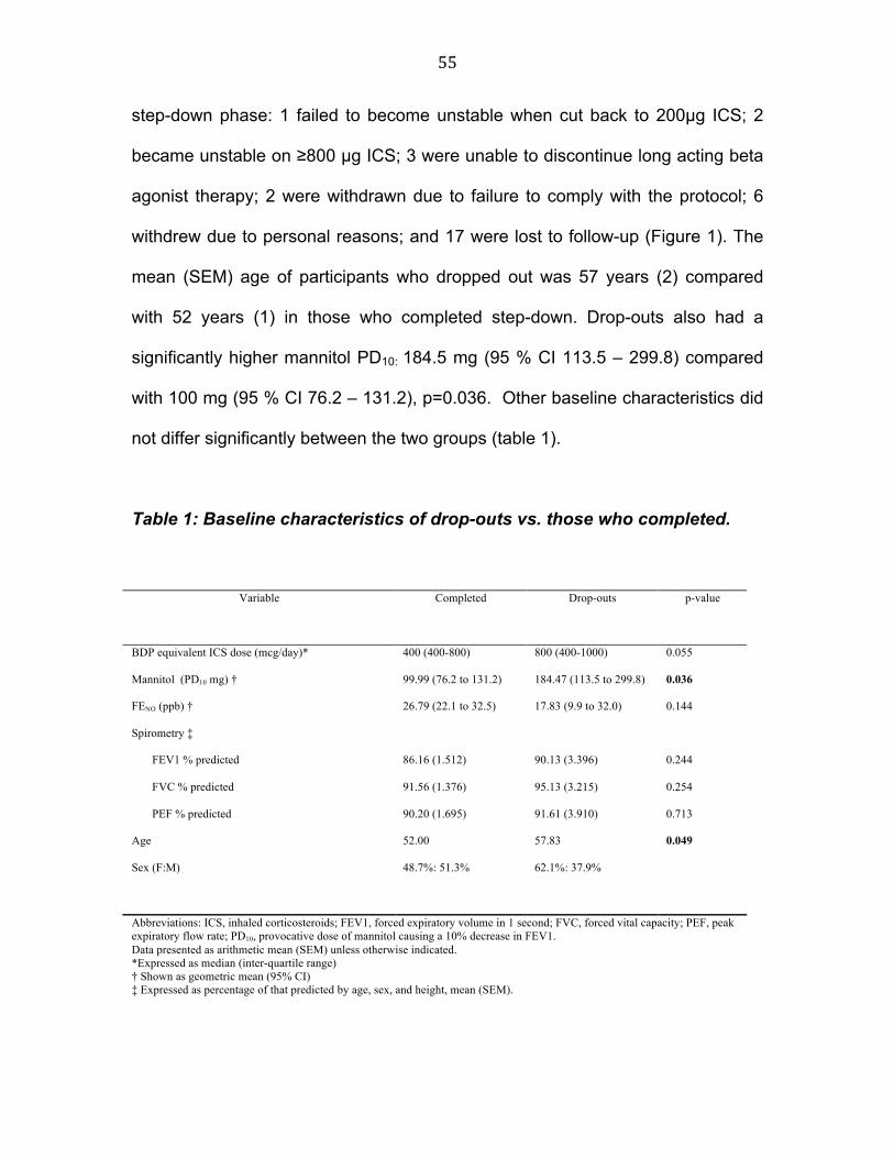
55
step-down phase: 1 failed to become unstable when cut back to 200µg ICS; 2
became unstable on ≥800 µg ICS; 3 were unable to discontinue long acting beta
agonist therapy; 2 were withdrawn due to failure to comply with the protocol; 6
withdrew due to personal reasons; and 17 were lost to follow-up (Figure 1). The
mean (SEM) age of participants who dropped out was 57 years (2) compared
with 52 years (1) in those who completed step-down. Drop-outs also had a
significantly higher mannitol PD10: 184.5 mg (95 % CI 113.5 – 299.8) compared
with 100 mg (95 % CI 76.2 – 131.2), p=0.036. Other baseline characteristics did
not differ significantly between the two groups (table 1).
Table 1: Baseline characteristics of drop-outs vs. those who completed.
Variable
Completed Drop-outs p-value
BDP equivalent ICS dose (mcg/day)* 400 (400-800) 800 (400-1000) 0.055
Mannitol (PD10 mg) † 99.99 (76.2 to 131.2) 184.47 (113.5 to 299.8) 0.036
FENO (ppb) † 26.79 (22.1 to 32.5) 17.83 (9.9 to 32.0) 0.144
Spirometry ‡
FEV1 % predicted 86.16 (1.512) 90.13 (3.396) 0.244
FVC % predicted 91.56 (1.376) 95.13 (3.215) 0.254
PEF % predicted 90.20 (1.695) 91.61 (3.910) 0.713
Age 52.00 57.83 0.049
Sex (F:M)
48.7%: 51.3% 62.1%: 37.9%
Abbreviations: ICS, inhaled corticosteroids; FEV1, forced expiratory volume in 1 second; FVC, forced vital capacity; PEF, peak expiratory flow rate; PD10, provocative dose of mannitol causing a 10% decrease in FEV1. Data presented as arithmetic mean (SEM) unless otherwise indicated. *Expressed as median (inter-quartile range) † Shown as geometric mean (95% CI) ‡ Expressed as percentage of that predicted by age, sex, and height, mean (SEM).
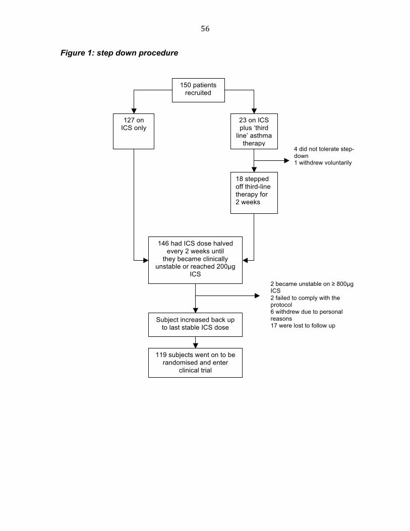
56
Figure 1: step down procedure
150 patients recruited
127 on ICS only
23 on ICS plus ‘third
line’ asthma therapy
18 stepped off third-line therapy for 2 weeks
4 did not tolerate step-down 1 withdrew voluntarily
146 had ICS dose halved every 2 weeks until
they became clinically unstable or reached 200µg
ICS
Subject increased back up to last stable ICS dose
2 became unstable on ≥ 800µg ICS 2 failed to comply with the protocol 6 withdrew due to personal reasons 17 were lost to follow up
119 subjects went on to be randomised and enter
clinical trial
57
Withdrawal of medication
23 participants were taking additional third line asthma therapy (long-acting β2-
agonists (n=21), theophylines (n=1) or leukotriene receptor antagonists (n=1)) at
the start of the step down. Only four participants did not tolerate withdrawal of
their third line therapy; one withdrew voluntarily. In patients who completed step-
down, the median (interquartile range) dose of BDP or equivalent was 400 µg
(400 – 800) at baseline, compared with 250 µg (200 – 400) post step-down,
giving a median reduction of 150 µg (p < 0.0001) (Figure 2). Subjects underwent
an average of 1.49 (0.07) step-downs prior to either becoming unstable or
reaching 200µg BDP equivalent.
Figure 2: median change in ICS dose (µg) pre and post step-down
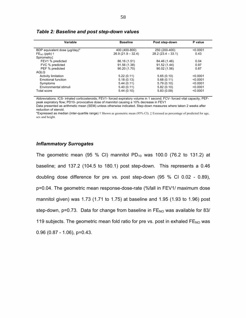
58
Table 2: Baseline and post step-down values
Variable Baseline Post step-down P value
BDP equivalent dose (µg/day)* 400 (400-800) 250 (200-400) <0.0001 FENO (ppb) † 26.9 (21.9 – 32.4) 28.2 (23.4 – 33.1) 0.43 Spirometry‡ FEV1 % predicted 86.16 (1.51) 84.46 (1.46) 0.04 FVC % predicted 91.56 (1.38) 91.52 (1.44) 0.97 PEF % predicted 90.20 (1.70) 90.02 (1.56) 0.87 AQLQ Activity limitation 5.22 (0.11) 5.65 (0.10) <0.0001 Emotional function 5.18 (0.13) 5.68 (0.11) <0.0001 Symptoms 5.44 (0.11) 5.79 (0.10) <0.0001 Environmental stimuli 5.40 (0.11) 5.82 (0.10) <0.0001 Total score 5.44 (0.10) 5.83 (0.09) <0.0001 Abbreviations: ICS- inhaled corticosteroids; FEV1- forced expiratory volume in 1 second; FCV- forced vital capacity; PEF- peak expiratory flow; PD10- provocative dose of mannitol causing a 10% decrease in FEV1 Data presented as arithmetic mean (SEM) unless otherwise indicated. Step-down measures where taken 2 weeks after reduction of steroid. *Expressed as median (inter-quartile range) † Shown as geometric mean (95% CI). ‡ Exressed as percentage of predicted for age, sex and height.
Inflammatory Surrogates
The geometric mean (95 % CI) mannitol PD10 was 100.0 (76.2 to 131.2) at
baseline; and 137.2 (104.5 to 180.1) post step-down. This represents a 0.46
doubling dose difference for pre vs. post step-down (95 % CI 0.02 - 0.89),
p=0.04. The geometric mean response-dose-rate (%fall in FEV1/ maximum dose
mannitol given) was 1.73 (1.71 to 1.75) at baseline and 1.95 (1.93 to 1.96) post
step-down, p=0.73. Data for change from baseline in FENO was available for 83/
119 subjects. The geometric mean fold ratio for pre vs. post in exhaled FENO was
0.96 (0.87 - 1.06), p=0.43.
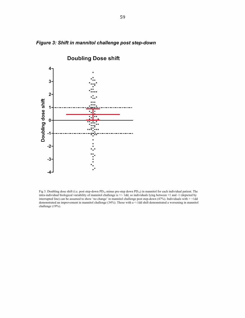
59
Figure 3: Shift in mannitol challenge post step-down
Fig 3. Doubling dose shift (i.e. post step-down PD10 minus pre-step down PD10) in mannitol for each individual patient. The intra-individual biological variability of mannitol challenge is +/- 1dd, so individuals lying between +1 and -1 (depicted by interrupted line) can be assumed to show ‘no change’ in mannitol challenge post step-down (47%). Individuals with > +1dd demonstrated an improvement in mannitol challenge (34%). Those with a <-1dd shift demonstrated a worsening in mannitol challenge (19%).
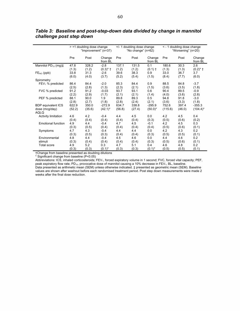
60
Table 3: Baseline and post-step-down data divided by change in mannitol challenge post step down > +1 doubling dose change
“Improvement” (n=37)
+/- 1 doubling dose change “No change” (n=62)
< - 1 doubling dose change “Worsening” (n=20)
Pre Post Change from BL
Pre Post Change from BL
Pre Post Change from BL
Mannitol PD10 (mg)‡ 47.8 (1.3)
328.2 (1.2)
-2.8 (0.3)* †
137.1 (1.2)
131.5 (1.2)
0.1 (0.1) †
180.6 (1.3)
30.3 (1.3)
2.6 (0.2)* †
FENO (ppb) 33.8 (6.0)
31.3 (4.0)
-2.6 (3.7)
39.6 (5.2)
38.3 (5.4)
0.9 (1.5)
33.0 (8.4)
36.7 (7.7)
3.7 (6.0)
Spirometry FEV1 % predicted 86.4
(2.5) 84.4 (2.8)
-2.0 (1.3)
85.3 (2.3)
84.4 (2.1)
0.9 (1.5)
88.5 (3.6)
84.8 (3.5)
-3.7 (1.8)
FVC % predicted 91.2 (2.2)
91.2 (2.9)
-0.03 (1.7)
93.7 (2.1)
93.1 (2.1)
0.6 (1.4)
90.4 (4.0)
89.5 (3.6)
-0.9 (2.8)
PEF % predicted 88.1 (2.8)
90.0 (2.7)
1.9 (1.8)
89.8 (2.8)
89.3 (2.4)
0.5 (2.1)
94.8 (3.6)
91.8 (3.3)
-3.0 (1.8)
BDP equivalent ICS dose (mcg/day)
622.9 (52.2)
350.0 (35.6)
-272.9 (42.1)*
634.7 (56.8)
338.8 (27.4)
-295.9 (50.0)*
752.6 (115.6)
397.4 (46.0)
-355.3 (104.4)*
AQLQ Activity limitation 4.6
(0.4) 4.2 (0.4)
-0.4 (0.4)
4.4 (0.4)
4.5 (0.4)
0.0 (0.3)
4.2 (0.5)
4.5 (0.6)
0.4 (0.2)
Emotional function 4.9 (0.3)
4.4 (0.5)
-0.4 (0.4)
4.7 (0.4)
4.5 (0.4)
-0.1 (0.4)
4.2 (0.5)
4.5 (0.6)
0.3 (0.1)
Symptoms 4.7 (0.3)
4.3 (0.5)
-0.4 (0.3)
4.4 (0.4)
4.4 (0.4)
0.0 (0.3)
4.2 (0.5)
4.3 (0.5)
0.2 (0.1)
Environmental stimuli
4.8 (0.3)
4.4 (0.4)
-0.4 (0.4)
4.5 (0.4)
4.6 (0.4)
0.0 (0.3)
4.4 (0.5)
4.6 (0.6)
0.2 (0.1)
Total score 4.9 (0.3)
5.2 (0.3)
0.3 (0.1)*
4.7 (0.3)
5.1 (0.3)
0.4 (0.1)*
4.6 (0.5)
4.8 (0.5)
0.2 (0.1)
†Change from baseline presented as doubling dilutions * Significant change from baseline (P<0.05) Abbreviations: ICS, inhaled corticosteroids; FEV1, forced expiratory volume in 1 second; FVC, forced vital capacity; PEF, peak expiratory flow rate; PD10, provocative dose of mannitol causing a 10% decrease in FEV1, BL, baseline. Data presented as arithmetic mean (SEM) unless otherwise indicated. ‡ presented as geometric mean (SEM). Baseline values are shown after washout before each randomised treatment period. Post step down measurements were made 2 weeks after the final dose reduction.
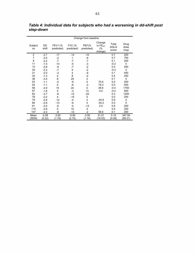
61
Table 4: Individual data for subjects who had a worsening in dd-shift post step-down
Change from baseline
Subject no
DD shift
FEV1 (% predicted)
FVC (% predicted)
PEF(% predicted)
Change in FENO
(% change)
Total AQLQ score
Drug dose (mg)
2 -2.7 -17 -14 -16 0.7 200 7 -3.5 -2 1 -9 0.7 750 8 -2.2 -7 -7 -7 0.1 200
11 -1.5 -13 -5 -3 -0.3 0 13 -2.8 -9 -7 -2 0.0 200 30 -2.3 -7 9 -2 -0.3 0 31 -2.0 -3 4 -9 0.1 400 32 -1.2 4 8 -2 0.4 250 36 -3.4 8 29 2 0.7 0 53 -1.1 -5 -9 0 70.6 0.0 200 54 -1.1 0 -6 -3 79.3 0.0 400 56 -4.9 15 24 0 28.6 -0.4 1750 57 -1.8 5 -3 13 0.0 -0.4 500 62 -3.7 -8 -12 -22 0.3 1250 78 -3.2 4 -15 5 0.0 200 79 -2.6 -12 -4 2 -34.8 0.9 0 80 -2.6 -13 -9 4 -34.2 0.0 0 91 -3.0 -6 -5 -13 2.0 0.9 200
110 -3.8 0 15 4 0.5 250 147 -2.2 -8 -12 -2 59.0 0.1 200
Mean (SEM)
-2.58 (0.22)
-3.65 (1.78)
-0.90 (2.75)
-3.00 (1.78)
21.21 (16.03)
0.19 (0.09)
347.50 (99.31)
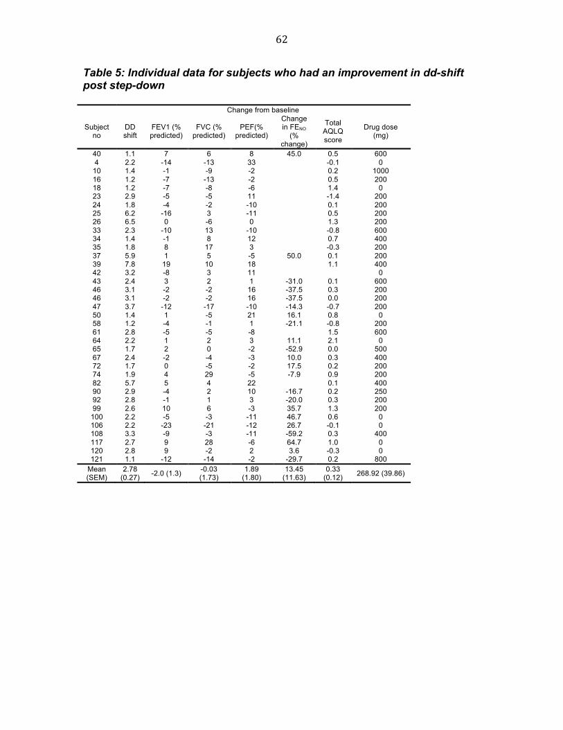
62
Table 5: Individual data for subjects who had an improvement in dd-shift post step-down
Change from baseline
Subject no
DD shift
FEV1 (% predicted)
FVC (% predicted)
PEF(% predicted)
Change in FENO
(% change)
Total AQLQ score
Drug dose (mg)
40 1.1 7 6 8 45.0 0.5 600 4 2.2 -14 -13 33 -0.1 0
10 1.4 -1 -9 -2 0.2 1000 16 1.2 -7 -13 -2 0.5 200 18 1.2 -7 -8 -6 1.4 0 23 2.9 -5 -5 11 -1.4 200 24 1.8 -4 -2 -10 0.1 200 25 6.2 -16 3 -11 0.5 200 26 6.5 0 -6 0 1.3 200 33 2.3 -10 13 -10 -0.8 600 34 1.4 -1 8 12 0.7 400 35 1.8 8 17 3 -0.3 200 37 5.9 1 5 -5 50.0 0.1 200 39 7.8 19 10 18 1.1 400 42 3.2 -8 3 11 0 43 2.4 3 2 1 -31.0 0.1 600 46 3.1 -2 -2 16 -37.5 0.3 200 46 3.1 -2 -2 16 -37.5 0.0 200 47 3.7 -12 -17 -10 -14.3 -0.7 200 50 1.4 1 -5 21 16.1 0.8 0 58 1.2 -4 -1 1 -21.1 -0.8 200 61 2.8 -5 -5 -8 1.5 600 64 2.2 1 2 3 11.1 2.1 0 65 1.7 2 0 -2 -52.9 0.0 500 67 2.4 -2 -4 -3 10.0 0.3 400 72 1.7 0 -5 -2 17.5 0.2 200 74 1.9 4 29 -5 -7.9 0.9 200 82 5.7 5 4 22 0.1 400 90 2.9 -4 2 10 -16.7 0.2 250 92 2.8 -1 1 3 -20.0 0.3 200 99 2.6 10 6 -3 35.7 1.3 200
100 2.2 -5 -3 -11 46.7 0.6 0 106 2.2 -23 -21 -12 26.7 -0.1 0 108 3.3 -9 -3 -11 -59.2 0.3 400 117 2.7 9 28 -6 64.7 1.0 0 120 2.8 9 -2 2 3.6 -0.3 0 121 1.1 -12 -14 -2 -29.7 0.2 800
Mean (SEM)
2.78 (0.27) -2.0 (1.3) -0.03
(1.73) 1.89
(1.80) 13.45
(11.63) 0.33
(0.12) 268.92 (39.86)
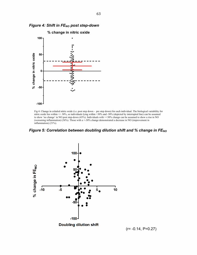
63
Figure 4: Shift in FENO post step-down
Figure 5: Correlation between doubling dilution shift and % change in FENO
(r= -0.14, P=0.27)
Fig 4. Change in exhaled nitric oxide (i.e. post step down - pre step down) for each individual. The biological variability for nitric oxide lies within +/- 30%, so individuals lying within +30% and -30% (depicted by interrupted line) can be assumed to show ‘no change’ in NO post step-down (43%). Individuals with >+30% change can be assumed to show a rise in NO (worsening inflammation) (36%). Those with a <-30% change demonstrated a decrease in NO (improvement in inflammation) (21%).
64
Lung function
Mean (SEM) baseline spirometry values were: FEV1 of 86.2% (1.5), FVC 91.6%
(1.4), PEF 90.2% (1.7). FEV1 post step-down was 84.5 % (1.5), p < 0.05. FVC
and PEF showed no significant change post step-down (91.5 % (1.4), and 90.0
% (1.6), p = 0.87).
AQLQ
The median (inter-quartile range) mini-AQLQ composite scores pre and post step
down were 5.7 (4.7 – 6.3) vs. 6.1 (5.3 – 6.5) respectively, representing a median
improvement of 0.4, p <0.001.
Discussion
The present study demonstrates that supervised step down of ICS in a
community setting can be achieved without any worsening of airways
inflammation, as measured by FENO and mannitol challenge. This is in
agreement with work carried out by Leuppi et al, who determined that the
majority of patients could undergo a halving of their ICS dose without
exacerbation [41]. We hypothesize that this may be due to improved adherence
with supervised treatment during step down. Whilst a significant reduction in ICS
could also be achieved if patients had initially been over treated, there should
have been a commensurate increase in airways inflammation on withdrawal of
65
treatment, as has been reported elsewhere [78]. In the absence of such a
change we feel that enhanced adherence is the logical conclusion. Adherence
with asthma medications, particularly ICS, is known to be poor [195]. Patient-
reported adherence rates are as low as 38% [196, 197], and actual drug use is
often even lower. Rand et al compared patient-reported adherence to change in
canister weight and demonstrated that although 73% of participants reported
using their ICS three times daily, this was only confirmed in 15% by canister
weight [197].
The commonest reasons to explain non-adherence include: patients’ perception
of their disease; such that they only take ICS when they become unwell as
opposed to using it as ‘preventer’ therapy [196]; and concerns over potential side
effects [195]. There is evidence to suggest that regular disease monitoring
addresses these issues and leads to benefits such as improved inhaler technique
and adherence [198, 199]. In this regard, patients in the present study were
reviewed fortnightly and may have additionally benefited from the positive
reinforcement of regular spirometry, peak flow and symptom monitoring. Whilst
this high level of supervision is too intensive to be reproduced in primary care, it
suggests that the widespread practice of annual review may be too infrequent to
adequately educate and support our asthmatic population.
Control of airways inflammation is of critical importance in asthma, as if left
uncontrolled long-term it can lead to airway remodelling, even in asymptomatic
66
patients [31]. Furthermore, studies in which asthma therapy was titrated to
achieve suppression of surrogates of inflammation demonstrate that
exacerbation rates can be reduced by up to five-fold, when compared with
treatment guided by symptoms and lung function alone [37, 38]. To address this,
the present study utilised bronchial challenge with mannitol and FENO as suitable
surrogates of asthmatic inflammation. Bronchial challenge tests are traditionally
used to diagnose the presence of AHR, and to monitor response to treatment
[200]. AHR is a key feature of persistent asthma, which is considered to correlate
well with underlying airways inflammation [21]. All participants in this study were
required to exhibit AHR to mannitol as one of the inclusion criteria. Hence, we
believe this makes an alternative diagnosis to asthma such as eosinophilic
bronchitis unlikely. Nonetheless, we appreciate that AHR may vary over time,
often increasing during exacerbations and decreasing during treatment with anti-
inflammatory therapy [200].
Mannitol challenge is a relatively novel indirect bronchial challenge test, which is
known to be reproducible, and to correlate well with other surrogate markers of
airways inflammation [136, 201]. Unlike other challenge tests, it is portable,
simple, and quick to perform, and therefore ideally suited for use in primary care.
Moreover, mannitol is an indirect stimulus, which induces AHR by osmotically
stimulating inflammatory cells to release mediators which constrict airway smooth
muscle. Fardon et al showed that PD10 correlates well with PD15, allowing an
abbreviated challenge to be utilised without loss of sensitivity, whilst exposing the
67
subject to less provocation agent [202]. The overall mean improvement
observed in mannitol challenge (0.46 doubling dose shift in mannitol PD10) was
statistically significant, but would not be considered clinically significant.
Examination of change in PD10 in individual patients, as depicted in Figure 3, is
more revealing than the magnitude of the overall mean shift for the group as a
whole. A change in mannitol PD10 of +/- 1 doubling dose in a given patient is
within the limits of intra-individual biological variability. Using these criteria we
identified 31% of patients who had an improvement in PD10 (i.e. a change > +1
doubling dose), 52 % who had no change (i.e. a change within +/- 1 doubling
dose), and 17 % who had a worsening (i.e. change < - 1 doubling dose) when
comparing pre vs. post step down (Fig 3). In other words it was evident that the
majority of patients (i.e. 81%) either had no change in AHR or improved. A
minority of patients showed a worsening of AHR, which would be expected if
subjects were initially adherent with treatment. A rate of 19% is consistent with
adherence rates reported in previous studies [197]. A comparison of the
baseline and post step down characteristics of these three groups revealed no
significant differences in terms of spirometry, FENO or ICS dose. Analysis of
mannitol PD10 showed that those who improved post step down had significantly
greater bronchial hyper-reactivity at baseline than the other two groups. Those
who worsened post-step down had significantly more bronchial hyper-reactivity at
the end of step down. This correlates well with data obtained by Leuppi et al who
determined that developing AHR to mannitol during dose-reduction was a good
predictor of step-down failure [41]. Changes in AHR correlated very poorly with
68
changes in symptoms, as subjects who either “improved” or had “no change” in
AHR had statistically significant, albeit clinically insignificant, worsening of their
symptom scores, whereas those who had a “worsening” had no change (table 3).
Individual data for those with “improved” and “worsened” AHR post-step down
are included in tables 4 and 5.
FENO is an established surrogate of airways inflammation in asthma [59] and
correlates closely to sputum eosinophil counts [60, 203] and mucosal
eosinophilic markers [204, 205]. When used on its own as an inflammatory
surrogate to titrate ICS, the results have been disappointing [76], indeed, a
recent Cochrane review has determined that FENO cannot be recommended for
tailoring the dose of inhaled corticosteroids in clinical practice [206]. However, its
rapid response to changes in inflammatory status [71] and acute sensitivity to
ICS make it helpful when used in conjunction with other measures. It has been
used as an early predictor of under-treatment (predicting exacerbation with FENO
and dose reduction) [78]. In this regard our cohort showed no net change in FENO
during step-down. If the biological variability of FENO is assumed to be <30%
[207]: 64% of subjects demonstrated no significant increase in FENO (figure 4),
These results correlate well with those of Leuppi et al who found no significant
increase in FENO during dose reduction or exacerbation [41]. Similarly, we found
no correlation between doubling dilution shift in mannitol challenge and %
change in FENO (r= -0.14, P=0.27). We know from previous studies that 200µg
BDP is sufficient to suppress FENO, even in the presence of persisting AHR,
69
which may explain the disconnect with AHR, and it’s lack of response in these
circumstances [200]. However, when this observation is taken in conjunction with
mannitol AHR, we believe it offers good evidence that airway inflammation was
well controlled in this study, despite the relatively rapid rate at which ICS was
reduced. Larger studies with a longer duration of follow-up are required to
determine whether these improvements are maintained long-term.
Despite evidence to support the use of inflammatory markers in asthmatic
assessment, current clinical practice dictates that ICS should be adjusted
according to patient reported symptoms and spirometry [8]. In this respect, we
observed a statistically significant improvement in quality of life scores, despite
the reduction in ICS dose. There were no clinically significant changes in
spirometry.
Treatment of asthmatic patients in primary care has been driven by results
obtained from randomised controlled trials, during which treatment compliance is
reinforced. These therefore, do not reflect a real life scenario. The current paper
highlights the potential pit falls of extrapolating data to a primary care setting
where compliance rates may be poor. Perhaps there is a need for the
development of more effective strategies which support asthmatics to use
established therapies correctly rather than continue a culture of escalating
therapy, without frequent review and step-down if appropriate. Improvement of
asthma care in the community may require an approach that includes more
70
effective supervision and targeted assessment to enhance adherence and
reduce airways inflammation.
In conclusion we have demonstrated that achieving a significant reduction in ICS
dose is possible, without any deterioration in surrogate markers of inflammation,
or quality of life. This apparent disconnect between reduction in anti-inflammatory
therapy and measured inflammation may reflect enhanced adherence through
frequent supervision.
71
Critique
This is the first study to utilise mannitol challenge in a community setting,
confirming that it is safe and easy to use. Furthermore, we have demonstrated
that inhaled steroids can be safely stepped down in general practice without a
concomitant increase in airway inflammation. Whilst these results are
encouraging, we would not advocate the back-titration of inhaled steroids in all
community-based patients. We recognise that this observational study has
several limitations, namely to do with study design: Firstly, we were unable to
follow subjects up long term to ensure that their inflammation remained
suppressed. Secondly, measuring FENO pre and post step-down was only
introduced once recruitment had commenced; consequently FENO measures are
not available for all subjects. Thirdly, as this was an observational study there
was no control group. In order to verify our suspicions that the improvements
seen were due to enhanced compliance we would like to repeat the study in a
prospective fashion measuring canister weight. This study suggests that
concordance with medication improves in patients who are entered into a clinical
trial, indicating that perhaps a more targeted approach to asthma therapy, which
includes more effective supervision, alongside the use of an appropriate
“inflammometer”, such as mannitol (so patients can actually see an improvement
following treatment) may be beneficial. A prospective study investigating the
benefits of such a treatment strategy, as well as evaluating the benefits of
mannitol as a tool for titrating asthma therapy, is required.
72
Part b: Determining which outcomes provide
sufficient assay sensitivity for detecting dose
response effects on airway and systemic
markers.
Study aims:
1. To compare the pharmacodynamic characteristics of
Hydrofluoroalkane (HFA) and Chlorofluorocarbon (CFC) Budesonide
utilising methacholine challenge as the primary endpoint in a cross
over study.
2. To determine which outcomes measures provide sufficient assay
sensitivity for detecting dose response.
73
Pharmacodynamic Comparison of Hydrofluoroalkane and
Chlorofluorocarbons Budesonide
Introduction
Inhaled corticosteroids (ICS) are the mainstay of treatment in persistent asthma
[8, 192]. Budesonide is one of the most commonly prescribed ICS, and has been
used effectively in the long-term management of persistent asthma for many
years. A suspension aerosol formulation with chlorofluorocarbon (CFC) as the
propellant was previously available for delivery of budesonide via pressurized
metered-dose inhaler (pMDI). In recent years CFCs have been implicated in
damage to the ozone layer, and are due to be phased out in accordance with the
Montreal Protocol [208]. Hydrofluoroalkane-134a (HFA) has been found to act as
adequate propellant for pMDI delivery systems without the same deleterious
environmental effects. Budesonide has therefore been re-formulated in
suspension with HFA as the propellant. Two strengths of budesonide HFA pMDI
(100µg and 200µg per actuation) have been developed. Determination of
therapeutic equivalence is essential for new drug formulations. Pharmacokinetic
studies have shown the two formulations to be bioequivalent [209]. The aim of
this study is therefore to present pharmacodynamic data comparing HFA and
CFC formulations of budesonide delivered via pMDI.
74
Methods
This was a single-centre, randomized, open-label, crossover study (Figure 6). At
screening, entry requirements were checked and informed consent was
obtained. Participants were considered eligible if they were aged 18-65 years,
and had a diagnosis of stable persistent asthma while receiving ≤1000µg
beclomethasone dipropionate (BDP) or equivalent. Subjects were excluded if
they were pregnant or lactating, had concomitant respiratory disease or had been
prescribed oral steroid or a change to their asthma therapy in the three months
preceding the screening visit. Eligible patients then entered a step-down phase
during which long-acting β2-agonists, theophyllines, cromones and leukotriene
inhibitors were stopped. Participants then had their steroid dose gradually
reduced in a manner similar to previously published studies from our department
[210]. They then entered a 1-week pre-treatment (ICS-free) washout period. A
methacholine PC20 of ≤4mg/ml and FEV1 ≥60% predicted post step-down was
required for entry into the treatment period. Subjects not meeting the entry
requirements at the end of this phase were given a further 1-2 weeks of washout.
The study consisted of two treatment periods. Subjects received low-dose
(200µg/day) budesonide, either via CFC or HFA pMDI, for 2 weeks followed by a
further 2 weeks of medium-dose (800µg/day) budesonide. Participants then
crossed over treatment arm, effectively acting as their own control. Treatment
periods were separated by a 1–2 week washout period. Subjects were required
to be within 1 doubling dilution of baseline methacholine PC20 at the end of
75
washout. During all study visits participants underwent pulmonary function testing
(including FEV1, forced vital capacity, PEF, and FEF25-75%), FENO, and
methacholine challenge. Methacholine challenge was performed using the five-
breath dosimeter technique in accordance with American Thoracic Society (ATS)
recommendations [183]. The PC20 (provocative dose required to cause a 20%
drop in FEV1) was calculated using log-linear interpolation of the dose response
[123]. FENO was measured in line with current ATS recommendations using a
NIOX analyzer (NIOX® Nitric Oxide Monitoring System, Aerocrine AB, Solna,
Sweden) [194]. Subjects were asked to withhold short-acting β2-agonists and
caffeine for 6 hours prior to study visits, and all study visits took place at the
same time of day. At each visit inhaler technique was checked and peak
flow/symptom diaries were reviewed. Subjects were asked to rate their asthma
symptoms twice daily. Daytime scores were recorded each evening, and night
time scores were recorded on waking. The following rating scales were used: 0 =
no asthma symptoms, 1= mild (easily tolerable) symptoms, 2= moderate
(interferes with normal activities/sleep) symptoms, 3= severe (prevents normal
activities/sleep). Overnight (i.e. 10pm to 8am) urine was collected for cortisol and
creatinine the night prior to study visits. Urinary cortisol was measured using a
commercial radioimmunoassay kit (DiaSorin Ltd, Wokingham, Berkshire, UK).
The intra-assay coefficient of variation was 4% and the inter-assay coefficient of
variation was 8%. Urinary creatinine was measured on a Cobas-Bio auto
analyzer (Roche Products, Welwyn Garden City, UK). The intra-assay and inter-
assay coefficients of variation were 4.6% and 3.0% respectively.
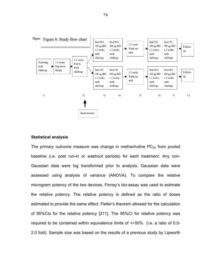
76
Statistical analysis
The primary outcome measure was change in methacholine PC20 from pooled
baseline (i.e. post run-in or washout periods) for each treatment. Any non-
Gaussian data were log transformed prior to analysis. Gaussian data were
assessed using analysis of variance (ANOVA). To compare the relative
microgram potency of the two devices, Finney’s bio-assay was used to estimate
the relative potency. The relative potency is defined as the ratio of doses
estimated to provide the same effect. Fieller’s theorem allowed for the calculation
of 95%CIs for the relative potency [211]. The 95%CI for relative potency was
required to be contained within equivalence limits of +/-50% (i.e. a ratio of 0.5-
2.0 fold). Sample size was based on the results of a previous study by Lipworth
Figure 6: Study flow chart
77
et al [212]. A comparison was also made between the two formulations at each
dose using the EMEA equivalence limits of +/- 33% (ratio of 0.67-1.50).
Other outcome variables were considered as secondary and were analyzed
using a similar model but without log transformation (except FENO and OUCC).
Results
133 adults with mild-moderate asthma were screened; 99 were randomized, and
68 completed the study according to protocol. The demographic characteristics
of subjects randomized to first treatment at screening are given in Table 4.
Reasons for non-randomization included: FEV1 ≤60% predicted (n=4); PC20
>4mg/ml (n=13); instability in step-down (n=8); voluntary discontinuation (n=8);
lost to follow-up (n=1). During the study 31 patients were withdrawn for the
following reasons: failure to wash-out (n=14); worsening of asthma (n=10);
personal reasons (n=4); adverse events (n=3). Both treatment sequences were
well matched for age, gender, height, weight and race.
Primary efficacy variable
There were no significant differences between baseline values prior to each
treatment (i.e. after each wash-out period) for any of the primary or secondary
outcome measures (table 6). Therefore, all analyses were performed compared
with pooled baseline values. There was also no significant difference in
compliance between the two treatments, or between doses.
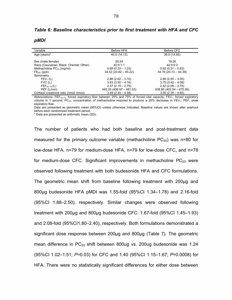
78
Table 6: Baseline characteristics prior to first treatment with HFA and CFC
pMDI
Variable Before HFA Before CFC Age (years)* 40.0 (14.13)
39.5 (14.65)
Sex (male:female) 20:24 19:26 Race (Caucasian: Black: Oriental: Other) 42:0:1:1 42:0:0:3 Methacholine PC20 (mg/ml) 0.68 (0.33 – 1.03) 0.62 (0.31 – 0.93) FENO (ppb) 34.42 (23.62 – 45.22) 34.76 (25.13 – 44.39) Spirometry FEV1 (L) 2.86 (2.62 – 3.10) 2.80 (2.55 – 3.05) FVC (L) 3.83 (3.50 – 4.16) 3.75 (3.42 – 4.08) FEF25–75 (L) 2.47 (2.15 – 2.79) 2.42 (2.09 – 2.75) PEF (L/min) 445.25 (408.97 – 481.53) 438.80 (402.54 – 475.06) Cortisol/ creatinine ratio (nmol/ mmol) 3.49 (2.40 – 4.58) 3.50 (2.35 – 4.65) Abbreviations: FEF25–75, forced expiratory flow between 25% and 75% of forced vital capacity; FEV1, forced expiratory volume in 1 second; PC20, concentration of methacholine required to produce a 20% decrease in FEV1; PEF, peak expiratory flow. Data are presented as geometric mean (95%CI) unless otherwise indicated. Baseline values are shown after washout before each randomized treatment period. * Data are presented as arithmetic mean (SD).
The number of patients who had both baseline and post-treatment data
measured for the primary outcome variable (methacholine PC20) was n=80 for
low-dose HFA, n=79 for medium-dose HFA, n=79 for low-dose CFC, and n=78
for medium-dose CFC. Significant improvements in methacholine PC20 were
observed following treatment with both budesonide HFA and CFC formulations.
The geometric mean shift from baseline following treatment with 200µg and
800µg budesonide HFA pMDI was 1.55-fold (95%CI 1.34–1.78) and 2.16-fold
(95%CI 1.88–2.50), respectively. Similar changes were observed following
treatment with 200µg and 800µg budesonide CFC: 1.67-fold (95%CI 1.45–1.93)
and 2.08-fold (95%CI1.80–2.40), respectively. Both formulations demonstrated a
significant dose response between 200µg and 800µg (Table 7). The geometric
mean difference in PC20 shift between 800ug vs. 200ug budesonide was 1.24
(95%CI 1.02–1.51; P=0.03) for CFC and 1.40 (95%CI 1.15–1.67; P=0.0008) for
HFA. There were no statistically significant differences for either dose between
79
the two propellant types (Table 8): 200ug/day: HFA vs. CFC 0.93 (95%CI 0.76–
1.13; P=0.44), 800ug/day: HFA vs. CFC 1.04 (95%CI 0.85–1.27; P=0.70). Both
of these 95%CI were contained within +/- 33% equivalence limits (ratio of 0.67-
1.50)
The log-linear dose-response curves for HFA and CFC were parallel and the
common slope was highly statistically significant (p<0.0001). The estimated
relative potency between HFA and CFC budesonide, for the full analysis set
(n=89), was 1.10 (95%CI 0.49–2.66). The 95% CI was not completely contained
within the +/- 50% equivalence limits of 0.5–2.0.
Secondary efficacy/safety variables
There were statistically significant differences between medium and low doses of
budesonide for both CFC and HFA formulations (except morning rescue
medication and pulmonary function) (Table 7). There were no statistically
significant differences when comparing low-dose budesonide CFC and HFA, and
medium-dose budesonide CFC and HFA (Table 8). Relative potencies were
calculated for secondary outcomes that were close to unity (Table 9). Only three
patients discontinued following adverse events: two lower respiratory tract
infections (CFC formulation); one oral candidiasis (HFA formulation). No serious
adverse events were reported in this study.
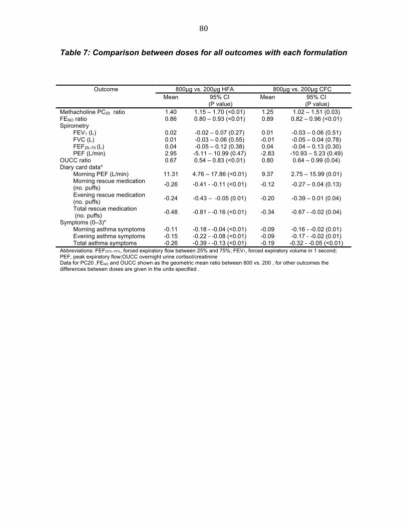
80
Table 7: Comparison between doses for all outcomes with each formulation
800µg vs. 200µg HFA 800µg vs. 200µg CFC Outcome Mean 95% CI
(P value) Mean 95% CI
(P value) Methacholine PC20 ratio 1.40 1.15 – 1.70 (<0.01) 1.25 1.02 – 1.51 (0.03) FENO ratio 0.86 0.80 – 0.93 (<0.01) 0.89 0.82 – 0.96 (<0.01) Spirometry FEV1 (L) 0.02 -0.02 – 0.07 (0.27) 0.01 -0.03 – 0.06 (0.51) FVC (L) 0.01 -0.03 – 0.06 (0.55) -0.01 -0.05 – 0.04 (0.78) FEF25–75 (L) 0.04 -0.05 – 0.12 (0.38) 0.04 -0.04 – 0.13 (0.30) PEF (L/min) 2.95 -5.11 – 10.99 (0.47) -2.83 -10.93 – 5.23 (0.49) OUCC ratio 0.67 0.54 – 0.83 (<0.01) 0.80 0.64 – 0.99 (0.04) Diary card data* Morning PEF (L/min) 11.31 4.76 – 17.86 (<0.01) 9.37 2.75 – 15.99 (0.01) Morning rescue medication (no. puffs) -0.26 -0.41 - -0.11 (<0.01) -0.12 -0.27 – 0.04 (0.13)
Evening rescue medication (no. puffs) -0.24 -0.43 – -0.05 (0.01) -0.20 -0.39 – 0.01 (0.04)
Total rescue medication (no. puffs) -0.48 -0.81 - -0.16 (<0.01) -0.34 -0.67 - -0.02 (0.04)
Symptoms (0–3)* Morning asthma symptoms -0.11 -0.18 - -0.04 (<0.01) -0.09 -0.16 - -0.02 (0.01) Evening asthma symptoms -0.15 -0.22 - -0.08 (<0.01) -0.09 -0.17 - -0.02 (0.01) Total asthma symptoms -0.26 -0.39 - -0.13 (<0.01) -0.19 -0.32 - -0.05 (<0.01) Abbreviations: FEF25%–75%, forced expiratory flow between 25% and 75%; FEV1, forced expiratory volume in 1 second; PEF, peak expiratory flow;OUCC overnight urine cortisol/creatinine Data for PC20 ,FENO and OUCC shown as the geometric mean ratio between 800 vs. 200 , for other outcomes the differences between doses are given in the units specified .
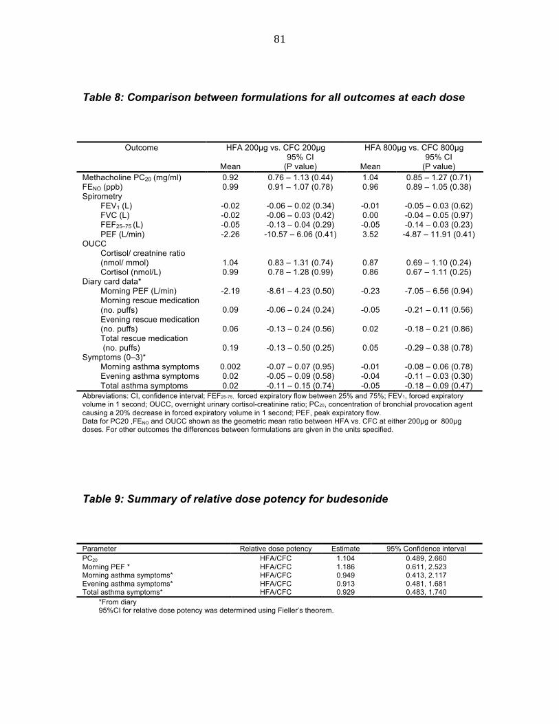
81
Table 8: Comparison between formulations for all outcomes at each dose
HFA 200µg vs. CFC 200µg HFA 800µg vs. CFC 800µg Outcome
Mean 95% CI
(P value) Mean 95% CI
(P value) Methacholine PC20 (mg/ml) 0.92 0.76 – 1.13 (0.44) 1.04 0.85 – 1.27 (0.71) FENO (ppb) 0.99 0.91 – 1.07 (0.78) 0.96 0.89 – 1.05 (0.38) Spirometry FEV1 (L) -0.02 -0.06 – 0.02 (0.34) -0.01 -0.05 – 0.03 (0.62) FVC (L) -0.02 -0.06 – 0.03 (0.42) 0.00 -0.04 – 0.05 (0.97) FEF25–75 (L) -0.05 -0.13 – 0.04 (0.29) -0.05 -0.14 – 0.03 (0.23) PEF (L/min) -2.26 -10.57 – 6.06 (0.41) 3.52 -4.87 – 11.91 (0.41) OUCC Cortisol/ creatnine ratio (nmol/ mmol) 1.04 0.83 – 1.31 (0.74) 0.87 0.69 – 1.10 (0.24) Cortisol (nmol/L) 0.99 0.78 – 1.28 (0.99) 0.86 0.67 – 1.11 (0.25) Diary card data* Morning PEF (L/min) -2.19 -8.61 – 4.23 (0.50) -0.23 -7.05 – 6.56 (0.94) Morning rescue medication (no. puffs) 0.09 -0.06 – 0.24 (0.24) -0.05 -0.21 – 0.11 (0.56) Evening rescue medication (no. puffs) 0.06 -0.13 – 0.24 (0.56) 0.02 -0.18 – 0.21 (0.86) Total rescue medication (no. puffs) 0.19 -0.13 – 0.50 (0.25) 0.05 -0.29 – 0.38 (0.78) Symptoms (0–3)* Morning asthma symptoms 0.002 -0.07 – 0.07 (0.95) -0.01 -0.08 – 0.06 (0.78) Evening asthma symptoms 0.02 -0.05 – 0.09 (0.58) -0.04 -0.11 – 0.03 (0.30) Total asthma symptoms 0.02 -0.11 – 0.15 (0.74) -0.05 -0.18 – 0.09 (0.47) Abbreviations: CI, confidence interval; FEF25-75, forced expiratory flow between 25% and 75%; FEV1, forced expiratory volume in 1 second; OUCC, overnight urinary cortisol-creatinine ratio; PC20, concentration of bronchial provocation agent causing a 20% decrease in forced expiratory volume in 1 second; PEF, peak expiratory flow. Data for PC20 ,FENO and OUCC shown as the geometric mean ratio between HFA vs. CFC at either 200µg or 800µg doses. For other outcomes the differences between formulations are given in the units specified.
Table 9: Summary of relative dose potency for budesonide
Parameter Relative dose potency Estimate 95% Confidence interval PC20 HFA/CFC 1.104 0.489, 2.660 Morning PEF * HFA/CFC 1.186 0.611, 2.523 Morning asthma symptoms* HFA/CFC 0.949 0.413, 2.117 Evening asthma symptoms* HFA/CFC 0.913 0.481, 1.681 Total asthma symptoms* HFA/CFC 0.929 0.483, 1.740
*From diary 95%CI for relative dose potency was determined using Fieller’s theorem.
82
Discussion
The aim of this study was to determine whether budesonide HFA and
budesonide CFC pMDI were therapeutically equivalent. Suppression of airway
hyper-responsiveness (AHR) to methacholine was selected as the primary
endpoint as it is a characteristic feature of asthma, which varies over time, often
increasing during exacerbations and decreasing during treatment with anti-
inflammatory medications [31]. AHR has been shown to correlate well with
airways inflammation and to be a reliable surrogate of disease activity [34, 37].
Since asthma is an inflammatory condition and the role of ICS is to suppress
inflammation, we feel that using methacholine challenge as a primary outcome is
justified. The results of this study demonstrated that there was no significant
difference in response between budesonide formulations in methacholine PC20
with either 200ug or 800ug per day doses. The ratio of HFA to CFC was close to
unity, and the 95%CIs lay within +/- 33% equivalence limits (0.67-1.50) at each
dose level. We also demonstrated sensitivity of the methacholine bioassay, as
there was a significant dose-response relationship for both formulations.
Significant dose separation has previously been reported using methacholine
PC20 with 100µg vs. 500µg fluticasone propionate [213]. The common slope for
the overall log dose-response relationship was highly significant, demonstrating
that the doses selected coincided with the steep part of the dose-response curve
for BHR. The 95%CIs for the relative dose potency ratio of HFA vs. CFC were
close to unity at 1.10, however, the 95%CI (0.49–2.66) was outside of the +/-
50% limits of 0.5-2.0 [212]. Comparable results have been shown in a similar
83
dose-response study published previously from our own laboratory [212]. It could
be argued that the change observed in the step up from 200µg to 800µg could be
a time effect, however we feel this is extremely unlikely. Sovijarvi et al found the
doubling dilution difference between fluticasone and placebo to be 1.19, 1.33 and
1.27 after 72 hours, 2 weeks and 4 weeks respectively, indicating that near
maximal response in methacholine PC20 is seen after 2 weeks treatment [214].
The differences observed in the present study are too large to be due to a time
effect alone. FENO is another reliable surrogate of airway inflammation [59, 60,
203]. Results revealed no significant difference between products, indicating that
airway inflammation was equally controlled by both formulations.
This improvement in airway inflammation was mirrored by an improvement in
diary data, including symptoms and rescue medication. All measured outcomes,
with the exception of spirometric measures, demonstrated a significant dose-
response relationship for budesonide with both the HFA and CFC formulations.
Comparison of HFA and CFC formulations at each dose demonstrated no
significant differences between products for any measure. The lack of detectable
dose-response for spirometric measures is likely due to our patients having mild
to moderate asthma with relatively well-preserved airway calibre and little room
for improvement. Previous studies have successfully demonstrated small,
clinically insignificant, improvements in lung function within this time frame [200].
Spirometric indices are known to be relatively insensitive measures for assessing
response to ICS in mild-to-moderate persistent asthmatic patients [147], studies
84
utilising these as primary endpoints have typically required large numbers of
patients with prolonged follow-up [215]. Despite these issues current EMEA
guidelines still recommend the use of spirometry as the primary outcome when
determining the equivalent efficacy of ICS. Additionally, they recommend the use
of a parallel-group study design, which requires careful matching of study groups.
This raises further problems due to the inherent heterogeneity of asthma. The
study design is also far more robust provided reliable, repeatable endpoints are
used. The only potential concern regarding crossover studies is unequal carry-
over of steroid effect between treatment periods. In this respect, for all endpoints
we found no important differences between the respective pre-treatment baseline
values at run-in and washout periods.
In order to be considered equivalent products also need to demonstrate similar
safety profiles. HPA-axis suppression has been shown to be one of the most
sensitive markers of systemic bioavailability for ICS [216]. It has also been used
as a surrogate marker for potential adverse effects in other tissues. In this
respect we found no significant difference between formulations at either dose.
Furthermore, assay sensitivity was demonstrated in terms of a significant dose
response between 200 and 800ug/day for levels of OUCC. A review of adverse
events also revealed no significant differences between the two products.
85
Conclusion
In conclusion, pharmacodynamic assessments of HFA vs. CFC pMDI
formulations of budesonide have demonstrated therapeutic equivalence in terms
of airway efficacy and systemic effects. This suggests in turn that both products
were therapeutically interchangeable when used in clinical practice on a puff per
puff basis.
Critique
This was an extremely well designed study, which utilised a reproducible primary
endpoint and a robust study design. Furthermore, it included measuring canister
weight as a measure of compliance.

86
Chapter 4: Tailoring treatment according to
clinical asthma phenotype
Part a: Smoking asthmatics
Study aims:
1. To evaluate the benefits of adding a long-acting ß-2 agonist to low dose
steroid vs. doubling the dose of steroid in smoking asthmatics, and
compare this to non-smoking asthmatics.
2. To dissect the underlying mechanism by employing both methacholine
and mannitol challenge.
87
Fluticasone/ Salmeterol combination confers benefits in
smoking asthmatics
Introduction
Smoking is known to greatly increase the morbidity and mortality associated with
asthma [149-151]. Asthmatic smokers are reported to have poorer symptom
control [150] an accelerated decline in lung function over time [149], and more
emergency department visits and hospitalizations than non-smoking asthmatics
[151]. The reasons for this are two-fold: firstly smoking has been associated with
increased airway inflammation and airway hyper-reactivity (AHR) [153, 156].
Secondly, smokers have been found to develop relative resistance to the
beneficial effects of corticosteroids [157, 158, 160]. Whilst smoking cessation
remains the single most effective intervention in this groups of asthmatics [217],
many do not wish to consider this. Indeed, the prevalence of active smoking
among asthmatics is the same as in the population at large, with rates ranging
from 17-35% [218]. Alternative treatment strategies therefore need to be found.
Tomlinson et al reported that treatment with high doses of inhaled corticosteroid
(2000µg beclomethasone) reduced the disparity of response between smokers
and non-smokers with asthma [159]. However, at high doses the risk of
developing long term adverse effects from inhaled corticosteroid treatment (ICS)
is increased [13]. Additional controller therapy is therefore required as add on to
inhaled corticosteroid. In non-smoking asthmatics the addition of long-acting
88
beta-2 agonists (LABAs) have been shown to have a ‘steroid sparing effect’, and
current guidelines recommend the addition of these if control is not achieved with
200-800µg BDP.
A recent subgroup analysis of the GOAL study [163] has suggested that smokers
gained more benefit from fluticasone/salmeterol (FPSM) vs. a higher dose of FP
in terms of reduction in exacerbation rates over 12 months. Expressing the
magnitude of gains in a clinical context; for non-smokers it would take 25 years to
prevent an exacerbation by taking fluticasone/ salmeterol (FPSM) vs. fluticasone
(FP) alone, as compared to 6.7 years in smokers [164]. The greater benefit of
FPSM in smokers was thought to be due to either relative resistance to the FP
moiety on asthmatic inflammation, or greater benefit of the SM moiety on airway
smooth muscle.
The aim of the present study was to evaluate the effects on AHR of adding SM to
FP or doubling the dose of FP, comparing smoking and non-smoking asthmatics.
To further dissect the underlying mechanisms we employed two different
bronchial challenges, methacholine which is a direct cholinergic smooth muscle
stimulus, and mannitol that acts indirectly as an osmotic stimulus to release
inflammatory mediators.
89
Methods
Participants
38 mild to moderate persistent stable asthmatics (17 smokers and 21 non
smokers) which met the following inclusion criteria were recruited: 18-65 years,
stable persistent asthma with an FEV1 ≥ 60% (<30% PEF variablilty) on ≤
1000µg beclomethasone diproprionate (BDP) or equivalent, and a methacholine
PC20 <4mg/ml at the end of run-in. Smoking asthmatics were required to have an
FEV1 /FVC ratio of >70%. Those with a ratio <70% had to have a clear clinical
history of asthma (diurnal variation in symptoms, symptoms in relation to
exercise or allergen exposure, and lack of productive cough), and demonstrate
either bronchodilator reversibility or diurnal variation in peak flow of >15% during
the run-in period. Subjects with an exacerbation of asthma requiring oral steroids,
a change in their asthma medications or hospitalisation in the preceding three
months, or a respiratory tract infection in the preceding two months were
excluded from the trial. Non-smokers were required to have never smoked or to
have quit > 1 year previously. Smokers were given smoking cessation advice
prior to being recruited for the trial. Only smokers who did not wish to stop
smoking were recruited. They were requested to smoke a constant amount for
the duration of the trial.
Study design
A single centre, randomised, double-blind, double-dummy, cross-over design
was employed (see figure 6). The Tayside committee for medical research ethics
90
approved the study protocol, and all participants gave written informed consent.
(eudraCT number 2008-001027-59, ClinicalTrials.gov Identifier NCT00830505).
Subjects were required to stop all antihistamines, leukotriene receptor
antagonists, and theopyllines for the duration of the study. Following an initial
screening visit, all participants had their inhaled steroid dose halved at weekly
intervals. They then entered a 1-2 week steroid free run-in period. Subjects who
achieved a methacholine PC20 <4mg/ml, whilst remaining clinically stable, at the
end of this period (baseline) were randomised to receive either FPSM (Cipla,
Mumbai, India) 125/25µg pMDI two puffs BiD and placebo to FP (Neolab, UK) or
active FP (Allen and Hanbury’s, Middlesex, UK) 250µg pMDI two puffs BiD and
placebo to FPSM (Neolab, UK) for two weeks each. Both medications were
administered through spacer devices (FP and placebo to FP via Volumatic®
(GSK UK), FPSM and placebo to FPSM via Synchro-breathe® (Neolab, UK). In
this regard we have previously shown that the fine particle dose and lung
bioavailblity of FPSM are equivalent in vitro and in vivo for Neolab and GSK
formulations when used via pMDI alone or with spacer [209] while the Synchro-
breathe and Volumatic produce a similar lung bioavailability for both moieties
when used with FPSM via pMDI [219].
Each treatment period comprised of four study visits (see figure 7), which all took
place at 2pm. On the first visit subjects underwent FENO measurement,
spirometry, impulse oscillometry (IOS), methacholine challenge and blood was
taken for eosinophil count, and ECP. During visit 2, which was scheduled to take
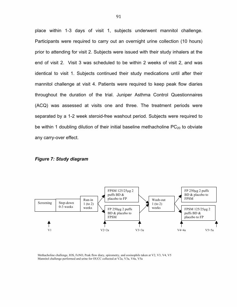
91
place within 1-3 days of visit 1, subjects underwent mannitol challenge.
Participants were required to carry out an overnight urine collection (10 hours)
prior to attending for visit 2. Subjects were issued with their study inhalers at the
end of visit 2. Visit 3 was scheduled to be within 2 weeks of visit 2, and was
identical to visit 1. Subjects continued their study medications until after their
mannitol challenge at visit 4. Patients were required to keep peak flow diaries
throughout the duration of the trial. Juniper Asthma Control Questionnaires
(ACQ) was assessed at visits one and three. The treatment periods were
separated by a 1-2 week steroid-free washout period. Subjects were required to
be within 1 doubling dilution of their initial baseline methacholine PC20 to obviate
any carry-over effect.
Figure 7: Study diagram
Screening Step-down 0-3 weeks
Run-in 1 (to 2) weeks
FPSM 125/25µg 2 puffs BD & placebo to FP
FP 250µg 2 puffs BD & placebo to FPSM
Wash-out 1 (to 2) weeks FPSM 125/25µg 2
puffs BD & placebo to FP
FP 250µg 2 puffs BD & placebo to FPSM
V1 V2+2a V3+3a V4+4a V5+5a
Methacholine challenge, IOS, FeNO, Peak flow diary, spirometry, and eosinophils taken at V2, V3, V4, V5 Mannitol challenge performed and urine for OUCC collected at V2a, V3a, V4a, V5a
92
Measurements
FENO was recorded prior to any pulmonary function measurements using a Niox
Nitric oxide analyser (Aerocrine 2 AB, Sweden) using ATS criteria [194].
Bronchial provocation testing using methacholine was performed using the five
breath dosimeter technique in accordance with ATS recommendations to
determine the PC20 threshold at baseline, after each treatment period, and after a
corticosteroid free washout using log-linear interpolation [183]. Mannitol
challenge was performed by administering spray-dry mannitol powder in gelatine
capsule form (AridolTM Pharmaxis Ltd, Sydney, Australia), inhaled from a dry
powder device (Osmohaler, Pharmaxis Ltd. French’s Forest, NSW, Australia) as
previously described by Anderson and colleagues [136]. A Jaeger Masterscreen
(Erich Jaeger, Hoechberg, Germany) was used to measure impulse oscillometry
according to the guidelines [188]. Subjects supported their cheeks to reduce
shunting, whilst impulses were applied for 30secs during tidal breathing. All
manoeuvres were performed in triplicate and means taken. Adrenal suppression
from systemic absorption of the inhaled corticosteroid treatment was assessed
by measuring overnight 10h urinary free cortisol and creatinine ratios as
described previously in literature [220].
93
Laboratory assays
All assays were performed in duplicate in a blinded fashion. The serum ECP was
measured using an enzyme linked immunoassay technique (UniCAP; Sweden
Diagnostics UK Ltd, Milton Keyes, UK) with an intra-assay co-efficient of variation
of 3.3%. Blood Eosinophil count was analysed using the Sysmex XE 2100
Hematology auto analyzer. The urinary cortisol was measured using a
commercial radioimmunoassay kit (DiaSorin Ltd, Wokingham, Berkshire, UK),
which has no cross reactivity with fluticasone. The intra assay coefficient of
variation was 4% and the inter assay coefficient of variation was 8%. Urinary
creatinine was measured on a Cobas-Bio auto analyser (Roche Products,
Welwyn Garden City, UK). The intra assay and inter assay co-efficient of
variation was 4.6% and 3% respectively.
Statistical analysis
SPSS version 16 (SPSS Inc, Chicago, IL, USA) was used to carry out the
statistical analysis. A sample size of 13 completed patients was estimated to give
>80% power to detect a one doubling dilution (dd) difference (minimal important
difference) in methacholine PC20. The sample size estimations were supported
by a previous studies from our department where the within subject standard
deviation was 0.95dd [221]. Data were analysed within each group for patients
who completed the crossover study per protocol. Any non-Gaussian data were
log transformed prior to analysis. Gaussian data were assessed using paired and
unpaired t-tests. Post run-in and washout were compared with a paired t-test to
94
demonstrate no carry over effect. The primary outcome measure was change in
methacholine PC20 from respective baseline (i.e. post run-in or washout periods)
for each treatment. All other outcomes were considered secondary. Response-
dose-ratios and ACQ were subjected to non-parametric analysis. Median
differences were calculated within group using the Wilcoxon Rank-Sum test for
paired data, and between group using the Mann-Whitney test.
Results
38 participants were randomised, of which 31 completed per protocol (16 non-
smokers, 15 smokers). 5 withdrew because of personal reasons, 2 had an FEV1
<60% following wash-out. The mean (SEM) cotinine levels for non-smokers was
4.37 (1.26) ng/ml, while all smokers had cotinine levels above 50 ng/ml. Baseline
data for smokers and non-smokers are shown in table 10. 13/15 smokers had no
baseline obstruction (i.e.FEV1/FVC ratio of >70%), the 3/15 who did not
demonstrated >15% variability in PEFR. There was no significant difference in
pre-treatment baseline vs. post washout values for the primary outcome.
Similarly, there were no differences in pre-treatment baselines for all secondary
outcomes, except FENO.
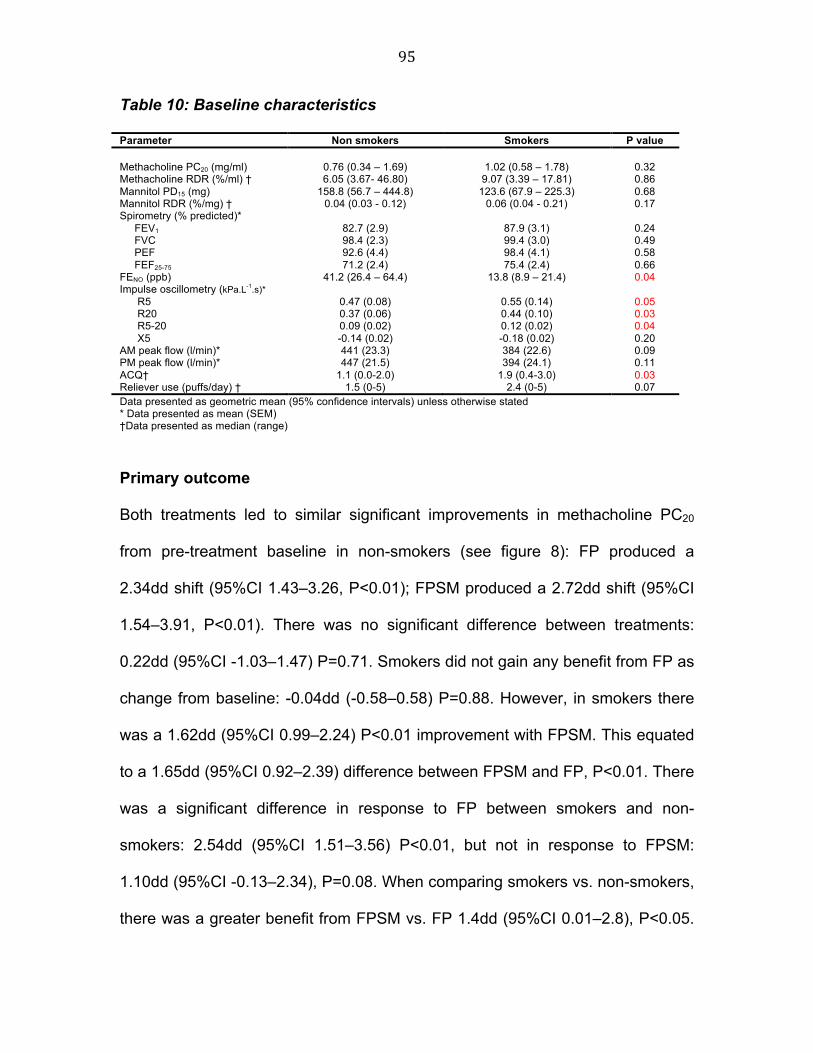
95
Table 10: Baseline characteristics
Parameter Non smokers Smokers P value Methacholine PC20 (mg/ml)
0.76 (0.34 – 1.69)
1.02 (0.58 – 1.78)
0.32
Methacholine RDR (%/ml) † 6.05 (3.67- 46.80) 9.07 (3.39 – 17.81) 0.86 Mannitol PD15 (mg) 158.8 (56.7 – 444.8) 123.6 (67.9 – 225.3) 0.68 Mannitol RDR (%/mg) † 0.04 (0.03 - 0.12) 0.06 (0.04 - 0.21) 0.17 Spirometry (% predicted)* FEV1 82.7 (2.9) 87.9 (3.1) 0.24 FVC 98.4 (2.3) 99.4 (3.0) 0.49 PEF 92.6 (4.4) 98.4 (4.1) 0.58 FEF25-75 71.2 (2.4) 75.4 (2.4) 0.66 FENO (ppb) 41.2 (26.4 – 64.4) 13.8 (8.9 – 21.4) 0.04 Impulse oscillometry (kPa.L-1.s)* R5 0.47 (0.08) 0.55 (0.14) 0.05 R20 0.37 (0.06) 0.44 (0.10) 0.03 R5-20 0.09 (0.02) 0.12 (0.02) 0.04 X5 -0.14 (0.02) -0.18 (0.02) 0.20 AM peak flow (l/min)* 441 (23.3) 384 (22.6) 0.09 PM peak flow (l/min)* 447 (21.5) 394 (24.1) 0.11 ACQ† 1.1 (0.0-2.0) 1.9 (0.4-3.0) 0.03 Reliever use (puffs/day) † 1.5 (0-5) 2.4 (0-5) 0.07 Data presented as geometric mean (95% confidence intervals) unless otherwise stated * Data presented as mean (SEM) †Data presented as median (range)
Primary outcome
Both treatments led to similar significant improvements in methacholine PC20
from pre-treatment baseline in non-smokers (see figure 8): FP produced a
2.34dd shift (95%CI 1.43–3.26, P<0.01); FPSM produced a 2.72dd shift (95%CI
1.54–3.91, P<0.01). There was no significant difference between treatments:
0.22dd (95%CI -1.03–1.47) P=0.71. Smokers did not gain any benefit from FP as
change from baseline: -0.04dd (-0.58–0.58) P=0.88. However, in smokers there
was a 1.62dd (95%CI 0.99–2.24) P<0.01 improvement with FPSM. This equated
to a 1.65dd (95%CI 0.92–2.39) difference between FPSM and FP, P<0.01. There
was a significant difference in response to FP between smokers and non-
smokers: 2.54dd (95%CI 1.51–3.56) P<0.01, but not in response to FPSM:
1.10dd (95%CI -0.13–2.34), P=0.08. When comparing smokers vs. non-smokers,
there was a greater benefit from FPSM vs. FP 1.4dd (95%CI 0.01–2.8), P<0.05.
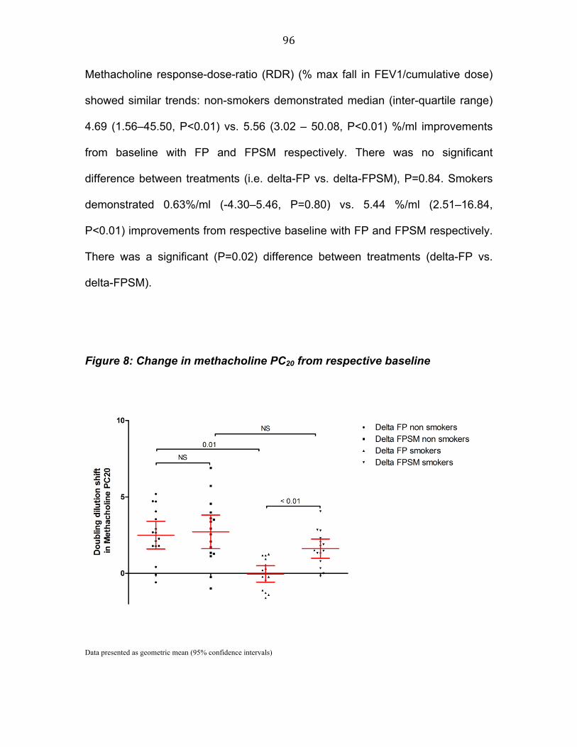
96
Methacholine response-dose-ratio (RDR) (% max fall in FEV1/cumulative dose)
showed similar trends: non-smokers demonstrated median (inter-quartile range)
4.69 (1.56–45.50, P<0.01) vs. 5.56 (3.02 – 50.08, P<0.01) %/ml improvements
from baseline with FP and FPSM respectively. There was no significant
difference between treatments (i.e. delta-FP vs. delta-FPSM), P=0.84. Smokers
demonstrated 0.63%/ml (-4.30–5.46, P=0.80) vs. 5.44 %/ml (2.51–16.84,
P<0.01) improvements from respective baseline with FP and FPSM respectively.
There was a significant (P=0.02) difference between treatments (delta-FP vs.
delta-FPSM).
Figure 8: Change in methacholine PC20 from respective baseline
Data presented as geometric mean (95% confidence intervals)
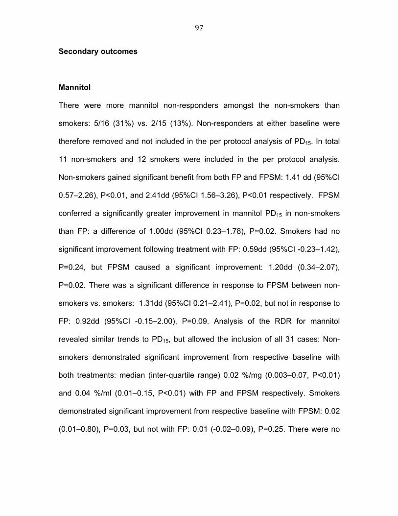
97
Secondary outcomes
Mannitol
There were more mannitol non-responders amongst the non-smokers than
smokers: 5/16 (31%) vs. 2/15 (13%). Non-responders at either baseline were
therefore removed and not included in the per protocol analysis of PD15. In total
11 non-smokers and 12 smokers were included in the per protocol analysis.
Non-smokers gained significant benefit from both FP and FPSM: 1.41 dd (95%CI
0.57–2.26), P<0.01, and 2.41dd (95%CI 1.56–3.26), P<0.01 respectively. FPSM
conferred a significantly greater improvement in mannitol PD15 in non-smokers
than FP: a difference of 1.00dd (95%CI 0.23–1.78), P=0.02. Smokers had no
significant improvement following treatment with FP: 0.59dd (95%CI -0.23–1.42),
P=0.24, but FPSM caused a significant improvement: 1.20dd (0.34–2.07),
P=0.02. There was a significant difference in response to FPSM between non-
smokers vs. smokers: 1.31dd (95%CI 0.21–2.41), P=0.02, but not in response to
FP: 0.92dd (95%CI -0.15–2.00), P=0.09. Analysis of the RDR for mannitol
revealed similar trends to PD15, but allowed the inclusion of all 31 cases: Non-
smokers demonstrated significant improvement from respective baseline with
both treatments: median (inter-quartile range) 0.02 %/mg (0.003–0.07, P<0.01)
and 0.04 %/ml (0.01–0.15, P<0.01) with FP and FPSM respectively. Smokers
demonstrated significant improvement from respective baseline with FPSM: 0.02
(0.01–0.80), P=0.03, but not with FP: 0.01 (-0.02–0.09), P=0.25. There were no
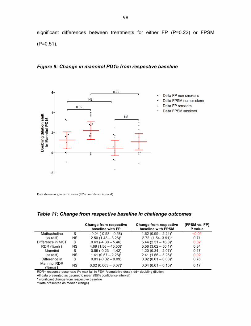
98
significant differences between treatments for either FP (P=0.22) or FPSM
(P=0.51).
Figure 9: Change in mannitol PD15 from respective baseline
Data shown as geometric mean (95% confidence interval)
Table 11: Change from respective baseline in challenge outcomes
Change from respective baseline with FP
Change from respective baseline with FPSM
(FPSM vs. FP) P value
S -0.04 (-0.58 – 0.58) 1.62 (0.99 – 2.24)* <0.01 Methacholine (dd shift) NS 2.50 (1.43 – 3.26)* 2.72 (1.54- 3.91)* 0.71
S 0.63 (-4.30 – 5.46) 5.44 (2.51 – 16.8)* 0.02 Difference in MCT RDR (%/ml) † NS 4.69 (1.56 – 45.50)* 5.56 (3.02 – 50.1)* 0.84
S 0.59 (-0.23 – 1.42) 1.20 (0.34 – 2.07)* 0.17 Mannitol (dd shift) NS 1.41 (0.57 – 2.26)* 2.41 (1.56 – 3.26)* 0.02
S 0.01 (-0.02 – 0.09) 0.02 (0.01 – 0.08)* 0.76 Difference in Mannitol RDR
(%/mg) † NS 0.02 (0.003 – 0.07)* 0.04 (0.01 – 0.15)* 0.17
RDR= response-dose-ratio (% max fall in FEV1/cumulative dose), dd= doubling dilution All data presented as geometric mean (95% confidence interval) * significant change from respective baseline †Data presented as median (range)
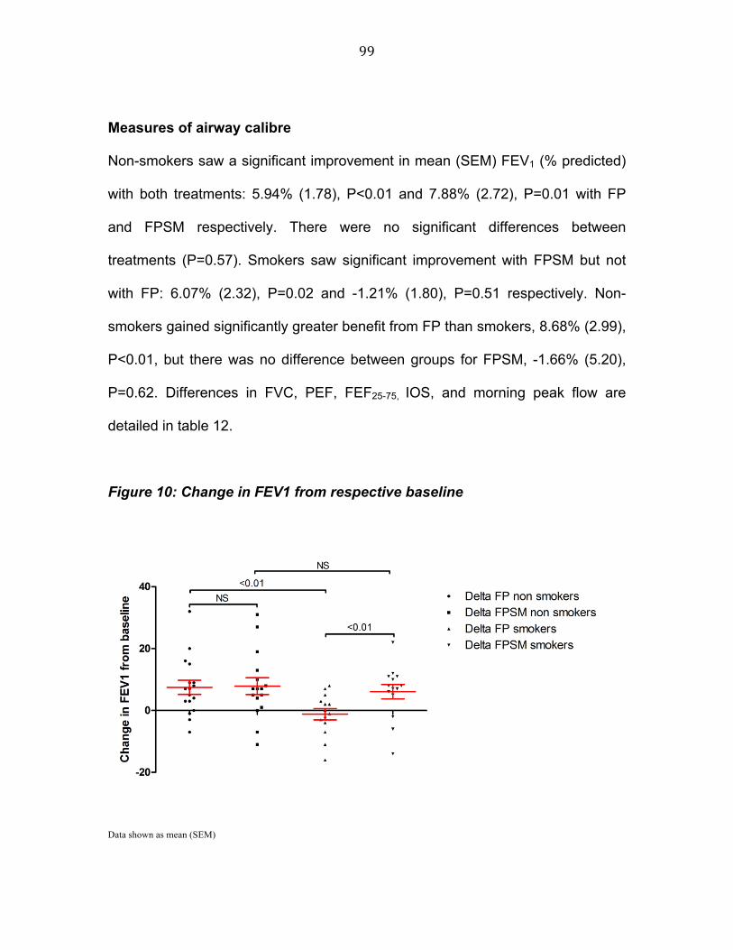
99
Measures of airway calibre
Non-smokers saw a significant improvement in mean (SEM) FEV1 (% predicted)
with both treatments: 5.94% (1.78), P<0.01 and 7.88% (2.72), P=0.01 with FP
and FPSM respectively. There were no significant differences between
treatments (P=0.57). Smokers saw significant improvement with FPSM but not
with FP: 6.07% (2.32), P=0.02 and -1.21% (1.80), P=0.51 respectively. Non-
smokers gained significantly greater benefit from FP than smokers, 8.68% (2.99),
P<0.01, but there was no difference between groups for FPSM, -1.66% (5.20),
P=0.62. Differences in FVC, PEF, FEF25-75, IOS, and morning peak flow are
detailed in table 12.
Figure 10: Change in FEV1 from respective baseline
Data shown as mean (SEM)
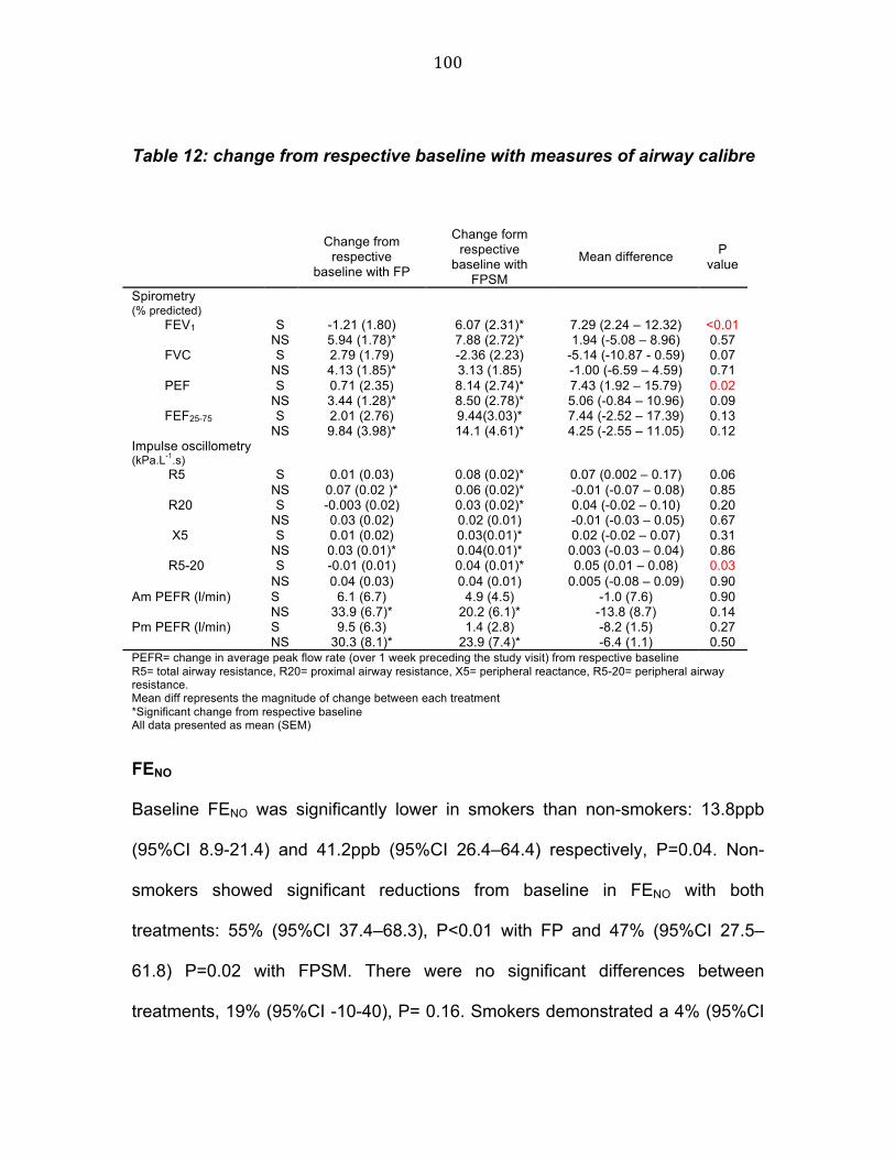
100
Table 12: change from respective baseline with measures of airway calibre
Change from
respective baseline with FP
Change form respective
baseline with FPSM
Mean difference P value
Spirometry (% predicted)
S -1.21 (1.80) 6.07 (2.31)* 7.29 (2.24 – 12.32) <0.01 FEV1 NS 5.94 (1.78)* 7.88 (2.72)* 1.94 (-5.08 – 8.96) 0.57 S 2.79 (1.79) -2.36 (2.23) -5.14 (-10.87 - 0.59) 0.07 FVC
NS 4.13 (1.85)* 3.13 (1.85) -1.00 (-6.59 – 4.59) 0.71 S 0.71 (2.35) 8.14 (2.74)* 7.43 (1.92 – 15.79) 0.02 PEF
NS 3.44 (1.28)* 8.50 (2.78)* 5.06 (-0.84 – 10.96) 0.09 S 2.01 (2.76) 9.44(3.03)* 7.44 (-2.52 – 17.39) 0.13 FEF25-75
NS 9.84 (3.98)* 14.1 (4.61)* 4.25 (-2.55 – 11.05) 0.12 Impulse oscillometry (kPa.L-1.s)
S 0.01 (0.03) 0.08 (0.02)* 0.07 (0.002 – 0.17) 0.06 R5 NS 0.07 (0.02 )* 0.06 (0.02)* -0.01 (-0.07 – 0.08) 0.85 S -0.003 (0.02) 0.03 (0.02)* 0.04 (-0.02 – 0.10) 0.20 R20
NS 0.03 (0.02) 0.02 (0.01) -0.01 (-0.03 – 0.05) 0.67 S 0.01 (0.02) 0.03(0.01)* 0.02 (-0.02 – 0.07) 0.31 X5
NS 0.03 (0.01)* 0.04(0.01)* 0.003 (-0.03 – 0.04) 0.86 S -0.01 (0.01) 0.04 (0.01)* 0.05 (0.01 – 0.08) 0.03 R5-20
NS 0.04 (0.03) 0.04 (0.01) 0.005 (-0.08 – 0.09) 0.90 Am PEFR (l/min) S 6.1 (6.7) 4.9 (4.5) -1.0 (7.6) 0.90 NS 33.9 (6.7)* 20.2 (6.1)* -13.8 (8.7) 0.14 Pm PEFR (l/min) S 9.5 (6.3) 1.4 (2.8) -8.2 (1.5) 0.27 NS 30.3 (8.1)* 23.9 (7.4)* -6.4 (1.1) 0.50 PEFR= change in average peak flow rate (over 1 week preceding the study visit) from respective baseline R5= total airway resistance, R20= proximal airway resistance, X5= peripheral reactance, R5-20= peripheral airway resistance. Mean diff represents the magnitude of change between each treatment *Significant change from respective baseline All data presented as mean (SEM)
FENO
Baseline FENO was significantly lower in smokers than non-smokers: 13.8ppb
(95%CI 8.9-21.4) and 41.2ppb (95%CI 26.4–64.4) respectively, P=0.04. Non-
smokers showed significant reductions from baseline in FENO with both
treatments: 55% (95%CI 37.4–68.3), P<0.01 with FP and 47% (95%CI 27.5–
61.8) P=0.02 with FPSM. There were no significant differences between
treatments, 19% (95%CI -10-40), P= 0.16. Smokers demonstrated a 4% (95%CI
101
-33.3–37.1) reduction in FENO post FP (P=0.49), and a 47% (95%CI 34.1–57.5)
reduction post FPSM (P<0.01). However, the pre-FPSM baseline in smokers was
significantly higher than the pre-FP baseline: 13.7ppb (95%CI 8.9–21.4) P= 0.04.
There was a 54% (25-71) difference between the response to FP in non-smokers
vs. smokers, P<0.01, but no significant difference for response to FPSM, 0.3%
(95%CI -31-44), P=0.99.
ACQ
Non-smokers had significantly lower scores at baseline than smokers: median
(range) 1.1 (0.0-2.0) and 1.9 (0.4-3.0) respectively. Non-smokers showed
significant improvements in ACQ with both FP (P=0.01) and FPSM (P=0.02).
There was no significant difference between treatments (P=0.57). No
improvements were seen in smokers with either treatment: P=0.31 and P=0.32.
Laboratory measures
Neither treatment caused significant suppression of blood eosinophil counts from
baseline: -0.01 (95%CI -0.05–0.04), P=0.13 vs. 0.03 (95%CI -0.03–0.09), P=0.53
in non-smokers, and 0.01 (-0.07–0.09), P=0.61 vs. 0.04 (-0.01-0.09), P=0.71 in
smokers, with FP and FPSM respectively. In keeping with the eosinophil data,
neither treatment led to a significant reduction in ECP from respective baselines:
-0.04 (95%CI -0.18–0.11), P=0.60 vs 0.05 (95%CI -0.07–0.17), P=0.41 in non-
smokers, and -0.04 (95%CI -0.16–0.08), P=0.51 vs 0.04 (95%CI -0.13–0.21),
P=0.63 in smokers, with FP and FPSM respectively. In smokers FP produced
102
significant adrenal suppression of OUCC compared to baseline: -2.82 (1.24)
nmol/mmol (P=0.03), but not with FPSM, -0.90 (0.91) nmol/mmol, P=0.34. In
non-smokers neither treatment produced a significant fall in OUCC: -1.24 (1.01)
nmol/mmol (P=0.24) and 0.12 (0.74) nmol/mmol (P=0.88) with FP and FPSM
respectively.
Discussion
Studies in non-smokers have shown LABA’s to have a relative ‘steroid sparing
effect’, allowing comparative improvements in lung function and reductions in
exacerbations to doubling the dose of inhaled steroid [221-223]. This is in
keeping with our non-smoking cohort, which demonstrated significant
improvements in AHR, FENO, and spirometric indices that were comparable
between treatments. Smokers on the other hand, showed significantly greater
improvements from the addition of SM to FP (500ug/day) compared to doubling
the dose of FP alone (1000ug/day), in terms of AHR (methacholine PC20 and
RDR), spirometry (FEV1) and IOS (R5-R20). Furthermore, smokers appeared to
gain proportionally greater benefit from the addition of SM than non-smokers on
methacholine AHR. These results support the observation that smokers would
need 18 years less treatment, by taking FP/SM vs. FP alone, to prevent an
exacerbation than non-smokers [164].
Current asthma guidelines emphasize the importance of establishing asthmatics
on ICS prior to addition of long-acting b2-agonist [8]. The OPTIMA trial clearly
demonstrated that the addition of low dose ICS in steroid naïve individuals led to
103
a significant reduction in severe exacerbations and improvements in asthma
control. Whereas the addition of a LABA led to improvements in lung function,
but did not confer any additional clinical benefit [192]. In the present study we
found no significant benefit, in any primary or secondary outcome, from high
dose inhaled FP (1000ug/day) in smokers. Whilst smoking asthmatics are known
to develop relative steroid resistance [157], it is currently thought that this can be
partially overcome through increased doses [159]. This assumption is based on a
study by Tomlinson et al, who demonstrated that high dose ICS decreased peak
flow variability to a similar degree in both smokers and non-smokers [159]. Peak
flow, like spirometry, reflects airway caliber and as such is relatively distant from
the underlying asthmatic inflammatory process [21]. In this respect we saw no
significant improvement in either peak flow dairy cards or ACQ in smokers. It
could be argued that two weeks duration of treatment with FP may not have been
sufficient to observe the changes in PEF seen by Tomlinson et al, however, it
has previously been shown by Sovijarvi et al that 2 weeks is sufficient in
achieving near maximal effects on AHR in response to FP 250ug bid via spacer
[214].
Our result has important implications as it suggests that higher doses of ICS fail
to control inflammation in smokers, exposing them to significant risk of side
effects. Addition of LABA on the other hand, allows improvements in AHR similar
to those seen in non-smokers. We showed a significant systemic effect on
adrenal suppression in smokers with FP 500µg bid but not with FPSM 250µg bid.
This further emphasizes the importance of adding salmeterol rather than
104
increasing the dose of FP, in terms of optimizing the risk-benefit. Whilst smoking
cessation should always be advocated as the best and most effective treatment
for persistent asthma [217], we would advocate that smoking asthmatics who do
not wish to consider cessation be started on combination therapy as first line
rather than being treated with inhaled steroid alone.
Some mechanistic insights can also be gleaned by examining the relative effects
on mannitol and methacholine challenges. Mannitol is an indirect osmotic
challenge, which is thought to correlate more closely with airway inflammation
than methacholine, a direct bronchial smooth challenge [90]. A study utilising
AMP, another indirect challenge, found FPSM 250µg bid to be inferior to 500µg
bid in non smokers [221]. Since mannitol has been shown to correlate closely
with AMP challenge [201], the results obtained in the present study were
somewhat surprising in that improvements in mannitol PD15 following FPSM were
greater than FP in non-smokers. Since SM exhibits no clinically meaningful in
vivo anti-inflammatory activity [224], the superiority of FP/SM vs. FP alone on
mannitol in non-smokers is likely due to an alternative mechanism: LABAs are
known to have a stabilizing effect on airway smooth muscle [225]; to increase
mast cell stabilization, thereby inhibiting the release of inflammatory mediators
such as histamine or cysteinyl leukotrienes [226] and to preserve the integrity of
the respiratory eithelium [227]. This in turn suggests than mannitol may be less
specific for inflammation than originally thought. However, we did not see any
difference in effect on methacholine AHR in non-smokers between FPSM vs. FP,
105
that is perhaps, counter intuitive since methacholine acts directly on airway
smooth muscle.
In smokers we saw a significant difference between FPSM vs. FP on
methacholine AHR, with a similar non-significant trend on mannitol. The greater
improvements with FP/SM in smokers were also seen on airway caliber on
outcomes of spirometry such as FEV1 and on imuplsue oscillometery on R5-
R20. The effect of salmeterol is due to a direct action on the airway smooth
muscle leading to both bronchodilation and a protective effect due to functional
antagonism against bronchoconstriction. This functional antagonism can be
considered to be a surrogate for stabilization of airway smooth muscle and
determines the degree of protection when exposed to a challenge stimulus. It
therefore appears that in the face of relative steroid resistance the smooth
muscle stabilization conferred by the LABA becomes relatively more important in
smokers than in non-smokers. Studies looking at the effect of smoking cessation
on AHR to AMP and methacholine have shown significant improvements in AMP
but not methacholine 6 months after smoking cessation in quitters [226],
suggesting that smokers may have a greater degree of smooth muscle
dysfunction than non-smokers.
CT studies have shown smokers to have a reduced airway lumen area compared
with non-smokers [228]. Similarly our results have shown that smokers have
higher peripheral resistance (R5-20) at baseline compared with non-smokers,
suggesting a greater degree of peripheral smooth muscle dysfunction, and
106
potentially increased peripheral inflammation. In combination products the
bronchodilator component might conceivably result in improved peripheral lung
deposition of the ICS, thereby producing enhanced anti-inflammatory activity in
smaller airways. It has been suggested that oscillometry is a sensitive measure
of small airway dysfunction and changes in R5-20, and X5 may reflect small
airway physiology [228]. Thus, the changes in IOS demonstrated in this study
with smokers may indicate beneficial small airway effects with FP/SM. The
mechanism by which smoking causes FENO reduction is not fully understood, but
may include reduction in NO synthesis due to feedback inhibition induced by high
concentrations of NO contained in cigarette smoke [229]. Pre-treatment
baselines for FP/SM vs. FP were significantly different, accounting for increased
suppression with FP/SM in smokers.
In conclusion, combination therapy with FP/SM conferred significant
improvements in AHR and airway caliber in smoking asthmatics as compared to
double the dose of FP alone. It is likely that in the face of the relative steroid
resistance seen in smokers, the bronchodilation and smooth muscle/ mast cell
stabilization conferred by the LABA become more important in terms of achieving
superior clinical control. Future guidelines should take into account the
differences in treatment response between smokers and non-smokers.
107
Critique
Therapeutic studies, which form the basis for current asthma guidelines, often
exclude current smokers due to concerns about recruiting participants with
chronic obstructive pulmonary disease (COPD). 87% of participants in the
present study had an FEV1/FVC ratio of ≥70% suggesting that they had no
baseline obstruction, making a diagnosis of COPD unlikely. However, due to the
difficulties that we encountered in recruiting smoking asthmatics, we also had to
include subjects with a ratio of <70%. These subjects were required to
demonstrate either >15% bronchodilator reversibility or >15% diurnal variation in
peak flow, alongside a clinical history suggestive of asthma (i.e. diurnal variation,
nocturnal, or exercise related symptoms). However, a recent study by Calverley
et al has clearly demonstrated that bronchodilator response is a poor diagnostic
test for COPD, as up to 52% of patients changed responder status between visits
[230]. Fortunately, all 3 patients with a ratio <70% demonstrated >15% diurnal
variation in peak flow during the run-in period. However, we acknowledge that
despite the above inclusion criteria, we may have included subjects with very
mild COPD. A further criticism that could be leveled at this study is that we did
not include a measure of compliance.
108
Part b: Elite swimmers
Study aims:
1. Determine the prevalence of EIB and rhinoconjunctivitis in adolescent elite
swimmers.
2. To determine what happens to airway inflammation following exposure to
chlorine and exercise.
3. To compare standardised field-based exercise challenge to mannitol
challenge,
109
Effects of chlorine and exercise on the unified airway in
adolescent elite Scottish swimmers
Introduction
Elite swimmers have higher rates of rhinoconjunctivitis and exercise induced
bronchospasm (EIB) compared with any other groups of athletes [168, 231].
Proposed mechanisms include a combination of chronic exposure to toxic
chlorine metabolites, and high ventilatory rates leading to osmotic degranulation
of mast cells and subsequent bronchoconstriction [232]. In adult elite swimmers,
significantly higher levels of airway inflammatory cells have been demonstrated
compared with healthy controls. This reverts back to normal following long-term
cessation of the sport [172]. There is a paucity of data on the prevalence of
rhinoconjunctivitis and exercise induced bronchoconstriction in adolescent elite
swimmers. This is of particular importance as rhinoconjunctivitis has been shown
to be an independent risk factor for developing asthma [233]. Indeed rhinitis and
asthma are considered to be part of the same disease continuum, the ‘unified
airway’ [234]. We therefore conducted a pilot study to assess the combined
effects of chlorine and exercise on the unified airway of adolescent elite
swimmers, including estimating the prevalence of rhinoconjunctivitis and EIB.
110
Methods
Participants and study design
The Scottish Midland District swimming squad (36 swimmers) were assessed
during a 2 hour training session. All swimmers underwent exhaled tidal (FENO)
and nasal (NNO) NO measurement peak nasal inspiratory flow rate (PNIF), and
FEV1 before and after swimming. A sport-specific exercise test was carried out
during an intensive aerobic set. All swimmers completed a health questionnaire.
Swimmers were asked to withhold anti-histamines, leukotriene receptor
antagonists (LRTAs), nasal steroid sprays, and long acting β2- agonists (LABAs)
for 1 week before the training session. Inhaled corticosteroid inhalers could be
taken as normal. Participants were asked to refrain from taking short acting β2-
agonists (SABAs) 12 hours before the session. This study was approved by the
local ethics committee (REC ref: 09/S1402/6) and all participants gave written
informed consent.
Measurements
NO measurement
All participants underwent measurement of FENO using a portable MINO (NIOX
MINO® Airway Inflammation Monitor; Aerocrine AB, Solna, Sweden). A single
reading was obtained in accordance with manufacturer’s guidelines. Nasal
exhaled NO was measured using an adapted MINO device, as previously
described [235].
111
Sport-specific field-based exercise challenge
The swimmers performed a sport-specific field-based exercise challenge
following a low intensity warm up. Swimmers were required to maintain >80%
maximum heart rate (220-age) for at least 8 minutes in order to optimally provoke
EIB. Heart rate was measured objectively using pulse oximetry (M-pulse™,
Merlin Medical, UK). FEV1 (forced expiratory volume in 1 second) was measured
using a Piko-6® portable spirometer (Ferraris Respiratory). Heart rate and FEV1
were measured at baseline, immediately following challenge and at 5 and 10
minutes during recovery. A positive challenge was defined as a fall in FEV1 of
≥10%.
Peak nasal inspiratory flow
Peak nasal inspiratory flow (PNIF) was measured using the In-Check® PNIF
meter (Clement Clarke International Ltd, Harlow, UK). After horizontal positioning
and restoration to zero, participants forcefully inhaled through their nose from
residual volume to total lung capacity. The best of three measurements was
taken.
Health questionnaire
A modified version of a questionnaire previously used in studies on elite athletes
was used to assess the presence of exercise-induced symptoms [176].
Symptoms of chlorine-induced rhinitis were assessed using a visual analogue
scale. Atopy was defined as a history of intermittent rhinoconjunctivis (in
112
accordance with current ARIA definitions) and/or a positive skin prick test or/ IgE
specific RAST test within the last 2 years.
Statistical analysis
SPSS version 15 (SPSS Inc, Chicago, Illinois) was used to perform the statistical
analysis. Non-Gaussian data were log transformed prior to analysis. Normalized
data were assessed using paired t-tests. A P value of less than 0.05 (two tailed)
was considered significant. As this was a pilot observational study, no formal
power calculation was used.
Results
Combined and free chlorine levels on the day were 1.66 and 0.3 mg/l
respectively. Baseline characteristics are described in Table 13. Complete
baseline data was available on 31/36 swimmers. Eight swimmers (22%) had
known asthma. 18 (50%) had rhinitis according to the 2008 ARIA guidelines (11
= intermittent rhinitis, 7 = persistent rhinitis). There were no significant differences
in FENO or NNO pre vs post exposure: mean (95% CI) for FENO 17.9 (13.5 to 23.5)
ppb vs 17.0 (12.8 to 22.5) ppb (P = 0.12); NNO 47.5 (36.3 to 62.1) ppb vs 45.4
(34.0 to 60.6) ppb respectively (P = 0.71). Mean PNIF increased from 124.4
L.min-1 (107.8 to 143.5) vs 136 L.min-1 (121.3 to 152.5): (P = 0.04). Baseline
FENO readings in asthmatics and non-asthmatics were 35.7 (8.57) and 18.5
(3.01) respectively.
113
13/36 (36%) of swimmers had a positive exercise challenge. 10/13 (77%) of
these were not previously known to asthmatic. 3/8 (38%) of asthmatic swimmers
had a positive challenge. During the challenge swimmers achieved a mean
(SEM) heart rate of 81.3% (1.5) maximum predicted.
36% (13) had a positive exercise challenge. 46% (6) of participants with a
positive challenge were symptomatic, 54% (7) were asymptomatic. 42% (15)
swimmers complained of worsening nasal symptoms post swimming, but only
13% (2) had a demonstrable fall in PNIF (mean fall 33 l/min), 87% (13) had no
change. 9/15 (60%) had a pre-existing diagnosis of either intermittent or
persistent rhinitis.
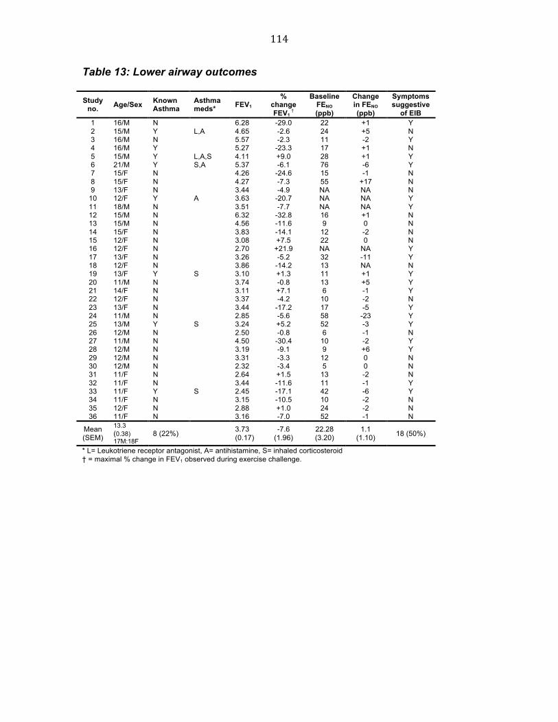
114
Table 13: Lower airway outcomes
Study no. Age/Sex Known
Asthma Asthma meds*
FEV1
% change FEV1
†
Baseline FENO (ppb)
Change in FENO (ppb)
Symptoms suggestive
of EIB 1 16/M N 6.28 -29.0 22 +1 Y 2 15/M Y L,A 4.65 -2.6 24 +5 N 3 16/M N 5.57 -2.3 11 -2 Y 4 16/M Y 5.27 -23.3 17 +1 N 5 15/M Y L,A,S 4.11 +9.0 28 +1 Y 6 21/M Y S,A 5.37 -6.1 76 -6 Y 7 15/F N 4.26 -24.6 15 -1 N 8 15/F N 4.27 -7.3 55 +17 N 9 13/F N 3.44 -4.9 NA NA N
10 12/F Y A 3.63 -20.7 NA NA Y 11 18/M N 3.51 -7.7 NA NA Y 12 15/M N 6.32 -32.8 16 +1 N 13 15/M N 4.56 -11.6 9 0 N 14 15/F N 3.83 -14.1 12 -2 N 15 12/F N 3.08 +7.5 22 0 N 16 12/F N 2.70 +21.9 NA NA Y 17 13/F N 3.26 -5.2 32 -11 Y 18 12/F N 3.86 -14.2 13 NA N 19 13/F Y S 3.10 +1.3 11 +1 Y 20 11/M N 3.74 -0.8 13 +5 Y 21 14/F N 3.11 +7.1 6 -1 Y 22 12/F N 3.37 -4.2 10 -2 N 23 13/F N 3.44 -17.2 17 -5 Y 24 11/M N 2.85 -5.6 58 -23 Y 25 13/M Y S 3.24 +5.2 52 -3 Y 26 12/M N 2.50 -0.8 6 -1 N 27 11/M N 4.50 -30.4 10 -2 Y 28 12/M N 3.19 -9.1 9 +6 Y 29 12/M N 3.31 -3.3 12 0 N 30 12/M N 2.32 -3.4 5 0 N 31 11/F N 2.64 +1.5 13 -2 N 32 11/F N 3.44 -11.6 11 -1 Y 33 11/F Y S 2.45 -17.1 42 -6 Y 34 11/F N 3.15 -10.5 10 -2 N 35 12/F N 2.88 +1.0 24 -2 N 36 11/F N 3.16 -7.0 52 -1 N
Mean (SEM)
13.3 (0.38) 17M:18F
8 (22%) 3.73 (0.17)
-7.6 (1.96)
22.28 (3.20)
1.1 (1.10) 18 (50%)
* L= Leukotriene receptor antagonist, A= antihistamine, S= inhaled corticosteroid † = maximal % change in FEV1 observed during exercise challenge.
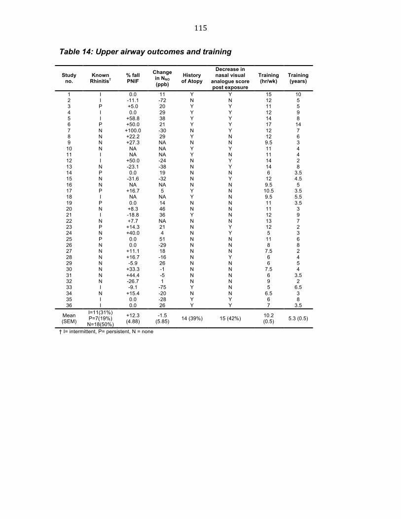
115
Table 14: Upper airway outcomes and training
Study no.
Known Rhinitis†
% fall PNIF
Change in NNO (ppb)
History of Atopy
Decrease in nasal visual
analogue score post exposure
Training (hr/wk)
Training (years)
1 I 0.0 11 Y Y 15 10 2 I -11.1 -72 N N 12 5 3 P +5.0 20 Y Y 11 5 4 I 0.0 29 Y Y 12 9 5 I +58.8 38 Y Y 14 8 6 P +50.0 21 Y Y 17 14 7 N +100.0 -30 N Y 12 7 8 N +22.2 29 Y N 12 6 9 N +27.3 NA N N 9.5 3
10 N NA NA Y Y 11 4 11 I NA NA Y N 11 4 12 I +50.0 -24 N Y 14 2 13 N -23.1 -38 N Y 14 8 14 P 0.0 19 N N 6 3.5 15 N -31.6 -32 N Y 12 4.5 16 N NA NA N N 9.5 5 17 P +16.7 5 Y N 10.5 3.5 18 I NA NA Y N 9.5 5.5 19 P 0.0 14 N N 11 3.5 20 N +8.3 46 N N 11 3 21 I -18.8 36 Y N 12 9 22 N +7.7 NA N N 13 7 23 P +14.3 21 N Y 12 2 24 N +40.0 4 N Y 5 3 25 P 0.0 51 N N 11 6 26 N 0.0 -29 N N 8 8 27 N +11.1 18 N N 7.5 2 28 N +16.7 -16 N Y 6 4 29 N -5.9 26 N N 6 5 30 N +33.3 -1 N N 7.5 4 31 N +44.4 -5 N N 6 3.5 32 N -26.7 1 N N 9 2 33 I -9.1 -75 Y N 5 6.5 34 N +15.4 -20 N N 6.5 3 35 I 0.0 -28 Y Y 6 8 36 I 0.0 26 Y Y 7 3.5
Mean (SEM)
I=11(31%) P=7(19%)
N=18(50%)
+12.3 (4.88)
-1.5 (5.85) 14 (39%) 15 (42%) 10.2
(0.5) 5.3 (0.5)
† I= intermittent, P= persistent, N = none
116
Discussion
This study aimed to assess the combined effects of chlorine and exercise on the
unified airway of adolescent elite swimmers. We found no significant change in
either tidal (FENO) or nasal (NNO) NO following a 2-hour training session, which
included a high intensity anaerobic set. Using linear regression analysis, this
change in NO was not influenced by asthma, presence of EIB or history of atopy.
Furthermore, there was no correlation between upper and lower airway NO
(r=0.12, P=0.81). We elected to use FENO as it is non-invasive, reproducible and
easy to perform at the poolside. FENO is an established surrogate in asthma,
which correlates closely with airway inflammation [60]. It is also used as a
surrogate marker of inflammatory diseases in the upper airway [236], enabling us
to examine effects on the unified airway. Additionally, FENO has been shown to
rise in elite runners following a marathon [237]. The absence of any acute rise in
FENO was therefore unexpected. Whilst forced vital manoeuvres are known to
‘washout’ FENO from the airways [194], there are no reports of exercise or
hyperventilation decreasing FENO. Indeed, our results did not show any reduction
post-swimming. It is also conceivable that the short duration of exposure was not
sufficient to induce NO synthase, however, FENO has previously been shown to
be elevated during an osmotic challenge suggesting that there is rapid induction
of this enzyme [238]. We did not assess whether a late phase response was
present in either the upper and lower airway. This is supported by anecdotal
reports from swimmers who frequently complain of nasal congestion and
symptoms, 4-6 hours after training.
117
The significant increase observed in PNIF post-swimming may not represent an
improvement in inflammation, but paradoxically may signify a ‘nasal douche
effect’. Saline irrigation is known to significantly improve nasal congestion
through a combination of improved mucociliary clearance and reduction of
mucosal oedema [239]. 18 (50%) of swimmers had a pre-existing diagnosis of
rhinitis, which is in line previously reported rates [240]. Rhinitis did not
necessarily predict worsening of nasal symptoms post swimming, however,
large-scale epidemiological studies are needed to investigate this issue further.
As part of this study we carried out a standardised field-based exercise
challenge. Traditional field based testing in which athletes perform a challenge
using their primary exercise, is usually limited by an inability to standardise both
the cardiovascular workload and environmental conditions [177]. We therefore
decided to use a standardised protocol based on the ATS (1999) guidelines for
exercise challenge, using an objective measure of heart rate monitoring. In the
current study, swimmers achieved an average heart rate of 81.3% of maximum,
which was maintained for an average of 8 minutes.
We found a high prevalence (36%) of EIB in our cohort of adolescent elite
swimmers. This is a unique finding, as previous figures are biased towards adult
swimmers [172]. Indeed, 77% of our swimmers with a positive exercise challenge
were not previously known to be asthmatic. Moreover, only 46% of swimmers
118
who were exercise-positive reported symptoms suggestive of EIB. The diagnosis
of EIB based on symptoms alone, is known to be highly inaccurate [241]. This
suggests that solely testing symptomatic athletes is grossly insufficient. At elite
level small changes in physiological reserve could potentially translate into
tangible improvements in performance. More importantly, adolescent swimmers
with undiagnosed EIB may not reach their full potential, preventing them from
progressing to compete at adult level. We therefore feel that universal screening
of all elite swimmers for EIB and rhinoconjunctivitis and should be advocated.
We recognize some limitations in our study. It could be argued that the non-
intensive exercise preceding the challenge may have induced a refractory period
in some swimmers, which may have led to an underestimate of the true
prevalence of EIB. However, previous studies have shown that low intensity
warm-ups to not induce refractoriness prior to challenge [242]. 4/5 asthmatic
swimmers with a negative exercise challenge were on inhaled corticosteroids,
which may explain the low incidence of EIB in this group. Ideally, these would
have been stopped; however, this would have interfered excessively with
training. As expected FENO was elevated at baseline in asthmatics, but not in
non-asthmatic swimmers. This is in keeping with previous studies. It is possible
that the swimmers in this cohort were too young to develop major airway
inflammatory changes. A larger study in a cohort of older more experienced
adolescent swimmers would help eliminate this as a confounding factor. Whilst
this study does not allow us to differentiate between the relative contributions of
119
exercise and chlorine on the airway we felt it was important to study swimmers in
a “real life” situation.
Conclusions
In conclusion, we found that a 2 hr training session, including a high intensity
anaerobic set, in a chlorinated indoor pool did not affect surrogate markers of
inflammation in the unified airway. There was a high prevalence of undiagnosed
EIB, highlighting the importance of screening of all elite swimmers for asthma.
120
Critique
In this study we were able to determine that there was a high prevalence of
undiagnosed EIB in adolescent elite swimmers, highlighting the need for an
effective screening test. Standardised field-based exercise testing in swimmers
did appear to be a viable option, as environmental conditions in the pool were
relatively constant. Although, one criticism, which was made during the
presentation of this paper, was that a single low FEV1 immediately post exercise
did not effectively differentiate between EIB and exhaustion. We therefore made
the decision to define EIB as a drop in FEV1 >10% at two separate time points
post exertion in subsequent studies.
The lack of response in FENO was somewhat surprising in view of the increase
that had been reported in marathon runners. We hypothesised that we had
perhaps missed a late phase response in airway inflammation, similar to that
seen following allergen challenge. The decision was therefore made to measure
this in a subsequent study. We also felt that it was important to compare field-
based exercise challenge to the gold standard test (EVH). However, EVH
requires a great deal of machinery, experienced technical staff and is not readily
available at the poolside. We therefore decided to use mannitol challenge in our
follow-up study, as this has been shown to correlate very closely with EVH.
121
Disconnect between standardised field based testing and
mannitol challenge in Scottish elite swimmers
Introduction
Exercise-induced bronchoconstriction is defined as a transient increase in airway
resistance that occurs after vigorous exercise [133]. Proposed aetiological
mechanisms include airway drying and cooling as a consequence of increased
ventilatory rates during strenuous exercise [243, 244]. Exercise is an indirect
airway challenge which has been found to have a high level of specificity, but a
low sensitivity for identifying EIB in cold weather and track athletes [179]. Sport-
specific exercise in the field (FBT) is limited with respect to the standardization of
both the workload and environmental conditions such as allergens, temperature,
and humidity. Eucapnic voluntary hyperpnoea (EVH), a laboratory based
challenge involving hyperventilation with dry air, has therefore been
recommended as the ‘gold standard’ test for the diagnosis of EIB by the
International Olympic Committee [175]. Mannitol, an osmotic challenge, has been
found to correlate strongly with EVH [146].
Elite swimmers have higher rates of rhinoconjunctivitis and exercise induced
bronchoconstriction (EIB) compared with any other groups of athletes [168, 231].
This may seem counterintuitive as they exercise in a warm moist environment. In
a recent study Pedersen et al evaluated airway response in swimmers following
four different challenge tests and found that EVH had a much lower sensitivity
122
than had been previously reported in other athletes [245]. Chlorine and chlorine
metabolites are known to induce bronchial hyper-reactivity following accidental
intense exposure [171]. Swimmers are repeatedly exposed to chlorine gases and
their metabolites, which accumulate at the water/gas interface on the swimming
pool surface. It has been reported that adult elite swimmers have significantly
higher levels of airway inflammatory cells compared with healthy controls [172].
In a recent preliminary study of young swimmers attending the district level
squad, we evaluated the acute response to chlorine and exercise using FENO, a
well recognised surrogate of airway inflammation, and found no increase
following a 2 hour training session [246]. We hypothesised that we may have
missed a late phase response and therefore performed a follow up study in elite
swimmers attending the national level training squad. In the present study we
have evaluated the early and late phase FENO response in both the upper and
lower airways. We have also compared the response to mannitol and sports
specific physiologic exercise challenges.
123
Methods
Participants and study design
The Scottish National swimming squad (61 swimmers) were assessed over a
three day residential training weekend at the National Swimming Academy in
Stirling. Swimmers are subdivided into 2 squads: the national squad, and the
national development squad. Swimmers underwent exhaled tidal (FENO) and
nasal (NNO) NO measurement, peak nasal inspiratory flow rate (PNIF), and FEV1
before, immediately after, and 4-6 hours post swimming. A sport-specific
exercise test was carried out during an intensive lactate set. All swimmers
underwent mannitol challenge, and completed a health questionnaire. Challenge
tests were incorporated into the training programme. Swimmers were asked to
withhold anti-histamines, leukotriene receptor antagonists (LRTAs), nasal steroid
sprays, and long acting β2- agonists (LABAs) for 1 week before the training
session. Inhaled cortico-steroid inhalers could be taken as normal to avoid
significant negative impact on training. Participants were asked to refrain from
taking short acting β2-agonists (SABAs) 12 hours before the training weekend.
This study was approved by the local ethics committee (REC ref: 09/S1402/6)
and all participants gave written informed consent. Combined (free) chlorine
levels on each of the three training days were: 1.66 (1.42), 1.58 (1.44), and 1.68
(1.40) mg/l, which are within UK recommended limits.
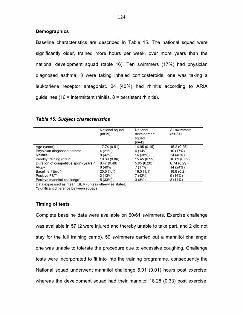
124
Demographics
Baseline characteristics are described in Table 15. The national squad were
significantly older, trained more hours per week, over more years than the
national development squad (table 16). Ten swimmers (17%) had physician
diagnosed asthma, 3 were taking inhaled corticosteroids, one was taking a
leukotriene receptor antagonist. 24 (40%) had rhinitis according to ARIA
guidelines (16 = intermittent rhinitis, 8 = persistent rhinitis).
Table 15: Subject characteristics
National squad (n=19)
National development squad (n=42)
All swimmers (n= 61)
Age (years)* 17.74 (0.51) 14.95 (0.15) 15.2 (0.25) Physician diagnosed asthma 4 (21%) 6 (14%) 10 (17%) Rhinitis 8 (42%) 16 (38%) 24 (40%) Weekly training (hrs)* 19.39 (0.86) 15.45 (0.55) 16.69 (0.52) Duration of competitive sport (years)* 8.47 (0.48) 5.95 (0.28) 6.74 (0.29) Atopy 6 (40%) 7 (17%) 14 (24%) Baseline FENO * 25.0 (1.1) 16.5 (1.1) 18.8 (0.2) Positive FBT* 2 (13%) 7 (42%) 9 (16%) Positive mannitol challenge* 5 (33%) 3 (8%) 8 (14%) Data expressed as mean (SEM) unless otherwise stated. *Significant difference between squads
Timing of tests
Complete baseline data were available on 60/61 swimmers. Exercise challenge
was available in 57 (2 were injured and thereby unable to take part, and 2 did not
stay for the full training camp). 59 swimmers carried out a mannitol challenge;
one was unable to tolerate the procedure due to excessive coughing. Challenge
tests were incorporated to fit into into the training programme, consequently the
National squad underwent mannitol challenge 5.01 (0.01) hours post exercise;
whereas the development squad had their mannitol 18.28 (0.33) post exercise.
125
Immediate post swim measures were performed after a 2 hour swimming
session. Delayed post swim measures were carried out 4.66 (0.18) hours after
swimming. These were performed prior to mannitol challenge. During the
challenge swimmers achieved a mean (SEM) heart rate of 85.0% (1.12)
maximum predicted.
Measurements
FENO
All participants underwent measurement of FENO using a portable MINO (NIOX
MINO® Airway Inflammation Monitor; Aerocrine AB, Solna, Sweden). A single
reading was obtained in accordance with manufacturer’s guidelines. Nasal
exhaled NO was measured using an adapted MINO device, as previously
described [235].
Sport-specific field-based exercise challenge
The swimmers performed a high intensity sport-specific field-based exercise
challenge during an intensive lactate set. This was preceded by a standard low
intensity warm up of approximately 15-20 minutes, to avoid muscular injury.
During the high intensity lactate set, swimmers were required to maintain >80%
maximum heart rate (220-age) for at least 8 minutes in order to optimally provoke
EIB. Heart rate was measured objectively using pulse oximetry (M-pulse™,
Merlin Medical, UK). FEV1 (forced expiratory volume in 1 second) was measured
using a Piko-6® portable spirometer (Ferraris Respiratory). Results from PIKO
126
devices have been found to be highly reproducible, and to correlate well with
results from other portable and office based spirometers [247]. Heart rate and
FEV1 were measured at baseline, immediately following challenge and at 5 and
10 minutes during recovery. The best of two reproducible readings was utilised.
A positive challenge was defined as a fall in FEV1 of ≥10% at ≥ 2 time points
during recovery (in accordance with recommendations by drug-free sport UK for
ATUE (therapeutic use exemption) certification)[174].
Mannitol challenge
Mannitol challenge was performed by administering spray-dry mannitol powder in
gelatine capsule form (AridolTM Pharmaxis Ltd, Sydney, Australia), inhaled from a
dry powder device (Osmohaler, Pharmaxis Ltd. French’s Forest, NSW, Australia)
as previously described by Anderson and colleagues [136][183]. FEV1 was
measured 60 seconds after delivery of each dose (5, 10, 20, 40, 80, 160, 160,
160 mg) using a Piko-6® portable spirometer (Ferraris Respiratory). This device
was used to ensure standardisation between both challenge tests. The test
continued until the FEV1 had fallen 15% or the maximal dose of 635 mg had
been administered. The provoking dose of mannitol to cause a 15% fall in FEV1
(PD15) was calculated by log linear interpolation. A positive challenge was taken
as being either a 15% fall in FEV1 at the threshold dose (PD15) or a 10% fall at
successive time points.
127
Peak nasal inspiratory flow
Peak nasal inspiratory flow (PNIF) was measured using the In-Check® PNIF
meter (Clement Clarke International Ltd, Harlow, UK). After horizontal positioning
and restoration to zero, participants forcefully inhaled through their nose and the
best of three measurements was taken.
Health questionnaire (symptoms)
A modified version of a questionnaire previously used in studies on elite athletes
was used to assess the presence of exercise-induced symptoms [176].
Symptoms of chlorine-induced rhinitis (blockage/ running) were assessed using a
visual analogue scale. Atopy was defined as a history of rhinoconjunctivis (in
accordance with current ARIA definitions) and/or a positive skin prick test or IgE
specific RAST test within the last 2 years.
Statistical analysis
SPSS version 15 (SPSS Inc, Chicago, Illinois) was used to perform the statistical
analysis. Non-Gaussian data were log transformed prior to analysis. Normalized
data were assessed using repeated measures ANOVA with Bonferroni
correction. Chi Squared test was used to check for associations in non-
parametric data. Pearson’s correlation was used for parametric data. A P value
of less than 0.05 (two tailed) was considered significant. As this was an
observational study, no formal power calculation was used.
128
Results
Challenge tests
8/59 (14%) of swimmers had a positive mannitol challenge 9/57 (16%) of
swimmers had a positive exercise test. Only one swimmer was positive to both
tests. There was no correlation between maximum FEV1 drop in mannitol
challenge and maximum FEV1 drop in exercise challenge. 3/10 asthmatics were
positive to mannitol, while only 2/10 were positive to exercise.
Results of subjects who achieved a ≥10% fall in FEV1 are detailed in table 16. 7/9
subjects deemed to have a positive exercise challenge had a fall immediately
post exercise which was sustained at 5 minutes (7/7) and 10 minutes (5/7)
recovery. The mean (SEM) %fall for these subjects immediately post exercise
was 18.9 (7.0), and 15.2 (6.0) at 5 minutes. 3 subjects had a fall of ≥10%, which
was not sustained; their mean (SEM) %fall was 11.1 (1.7) immediately and -0.47
(1.7) at 5 minutes recovery. Two subjects developed a sustained ≥10% fall
during recovery. The geometric mean (95% CI) mannitol PD15 was 209.9 (128.7
– 342.3). The degree of bronchial reactivity, measured in terms of response-dose
ratio (final % fall in FEV1 divided by the total administered dose of mannitol), was
similar in swimmers with EIB and those who did not respond to either test, 0.01
and 0.01 respectively. The response-dose ratio in those with a positive mannitol
was higher at 0.06.
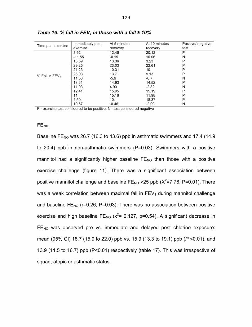
129
Table 16: % fall in FEV1 in those with a fall ≥ 10%
Time post exercise Immediately post-exercise
At 5 minutes recovery
At 10 minutes recovery
Positive/ negative test
8.92 12.45 20.12 P -11.55 -0.19 10.06 N 13.59 13.36 3.23 P 29.25 23.03 22.61 P 21.23 10.31 10 P 26.03 13.7 9.13 P 11.53 -5.9 -6.7 N 18.61 14.93 14.52 P 11.03 4.93 -2.82 N 12.41 15.95 15.19 P 11 15.16 11.98 P 4.59 10.1 18.37 P
% Fall in FEV1
10.67 -0.46 -2.09 N P= exercise test considered to be positive, N= test considered negative
FENO
Baseline FENO was 26.7 (16.3 to 43.6) ppb in asthmatic swimmers and 17.4 (14.9
to 20.4) ppb in non-asthmatic swimmers (P=0.03). Swimmers with a positive
mannitol had a significantly higher baseline FENO than those with a positive
exercise challenge (figure 11). There was a significant association between
positive mannitol challenge and baseline FENO >25 ppb (X2=7.76, P=0.01). There
was a weak correlation between maximal fall in FEV1 during mannitol challenge
and baseline FENO (r=0.26, P=0.03). There was no association between positive
exercise and high baseline FENO (x2= 0.127, p=0.54). A significant decrease in
FENO was observed pre vs. immediate and delayed post chlorine exposure:
mean (95% CI) 18.7 (15.9 to 22.0) ppb vs. 15.9 (13.3 to 19.1) ppb (P <0.01), and
13.9 (11.5 to 16.7) ppb (P<0.01) respectively (table 17). This was irrespective of
squad, atopic or asthmatic status.
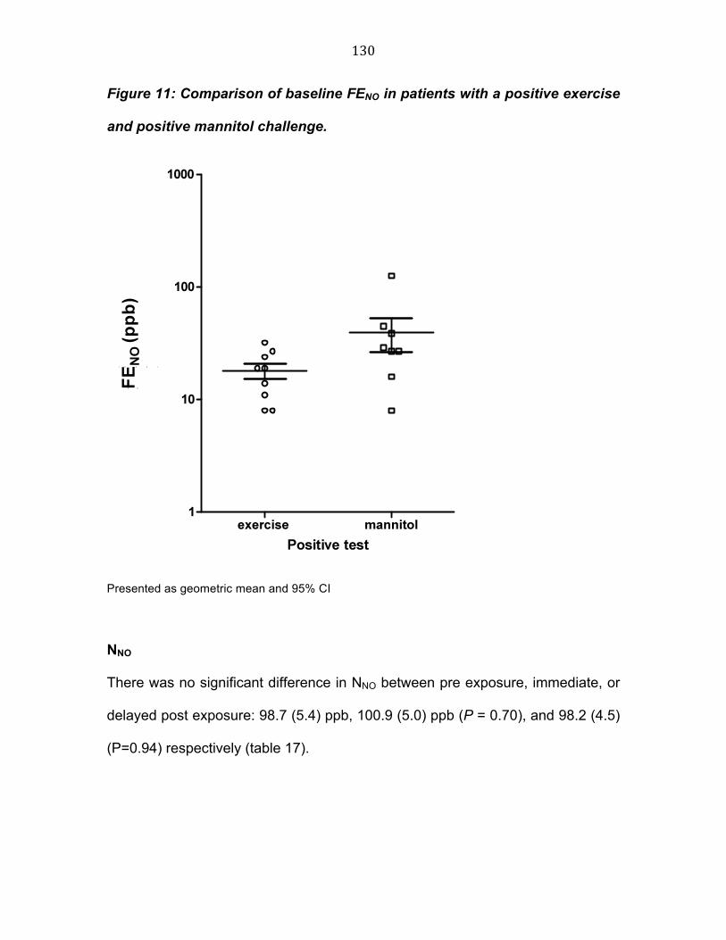
130
Figure 11: Comparison of baseline FENO in patients with a positive exercise
and positive mannitol challenge.
Presented as geometric mean and 95% CI
NNO
There was no significant difference in NNO between pre exposure, immediate, or
delayed post exposure: 98.7 (5.4) ppb, 100.9 (5.0) ppb (P = 0.70), and 98.2 (4.5)
(P=0.94) respectively (table 17).
131
PNIF
Mean PNIF increased from 142 L.min-1 (6) at baseline to 162 L.min-1 (6)
immediately post exposure (P <0.01). Delayed post exposure PNIF was not
significantly different from pre-exposure readings (P=0.90) (table 17). There was
no significant increase in visual analogue scale at any point post exposure.
Table 17: Effects of swimming on FENO, NNO and PNIF
Pre-swim Immediate post-swim
4-6 hrs post-swim
FENO (ppb) * 18.7 (15.9 - 22.0) 15.9 (13.3 - 19.1) ‡ 13.9 (11.5 - 16.7) ‡
NNO (ppb) † 98.68 (5.39) 100.93 (5.00) 98.22 (4.45)
PNIF (l/min) † 142 (6) 162 (6) ‡ 143 (6)
* Shown as geometric mean (95% CI) †Expressed as mean (SEM) ‡ Statistically significant change from pre-swim measure
Symptoms
43% (26) of swimmers complained of exercise related symptoms. 36% (8/22) of
these had a positive exercise challenge (4 symptomatic swimmers were unable
to take part in the challenge). 30% (7/26) of symptomatic swimmers had a
positive mannitol challenge. 44% (4) of participants with a positive exercise
challenge were symptomatic, 56% (5) were asymptomatic. 75% (6) of
participants with a positive mannitol challenge were symptomatic, 25% (2) were
asymptomatic.
132
Discussion
This study aimed to compare the response to standardised exercise testing and
mannitol challenge in elite national level, swimmers. We also aimed to
investigate the combined effects of chlorine and exercise on the unified airway.
No association was found between mannitol and exercise challenge. 14%
swimmers were positive to mannitol, 15% were positive to exercise challenge,
while only one swimmer was positive to both. Whilst these findings are not
supported by studies performed in cold weather or track athletes [177, 178], they
are consistent with the findings of Pedersen et al who reported that EVH had a
relatively low sensitivity in swimmers [245]. This is not entirely unexpected as the
humidified air inspired by swimmers during exercise should decrease the rate of
expired water loss during hyperpnoea. Despite this, swimmers have one of the
highest prevalences of EIB, which would suggest that the theories of heat and
water loss as a cause of EIB might not be totally applicable and other factors
(such as the presence of chlorine) may be pertinent.
In the current study a disconnect was observed between AHR detected by
standardised-FBT and mannitol challenge. Traditionally, FBT in athletes has
been non-standardised and centred on a challenge using their primary exercise
[177]. This will limit the ability to standardise both the cardiovascular workload
and environmental conditions for repeated challenges or between athletes,
making it inappropriate for direct comparison to highly standardised tests such as
EVH or methacholine challenge. For example, a sprinter whose main event is a
133
50m freestyle will be unlikely to sustain the adverse effects on the airways for
long enough to induce bronchoconstriction, however EIB in longer aerobic
training sets may still have a significant effect on training and may still therefore
be of importance to the swimmer. We therefore decided to use a standardised,
rather than event specific protocol based on the ATS (1999) guidelines for
exercise challenge and using an objective measure of heart rate monitoring
[183]. In the current study all athletes were required to achieve an average heart
rate of 80% of maximum, and maintain this for at least of 8 minutes.
Environmental conditions within the pool over the 3-day period were constant. A
positive test required the presence of a sustained fall in FEV1 post exercise (i.e.
a fall of ≥10% at two time points post exercise) in order to exclude those who
may have had a drop secondary to exhaustion.
In this study mannitol challenge was positively associated with high baseline
FENO, whereas exercise challenge was not, suggesting that mannitol challenge
may be more sensitive at picking out swimmers with a traditional ‘inflammatory’
asthmatic phenotype, rather than EIB. Leukotrienes are known to play a
significant part in sustaining the contraction of airway smooth muscle and airway
narrowing in EIB. It is therefore also of interest that mannitol positive swimmers,
who have demonstrated that their airway is susceptible to endogenous release of
leukotrienes, do not respond to exercise. This suggests that a different
mechanism is responsible for the fall in FEV1 with high intensity swimming.
Mannitol challenge has previously been shown to correlate very closely with
134
eosinophils in induced sputum [248]. Swimmers are thought to have higher levels
of eosinophils and neutrophils in their sputum [172]. However, closer inspection
of the data by Helenius et al reveals that most swimmers with sputum
eosinophila were in fact known asthmatics, whilst the greater majority of study
population had eosinophil levels comparable to controls. Findings in participants
without known asthma or AHR to methacholine (14/29) were not separately
reported. A study by Boulet et al suggested that sputum neutrophils are up-
regulated in swimmers following high intensity training [249]. This is of interest as
asthmatic subjects with high sputum neutrophil counts have been found to have
milder AHR to mannitol compared to those with a preponderance of other
inflammatory cell types [250]. However, high sputum neutrophil counts would not
serve to explain the disconnect observed between the two challenge tests.
In this regard, the current study used FENO as a surrogate of airway inflammation
[194]. There was a significant fall in FENO following a 2-hour swim, and a trend
towards a continuing drop at 4-6 hours post exposure. This was seen in both
squads, and was not affected by asthmatic or atopic status. Forced vital
manoeuvres are known to ‘wash-out’ FENO from the airways, however this effect
has not been established in relation to exercise or hyper-ventilation [194].
Indeed, the continued decrease in FENO 4-6 hours post-exposure would not
support a ‘wash-out’ effect and suggests another action. Case series describing
airway changes following accidental high intensity chlorine exposure have
described significant drops in FENO which persist for up to 2 months post-
135
exposure [171]. It has been suggested that acute chlorine exposure causes
widespread oedema of the respiratory mucosa, degeneration and desquamation
of the bronchial epithelium, resulting in exposure of vagal receptors and
consequently enhanced vagal activity with bronchial hyper-reactivity [251].
Therefore, it seems plausible that repeated low-intensity exposure to chlorine
may cause bronchial hyper-reactivity by a non-cellular inflammatory response.
This would explain the high prevalence of EIB diagnosed by standardise-FBT,
even in swimmers with normal baseline FENO. There have been several case
reports of acute pulmonary oedema in swimmers and divers. In 2004 Adir et al
published a case series of 70 swimmers with “swimming-induced acute
pulmonary oedema” [252]. However, they reported clinical haemoptysis and a
decrease in mean oxygen saturations to 76%. Adir and colleagues hypothesised
that the pulmonary oedema may have been due to exercising in the prone
position, as this will have affected the distribution of blood throughout the lung.
Whilst no decrease in oxygen saturations was noted in our swimmers it is
possible that a combination of exercising in the prone position and exposure to
toxic metabolites may have caused a mild degree of airway oedema, which led to
a decrease in bronchial flux. An alternative mechanism for the suppression of
FENO is direct blockage of production through inhibition of NO synthase (the
enzyme responsible for NO production).
Unlike FENO, there was no change in NNO following a 2 hour swim. The cause for
this is unclear, however, a possible explanation is that at exercise capacity
136
athletes mouth breath rather than breathing through their nose. Given this, the
paranasal sinuses will have comparatively little gas exchange or chlorine
exposure. There was also no change 4-6 hours post exposure, suggesting that
there is no late phase response in NO. This is substantiated by the PNIF results
which increase immediately post exposure but returns to baseline after 4-6
hours. The immediate improvement is most likely due to a ‘nasal douche effect’,
given that saline irrigation is known to significantly improve nasal congestion
[239]. There was additionally no significant change in visual analogue scale
either immediately or 4-6 hours post-exposure. 27/60 swimmers had a PNIF
≤120 l/min at baseline. Given that national elite swimmers train on a daily basis
during the season, it was not possible to establish a truly chlorine free baseline.
The diagnosis of EIB based on symptoms alone, is known to be highly inaccurate
[241]. In the current study 43% of swimmers complained of exercise related
symptoms. However, only 38% of these had a positive challenge, which is in
keeping with the results of our previous study [246].
There were significant differences in baseline values between the two squads.
The National squad (NS) were significantly older, trained more hours per week,
over a greater number of years compared to the National Development squad
(NDS). They also had a greater proportion of asthmatics, and a higher mean
baseline FENO. It has been suggested that regular pool attendance is associated
with an increased risk of asthma [253], which could explain why the older more
137
experienced team has a higher prevalence. However, this discrepancy may
simply be due to the smaller number of swimmers in the NS.
We recognize some limitations in our study. Ideally both challenges would have
been done on separate days; however, due to constraints of the training
timetable, this was only possible for the national development squad. Edmunds
et al found little evidence of a refractory period 4 hours post exercise [254],
therefore the mean of 5.05hrs between challenges in the national squad should
have been sufficient to re-establish stored inflammatory mediators. In addition,
Anderson et al noted that exercising whilst breathing in warmed humidified air,
did not induce mast cell degranulation, bronchoconstriction or not prevent the
development of severe exercise induced asthma in response to a standard
exercise challenge 30 minutes later [255]. These results are further substantiated
by those of Hahn et al who found that exercise challenge with warm humidified
air led to significantly lower fall in FEV1 post challenge, and did not induce a
refractory period [256]. The national development squad performed mannitol
challenge 18 hours post exercise, therefore refractory period is not relevant in
this group. Although, the national squad performed mannitol challenge 5 hours
after exercise, only one swimmer had a positive exercise challenge subsequently
followed by a negative mannitol challenge.
In the current study we measured FEV1 for 10 minutes post exercise. It has been
reported that peak fall in FEV1 may occasionally occur up to 30 minutes [257].
138
However, the ATS guidelines on exercise testing conclude that “including a 30-
minute post-exercise observation is controversial, because such a delay is
infrequently seen” [183]. As a compromise, to minimise the interference with the
athlete’s intensive training session, we decided that 10 minutes should be
sufficient to pick up the majority of cases. It could additionally be argued that the
non-intensive exercise preceding the challenge may have induced a refractory
period in some swimmers, which may have led to an underestimate of the true
prevalence of EIB. The low incidence of positive mannitol challenge in swimmers
with a physician diagnosis of asthma was unexpected. Of those that were
negative (7/10), 2 were taking regular ICS. Ideally, all preventer therapy would
have been stopped prior to assessment, however it was felt that this would have
interfered excessively with training. Of the remaining 5 swimmers, 2 had a high
FENO (>30), and 3 had normal readings (<20). A normal FENO in the presence of
a negative mannitol and exercise challenge suggests that these patients were
wrongly diagnosed with asthma.
139
Conclusion
We have found no association between mannitol and standardised-FBT in elite
swimmers. Mannitol challenge was associated with a high baseline FENO,
however exercise challenge was not. This suggests that whilst mannitol may
select swimmers with a ‘traditional’ inflammatory asthmatic phenotype, there may
still be a role for a standardised field based challenge in high performance
swimming, where the bronchoconstrictor response may have a more complex
aetiology. Swimmers show a sustained fall in FENO following chlorine exposure
suggesting that a non-cellular, perhaps neurogenic, type-inflammation may be
important in airway dysfunction in this group of athletes. Further large scale
studies are warranted to understand the pathophysiology of lose dose chronic
chlorine exposure in elite swimmers.
140
Critique
The results of this study were unexpected, and led us to realise that several
potential aetiologies may be at play in elite swimmers, making the use of a single
diagnostic test problematic. We recognise that further studies are required to
confirm the existence of ‘a swimming-specific bronchoconstrictor response’,
however such studies would be inherently difficult to perform. Ideally,
standardised field-based challenge would be performed in a chlorine-rich and
chlorine-free environment; unfortunately there are no chlorine-free pools in
Scotland. If such a comparison were to be made, it would be important to find a
group of elite swimmers who exercise solely in a chlorine-free environment, in
order to exclude the effects of chronic low-grade chlorine exposure on the
respiratory epithelium. It has been suggested that sea swimmers could constitute
such a group, however such a comparison is problematic in that the focus of sea
swimming is endurance whereas indoor swimming requires high intensity
exercise of relatively short duration. In other words, elite indoor swimmers are
therefore far more likely to exercise at the physiological extremes required to
trigger EIB. Furthermore, the different strokes utilised in the two disciplines allow
for the development of different muscle groups, making a direct comparison
impossible. As an alternative, field-based challenge could be compared to
standardised exercise challenge in a different discipline, e.g. CPET (which
involves exercising on a treadmill or static cycle whilst breathing in air of a fixed
temperature/ humidity). The difficulty with this is that swimmers develop certain
muscle groups, and are frequently unable to maintain similar exercise intensities
141
whilst running or cycling. It has been suggested that ’chlorine challenge’ could be
employed, however as chlorine is known to induce reactive airways dysfunction
syndrome (RADS) the ethics of this would have to be closely debated. It is likely
that even in swimmers a battery of tests would need to be performed in order to
determine the most appropriate test to monitor response in clinical trials.

142
Chapter 5:
Discussion and
conclusions
143
This work aimed to investigate potential mechanisms for improving the
management of asthma utilising current available therapies. To this end the
thesis is divided into two distinct sections: Establishing the role of surrogate
markers of inflammation in clinical and research settings and tailoring treatment
according to asthma phenotype.
It is estimated that as many as 300 million people of all ages, and all ethnic
backgrounds, suffer from asthma, and that the burden of this disease to
governments, health care systems, families, and patients is increasing [1].
Scotland has been reported to have the highest proportion of asthma per head of
population worldwide, with 18.4% of inhabitants affected [1]. Worryingly, Scotland
has been also ranked in the top third of countries with regard to asthma mortality,
with 0.6 asthma deaths per 100 000 5-35 year olds [1]. This ranking is
significantly higher than countries with lower GDPs such as Ecuador and Latvia,
which is surprising, as >95% of the Scottish population have access to essential
drugs [1]. National guidelines were introduced with the aim of standardising the
management of asthma, however, the majority of asthmatics remain
symptomatic. In 2006 an observational study of 9467 asthmatic patients, of all
ages and clinical severities, in 319 general practices throughout Scotland was
carried out [258]. They aimed to calculate the ‘human cost’ of asthma though the
assessment of symptoms and lifestyle disruption, and to assess direct health
care costs through a study of health service utilization. It was found that only 1/3
of patients were free from asthma symptoms. 4211 (66%) had experienced
144
symptoms due to asthma in the past month; with 12% (770) reporting lost time
from work/school due to asthma. In the preceding 12 months 20% (1916) had
experienced an acute exacerbation, 321 (3%) patients had attended an Accident
and Emergency (A&E) department because of an asthma related problem, 455
(5%) had attended out patients, and 237 (2.5%) were admitted to hospital.
Fifteen of these patients experienced a total of 28 days stay in an intensive care
unit. These results indicate that the human and economic costs of asthma remain
a substantial burden to the Scottish population. It is clear that a new approach to
asthma management is urgently required.
Currently asthma therapy is titrated on the basis of symptoms and markers of
airflow limitation, both of which are known to correlate poorly with the underlying
asthmatic inflammation. Strategies in which asthma therapy has been targeted to
suppress surrogate markers of inflammation have proved very successful [37,
38]. However, applying this strategy to a wider ‘community’ setting has proved
problematic. The main difficulties lie with the identification of an appropriate
surrogate marker of inflammation. Both sputum eosinophils and methacholine
bronchial challenge are time consuming to perform and require the presence of
highly trained staff and expensive machinery, making them impractical for use in
the community. FENO was a promising candidate, especially following the
development of the portable MINO device. However, a recent community based
study by Menzies et al found that FENO was an insensitive method (sensitivity,
66.7%; specificity, 51.9% at a cutoff value of 20 ppb) for identifying patients who
145
subsequently exacerbated [76]. This was likely due to frequently exacerbating
patients receiving higher doses of maintenance inhaled corticosteroids, leading
to suppression of FENO. Bronchial challenge is a well-established surrogate
marker of inflammation. The development of a challenge test utilizing dry powder
mannitol offers an exciting opportunity, as only a portable spirometer and dry
power inhaler are required [136]. In this respect, the first study to be included in
this thesis looked at the use of mannitol challenge to identify changes in airway
inflammation during community-based down titration of asthma medication.
Establishing patients on the lowest dose of inhaled steroids, in order to reduce
the risk of dose related side effects, is recommended in current guidelines, but
rarely carried out in clinical practice. In this study we were able to demonstrate
that mannitol challenge can easily and safely be used in a community setting. In
addition we showed that a significant reduction in ICS dose could be achieved in
a without any worsening of airways inflammation, lung function, or quality of life.
34% of subjects demonstrated an improvement (and 41% showed no change) in
AHR post step-down, suggesting that compliance improved on entry into the
clinical trial. Only a minority of patients showed a worsening of AHR, which would
be expected if subjects were initially concordant with treatment. A rate of 19% is
consistent with concordance rates reported in previous studies [197]. Treatment
of asthmatic patients in primary care has been driven by results obtained from
randomised controlled trials, during which treatment compliance is reinforced.
These therefore, do not reflect a real life scenario. This study highlights the
potential pit falls of extrapolating data to a primary care setting where compliance
146
rates may be poor. Perhaps there is a need for the development of more
effective strategies which support asthmatics to use established therapies
correctly rather than continue a culture of escalating therapy, without frequent
review and step-down if appropriate. Improvement of asthma care in the
community may require an approach that includes more effective supervision and
targeted assessment to enhance adherence and reduce airways inflammation.
Further studies are required to assess the impact of such a strategy in a
prospective manner. Large randomised control trials are required to further
evaluate the use of mannitol challenge as a tool by which to titrate inhaled
therapy in a community setting.
The importance of identifying appropriate surrogate markers of inflammation is
not restricted to clinical practice, and is equally important in the research setting.
This is particularly true when determining therapeutic equivalence between
inhaled products. A successful therapeutic equivalence study requires
demonstration of a significant dose-response relationship with at least two doses
of the test compared with, if possible, two doses of the reference product [259].
An ideal clinical efficacy study design for establishing a dose response needs to
be sensitive (i.e. steep-dose-response slope), reproducible, have low inter- and
intra-subject variability, and be achievable with low patient numbers. Current
EMEA guidelines state that the most well-used study design is the double-blind,
randomised, parallel group comparison of test and reference product [259].
However, this design requires the careful matching of cases and controls, which
147
is problematic due to the inherent heterogeneity of asthma. The alternative is a
crossover study, which has the advantages of allowing cases to act as their own
controls, and allows the use of a much smaller population, however this requires
the use of a reliable, repeatable endpoint measure. In addition, the lack of a
carry-over effect much be demonstrated. The EMEA currently recommend that
the primary efficacy variable should be a pulmonary function measure, preferably
FEV1 measured regularly, if possible daily at home or at least every two weeks in
the clinic [259]. Alternative primary variables can be considered but their
sensitivity to detect differences between adjacent doses of inhaled corticosteroid
must be demonstrated. The purpose of utilizing inhaled steroids in the
management of asthma is to suppress the underlying airway inflammation. It
therefore appears nonsensical to select a marker that is distant from this
process. The aim of the second study was therefore to determine which
inflammatory outcome measures provided sufficient assay sensitivity, as part of a
crossover design, for detecting dose response effects on airway and systemic
markers. Methacholine challenge was selected as the primary outcome variable
as significant dose separation has previously been reported with 100µg vs.
500µg fluticasone propionate [213]. In this respect, we also demonstrated
sensitivity of the methacholine bioassay, as there was a significant dose-
response relationship for both formulations. The common slope for the overall log
dose-response relationship was highly significant, demonstrating that the doses
selected coincided with the steep part of the dose-response curve for BHR. The
95%CIs for the relative dose potency ratio of HFA vs. CFC were close to unity at
148
1.10, however, the 95%CI (0.49–2.66) was outside predetermined equivalence
limits of +/-50% (0.5-2.0) [212]. Current EMEA guidelines state that
bioequivalence in respect of systemic exposure can be demonstrated if the 90%
confidence interval is entirely contained within 80 – 125%. However, the FDA
also accept a +/-33% limit (i.e. 0.67-1.5) for PD studies. Whilst we accept that
these limits are valid when comparing across doses (e.g. 200ug HFA vs CFC or
800ug HFA vs CFC), such limits are not appropriate for calculating relative
potency from the slope of the DRC from a Finney bio-assay. We have found,
from previous experience, that it is not possible to achieve confidence intervals
within these extremely tight boundaries for pharmacodynamic endpoints [260]. In
a previous study from our department involving 27 patients we found that the
95% confidence interval for the relative dose potency of two formulations of
budesonide was 0.5-2.46. In order to increase the power in the present study we
doubled the number of patients, and utilized methacholine challenge as opposed
to adenosine-5-monophosphate (AMP) challenge, as this is considered to yield
lower variability. The decision to aim for a relative potency of 0.5 – 2 from Finney
was based on this study [212]. However, even with more than double the number
of patients, lower equivalence limits were not achieved.
Several studies have suggested that FENO may be able to discriminate doses of
ICS [69, 71, 81, 261] however; significant limitations in their study design put
these results into question. For example, a number of these studies utilized a
cumulative-dose design, suggesting that the observed dose–response
149
relationship may have been due to a time effect rather than a dose effect. In the
current study a significant dose response relationship was seen for both HFA and
CFC, however, the 95% confidence interval was also outside of predetermined
equivalence limits. In this study, cumulative dose was also an issue for FENO,
however the same cannot be said for methacholine challenge. Sovijvari et al
demonstrated that the doubling dilution difference between FP and placebo was
1.19, 1.33 and 1.27 after 72 hours, 2 weeks and 4 weeks respectively [214].
Thereby demonstrating that near-maximal effects on methacholine PC20 are
obtained within the first two weeks of treatment. In the present study we
observed PC20 shifts of 1.55dd and 2.16dd following 200µg and 800 µg of HFA
budesonide, giving a mean difference of 0.51dd which is too large to be simply
due to a time effect. The current evidence suggests that the tight equivalence
limits recommended by both the EMEA and FDA cannot be achieved in PD
studies, without the recruitment of hundreds of participants. Among the study
designs that have been examined to date, a crossover design utilizing
methacholine challenge as the primary endpoint seems to hold the most promise;
but even for this primary endpoint, more than doubling of the study subjects, did
not achieve the required confidence intervals. This suggests that wider
confidence interval will have to be accepted if bioequivalence is to be
demonstrated using pharmacodynamic endpoints.
These two studies have demonstrated that the selection of appropriate surrogate
markers of inflammation can potentially improve the clinical management of
150
asthma and play an important role in asthma research. Clearly the titration of
steroid therapy in asthma is of vital importance, however, it must be noted that no
two individuals have the same response to the same dose of the same steroid.
This is in effect a reflection of the degree of steroid responsiveness, or
resistance, and any understanding of the individual’s responsiveness to
proposed steroid therapy should allow improved therapeutic options, tailored to
the individual. It is likely that there is a spectrum of steroid responsiveness in
asthma, with rare complete resistance at one end, but a relative resistance in
patients who require high doses of inhaled or oral corticosteroids [262]. True
(complete) corticosteroid resistance is very rare, with an estimated prevalence of
1:1000 asthmatics [262]. The reasons for the variability in individual therapeutic
responses to drugs used to treat asthma are complex, involving both genetic and
environmental factors as well as levels of adherence with therapy [263, 264].
One of the most influential environmental factors is cigarette smoking, which is
known to significantly worsen asthma control though a combination of epithelial
toxicity, oxidative damage, and recruitment of inflammatory cells [152, 153]. In
addition, smokers with chronic asthma are less sensitive to the beneficial effects
of both inhaled and oral corticosteroids compared with nonsmokers with asthma
[157, 158, 160]. Therapeutic studies, which form the basis for current asthma
guidelines, often exclude current smokers due to concerns about recruiting
participants with chronic obstructive pulmonary disease (COPD). Consequently
information on which drugs are most appropriate to treat smokers with asthma is
severely lacking. Knowing how best to manage this group of patients is of
151
considerable importance because smoking is common among patients with
asthma, current smoking rates among asthmatic patients have been reported as
ranging from 17-35% [148, 150, 151, 161, 162]. Although smoking cessation has
been shown to improve asthma control, lung function and lead to a decline in
sputum neutrophil counts many asthmatics are either unable or unwilling to stop
smoking [217]. The lack of available evidence means that guidelines do not
recommend alternative treatment strategies for smokers, other than suggesting
that ‘higher doses of inhaled steroids may be required’ [8].
Only a handful of studies to date have investigated this issue. In 2007, Lazarus
and colleagues compared the response to low-dose inhaled steroid
(becomethasone 400mcg daily) with an oral leukotriene receptor antagonist
(montelukast 10mg daily) for 8 weeks in smokers and non-smokers [265]. Their
main findings were that inhaled steroid significantly increased pre-bronchodilator
FEV1 in non-smokers only, whereas montelukast increased morning peak flow in
smokers only. Unfortunately, these changes were only seen within group and not
between groups. Furthermore, neither montelukast nor beclomethasone lead to
improvements in either sputum eosinophils or methacholine challenge. A more
recent study by Spears and colleagues investigated the potential additive effects
of theophyllines on ICS [266]. The recruitment of histone deacetylase (HDAC) is
known to partly mediate the anti-inflammatory actions of corticosteroids. HDAC is
a nuclear enzyme involved in the switching off of activated inflammatory genes
[267]. Cigarette smoke has been shown to reduce HDAC activity in vitro [268],
152
which could explain corticosteroid insensitivity seen in smokers with asthma. In
vitro studies have shown that HDAC activity can be restored by low-doses of
theophylline [269]. Spears et al therefore set out to determine whether
theophyllines, in combination with low-dose ICS had any additive effects on lung
function over ICS or theophyllines alone [266]. They found that subjects treated
with the combination demonstrated significant improvements in morning PEF and
mean pre-bronchodilator FEV1. These improvement were noted to be superior to
those seen with montelukast [265] and high dose inhaled steroid [159]. However,
no differences were observed in sputum inflammatory cells, other than a
decrease in lymphocyte count, the clinical significance of which was unclear. In
our study we investigated the benefits of combination therapy with LABA and ICS
over doubling the dose of inhaled steroid in smokers and non-smokers, and were
able to demonstrate an improvement in lung function similar to that seen in non-
smokers. Furthermore, smokers appeared to gain proportionally greater benefit
from the addition of salmeterol than non-smokers on methacholine AHR. These
results support the observation made by Pedersen and colleagues during a post-
hoc analysis of the Gaining Optimal Asthma controL (GOAL) study’ that smokers
would need 18 years less treatment, by taking FPSM vs. FP alone, to prevent an
exacerbation than non-smokers [163]. In our study we found no significant
benefit, in any primary or secondary outcome, from high dose inhaled FP
(1000ug/day) in smokers, which goes against current recommendations that
smokers may benefit from higher doses of ICS [8]. This assumption was based
on a study by Tomlinson et al, who demonstrated that high dose ICS decreased
153
peak flow variability to a similar degree in both smokers and non-smokers [159].
Peak flow, like spirometry, reflects airway caliber and as such is relatively distant
from the underlying asthmatic inflammatory process [21]. In this respect we saw
no significant improvement in either peak flow dairy cards or ACQ in smokers.
Whilst the results of our study suggest that smoking asthmatics gain no benefit
from inhaled steroids, we would not advocate that they be treated with salmeterol
alone. The use of LABAs as monotherapy has been shown to lead to an increase
in asthma related deaths [270]. Further safety research would be required prior to
making such a declaration. Similarly, although our results suggest similar
improvements in spirometry to those seen during smoking cessation, we would
still advocate smoking cessation as the first line intervention in smoking
asthmatics. The detrimental effects of smoking are not solely limited to poorer
asthma control, significant increases in cardiovascular risk and increased risk of
malignancy dictate that smoking cessation be discussed on a regular basis.
We had hoped to provide a mechanistic explanation for this improvement by
including both methacholine and mannitol challenge. The intention was that
mannitol would provide a sensitive marker of inflammation, whereas methachline
would provide a measure of airway tone. Interestingly, similar results were seen
with both challenge tests (although differences between treatments in smokers
did not reach significance with mannitol). We would have expected to see a
greater improvement in mannitol PD15 following treatment with double dose FP
compared to FPSM, as was observed during a similar study which utilised AMP
154
challenge [271]. In view of the fact that salmeterol has been shown to have no
meaningful in vivo activity we have to hypothesise that the improvements
conferred by LABAs was either due to their stabilizing effect on airway smooth
muscle [225] and mast cells [272], or to their ability to preserve the integrity of the
respiratory epithelium [227]. These findings suggest that mannitol may be less
may be less specific for inflammation than originally thought. Further studies are
required to determine the exact mechanism of action of this indirect bronchial
challenge test. However, despite this, we have successfully demonstrated that
alternative, treatment approaches tailored to different subgroups of asthmatics
may be beneficial.
Another group of individuals in whom the traditional approach to asthma
management may not be appropriate is elite athletes. It is has been well
documented that the mechanism of bronchoconstriction in athletes involves the
drying and/or cooling of airways [134]. However, even within this category there
are athletes to which this mechanism of action is unlikely to apply. Swimmers, for
example, exercise in a warm, humid environment and are therefore not subject to
the same environmental strains as cold weather or endurance athletes. It
therefore seems unlikely that tests designed to reproduce hyperosmolar shifts
will have the same diagnostic sensitivity in swimmers as they do in these other
groups of athletes. The aim of the fourth and fifth studies included in this thesis
was therefore to compare various diagnostic tests in elite swimmers, in order to
determine which were the most sensitive. This would allow us to determine which
155
test would be most specific for future use in studies monitoring benefit from
medical therapy.
We hypothesised that exposure to chlorine had to play an important part in the
underlying pathogenesis of EIB in swimmers, as traditional explanations did not
fit, and wanted to determine the effect of chlorine and exercise on the unified
airway prior to undertaking studies comparing the different tests. Adult elite
swimmers have been shown to have significantly higher levels of airway
inflammatory cells compared with healthy controls [172]. We therefore conducted
a pilot study to assess the combined effects of chlorine and exercise on the
unified airway of adolescent elite swimmers, including estimating the prevalence
of rhinoconjunctivitis and EIB. We elected to use FENO as it is a non-invasive,
reproducible, well-established marker of airway inflammation, which can be used
to assess both the upper and lower airways [236], In addition, FENO had been
shown to rise following a marathon [237]. The absence of any acute rise in either
tidal or nasal FENO post exercise challenge was therefore unexpected. We
hypothesised that we may have missed a late phase response, similar to that
seen during allergen challenge. This assumption was supported by anecdotal
reports from swimmers who complained of nasal congestion and symptoms, 4-6
hours after training. The use of a standardised field based test was a novel
approach. Traditional field based testing, in which athletes perform a challenge
using their primary exercise, was previously limited by an inability to standardise
both the cardiovascular workload and environmental conditions [177]. We
156
therefore decided to use a standardised protocol based on the ATS guidelines for
exercise challenge, using an objective measure of heart rate monitoring. Using
this test we were able to identify a high prevalence (36%) of EIB in our cohort of
adolescent elite swimmers.
The next step was therefore to compare our standardised exercise test to
mannitol challenge, as a surrogate for the gold-standard test, EVH. We found a
disconnect between the challenge tests, in that 14% swimmers were positive to
mannitol, 15% were positive to exercise challenge, while only one swimmer was
positive to both. Whilst these findings were not supported by studies performed in
cold weather or track athletes [146, 177, 178], they were consistent with the
findings of Pedersen et al who reported that EVH had a relatively low sensitivity
in swimmers [245]. Interestingly, swimmers with a positive mannitol challenge
had a significantly higher baseline FENO, than those who were positive to
exercise, suggesting that mannitol challenge may be more sensitive at picking
out swimmers with a traditional ‘inflammatory’ asthmatic phenotype, rather than
EIB. When we looked at FENO we found that there was a significant decrease
post exercise, which was maintained 4-6 hours post exercise. Case reports of
children exposed to high doses of chlorine have shown increased bronchial
hyper-reactivity and persistently low FENO levels. We therefore hypothesised that
a similar pathogenic mechanism may be at play in the swimmers, a sort of
‘swimmer-specific bronchoconstrictor response’ thereby explaining why they
didn’t respond to both challenges. Interestingly, similar results have been
157
reported previously. Bonsignore et al compared the effects of a 5km race on
spirometry and FENO in competitive outdoor pool swimmers and sea swimmers.
Although the chlorine levels in the outdoor pool were extremely low, they noted a
significant decrease in FENO post-exercise, which was not observed in sea
swimmers. No differences were seen in sputum inflammatory cell composition,
and no changes in FEV1 were noted post exercise, however, again the field-
based challenges were not standardised [273]. Furthermore, neither group was
considered to be ‘elite’, indicating that perhaps the intensities of exertion required
to trigger EIB were not reached.
Unfortunately, subsequent publications do not correlate these results. Castricum
et al recently published a study comparing standardised field-based challenge,
EVH and lab-based challenge in 33 adult swimmers [274]. They found that only 1
of the 33 subjects had a positive field swim challenge compared to 18 with a
positive EVH challenge, and 4 with a positive laboratory cycle challenge. Only 1
of the 33 subjects was positive to all 3 challenges. Whilst the disconnect between
the challenge tests reflects our results, the low prevalence of EIB diagnosed by
field challenge is somewhat surprising. The prevalence of EIB diagnosed in both
studies presented in this thesis were 36% and 15% respectively. Rates in the first
study may have been artificially high as positive challenge was defined as a
single >15% drop in FEV1 post exercise. In the second study these criteria were
tightened to >15% drop at two time points post exercise, with the aim of
excluding those with a drop secondary to exhaustion. In the study by Castricum
158
et al the field-based challenge took place in an ozone filtered pool, which would
have resulted in significantly lower concentrations of chlorine. Further studies are
therefore required to compare standardized field based challenge in high and low
chlorine environments.
Much controversy surrounds the diagnosis of EIB itself. It has been suggested
that EIB in elite athletes is simply a reflection of very mild asthma, which only
becomes symptomatic due to extremes of exercise. Whilst this is almost certainly
true in a subgroup of athletes, such as the swimmers who were positive to
mannitol in the 5th study, I believe that EIB occurs as a distinct clinical entity.
Exercise is the most common trigger of bronchospasm in those who are known
to be asthmatic, affecting between 50 and 90% of all individuals with asthma
[275]. However, EIB also occurs in up to 10% of subjects who are not known to
be atopic or asthmatic [276]. Elite athletes claim that exercise is the most
prominent trigger of asthma symptoms, and rarely complain of diurnal variation or
nocturnal symptoms [241]. The pathogenesis of asthma-like symptoms in elite
athletes is likely to be multifactorial, and is not completely understood. However,
during competitive sport the use of full lung capacity means that large volumes of
atmospheric air that is cold, dry and polluted are inhaled on a regular basis. This
overcomes the ability of the upper airways to warm up and humidify the air
reaching the smaller airways [277], bringing about airway narrowing through
water and heat loss [134]. These airway differences, together with some degree
of inflammation, lead to EIB or “sports asthma” [278]. Several studies have
159
suggested that athletes have different patterns of airway inflammation compared
to ‘traditional inflammatory’ asthmatics [279-281]. Furthermore, unlike traditional
asthma, EIB and airway inflammation have been shown to attenuate on
cessation of competitive sport [173]. However, no study to date has excluded
subjects with either a physician diagnosis of asthma or ‘typical’ asthma
symptoms (e.g. nocturnal waking or diurnal variation). It is therefore unclear
whether subjects who are responsive to exercise have a different pattern of
inflammation than those who are responsive to direct challenge, or what role
inflammation actually plays in the pathogenesis of EIB. Karjailinen et al carried
out a study in which elite skiers were classified according to their response to
methacholine challenge [279]. They found that all skiers, irrespective of
challenge, had significantly higher neutrophil counts on bronchial biopsy than
asthmatic controls. However, methacholine-responsive skiers had significantly
higher eosinophil counts than non-responders, suggesting that methacholine
challenge may select out those with more traditional ‘inflammatory’ asthma [279].
Unfortunately, this study did not compare response to exercise between the two
groups. Lab-based studies have shown that exposing healthy subjects to cold air
whilst running can induce an increase in the number of granulocytes and
macrophages in the BAL [282]. Repetitive hyperpnoea itself has been shown to
recruit eosinophils to the airways and to promote the release of inflammatory
cytokines [283]. Although inflammatory cells have been documented in the
airways of non-asthmatic subjects with EIB, it should be stressed that their role in
the development of airway hyper-responsivness is currently unknown. Smooth
160
muscle dysfunction on the other hand undoubtedly plays a large role in the
pathogenesis of traditional asthma, hence why bronchial challenge tests to direct
stimuli have such a strong negative predictive value. The same however, cannot
be said for EIB, and direct challenge has been shown to be a poor diagnostic tool
for EIB in athletes [176, 179], adding yet further weight to the argument that a
different pathogenesis is at play in this group of individuals.
The current gold standard test for the diagnosis of EIB is EVH. However, as a
positive results is seen in up to 50% of athletes [179, 245], one has to wonder
whether it is over-diagnosing EIB? Whilst the lack of environmental
standardisation makes field-based testing a relatively poor diagnostic tool in track
and cold weather athletes, the same cannot be said for elite swimmers. Which
begs the question: What is the clinical significance of a positive EVH challenge in
a swimmer who doesn’t bronchoconstrict during a standardised exercise
challenge? In other words, is EVH simply identifying a normal response at the
extremes of physiology? Further research is require to investigate the role of
inflammatory cells in the pathogenesis of EIB, and to investigate whether athletes
who bronchoconstrict to exercise, EVH or direct challenge have different
inflammatory cell compositions or airway dynamic effects.
161
Conclusions
1. The use of inflammatory surrogates has opened many possibilities in
both the fields of clinical management and asthma research. We have
demonstrated that mannitol challenge can safely be used in the
community, and that down-titration of inhaled steroid is possible
without a significant rise in airway inflammation. These findings
suggest that an approach that includes effective supervision and
targeted assessment, to enhance adherence and reduce airways
inflammation, may be beneficial.
2. Methacholine challenge has sufficient assay sensitivity to be used as
the primary outcome measure in crossover studies examining the
bioequivalence of inhaled corticosteroids. However, wider confidence
intervals will have to be accepted if bioequivalence is to be
demonstrated using pharmacodynamic endpoints.
3. Fluticasone/salmeterol combination affords more bronchoprotection,
and greater bronchodilation than doubling the dose of fluticasone in
smoking asthmatics. This study adds further weight to the argument
that smoking asthmatics require a different approach to treatment than
their non-smoking counterparts.
4. Mannitol challenge may not be as specific for inflammation as originally
thought, as it appears to be attenuated by LABA induced smooth
muscle stabilisation.
162
5. Adolescent elite swimmers have a high prevalence of asthma and
rhinitis.
6. Swimmers are not exposed to the same environmental strains as other
cohorts of elite athletes, and the pathogenesis of their EIB is likely to
be different. The disconnect observed between swimmers with high
FENO and a positive mannitol, and those with a positive field-based
challenge, suggests that a ‘swimmer specific bronchoconstrictor
response’ may be at play. This is substantiated by the persistent drop
in FENO observed 4-6 hours post exercise.
163
Publications arising from this thesis
Clearie K, Vaidyanathan S, Williamson P.A, Goudie A, Short, P, Lipworth, B.J.
Effects of chlorine and exercise on the unified airway in adolescent elite
Scottish swimmers. Allergy 2010 Feb; 65(2): 269-73
Clearie KL, Williamson PA, Vaidyanathan S, Short P, Goudie A, Burns P,
Hopkinson P, Meldrum K, Howaniec L, Lipworth BJ. Disconnect between
standardized field-based testing and mannitol challenge in Scottish elite
swimmers. Clinical Experimental Allergy. 2010 May;40(5):731-7.
Clearie KL, Jackson CM, Fardon TC, Williamson PA, Vaidyanathan S, Burns P,
Lipworth BJ Supervised step-down of inhaled corticosteroids in the
community – an observational study. Respiratory Medicine. 2010 (epub ahead
of print).
Clearie KL, Williamson PA, Meldrum K, Gillen M, Carlsson LG, Carlholm M,
Ekelund J, Lipworth BJ. Pharmacokinetic and Pharmacodynamic
Comparison of Hydrofluoroalkane and Chlorofluorocarbon Budesonide.
British Journal of Clinical Pharmacology. 2010 (epub ahead of print)
164
Poster presentations arising from this thesis
Disconnect between standardised field based testing and mannitol challenge in
Scottish elite swimmers.
• Association of Physicians of Great Britain and Ireland (APAM)
Annual Meeting (Dundee, 16th April 2010)
Establishing equivalence of inhaled corticosteroids using pharamacokinetic and
pharmacodynamic outcomes
• Scottish Thoracic Society meeting (Stirling, October 2009)
• European respiratory Society meeting (Barcelona, September 2010)
Effects of chlorine and exercise on the unified airway in young elite Scottish
Swimmers.
• Scottish Thoracic Society meeting (Scone, April 2009)
• American Thoracic Society meeting (San Diago, May 2009)
• European Thoracic Society meeting (Vienna, September 2009)
Supervised step-down in a community setting does not increase airways
inflammation in mild-to-moderate asthmatics.
• American Thoracic Society meeting (San Diago, 16th May 2009)
165
Fluticasone/Salmeterol combination confers significant benefits in smoking
asthmatics
European respiratory Society meeting (Barcelona, September 2010)
British Thoracic Society meeting (London, December 2010)
Scottish Thoracic Society meeting (Stirling, October 2010)
Oral Presentations arising from this thesis
Disconnect between standardised field based testing and mannitol challenge in
Scottish elite swimmers.
British Thoracic Society meeting (London, December 09)
Scottish Thoracic Society meeting (Stirling, October 09)
Supervised Step-down In a Community Setting Does Not Increase Airways
Inflammation In Mild-to-Moderate Asthmatics
British Thoracic Society meeting (London, December 09)
Scottish Thoracic Society meeting (Stirling, April 08)

166
Bibliography
1. Masoli, M., et al., The global burden of asthma: executive summary of the GINA Dissemination Committee report. Allergy, 2004. 59(5): p. 469-78.
2. Viinanen, A., et al., Prevalence of asthma, allergic rhinoconjunctivitis and allergic sensitization in Mongolia. Allergy, 2005. 60(11): p. 1370-1377.
3. Perzanowski, M.S., et al., Endotoxin in inner-city homes: associations with wheeze and eczema in early childhood. Journal of allergy and clinical immunology, 2006. 117(5): p. 1082-1089.
4. Yemaneberhan, H., et al., Prevalence of wheeze and asthma and relation to atopy in urban and rural Ethiopia. Lancet, 1997. 350(9071): p. 85-90.
5. Barnes, P.J., B. Jonsson, and J.B. Klim, The costs of asthma. European Respiratory Journal, 1996. 9(4): p. 636-641.
6. Action Asthma. The occurance and cost of asthma Worthing, UK, Cambridge Medical Publications, 1990
7. Jones, P.W., et al., The impact of asthma on an employed population (The Zeneca Asthma Study) European respiratory journal, 1995. 8(19): p. 153s.
8. British Guideline on the management of asthma. BTS/SIGN guidelines revised edition 2008. www.brit-thoracic.org.uk.
9. Barnes, P.J., S. Pedersen, and W.W. Busse, Efficacy and safety of inhaled corticosteroids. New developments. . American journal of respiratory and critical care medicine, 1998. 157(3): p. S1-53.
10. Laitinen, L.A., A. Laitinen, and T. Haahtela, A comparative study of the effects of an inhaled corticosteroid, budesonide, and a beta-2 agonist, terbutaline, on airway inflammation in newly diagnosed asthma: a randomized, double-blind, parallel-group controlled trial. Journal of allergy and clinical immunology, 1992. 90(1): p. 32-42.
11. Pauwels, R.A., et al., Early intervention with budesonide in mild persistent asthma: a randomised, double-blind trial. Lancet, 2003. 361(9363): p. 1071-1076.
12. O'Byrne, P.M., et al., Low dose inhaled budesonide and formoterol in mild persistent asthma: the OPTIMA randomized trial. American journal of resipratory and critical care medicine, 2001. 164(8): p. 1392-1397.
13. Lipworth, B.J., Systemic adverse effects of inhaled corticosteroid therapy: a systematic review and meta-analysis. Archives of Internal Medicine, 1999. 159: p. 941-955.
14. Rabe, K.F., et al., Clinical management of asthma in 1999: the asthma insights and reality in Europe (AIRE) study. European respiratory journal, 2000. 16(5): p. 802-807.
15. The global initiative for asthma (GINA). Global Strategy for Asthma Management and Prevention. National Institute of Health Publication 02-3659 issued January 1995 (Updated 2009) At http://www.ginasthma.com
16. Mathers, C.D. and D. Loncar, Projections of global mortality and burden of disease from 2002 to 2030. PLoS Medicine, 2006. 3(11): p. e442.
17. Houston, J.C., S. De Navasquez, and J.R. Trounce, A clinical and pathological study of fatal cases of status asthmaticus. . Thorax, 1953. 8(3): p. 207-213.
167
18. Carroll, N.G., S. Mutavdzic, and A.L. James, Increased mast cells and neutrophils in submucosal mucous glands and mucus plugging in patients with asthma. Thorax, 2002. 57(8): p. 677–682.
19. de Magalhaes Simoes, S., et al., Inflammatory cell mapping of the respiratory tract in fatal asthma. Clinical and experimental allergy, 2005. 35(5): p. 602-611.
20. Bradley, B.L., et al., Eosinophils, T-lymphocytes, mast cells, neutrophils, and macrophages in bronchial biopsy specimens from atopic subjects with asthma: comparison with biopsy specimens from atopic subjects without asthma and normal control subjects and relationship to bronchial hyperresponsiveness. Journal of allergy and clinical immunology, 1991. 88(4): p. 661-674.
21. Sont, J.K., et al., Relationship between the inflammatory infiltrate in bronchial biopsy specimens and clinical severity of asthma in patients treated with inhaled steroids. Thorax, 1996. 51(5): p. 496-502.
22. Booth, H., et al., Effect of high dose inhaled fluticasone proprionate on asthma inflammation in asthma. American journal of respiratory and critical care medicine, 1995. 152(1): p. 45-52.
23. Carroll, N., C. Cooke, and A. James, The distribution of eosinophils and lymphocytes in the large and small airways of asthmatics. European respiratory journal, 1997. 10 (2): p. 292–300.
24. Louis, R., et al., The relationship between airways inflammation and asthma severity. American Journal of resipratory and critical care medicine, 2000. 161(1): p. 9-16.
25. Jatakanon, A., S.A.M. Lim, and P.J. Barnes, Changes in sputum eosinophils predict loss of asthma control. American journal of respiratory and critical care medicine, 2000. 161(1): p. 64-72.
26. Pin, I., et al., Changes in the cellular profile of induced sputum after allergen-induced asthmatic responses. American review of respiratory disease, 1992. 145(2): p. 1265-1269.
27. Pizzichini, M.M., et al., Prednisolone dependent asthma: inflammatory indices in induced sputum. European respiratory journal, 1999. 13(1): p. 15-21.
28. Green, R.H., et al., Comparison of asthma treatment given in addition to inhaled corticosteroids on airway inflammation and responsiveness. European respiratory journal, 2006. 27(6): p. 1144-1151.
29. Louis, R., et al., Theophylline decreases sputum eosinophilia of asthmatics. International archives of allergy and immunology, 1999. 118(2-4): p. 343-344.
30. Pizzichini, E., et al., Montelukast reduces airway eosinophilic inflammation in asthma: a randomized, controlled trial. European respiratory journal, 1999. 14(1): p. 12-18.
31. Laprise, C., et al., Asymptomatic airway hyperresponsiveness: relationships with airway inflammation and remodelling. European respiratory journal, 1999. 14(1): p. 63–73.
32. Davies, D.E., et al., Airway remodeling in asthma: new insights. . Journal of allergy and clinical immunology, 2003. 111(2): p. 215–225.
33. Jeffery, P.K., Remodeling in asthma and chronic obstructive lung disease. American journal of resipratory and critical care medicine, 2001. 164(10): p. S28–S38.
168
34. Ward, C., et al., Inter-relationships between airway inflammation, reticular basement membrane thickening and bronchial hyper-reactivity to methacholine in asthma; a systematic bronchoalveolar lavage and airway biopsy analysis Clinical experimental allergy, 2005. 35(12): p. 1565-1571.
35. Boser, S.R., et al., Fractal geometry of airway remodeling in human asthma. . American Journal of resipratory and critical care medicine, 2005. 172(7): p. 817–823.
36. Haahtela, T., et al., Effects of Reducing or Discontinuing Inhaled Budesonide in Patients with Mild Asthma. New england journal of medicine, 1994. 331(11): p. 700-705.
37. Sont, J.K., et al., Clinical control and histopathologic outcome of asthma when using airway hyperresponsiveness as an additional guide to long-term treatment. The AMPUL Study Group. American journal of respiratory and critical care medicine, 1999. 159(4): p. 1043–1051.
38. Green, R.H., et al., Asthma exacerbations and sputum eosinophil counts: a randomised control trial. Lancet, 2002. 360(17): p. 15-21.
39. Adamko, D.J., et al., The rise of the phoenix: the expanding role of the eosinophil in health and disease. Allergy, 2005. 60(1).
40. Little, S.A., et al., Non-invasive markers of airway inflammation as predictors of oral steroid responsiveness in asthma. Thorax, 2000. 55(3): p. 232-234.
41. Leuppi, J.D., et al., Predictive markers of asthma exacerbation during stepwise dose reduction of inhaled corticosteroids. American Journal of Respiratory and Critical Care Medicine, 2001. 163: p. 406-412.
42. Jones, S.L., et al., The predictive value of exhaled nitric oxide measurements in assessing changes in asthma control. American Journal of resipratory and critical care medicine, 2001. 164(5): p. 738-743.
43. Currie, G.P., et al., Effects of montelukast on surrogate inflammatory markers in corticosteroid-treated patients with asthma. American Journal of respiratory and Critical Care Medicine 2003. 167 (9): p. 1232-1238.
44. Currie, G.P., et al., Effects of low dose fluticasone/salmeterol combination on surrogate inflammatory markers in moderate persistent asthma. Allergy, 2003. 58(7): p. 602-607.
45. Belda, J., et al., Predictors of loss of asthma control induced by corticosteroid withdrawal. Canadian Respiratory Journal, 2006. 13(3): p. 129-133.
46. Koh, Y.Y., H. Kang, and C.K. Kim, Ratio of serum eosinophil cationic protein/blood eosinophil counts in children with asthma: comparison between acute exacerbation and clinical remission. Allergy and asthma proceedings, 2003. 24(4): p. 269-274.
47. Venge, P., et al., Eosinophil cationic protein (ECP): molecular and biological properties and the use of ECP as a marker of eosinophil activation in disease. Clinical and experimental allergy, 1999. 29(9): p. 1172-1186.
48. Venge, P., Soluble markers of allergic inflammation. Allergy, 1994. 49(1): p. 1-8. 49. De Monchy, J.G., et al., Bronchoalveolar eosinophilia during allergen-induced
late asthmatic reactions. American review of respiratory disease, 1985. 131(3): p. 373-376.
169
50. Adelroth, E., et al., Inflammatory cells and eosinophilic activity in asthmatics investigated by bronchoalveolar lavage. The effects of anti-asthmatic treatment with budesonide or terbutaline. American review of respiratory disease, 1990. 142(1): p. 91-99.
51. Bacci, E., et al., Comparison between eosinophilic markers in induced sputum and blood in asthmatic patients. Clinical experimental allergy, 1998. 28(10): p. 1237-1243.
52. Wever, A.M., J. Wever-Hess, and J. Hermans, The use of serum eosinophil cationic protein (ECP) in the management of steroid therapy in chronic asthma. Clinical experimental allergy, 1997. 27(5): p. 519-529.
53. Ferguson, A.C., et al., Evaluation of serum eosinophil cationic protein as a marker of disease in chronic asthma. Journal of allergy and clinical immunology, 1995. 95(1): p. 23-28.
54. Lowhagen, O., et al., The inflammatory marker serum eosinophil cationic protein (ECP) compared with PEF as a tool to decide inhaled corticosteroid dose in asthmatic patients. Respiratory medicine, 2002. 96(2): p. 95-101.
55. Ricciardolo, F.L., Multiple roles of nitric oxide in the airways. Thorax, 2003. 58(2): p. 175-182.
56. Förstermann, U., et al., Isoforms of nitric oxide synthase: characterization and purification from different cell types. . Biochemical Pharmacology, 1991. 42(10): p. 1849-1857.
57. Gustafsson, L.E., et al., Endogenous nitric oxide is present in the exhaled air of rabbits, guinea pigs and humans. . Biochemical and biophysical research communications, 1991. 181(2): p. 852-857.
58. Saleh, D., et al., Increased formation of the potent oxidant peroxynitrite in the airways of asthmatic patients is associated with induction of nitric oxide synthase: effect of inhaled glucocorticoid. The FASEB journal, 1998. 12(11): p. 929-237.
59. Bates, C.A. and P.E. Silkoff, Exhaled nitric oxide in asthma: from bench to bedside. Journal of allergy and clinical immunology, 2003. 111(2): p. 256 –262.
60. Jatakanon, A., et al., Correlation between exhaled nitric oxide, sputum eosinophils, and methacholine responsiveness in patients with mild asthma. Thorax, 1998. 53(2): p. 91-95.
61. Massaro, A.F., et al., Expired nitric oxide levels during treatment of acute asthma. American Journal of resipratory and critical care medicine, 1995. 152(2): p. 800-803.
62. Folkerts, G., et al., Virus-induced airway hyper-responsiveness and asthma American Journal of resipratory and critical care medicine, 1998. 157(6): p. 1708-1720.
63. Dupont, L.J., et al., Exhaled nitric oxide correlates with airway hyperresponsiveness in steroid-naive patients with mild asthma. American Journal of resipratory and critical care medicine, 1998. 157(3): p. 894-898.
64. Mattes, J., et al., NO in exhaled air is correlated with markers of eosinophilic airway inflammation in corticosteroid-dependent childhood asthma. European respiratory journal, 1999. 13(6): p. 1391-1395.
170
65. Ketai, L., et al., Exhaled nitric oxide and bronchial wall thickening in asthmatics during and after acute exacerbation: evidence of bronchial wall remodeling. Journal of asthma, 2005. 42(8): p. 667-671.
66. Strunk, R.C., et al., Relationship of exhaled nitric oxide to clinical and inflammatory markers of persistent asthma in children. Journal of allergy and clinical immunology, 2003. 112(5): p. 883-892.
67. Berry, M.A., et al., The use of exhaled nitric oxide concentration to identify eosinophilic airway inflammation: an observational study in adults with asthma. Clinical and experimental allergy, 2005. 35(9): p. 1175-1179.
68. Berkman, N., et al., Exhaled nitric oxide in the diagnosis of asthma: comparison with bronchial provocation tests. Thorax, 2005. 60(5): p. 383-388.
69. Kharitonov, S.A., et al., Dose- dependent onset and cessation of action of inhaled budesonide on exhaled nitric oxide and symptoms in mild asthma. Thorax, 2002. 57(10): p. 889-896.
70. Kharitonov, S.A., P.J. Barnes, and B.J. O’Connor, Reduction in exhaled nitric oxide after a single dose of nebulized budesonide in patients with asthma. American Journal of resipratory and critical care medicine, 1996. 153: p. A799.
71. Silkoff, P.E., et al., Dose-response relationship and reproducibility of the fall in exhaled nitric oxide after inhaled beclomethasone dipropionate therapy in asthma patients. Chest, 2001. 119(5): p. 1322–1328.
72. Lee, D.K., et al., Airway and systemic effects of hydrofluoroalkane formulations of high-dose ciclesonide and fluticasone in moderate persistent asthma. Chest, 2005. 127(3): p. 851-860.
73. Kharitonov, S.A., et al., Changes in the dose of inhaled steroid affect exhaled nitric oxide levels in asthmatic patients. European respiratory journal, 1996. 9(2): p. 196-201.
74. Michils, A., S. Baldassarre, and A. Van Muylem, Exhaled nitric oxide and asthma control: a longitudinal study in unselected patients. European respiratory journal, 2008. 31: p. 539-546.
75. van Veen, I.H., et al., Exhaled nitric oxide predicts lung function decline in difficult-to-treat asthma. European respiratory journal, 2008. 32(2): p. 344-349.
76. Menzies, D., et al., Symptoms, spirometry, exhaled nitric oxide and asthma exacerbation in clinical practice. Annals of allergy asthma and immunology., 2008. 101(3): p. 248-255.
77. Shaw, D.E., et al., The use of exhaled nitric oxide to guide asthma management: A randomised control trial. American journal of respiratory and critical care medicine, 2007. 176(3): p. 231-237.
78. Smith, A.D., et al., Use of exhaled nitric oxide measurements to guide treatment in chronic asthma. New England Journal of Medicine, 2005. 352(21): p. 2163- 2173.
79. Pijnenburg, M.W., et al., Titrating steroids on exhaled nitric oxide in children with asthma: a randomized controlled trial American Journal of resipratory and critical care medicine, 2005. 172(7): p. 831-836.
80. Wilson, A.M. and B.J. Lipworth, Dose-response evaluation of the therapeutic index for inhaled budesonide in patients with mild-to-moderate asthma. American journal of medicine, 2000. 108(4): p. 269-275.
171
81. Jatakanon, A., et al., Effect of differing doses of inhaled budesonide on markers of airway inflammation in patients with mild asthma Thorax, 1999. 54(2): p. 108-114.
82. Hoyt, J.C., et al., Cigarette smoke decreases inducible nitric oxide synthase in lung epithelial cells. Experimental Lung Research, 2003. 29(1): p. 17-28.
83. Kharitonov, S.A., et al., Elevated levels of exhaled nitric oxide in bronchiectasis. American journal of resipratory and critical care medicine, 1995. 151(6): p. 1889-1893.
84. Henriksen, A.H., et al., Exhaled and nasal NO levels in allergic rhinitis: relation to sensitization, pollen season and bronchial hyperresponsiveness. European respiratory journal, 1999. 13(2): p. 301-306.
85. Gratziou, C., et al., Influence of atopy on exhaled nitric oxide in patients with stable asthma and rhinitis. European respiratory journal, 1999. 14(4): p. 897–901.
86. Cockcroft, D.W. and F.E. Hargreave, Airway hyperresponsiveness: definition, measurement and clinical relevance. In: Kaliner MA, Barnes, PJ, Persson CGA, eds. Asthma: Its pathology and treatment. New York: Marcel Dekker, 1991.
87. Cockcroft, D.W. and B.E. Davis, Mechanisms of airway hyperresponsiveness. Journal of allergy and clinical immunology, 2006. 118(3): p. 551-559.
88. Reiss, T.F., et al., Increased urinary excretion of LTE4 after exercise and attenuation of exercise-induced bronchospasm by montelukast, a cysteinyl leukotriene receptor antagonist. Thorax, 1997. 52(12): p. 1030–1035.
89. Brannan, J.D., et al., Inhibition of mast cell PGD2 release protects against mannitol-induced airway narrowing. European respiratory journal, 2006. 27(5): p. 944-950.
90. Van Den Berge, M., et al., PC20 adenosine 5'-monophosphate is more closely related with airways inflammatin in asthma than PC20 methacholine. American Journal of Respiratory and Critical Care Medicine, 2001. 163(7): p. 1546.
91. Brannan, J.D., et al., Fexofenadine decreases sensitivity to and montelukast improves recovery from inhaled mannitol. American journal of respiratory and critical care medicine, 2001. 163(6): p. 1420–1425.
92. Brannan, J.D., et al., Evidence of mast cell activation and leukotriene release after mannitol inhalation. European respiratory journal, 2003. 22: p. 491-496.
93. O’Sullivan, S., et al., Evidence for mast cell activation during exercise-induced bronchoconstriction. European respiratory journal, 1998. 12(2): p. 345–350.
94. Phillips, G.D., et al., The response of plasma histamine to bronchoprovocation with methacholine, adenosine 5'-monophosphate and allergen in atopic non asthmatic subjects. . American review of respiratory diseases, 1990. 141(1): p. 9–13.
95. Van den Berge, M., et al., Corticosteroid-induced improvement in the PC20 of adenosine monophosphate is more closely associated with reduction in airway inflammation than improvement in the PC20 of methacholine. American journal of respiratory and critical care medicine, 2001. 164(7): p. 1127–1132.
96. Waalkans, H.J., et al., The effect of an inhaled corticosteroid (budesonide) on exercise-induced asthma in children. Dutch CNSLD Group. European respiratory journal, 1993. 6(5): p. 652-656.
172
97. Jonasson, G., K.H. Carlsen, and C. Hultquist, Low-dose budesonide improves exercise induced bronchospasm in schoolchildren. Paediatric journal of allergy and immunology, 2000. 11(2): p. 120–125.
98. Henriksen, J.M. and R. Dahl, Effects of inhaled budesonide alone and in combination with low-dose terbutaline in children with exercise-induced asthma. American review of respiratory diseases, 1983. 128(6): p. 993–997.
99. Pedersen, S. and O.R. Hansen, Budesonide treatment of moderate and severe asthma in children: a dose-response study. Journal of allergy and clinical immunology, 1995. 95(1): p. 29-33.
100. Anderson, S.D., et al., Acute effect of sodium cromoglycate on airway narrowing induced by 4.5 percent saline aerosol. Outcome before and during treatment with aerosol corticosteroids in patients with asthma. . Chest, 1994. 105(3): p. 673–680.
101. Du Toit, J.L., et al., Airway responsiveness in asthma: bronchial challenge with histamine and 4.5% sodium chloride before and after budesonide. Allergy and asthma proceedings, 1997. 18(1): p. 7-14.
102. Rodwell, L.T., et al., Nedocromil sodium inhibits the airway response to hyperosmolar challenge in patients with asthma. American review of respiratory diseases, 1992. 146(5): p. 1149–1155.
103. Brannan, J.D., et al., Budesonide reduces sensitivity and reactivity to inhaled mannitol in asthmatic subjects. Respirology, 2002. 7(1): p. 37-44.
104. Doull, J., et al., Differential inhibitory effect of regular inhaled corticosteroid on airway responsiveness to adenosine 5' monophosphate, methacholine, and bradykinin in symptomatic children with recurrent wheeze. Paediatric pulmology, 1997. 23(6): p. 404–411.
105. O’Connor, B.J., et al., Greater effect of inhaled budesonide on adenosine 5'-monophosphate induced than on sodium-metabisulfite-induced bronchoconstriction in asthma. American review of respiratory diseases, 2001. 146(3): p. 560–564.
106. Juniper, E.F., et al., Effect of long-term treatment with inhaled corticosteroids on airway hyperresponsiveness and clinical asthma in non-steroid dependent asthmatics. American review of respiratory diseases, 1990. 142(4): p. 832–836.
107. Reddel, H.K., et al., Optimal asthma control, starting with high doses of inhaled budesonide. European respiratory journal, 2000. 16(2): p. 226–235.
108. Cockcroft, D.W., et al., Allergen-induced increase in nonallergic bronchial reactivity. Clinical allergy, 1977. 7: p. 503–513.
109. Brusasco, V. and E. Crimi, Methacholine provocation test for diagnosis of allergic respiratory diseases. Allergy, 2001. 56(12): p. 1114–1120.
110. Killian, D., et al., Factors in allergen induced asthma: relevance of the intensity of the airways allergic reaction and non-specific bronchial reactivity. . Clinical allergy, 1976. 6(3): p. 219–225.
111. Hill, D.J., M.J. Shelton, and C.S. Hosking, Predicting the results of allergen bronchial challenge by simple clinical methods. Clinical allergy, 1982. 12(3): p. 295–301.
112. Cockcroft, D.W., et al., Determinants of allergen-induced asthma: dose of allergen, circulating IgE antibody concentration and bronchial responsiveness to
173
inhaled histamine. American review of respiratory diseases, 1979. 120(5): p. 1053–1058.
113. Cockcroft, D.W., et al., The links between allergen skin test sensitivity, airway responsiveness and airway response to allergen. Allergy, 2005. 60(1): p. 56–59.
114. Cockcroft, D.W. and B.E. Davis, Lack of Tachyphylaxis to Methacholine at 24 h. Chest, 2005. 128(3): p. 1248–1251.
115. Juniper, E.F., et al., Reproducibility and comparison of responses to inhaled histamine and methacholine. Thorax, 1978 33(6): p. 705-710.
116. Currie, G.P., et al., Airway and systemic effects of hyrdofluroalkane fluticasone and beclomethasone in patients with asthma. Thorax, 2002. 57(10): p. 865-868.
117. Backer, V., et al., Sensitivity and specificity of the histamine challenge test for the diagnosis of asthma in an unselected sample of children and adolescents. European respiratory journal, 1991. 4(9): p. 1093-1100.
118. Du Toit, J.I., et al., Characteristics of bronchial hyperresponsiveness in smokers with chronic air-flow limitation. American review of respiratory disease, 1986. 134(3): p. 498-501.
119. Cockcroft, D.W., B.A. Berscheid, and K.Y. Murdock, Unimodal distribution of bronchial responsiveness to inhaled histamine in a random population. Chest, 1983. 83(5): p. 751-754.
120. Gibson, P.G., et al., Epidemiological association of airway inflammation with asthma symptoms and airway hyperresponsiveness in childhood. American Journal of resipratory and critical care medicine, 1998. 158(1): p. 36-41.
121. Yan, K., C.M. Salome, and A.J. Woolcock, Prevalence and nature of bronchial hyperresponsiveness in subjects with chronic obstructive pulmonary disease. American review of respiratory disease, 1985. 132(1): p. 25-29.
122. Braman, S.S., et al., Airway hyperresonsiveness in allergic rhinitis. A risk factor for asthma. Chest, 1987. 91(5): p. 671-174.
123. Crapo, R.O., et al., Guidelines for methacholine and exercise challenge testing-1999. This official statement of the American Thoracic Society was adopted by the ATS Board of Directors, July 1999. American journal of resipratory and critical care medicine, 2000. 161(1): p. 309-29.
124. Hopp, R.J., et al., The presence of airway reactivity before the development of asthma. American review of respiratory disease, 1990. 141(1): p. 2-8.
125. Woolcock, A.J., et al., Prevalence of bronchial hyperresponsiveness and asthma in a rural adult population. Thorax, 1987. 42(5): p. 361-368.
126. Josephs, L.K., et al., Non- specific bronchial reactivity and its relationship to the clinical expression of asthma. A longitudinal study. American review of respiratory disease, 1989. 140(2): p. 350-357.
127. Chhabra, S.K., S.N. Gaur, and A.K. Khanna, Clinical significance of nonspecific bronchial hyperresponsiveness in asthma. Chest, 1989. 96(3): p. 596-600.
128. Arm, J.P., B.W. Spur, and L. T.H, The effects of inhaled leukotriene E4 on the airway responsiveness to histamine in subjects with asthma and normal subjects. Journal of allergy and clinical immunology, 1988. 82(4): p. 654–660.
129. Barnes, N.C., P.J. Piper, and J.F. Costello, Comparative effects of inhaled leukotriene C4, leukotriene D4, and histamine in normal human subjects. Thorax, 1984. 39(7): p. 500–504.
174
130. Haby, M.M., et al., An exercise challenge for epidemiological studies of childhood asthma: validity and repeatablity. European respiratory journal, 1995. 8(5): p. 729–736.
131. Backer, V., et al., The distribution of bronchial responsiveness to histamine and exercise in 527 children and adolescents. Journal of allergy and clinical immunology, 1991. 88(1): p. 68-76.
132. Silverman, M. and S.D. Anderson, Standardization of exercise tests in asthmatic children. Archives of disease in childhood, 1972. 47(256): p. 882–889.
133. Anderson, S.D., Exercise-induced asthma Principles and Practice, 1993. 2(4th ed): p. 1343-1367.
134. Anderson, S.D., Is there a unifying hypothesis for exercise-induced asthma? Journal of allergy and clinical immunology, 1984. 73(5): p. 660–665.
135. Phillips, Y.Y., et al., Eucapnic voluntary hyperventilation of compressed gas mixture. A simple system for bronchial challenge by respiratory heat loss. American review of respiratory disease, 1985. 131(1): p. 31-35.
136. Anderson, S.D., et al., A new method for bronchial-provocation testing in asthmatic subjects using a dry powder of mannitol. American journal of respiratory and critical care medicine, 1997. 156(3): p. 758–765.
137. Polosa, R. and S.T. Holgate, Adenosine bronchoprovocation: a promising marker of allergic inflammation in asthma? . Thorax, 1997. 52(10): p. 919–923.
138. Riedler, J., et al., Hypertonic saline challenge in an epidemiological survey of asthma in children. American journal of respiratory and critical care medicine, 1994. 150(6): p. 1632-1639.
139. Riedler, J., et al., Prevalence of bronchial hyperresponsiveness to 4.5% saline and its relation to asthma and allergy symptoms in Austrian children. European respiratory journal, 1998. 11(2): p. 355–360.
140. Rabone, S., et al., Hypertonic saline challenge in an adult epidemiological field survey. Occupational medicine, 1996. 46(3): p. 177–185.
141. Brannan, J.D., et al., Inhaled mannitol does not cause bronchoconstriction in non-asthmatic healthy persons. Respirology, 2002. 7: p. A16.
142. Hurwitz, K.M., et al., Interpretation of eucapnic voluntary hyperventilation in the diagnosis of asthma. Chest, 1995. 108(5): p. 1240–1245.
143. Godfrey, S., et al., Cut-off points defining normal and asthmatic bronchial reactivity to exercise and inhalation challenges in children and young adults. European respiratory journal, 1999. 14(3): p. 659–668.
144. Cushley, M.J., A.E. Tattersfield, and S.T. Holgate, Inhaled adenosine and guanosine on airway resistance in normal and asthmatic subjects. British journal of clinical pharmacology, 1983. 15(2): p. 161–165.
145. Currie, G.P., et al., Effects of mediator antagonism on mannitol and adenosine monophosphate challenges. Clinical and Experimental Allergy, 2003. 33: p. 783-788.
146. Brannan, J.D., et al., Responsiveness to mannitol in asthmatic subjects with exercise and hyperventillation induced asthma. American journal of respiratory and critical care medicine, 1998. 158: p. 1120-1126.
147. Masoli, M., et al., Clinical dose-response relationship of fluticasone proprionate in adults with asthma. Thorax, 2004. 59(1): p. 16-20.
175
148. Althuis, M., M. Sexton, and D. Prybylski, Cigarette smoking and asthma symptom severity among adult asthmatics. Journal of asthma, 1999. 36(3): p. 257-264.
149. Lange, P., et al., A 15 year follow-up study of ventilatory function in adults with asthma. New england journal of medicine, 1998. 339(17): p. 1194-1200.
150. Siroux, V., et al., Relationships of active smoking to asthma and asthma severity in the EGEA study. Epidemiological study on the Genetics and Environment of Asthma. European respiratory journal, 2000. 15(3): p. 470-477.
151. Sippel, J.M., et al., Associations of smoking with hospital-based care and quality of life in patients with obstructive airway disease. . Chest, 1999. 115(3): p. 691-696.
152. Rahman, I. and W. McNee, Oxidant/antioxidant imbalance in smokers and chronic obstructive pulmonary disease. Thorax, 1996. 51(4): p. 348-350.
153. Chalmers, G.W., et al., Smoking and Airway Inflammation in Patients With Mild Asthma Chest, 2001. 120(6): p. 1917-1922.
154. Sunyer, J., et al., Smoking and bronchial hyperresponsiveness in non-atopic and atopic young adults. Spanish Group of the European Study of Asthma. Thorax, 1997. 52(3): p. 235-238.
155. Gerrard, J.W., et al., Increased nonspecific bronchial reactivity in cigarette smokers with normal lung function American review of respiratory disease, 1980. 122(4): p. 577-581.
156. Willemse, B.W., et al., Smoking cessation improves both direct and indirect airway hyperresponsiveness in COPD. European respiratory journal, 2004. 24(3): p. 391-396.
157. Chalmers, G.W., et al., Influence of cigarette smoking on inhaled corticosteroid treatment in mild asthma. Thorax, 2002. 57(3): p. 226-30.
158. Chaudhuri, R., et al., Cigarette smoking impairs the therapeutic response to oral corticosteroids in chronic asthma American journal of resipratory and critical care medicine, 2003. 168(11): p. 1308-1311.
159. Tomlinson, J.E., et al., Efficacy of low and high dose inhaled corticosteroid in smokers versus non-smokers with mild asthma. . Thorax, 2005. 60(4): p. 282-287.
160. Livingston, E., et al., Systemic sensitivity to corticosteroids in smokers with asthma. European respiratory journal, 2007. 29(1): p. 64-71.
161. Silverman, R.A., et al., Cigarette smoking among asthmatic adults presenting to 64 emergency departments. Chest, 2003. 123(5): p. 1472-1479.
162. Walsh, L., et al., Morbidity from asthma in relation to regular treatment: a community based study. Thorax, 1999. 54(4): p. 296-300.
163. Pedersen, S.E., et al., Determinants of response to fluticasone propionate and salmeterol/fluticasone propionate combination in the Gaining Optimal Asthma controL study. Journal of allergy and clinical immunology, 2007. 120(5): p. 1036-1042.
164. Jackson, C. and B. Lipworth, Benefits of combination therapy on exacerbations in nonsmoking patients with asthma. Journal of allergy and clinical immunology, 2008. 121(3): p. 780.
176
165. Barry, D.M., M.L. Burr, and E.S. Limb, Prevalence of asthma among 12 year old children in New Zealand and New South Wales: a comparative survey. Thorax, 1991. 46(6): p. 405-409.
166. Nish, W.A. and L.A. Schwietz, Underdiagnosis of asthma in young adults presenting for USAF basis training. Annals of allergy, 1992. 69(3): p. 239-242.
167. Deal, E.C., et al., Role of respiratory heat exchange in production of exercise-induced asthma. Journal of applied physiology, 1979. 46(3): p. 467-475.
168. Katelaris, C.H., et al., Patterns of allergic reactivity and disease in Olympic athletes Clinical journal of sports medicine, 2006. 16(5): p. 401-405.
169. Bernard, A. and M. Nickmilder, Respiratory health and baby swimming Archives of disease in childhood, 2006. 91(7): p. 620-621.
170. Nickmilder, M. and A. Bernard, Ecological association between childhood asthma and availability of indoor chlorinated swimming pools in Europe. Journal of occupational and environmental medicine, 2007. 64(1): p. 37-46.
171. Bonetto, G., et al., Longitudinal monitoring of lung injury after acute cholrine exposure in a swimming pool. American journal of respiratory and critical care medicine, 2006. 174(5): p. 545-549.
172. Helenius, I.J., et al., Respiratory symptoms, bronchial responsiveness, and cellular characteristics of induced sputum in elite swimmers. Allergy, 1998. 53(4): p. 346-352.
173. Helenius, I.J., et al., Effect of continuing or finishing high-level sports on airway inflammation, bronchial hyperresponsiveness, and asthma: a 5-year prospective follow-up study of 42 highly trained swimmers. Journal of allergy and clinical immunology, 2002. 109(6): p. 962-968.
174. WADA. The world anti-doping code. The 2004 prohibited list. International standard (effective January 1, 2004). http://www.wada-ama.org.
175. International Olympic Medical Commision. Beta2 adrenoreceptor agonists and the winter olympic games in Beijing. Available at: http://www.olympic.org, 2008.
176. Holzer, K., S.D. Anderson, and J. Douglass, Exercise in elite summer athletes: challenges for diagnosis. Journal of allergy and clinical immunology, 2002. 110(3): p. 374–380.
177. Dickinson, J.W., et al., Screening elite winter athletes for exercise induced asthma: a comparison of three challenge methods. British journal of sports medicine, 2006. 40(2): p. 179-183.
178. Mannix, E.T., F. Manfredi, and M.O. Farber, A comparison of two challenge tests for identifying exercise-induced bronchospasm in figure skaters. Chest 1999. 115(3): p. 649-53.
179. Rundell, K.W., et al., Field exercise vs laboratory eucapnic ventilation to identify airway hyper-responsiveness in elite cold weather athletes. . Chest, 2004. 125(3): p. 909-15.
180. Holzer, K., et al., Mannitol as a challenge to identify exercise-induced bronchoconstriction in elite athletes. American journal of respiratory and critical care medicine, 2003. 167(4): p. 534-537.
181. American Thoracic Society, Standardization of Spirometry 1994 Update, Official statement of the american thoracic society. 1994.
177
182. Beach, J.R., et al., Measurement of airway responsiveness to methacholine: relative importance of the precision of drug delivery and the method of assessing response. Thorax, 1993. 48(3): p. 239.
183. American Thoracic Society, Guidelines for Methacholine and Exercise Challenge Testing. American Journal of Respiratory & Critical Care Medicine, 2000. 161(1): p. 309-329.
184. Jokic, R., E.E. Davis, and D.W. Cockcroft, Methacholine PC20 extrapolation. Chest, 1998. 114(6): p. 1796-1797.
185. Menzies, D., A. Nair, and B.J. Lipworth, Portable Exhaled Nitric Oxide Measurement: Comparison With the "Gold Standard" Technique. Chest, 2007. 131(2): p. 410-414.
186. Bruce, C., D.H. Yates, and P.S. Thomas, Caffeine decreases exhaled nitric oxide. Thorax, 2002. 57(4): p. 361-363.
187. ATS/ERS recommendations for standardized procedures for the online and offline measurement of exhaled lower respiratory nitric oxide and nasal nitric oxide. . American Journal of resipratory and critical care medicine, 2005. 171(8): p. 912-930.
188. Oostveen, E., et al., The forced oscillation technique in clinical practice: methodology, recommendations and future developments. European respiratory journal, 2003. 22(6): p. 1026-1041.
189. Juniper, E.F., et al., Development and validation of the Mini Asthma Quality of Life Questionnaire European respiratory journal, 1999 14(1): p. 32-38.
190. Juniper, E.F., et al., Development and validation of a questionnaire to measure asthma control. European respiratory journal, 1999. 14(4): p. 902-907.
191. Juniper E, F., et al., Determining a minimal important change in a disease specific Quality of Life Questionnaire. Journal of clinical epidemiology, 1994. 47(1): p. 81-87.
192. Selroos, O., et al., Asthma control and steroid doses 5 years after early or delayed introduction of inhaled corticosteroids in asthma: a real-life study. Respiratory medicine, 2004. 98: p. 254-262.
193. Hawkins, G., et al., Stepping down inhaled corticosteroids in asthma: randomised controlled trial. BMJ, 2003. 326: p. 1115-1120.
194. ATS Workshop proceedings: exhaled nitric oxide and nitric oxide oxidative metabolism in exhaled breath condensate. Proceedings of the American thoracic Society, 2006. 3: p. 131-145.
195. Horne, R., Compliance, adherence, and concordance: Implications for asthma treatment. Chest, 2006. 130: p. 65S-72S.
196. Chambers, C.V., et al., Health beliefs and compliance with inhaled corticosteroids by asthmatic patients in primary care practices. Respiratory medicine, 1999. 93(2): p. 88-94.
197. Rand, C.S., et al., Metered-dose inhaler adherence in a clinical trial. American review of respiratory disease, 1992. 146(6): p. 1559-1564.
198. Charlton, I., et al., Audit of the effect of a nurse run asthma clinic on workload and patient morbidity in a general practice. British journal of general practice, 1991. 41(347): p. 227-231.
178
199. Dickinson, J., et al., Reducing asthma morbidity in the community: the effect of a targetted nurse-run asthma clinic in an English general practice. Respiratory medicine, 1997. 91(10): p. 634-640.
200. Fardon, T.C., et al., A comparison of 2 extrafine hydrofluoroalkane-134a–beclomethasone formulations on methacholine hyperresponsiveness. Annals of allergy asthma and immunology, 2006. 96(3): p. 422-30.
201. Currie, G.P., et al., Relationship between airway hyperresponsiveness to mannitol and adenosine monophosphate. Allergy, 2003. 58: p. 762-766.
202. Fardon, T.C., et al., Comparative cutoff points for adenosine monophosphate and methacholine challenge testing. Annals of asthma and immunology, 2004. 93: p. 365-372.
203. Berlyne, G.S., et al., A comparison of exhaled nitric oxide and induced sputum as markers of airway inflammation. Journal of allergy and clinical immunology, 2000. 106(4): p. 638–644.
204. van den Toorn, L.M., et al., Airway inflammation is present during clinical remission of atopic asthma. American journal of resipratory and critical care medicine, 2001. 164(11): p. 2107–2113.
205. Payne, D.N., et al., Relationship between exhaled nitric oxide and mucosal eosinophilic inflammation in children with difficult asthma, after treatment with oral prednisolone. American journal of respiratory and critical care medicine, 2001. 164(8): p. 1376–1381.
206. Petsky, H.L., et al., Tailored interventions based on exhaled nitric oxide versus clinical symptoms for asthma in children and adults. Cochrane database of systematic reviews, 2009(4): p. CD006340.
207. Ekroos, H., J. Tuominen, and A.R.A. Sovijarvi, Exhaled nitric oxide and its long-term variation in healthy non-smoking subjects. Clinical Physiology, 2008. 20(6): p. 434-439.
208. Montreal Protocol. The Montreal Protocol on substances that deplete the ozone layer. Final act (Nairobi: UNEP, 1987). Federal Register 1994. 59: p. 56276-56298.
209. Clearie, K.L., et al., Pharmacokinetic and Pharmacodynamic Comparison of Hydrofluoroalkane and Chlorofluorocarbons Budesonide. British journal of clinical pharmacology, 2010 (epub ahead of print).
210. Fardon, T.C., et al., Adrenal suppression with dry powder formulations of fluticasone propionate and mometasone furoate. American journal of resipratory and critical care medicine, 2004. 170(9): p. 960-966.
211. Fieller, E.C., Some problems in interval estimation. Journal of the royal statistical society. , 1954 16(2): p. 175-185
212. Lipworth, B.J., et al., Dose response comparison of budesonide dry powder inhalers using adenosine monophosphate bronchial challenge. Annals of Allergy Asthma and Immunology., 2005. 94(6): p. 657-681.
213. Nair, A., et al., Steroid sparing effects of intranasal corticosteroids in asthma and allergic rhinitis. Allergy, 2009. 65(3): p. 359-367.
214. Sovijarvi, A.R., et al., Sustained reduction in bronchial hyperresponsiveness with inhaled fluticasone propionate within three days in mild asthma: time course after onset and cessation of treatment. Thorax, 2003. 58(6): p. 500 –504.
179
215. Busse, W., S. Brazinsky, and K. Jacobson, Efficacy response of inhaled beclomethasone dipropionate in asthma is proportional to dose and is improved by formulation with a new propellant. Journal of allergy and clinical immunology, 1999. 104: p. 1215–1222.
216. Wilson, A.M. and B.J. Lipworth, 24 hour and fractionated profiles of adrenocortical activity in asthmatic patients receiving inhaled and intranasal corticosteroids. Thorax, 1999. 54: p. 20 –26.
217. Chaudhuri, R., et al., Effects of smoking cessation on lung function and airway inflammation in smokers with asthma. American journal of resipratory and critical care medicine, 2006. 174: p. 127-133.
218. Thomson, N.C., R. Chaudhuri, and E. Livingston, Asthma and cigarette smoking. European respiratory journal, 2004. 24: p. 822-833.
219. Nair, A., et al., A novel breath-actuated integrated vortex spacer device increases relative lung bioavailability of fluticasone/salmeterol in combination. Pulmonary pharmacology & therapeutics, 2009. 22: p. 305–310.
220. Lipworth, B.J. and J.R. Seckl, Measures for detecting systemic bioactivity with inhaled and intranasal corticosteroids. Thorax, 1997. 52: p. 476-482.
221. Currie, G.P., S. Stenback, and B.J. Lipworth, Effects of Fluticasone vs. fluticasone/salmeterol on airway calibre and airway hyperresponsiveness in mild persistent asthma. British journal of clinical pharmacology, 2003. 56: p. 11-17.
222. Shrewsbury, S., S. Pyke, and M. Britton, Meta-analysis of increased dose of inhaled steroid or addition of salmeterol in symptomatic asthma (MIASMA). British medical journal, 2000. 320: p. 1368-1373.
223. Pauwels, R.A., et al., Effect of inhaled formoterol and budesonide on exacerbations of asthma. Formoterol and Corticosteroids Establishing Therapy (FACET) International Study Group. . New england journal of medicine, 1997. 337(20): p. 1405-1411.
224. Sindi, A., D.C. Todd, and P. Nair, Antiinflammatory Effects of Long-Acting β2 -Agonists in Patients With Asthma. Chest, 2009. 136(1): p. 145-154.
225. Currie, G.P., et al., Airway-stabilizing effect of long-acting beta-2 agonists as add-on therapy to inhaled corticosteroids. QJM, 2003. 96: p. 435-440.
226. Piccillo, G., et al., Changes in airway hyperresponsiveness following smoking cessation: Comparisons between Mch and AMP. Respiratory medicine, 2008. 102: p. 256–265.
227. Koopmans, J.G., et al., Adding salmeterol to an inhaled corticosteroid: long term effects on bronchial inflammation in asthma. Thorax, 2006. 61(4): p. 302-312.
228. Oppenheimer, B.W., et al., Distal airway function in symptomatic subjects with normal spirometry following World Trade Center dust exposure. Chest 2007. 132(4): p. 1275-1282.
229. Kharitonov, S.A., R.A. Robbins, and D. Yates, Acute and chronic effects of cigarette smoking on exhaled nitric oxide. American journal of resipratory and critical care medicine, 1995. 152: p. 609-612.
230. Calverley, P.M.A., et al., Bronchodilator reversibility testing in chronic obstructive pulmonary disease. Thorax, 2003. 58: p. 659-664.
180
231. Helenius, I.J., et al., Asthma and increased bronchial hyper-responsiveness in elite athletes: atopy and sport eventas risk factors. Journal of allergy and clinical immunology, 1998. 101: p. 646-652.
232. Anderson, S.D. and K. Holzer, Exercise-induced asthma: is it the right diagnosis in elite athletes? Journal of allergy and clinical immunology, 2000. 106(3): p. 419-128.
233. Shaaban, R., et al., Rhinitis and onset of asthma: a longitudinal population-based study. Lancet, 2008. 372(9643): p. 1049-1057.
234. Bonini, S., et al., Rhinitis and asthma in athletes: an ARIA document in collaboration with GA2LEN. Allergy, 2006 61: p. 681-692.
235. Maniscalco, M., et al., Validation study of nasal nitric oxide measurement using a hand-held electrochemical analyser. European respiratory journal, 2008. 38: p. 197-200.
236. Kharitonov, S.A., et al., Nasal nitric oxide is increased in patients with asthma and allergic rhinitis and may be modulated by glucocorticoids. Journal of allergy and clinical immunology, 1997. 99(1): p. 58-64.
237. Bonsignore, M.R., et al., Airway inflammation in nonasthmatic amateur runners. American journal of physiology, 2001. 281: p. L668–L676.
238. Hogman, M., et al., Inhaled mannitol shifts exhaled nitric oxide in opposite directions in asthmatics and healthy subjects. Respiration physiology 2001. 124 p. 141-150.
239. Harvey, R., et al., Nasal saline irrigations for the symptoms of chronic rhinosinusitis. Cochrane database of systematic reviews, 2007.
240. Zwick, H., et al., Increased sensitization to aeroallergens in competitive swimmers. Lung, 1990. 168(2): p. 111-115.
241. Rundell, K.W., J. Im, and L.B. Mayers, Self-reported symptoms and exercise-induced asthma in the elite athlete. Medical Science Sports and Excercise, 2001. 33(208-213).
242. Morton, A.R., K.D. Fitch, and T. Davis, The effect of "warm-up" on exercise-induced asthma. Annals of allergy, 1979. 42(2): p. 257-60.
243. Anderson, S.D. and E. Daviskas, Pathophysiology of exercise-induced asthma: role of respiratory water loss. Weiler J, ed. Allergic and respiratory disease in sports medicine. New York: Marcel Dekker, 1997: p. 87-114.
244. Anderson, S.D. and E. Daviskas, Airway drying and exercise induced asthma. McFadden ER, ed. Exercise induced asthma. New York: Marecl Dekker, 1999: p. 77-113.
245. Pedersen, L., et al., Airway responses to eucapnic hyperpnea, exercise, and methacholine in elite swimmers. Medicine and science in sports and exercise, 2008. 40(9): p. 1567-1572.
246. Clearie, K., et al., Effects of chlorine and exercise on the unified airway in adolescent elite Scottish swimmers. Allergy, 2009. 65(2): p. 269-273.
247. Fonseca, J.A., et al., Pulmonary Function Electronic Monitoring Devices. Chest 2005. 128: p. 1258-1265.
248. Gibson, P.G., N. Saltos, and T. Borgas, Airway mast cells and eosinophils correlate with clinical severity and airway hyperreaponsiveness in cortico-steroid asthma. Journal of allergy and clinical immunology, 2000. 105: p. 752-759.
181
249. Boulet, L.P., et al., Lower airway inflammatory responses to high-intensity training in athletes. Clinical and investigative medicine, 2005. 28: p. 15-22.
250. Porsbjerg, C., et al., Relationship between airway responsiveness to mannitol and to methacholine and markers of airway inflammation, peak flow variability and quality of life in asthma patients. Clinical and experimental allergy, 2008. 38(1): p. 43-50.
251. Benson, M.K., Bronchial hyper-reactivity British Journal of diseases of the chest, 1975. 69: p. 227-239.
252. Adir, Y., et al., Swimming-induced pulmonary edema: clinical presentation and serial lung function. Chest, 2004. 126(2): p. 394-399.
253. Bernard, A., N. Nickmilder, and C. Voisin, Outdoor swimming pools and the risks of asthma and allergies during adolescence European respiratory journal, 2008. 32(4): p. 979-988.
254. Edmunds, A.T., M. Tooley, and S. Godfrey, The refractory period after exercise-induced asthma: it's duration and relation to the severity of exercise. American journal of respiratory disease, 1978. 117: p. 247-254.
255. Anderson, S.D., et al., Prevention of severe exercise induced asthma with hot humid air. Lancet, 1979. 22(2): p. 629.
256. Hahn, A.G., et al., Absence of refractoriness in asthmatic subjects after exercise with warm, humid inspirate. Thorax, 1985. 40: p. 418-421.
257. Brudno, D.S., J.M. Wagner, and N.T. Rupp, Length of postexercise assessment in the determination of exercise-induced bronchospasm. Annals of allergy, 1994. 73(3): p. 227-231.
258. Neville, R.G., et al., The economic and human costs of asthma in Scotland. Primary Care Respiratory Journal, 2003. 12(4): p. 115-118.
259. EMEA, 2009. Guideline on the requirements for clinical documentation for orally inhaled products (OIP) including the requirements for demonstration of therapeutic equivalence between two inhaled products for use in the treatemnt of asthma and chronic obstructive pulmonary disease (COPD) in adults and for the use in the treatment of asthma in children and adolescents. Available at: http://www.emea.europa.eu/pdfs/human/ewp/4850108en.pdf.
260. Wilson, A.M., et al., Differences in lung bioavailability between different propellants for fluticasone proprionate. The Lancet, 1999. 354(9187): p. 1357 - 1358.
261. Jones, S.L., et al., Exhaled NO and assessment of anti-inflammatory effects of inhaled steroid: dose-response relationship. European respiratory journal, 2002. 20: p. 601-608.
262. Barnes, P.J., Corticosteroid resistance in airway disease. Proceedings of the american thoracic society, 2004. 1: p. 264-268.
263. Spears, M. and N.C. Thomson, Factors influencing individual variability to the therapeutic response to corticosteroids. Current respiratory medicine reviews, 2006. 2: p. 197-209.
264. Wechsler, M.E. and E. Israel, How pharmacogenetics will play a role in the management of asthma. American journal of resipratory and critical care medicine, 2005. 172(1): p. 12-18.
182
265. Lazarus, S.C., et al., Smoking affects response to inhaled corticosteroids or leukotriene receptor antagonists in asthma. American journal of respiratory and critical care medicine, 2007. 175(8): p. 783-790.
266. Spears, M., et al., Effect of low-dose theophylline plus beclometasone on lung function in smokers with asthma: a pilot study. European respiratory journal, 2009. 33: p. 1010-1017.
267. Barnes, P.J., I.M. Adcock, and K. Ito, Histone acetylation and deacetylation: importance in inflammatory lung diseases. European respiratory journal, 2005. 2(4): p. 445-455.
268. Ito, K., et al., Cigarette smoking reduces histone deacetylase 2 expression, enhances cytokine expression, and inhibits glucocorticoid actions in alveolar macrophages. FASEB J, 2001. 15: p. 1110-1112.
269. Ito, K., et al., A molecular mechanism of action of theophylline: induction of histone deacetylase to decrease inflammatory gene expression. Proceedings of the National Academy of Sciences, 2002. 99(13): p. 8921-8926.
270. Nelson, H.S., et al., The Salmeterol Multicentre Asthma Research Trial: a comparison of usual pharmacotherapy for asthma or usual pharmacotherapy plus salmeterol. Chest, 2006. 129: p. 15-26.
271. Currie, G.P., et al., Effects of fluticasone plus salmeterol versus twice the dose of fluticasone in asthmatic patients. European journal of clinical pharmacology, 2003. 59: p. 11-15.
272. Chong, L.K., et al., Salmeterol inhibition of mediator release from human lung mast cells by beta-adrenoceptor-dependent and independent mechanisms. British journal of clinical pharmacology, 1998. 123: p. 1009-1015.
273. Bonsignore, M.R., et al., Airway cells after swimming outdoors or in the sea in nonasthmatic athletes. Medicine and science in sports and exercise, 2003. 35(7): p. 1146-1152.
274. Castricum, A., et al., The role of the bronchial provocation challenge tests in the diagnosis of exercise-induced bronchoconstriction in elite swimmers. British journal of sports medicine, 2010. 44(10): p. 736-740.
275. Cotshall, R.W., Exercise indiced bronchoconstriction. Drugs, 2002. 62: p. 1725-1739.
276. Rundell, K.W. and D.M. Jenkinson, Exercise induced bronchospam in the elite athlete. Sports medicine, 2002. 32: p. 583-600.
277. Evans, T., K. Rundell, and K. Beck, Cold air inhalation does not affect the severity of EIB after exercise or eucapnic voluntary ventilation. Medicine and science in sports and exercise, 2005. 37: p. 544–549.
278. Parsons, J.P. and J.G. Mastronarde, Exercise induced bronchoconstriction in athletes. Chest, 2005. 128(6): p. 3966-3974.
279. Karjalainen, E.M., et al., Evidence of airway inflammation and remodeling in ski athletes with and without bronchial hyperresponsiveness to methacholine. American journal of resipratory and critical care medicine, 2000. 161(6): p. 2086-2091.
280. Sue-Chu, M., et al., Bronchoscopy and bronchoalveolar lavage findings in cross-country skiers with and without "ski asthma". European respiratory journal, 1999. 13(3): p. 626-632.
183
281. Sue-Chu, M., et al., Lymphoid aggregates in endobronchial biopsies from young elite cross-country skiers. American journal of resipratory and critical care medicine, 1998. 158(2): p. 597-601.
282. Larsson, K., et al., Inhalation of cold air increases the number of inflammatory cells in the lungs of healthy subjects. European respiratory journal, 1998. 12(4): p. 825-830.
283. Davis, M.S. and A.N. Freed, Repetitive hyperpnoea causes peripheral airways obstruction and eosinophilia. European respiratory journal, 1999. 14: p. 57-62.