Embed Size (px)
Citation preview

Clinical algorithms for the
surgical management of retro-
rectal tumours
P M Sagar
The John Goligher Colorectal Department
The General Infirmary at Leeds , UK.

The retrorectal space
• Potential space
• Anterior -
mesorectum
• Posterior - sacrum
• Inferior - rectosacral
fascia
• Lateral - ligaments,
ureters & iliac vessels

Which team ?

History
• Often non-specific or absent
• Pain - vague, long duration
• Constipation
• Urinary or fecal incontinence
• Perianal discharge
• Obstructed labor

Examination
• Extrarectal mass on digital rectal exam
• Assess fixation
• Determine level
•
• Presence of postanal dimple or previous scars

Classification

Classification
• Any tissue type within the retrorectal
space may give rise to benign or
malignant lesions

Tumour specific points
• Developmental cysts e.g. tail gut cysts
• Neurogenic tumours e.g. schwannoma
• Congenital neural abnormality e.g. meningocele
• Sacrococcygeal chordomas

Tailgut cysts

Tailgut cysts
• Remnants of
embryonic primitive
gut
• Multiloculated, low
lying lesions
• Rarely undergo
malignant change

Neurogenic tumours
• Second most
common
• 12% of retrorectal
tumours
• Arise from peripheral
nerves
• 2/3 are benign
• Tend to be large (>7
cm)

Anterior sacral meningocele
• Beware !!
• Scimitar sacrum -
rounded concave
border, no bony
destruction
• Avoid aspiration - risk
of meningitis

Chordoma
• Long standing vague
pain
• “Fang” sign due to
sacral bone
destruction
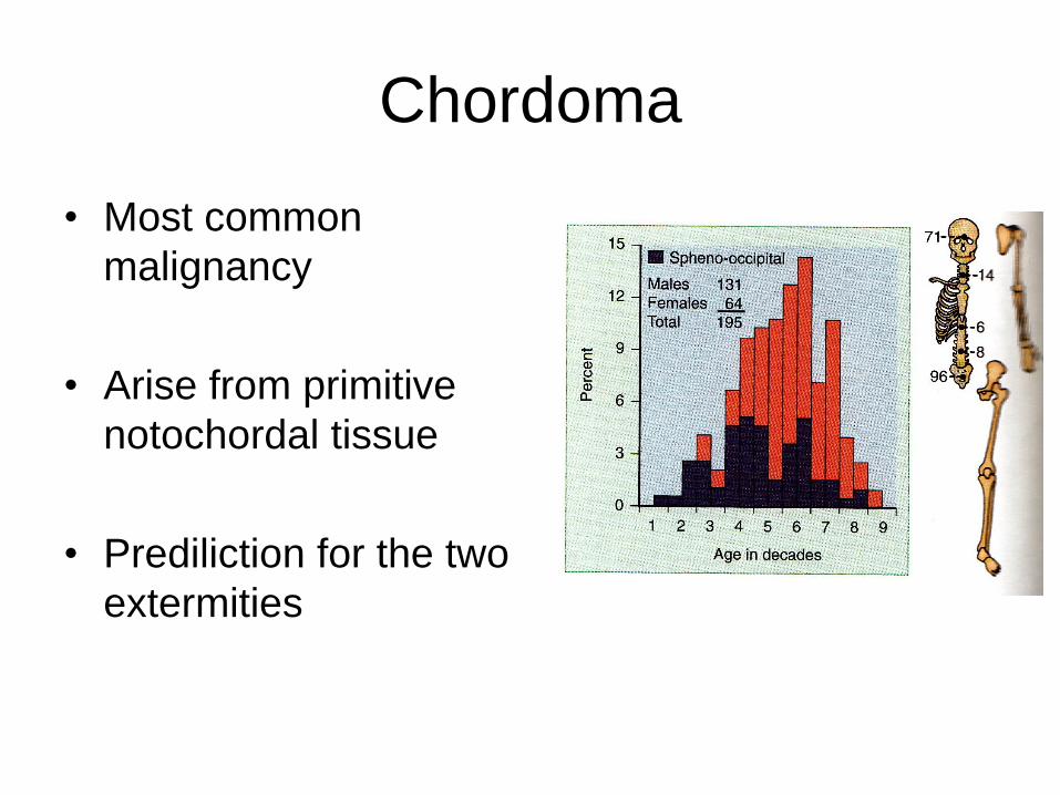
Chordoma
• Most common
malignancy
• Arise from primitive
notochordal tissue
• Prediliction for the two
extermities

Investigations - CT
• CT scan to distinguish
cystic, solid or mixed
• Involvement of
adjacent structures
• Bony destruction

Investigations - MRI
• Improved soft tissue
resolution helps plan
extent of resection
• Evaluation of marrow
involvement
• Identifies nerve root
and foraminal
involvement

Role of preoperative biopsy
• Will the biopsy change treatment ?
• Not needed if mass resectable
• Do not biopsy if lesion cystic (beware meningocele)
• Consider for osteogenic sarcomas

Well encapsulated fluid
intensity mass
Sagittal image showing the typical appearance of a tail gut cyst (cystic
hamartoma).
Surrounds the coccyx but
no overt bony destruction or
invasion

Sharply demarcated, fluid
containing mass with
internal septations
Coronal image showing the typical appearance of a tail gut cyst (cystic
hamartoma)
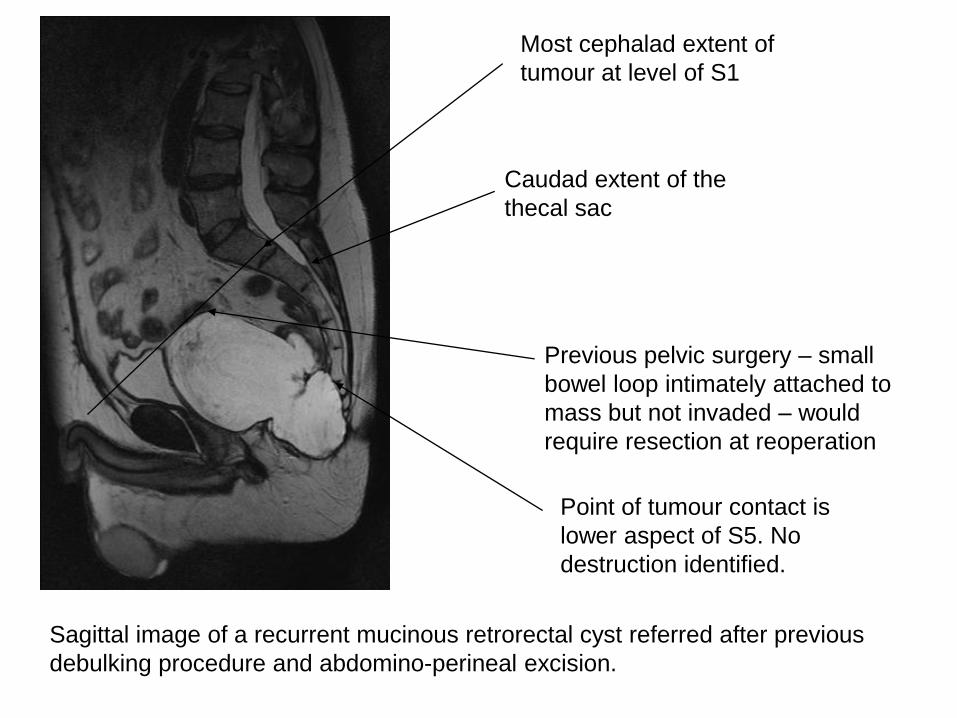
Sagittal image of a recurrent mucinous retrorectal cyst referred after previous
debulking procedure and abdomino-perineal excision.
Point of tumour contact is
lower aspect of S5. No
destruction identified.
Caudad extent of the
thecal sac
Previous pelvic surgery – small
bowel loop intimately attached to
mass but not invaded – would
require resection at reoperation
Most cephalad extent of
tumour at level of S1

Sagittal and coronal images showing the typical well demarcated
heterogenous appearance of a Schwannoma.

Sagittal image showing heterogenous
well-demarcated pre-sacral mass with no
gross evidence of bone invasion or
destruction.
Axial fat saturated post
gadolinium sequence showing
enhancing right sided pelvic mass
arising from the right S2 nerve
root
Schwannoma

Tumour abutting the coccyx and
lower sacrum
Uterus
Sagittal T2W image showing a heterogenous retrorectal mass (solitary fibrous
tumour) - well encapsulated, compresses the rectum and vagina anteriorly, and
abuts but does not invade the coccyx and lower sacrum posteriorly.
Bladder
Most cephalad extent of
tumour

T2W sagittal image showing a retrorectal tumour (chordoma). Although the
most cephalad tumour extent lies at the level of S1/2 (line
Most cephalad
limit of tumour
lying at the
level of S1/2
disc space
Sacral destruction
involving distal S2, S3
and S4. Note further
tumour extension
behind S2.
Retrorectal
tumour

Rectum displaced anteriorly
Tumour causing
destruction of S4 and
S5
Sagittal image showing the typical heterogenous high signal intensity of a
chordoma
Away from its sacral origin,
the tumour is encapsulated
and well demarcated.

chordoma
Coronal T2W image of a multi-septated heterogenous high signal intensity
retrorectal tumour (chordoma).

Axial CT image of chordoma showing anterior displacement of the rectum and
posterior bulge into the medial aspect right gluteal muscle.
Retrorectal tumour
(chordoma) displacing the
rectum anteriorly
Posterior tumour extension,
compressing the right gluteal
muscle.
Rectum

Axial T2W image showing a 2cm extension of a malignant retrorectal tumour
(myxoid liposarcoma) into the right greater sciatic notch (arrow).
Extension into greater sciatic notch

Important to identify
close proximity of
internal iliac vascular
bundle on pre-operative
imaging
Tumour exiting through
left greater sciatic notch
Axial post gadolinium fat saturated T1W image demonstrating crucial additional
information for the assessment of operability. Tumour extends into the left
greater sciatic notch. Appears well encapsulated rather than frankly infiltrative
so has potential to “shell out”.
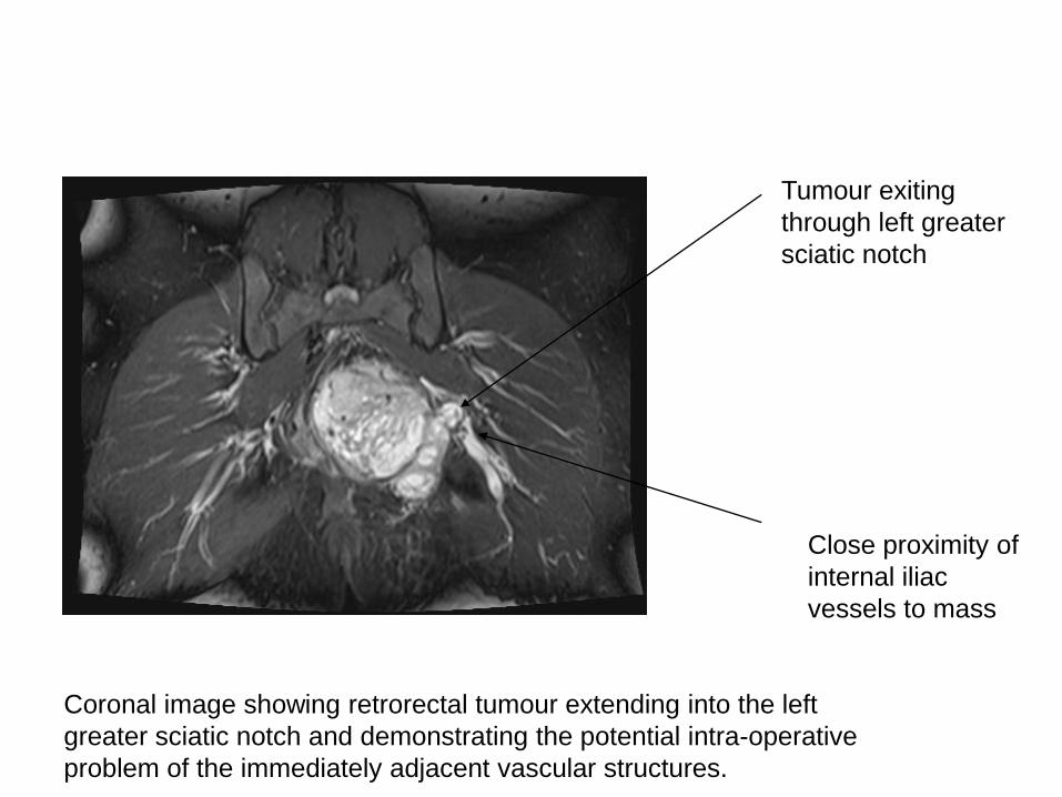
Tumour exiting
through left greater
sciatic notch
Close proximity of
internal iliac
vessels to mass
Coronal image showing retrorectal tumour extending into the left
greater sciatic notch and demonstrating the potential intra-operative
problem of the immediately adjacent vascular structures.

Algorithm for intra-operative decision-making for an adherent tumour with no radiological
evidence of sacral invasion
Circumferential excision
Benign features on MRI?
•Tumour in contact with adjacent pelvic side wall or viscera
•No invasion on MRI
•Tumour and adjacent structure do not separate on trial dissection
Yes
Extended circumferential excision with
En bloc wedge excision
No

Algorithm for surgical decision making in retrorectal tumours lying at S3 or above
Sacrum +/- Pelvic viscera involved
Adjacent structures involved?
Tumour lying at S3 or above
Yes
Abdominal approach
Circumferential excision
No
Pelvic viscera involved
YesYes No No
Invasion of pelvic visceraInvasion of sacrum?
Abdominal
approach
Abdominal
Approach
En bloc
Excision
Abdominal
Approach
Abdominal approach
Major sacral excision
+/- pelvic stabilisation
+/- en bloc excision
of viscera

Algorithm for surgical decision making in retrorectal tumours below S3
Sacrum only involved
Adjacent structures involved?
Tumour lying below S3
Yes
Size > 10cm
Or access difficult
NoPelvic side wall or viscera involved
Yes
Yes No
No
Invasion of pelvic viscera
Invasion of sacrum?Invasion of
Pelvic
Viscera
Composite
Abdomino-
Sacral
approach
Abdominal approach
Circumferential excision
Or perineal approach
+/- distal sacretomy
Perineal
approach
En bloc
excision
Yes No
Perineal
approach
Perineal
Approach
Excision of
Coccyx +/-
S4 and 5
Yes No
Abdominal
approach
Abdominal
approach
En bloc excision

Level of tumour and choice of
approach

Abdominal approach

Retrorectal schwanoma

Preservation of adjacent
structures

Posterior approach


Congenital cyst 23
Schwannoma 13
Chordoma 9
Ganglioneuroma 3
Liposarcoma 3
Gist 2
Solitary fibrous tumour 2
Angiomyxoma,Mucin
secreting,Leomyoma,Leomyosarcoma,
Rhabdomyosarcoma
5
Neuroendocrine , Neurofibroma,Dermoid with
extramammary paget’s,Myeolipoma
5
Others 10
Total 75

Tumor Abdominal Perineal Abdo-peri Total
Congenital cyst 7 15 1 23
Schwannoma 13 - - 13
Chordoma - 3 6 9
Ganglioneuroma 3 - - 3
Liposarcoma 1 2 - 3
GIST 1 1 - 2
Solitary fibrous tumour 2 -- - 2
Surgical approach

Summary
• Rare tumors but be aware of them
• Avoid biopsy if possible
• Imaging is crucial
• Approach determined by level of tumor

Leeds seriesTumor No of
cases
Sex ratio
F/M
Age group
Congenital cyst 23 18/5 20-88
Schwannoma 13 8/5 27-78
Chordoma 9 3/6 45-77
Ganglioneuroma 3 3/0 21-55
Liposarcoma 3 2/1 44-59
Gist 2 1/1 54-57
Solitary fibrous tumour 2 2/0 38-69
Angiomyxoma,Mucin
secreting,Leiomyoma,Leomyosarcoma,
Rhabdomyosarcoma
5
Neuroendocrine , Neurofibroma,Dermoid with
extramammary paget’s,Myeolipoma5
Others 10
Total 75 F>M