Embed Size (px)
Citation preview
Clinical Characteristics andTreatment of Langerhans
Cell HistiocytosisChalinee Monsereenusorn, MDa,Carlos Rodriguez-Galindo, MDa,b,*
KEYWORDS
� Langerhans cell histiocytosis � Neurodegeneration � Oncogenes � BRAF� Chemotherapy
KEY POINTS
� Langerhans cell histiocytosis (LCH) is a neoplasm of myeloid origin characterized by aclonal proliferation of CD1a1/CD2071 cells. In a little more than half of the cases,BRAFmutations, predominantly encoding BRAF V600E, are identified; mutations of othermembers of the MAPK/ERK pathway, such as MAP2K1 or ARAF, are present in another10% to 25%.
� LCH affects individuals of all ages, although infants more often present with multisystemdisease. The disease can affect many tissues and organs of the head and neck. Bonylesions are most common; but the skin, lymph nodes, and brain can also be involved.
� Patients with involvement of only one organ system can often be treated with surgeryalone and have excellent outcomes. Patients with multisystem disease, especially withrisk-organ involvement, need multimodality treatment and have variable prognoses.
� Treatment with BRAF inhibitors has shown to induce complete and durable responses,and the role of BRAF and MEK inhibitors is currently being investigated.
� LCH neurodegeneration, a devastating long-term complication of LCH, represents one ofthe major challenges in clinical and translational research of LCH.
INTRODUCTION
Langerhans cell histiocytosis (LCH) is a disease characterized by clonal proliferation ofCD1a1/CD2071 myeloid dendritic cells that presents at all ages and with differentdegrees of systemic involvement. Almost any organ can be affected, and the clinicalpresentation reflects the tissue-specific inflammatory phenomena. For several
a Department of Pediatric Oncology, Dana-Farber/Boston Children’s Cancer and Blood Dis-orders Center, 450 Brookline Avenue D3-133, Boston, MA 02215, USA; b Department ofPediatrics, Harvard Medical School, Boston, MA, USA* Corresponding author. Dana-Farber/Boston Children’s Cancer and Blood Disorders Center,450 Brookline Avenue D3-133, Boston MA 02215.E-mail address: [email protected]
Hematol Oncol Clin N Am 29 (2015) 853–873http://dx.doi.org/10.1016/j.hoc.2015.06.005 hemonc.theclinics.com0889-8588/15/$ – see front matter � 2015 Elsevier Inc. All rights reserved.
Monsereenusorn & Rodriguez-Galindo854
decades, LCH has been considered to be a reactive clonal proliferation of Langerhanscells; however, in recent years, LCH has been defined as a neoplasm of myeloid origin,with a significant inflammatory component that defines some of the acute and long-term manifestations. This evolution from an immune disorder to a neoplasia hasreframed the disease and opened the door for the development of targeted therapies.
BIOLOGY
The pathogenic cells are known to originate from a myeloid-derived precursor and areuniformly characterized by activation of the MAPK/ERK signaling pathway; ERK acti-vation is documented in all cases.1,2 In up to two-thirds of cases, pathway activationis secondary to a somatic mutation in BRAF (BRAFV600E); in other cases, mutations inMAP2K13 or less frequently in other members of the pathway, such as ARAF,4 havebeen described. About one-quarter of cases have no known genomic abnormalities.
EPIDEMIOLOGY
The estimated incidence of LCH is 8.9 cases per million children younger than 15 yearsper year, with a median age at diagnosis of 3 years old.5 The causes and risk factorsfor developing LCH are unclear.6 However, the unique patterns of presentation,ranging from localized bone lesions with spontaneous regression to disseminatedforms with multiorgan involvement, suggest a complex pathogenesis. Familial associ-ations, particularly the observation of increased incidence in monozygotic twins ofaffected patients, have suggested the presence of a germline predisposition at leastfor a proportion of cases.7 Also, population-based studies have shown differencesin the incidence of disseminated LCH by race and ethnic group; a higher incidencehas been reported for Hispanics and a lower incidence for blacks.8 Studies havealso shown a correlation with maternal and neonatal infections,6,9,10 lack of childhoodvaccinations,6,9 family history of thyroid disease,6 in vitro fertilization,11 and feedingproblems and transfusions during infancy.10 Finally, lower socioeconomic conditionshave been associated with an increased incidence of disseminated LCH.8
PATHOLOGY
Since pathologic Langerhans cells activate other immunologic cells, microscopicexamination of diseased tissue shows eosinophils, neutrophils, lymphocytes, andhistiocytes in addition to the LCH cells; this appearance is what has been traditionallydescribed as eosinophilic granuloma. Abscesses and necrosis may be present. LCHcells are large, oval , and mononuclear, with a prominent nucleus and eosinophiliccytoplasm. They do not have dendritic cell processes like cutaneous Langerhans cells.They stain positive for protein S-100, CD1a, and CD207 (langerin) and contain cyto-plasmic rod-shaped inclusions called Birbeck granules. A diagnosis of LCH is madeby typical positive staining with CD1a or CD207.12
With the development of new technology for accurate detection of cell-free DNA,BRAFV600E mutation analysis in plasma and urine has shown to be an effective toolfor diagnosis and monitoring of disease activity in patients with LCH.13
CLINICAL PRESENTATION
Classically, LCH was defined as 3 distinct diseases; eosinophilic granuloma, Hand-Schuller-Christian disease, and Abt-Letterer-Siwe disease were different clinical de-scriptions within the same spectrum of progressive system involvement. Eosinophilicgranuloma, whether solitary or multifocal, is found predominantly in older children as
Treatment of Langerhans Cell Histiocytosis 855
well as in young adults, with a peak incidence between 5 and 10 years of age. Eosin-ophilic granuloma is the most common form of LCH, accounting for approximately60% to 80% of the diagnoses. Hand-Schuller-Christian disease was historicallydescribed as a clinical triad of lytic bone lesions, exophthalmos caused by orbitalinvolvement, and diabetes insipidus (DI). It is most commonly described in the first4 to 7 years of life, and it may account for approximately 15% to 40% of all LCH cases.Abt-Letterer-Siwe disease is the most severe manifestation of LCH, albeit rare.Typically, patients are less than 2 years of age and present with a scaly seborrheicrash, ear discharge, and signs of severe systemic involvement with symptoms suchas cytopenias, pulmonary dysfunction, lymphadenopathy, or hepatosplenomegaly.14
Nowadays, this old terminology has been replaced by a classification system that isbased on the site of lesions, number of involved sites (single or multisystem/local ormultifocal), and whether the disease involves risk organs (hematopoietic system, liver,or spleen). This classification is the basis for the risk-adapted treatment used in theLCH-III protocol discussed later (Table 1).
SITES OF INVOLVEMENT
All organs can be affected by LCH; therefore, a comprehensive evaluation is indicated.The most commonly involved organs and systems are highlighted next:
Bone
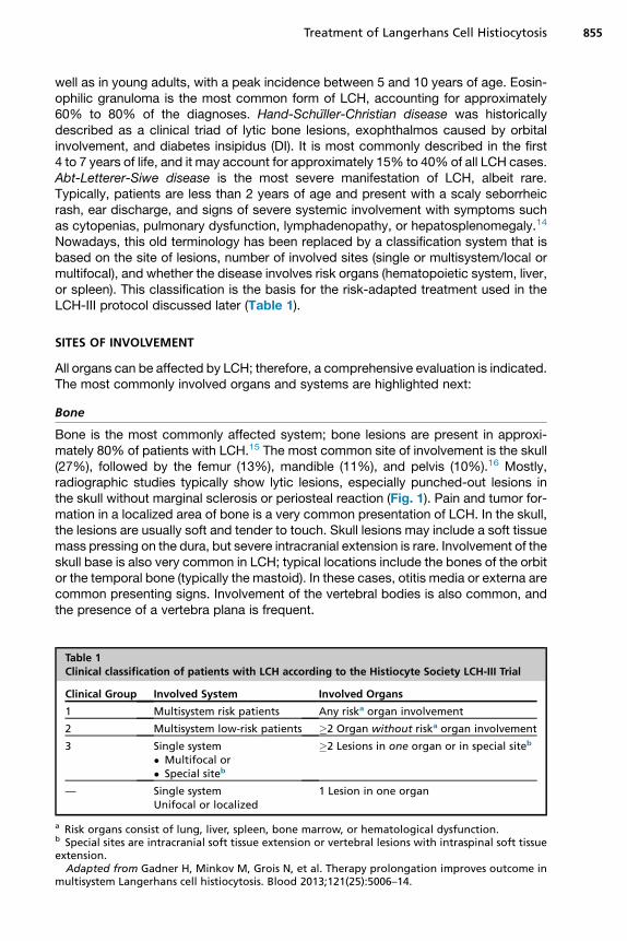
Bone is the most commonly affected system; bone lesions are present in approxi-mately 80% of patients with LCH.15 The most common site of involvement is the skull(27%), followed by the femur (13%), mandible (11%), and pelvis (10%).16 Mostly,radiographic studies typically show lytic lesions, especially punched-out lesions inthe skull without marginal sclerosis or periosteal reaction (Fig. 1). Pain and tumor for-mation in a localized area of bone is a very common presentation of LCH. In the skull,the lesions are usually soft and tender to touch. Skull lesions may include a soft tissuemass pressing on the dura, but severe intracranial extension is rare. Involvement of theskull base is also very common in LCH; typical locations include the bones of the orbitor the temporal bone (typically the mastoid). In these cases, otitis media or externa arecommon presenting signs. Involvement of the vertebral bodies is also common, andthe presence of a vertebra plana is frequent.
Table 1Clinical classification of patients with LCH according to the Histiocyte Society LCH-III Trial
Clinical Group Involved System Involved Organs
1 Multisystem risk patients Any riska organ involvement
2 Multisystem low-risk patients �2 Organ without riska organ involvement
3 Single system� Multifocal or� Special siteb
�2 Lesions in one organ or in special siteb
— Single systemUnifocal or localized
1 Lesion in one organ
a Risk organs consist of lung, liver, spleen, bone marrow, or hematological dysfunction.b Special sites are intracranial soft tissue extension or vertebral lesions with intraspinal soft tissueextension.
Adapted from Gadner H, Minkov M, Grois N, et al. Therapy prolongation improves outcome inmultisystem Langerhans cell histiocytosis. Blood 2013;121(25):5006–14.
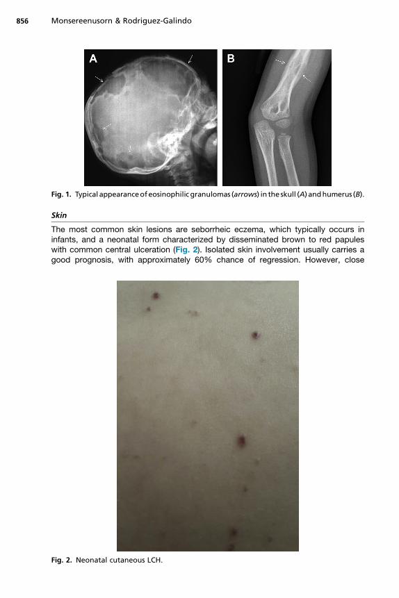
Fig. 1. Typical appearanceofeosinophilic granulomas (arrows) in the skull (A) andhumerus (B).
Monsereenusorn & Rodriguez-Galindo856
Skin
The most common skin lesions are seborrheic eczema, which typically occurs ininfants, and a neonatal form characterized by disseminated brown to red papuleswith common central ulceration (Fig. 2). Isolated skin involvement usually carries agood prognosis, with approximately 60% chance of regression. However, close
Fig. 2. Neonatal cutaneous LCH.

Treatment of Langerhans Cell Histiocytosis 857
monitoring is required, as reactivation or progression to multisystem involvement hasbeen observed in up to 40% of cases.12,17
Neuroendocrine and Central Nervous Systems
DI caused by involvement of the pituitary stalk occurs in approximately 25% of cases,usually after therapy. LCH is also a common diagnosis in patients with DI of unknowncause, and almost all patients with DI caused by LCH have involvement of other or-gans concurrently or subsequently.14 Anterior pituitary involvement, commonly repre-sented by growth hormone deficiency, is also a common complication and highlightsthe importance of a comprehensive endocrine monitoring in patients with LCH. Masslesions of the gray or white matter are less frequent (1%). Involvement of the sphenoid,orbital, ethmoid, zygomatic, or temporal bones confers a higher (25% overall) risk forcentral nervous system (CNS) involvement (CNS-risk lesions), including late neurode-generation (see later discussion), an inflammatory phenomenon of unclear pathogen-esis that is characterized by cerebellar dysfunction and neurocognitive deficits.18
Pulmonary
Involvement of the lungs usually occurs in the context of multisystem involvement,commonly limited to young children. Patients usually present with pulmonary dysfunc-tion including tachypnea, dyspnea, and cough. Radiographic findings are typical forthe presence of a reticulonodular pattern with bullae formation. Isolated pulmonaryinvolvement is a rare presentation that is almost exclusively found in adults with asmoking habit (discussed later).19
Hematopoietic System
The presence of hematopoietic dysfunction in the form of cytopenias is a poor prog-nostic sign. It occurs in the context of multisystem involvement, more frequently inyoung children. Its pathophysiology is multifactorial, including direct involvement ofthe bone marrow as well as peripheral destruction caused by hypersplenism fromLangerhans cell infiltrates in the spleen.20
Hepatobiliary System
Liver involvement, which typically occurs in infants with multisystem disease, alsocarries a poor prognosis. Patients present with hypoalbuminemia, edema, hepato-megaly, or conjugated hyperbilirubinemia. A well-described complication of hepaticinvolvement is the development of sclerosing cholangitis and hepatic fibrosis, whichcan result in liver failure and need for liver transplantation.14
TREATMENT OF LANGERHANS CELL HISTIOCYTOSIS
The treatment of LCH over the years has reflected the changing concepts of the dis-ease process. Indeed, the difficulties in developing more effective therapies are linkedto the deficiencies in the understanding of the pathogenesis of LCH. Retrospectivestudies of Lahey21 and Komp and colleagues22 showed that, although many organscan harbor proliferating pathogenic cells, only if organ function was disrupted wassuch involvement of prognostic significance. Patients could then be stratified intodifferent risk categories based on the extent of their disease and the degree of organdysfunction. Patients with single-system disease confined to a single site usuallyrequire only local therapy or observation. Patients with more extensive disease requiresystemic therapy; several groups have explored risk-based approaches, which aresummarized next.
Monsereenusorn & Rodriguez-Galindo858
Histiocyte Society’s Langerhans Cell Histiocytosis Studies
Langerhans Cell Histiocytosis-I (LCH-I)The first international randomized trial for multisystem LCH (MS-LCH), LCH-I (1991–1995), randomized patients to receive weekly vinblastine or etoposide every 3 weeksfor 24 weeks. Overall survival was close to 80%, with no significant differencesby treatment arm. Involvement of the hematopoietic system, lung, liver, and spleen,age at diagnosis less than 2 years old, and poor response at 6 weeks were signifi-cantly associated with an adverse outcome.23 In comparison with the contempora-neous DAL-HX 83 study, which explored a more intensive and longer regimen,LCH-I showed a lower 6-week response rate (50% vs 80%) and a higher reactivationrate (50% vs 30%).24
Langerhans Cell Histiocytosis-II (LCH-II)The LCH-II study (1996–2001) followed on the LCH-I findings and explored early inten-sification through a randomized design that investigated the addition of etoposide(arm B) to a standard 6-week induction with prednisone and vinblastine and continu-ation with 6-mercaptopurine and every-3-week pulses of prednisone and vinblastinefor a total of 24 weeks of therapy (arm A).25 Both arms produced similar outcomesin terms of 6-week response rates (63% arm A vs 71% arm B), 5-year survivalprobability (74% vs 79%), and disease reactivation rates (46% both arms); however,the more intensive arm B resulted in reduced mortality for patients with risk organinvolvement. The LCH-II study also showed that patients younger than 2 years withoutrisk-organ involvement have excellent response rates and a 100% survival, andpatients with risk-organ involvement and poor response at 6 weeks have the highestmortality.25 Those findings helped refine the risk-stratification and treatment for itssuccessor study, the LCH-III.
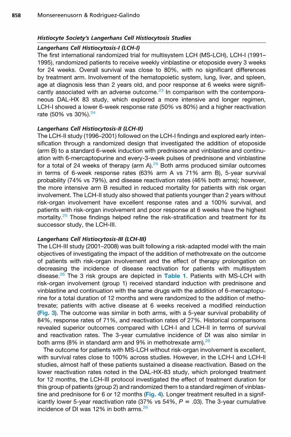
Langerhans Cell Histiocytosis-III (LCH-III)The LCH-III study (2001–2008) was built following a risk-adapted model with the mainobjectives of investigating the impact of the addition of methotrexate on the outcomeof patients with risk-organ involvement and the effect of therapy prolongation ondecreasing the incidence of disease reactivation for patients with multisystemdisease.26 The 3 risk groups are depicted in Table 1. Patients with MS-LCH withrisk-organ involvement (group 1) received standard induction with prednisone andvinblastine and continuation with the same drugs with the addition of 6-mercaptopu-rine for a total duration of 12 months and were randomized to the addition of metho-trexate; patients with active disease at 6 weeks received a modified reinduction(Fig. 3). The outcome was similar in both arms, with a 5-year survival probability of84%, response rates of 71%, and reactivation rates of 27%. Historical comparisonsrevealed superior outcomes compared with LCH-I and LCH-II in terms of survivaland reactivation rates. The 3-year cumulative incidence of DI was also similar inboth arms (8% in standard arm and 9% in methotrexate arm).26
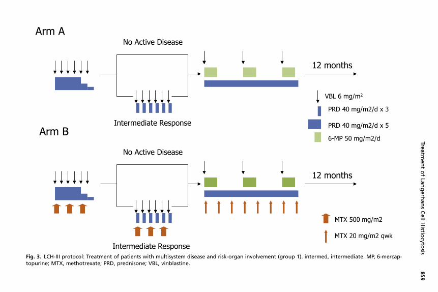
The outcome for patients with MS-LCH without risk-organ involvement is excellent,with survival rates close to 100% across studies. However, in the LCH-I and LCH-IIstudies, almost half of these patients sustained a disease reactivation. Based on thelower reactivation rates noted in the DAL-HX-83 study, which prolonged treatmentfor 12 months, the LCH-III protocol investigated the effect of treatment duration forthis group of patients (group 2) and randomized them to a standard regimen of vinblas-tine and prednisone for 6 or 12 months (Fig. 4). Longer treatment resulted in a signif-icantly lower 5-year reactivation rate (37% vs 54%, P 5 .03). The 3-year cumulativeincidence of DI was 12% in both arms.26
Arm A
12 months
No Active Disease
Intermediate Response
12 months
No Active Disease
Intermediate Response
Arm B
VBL 6 mg/m2
PRD 40 mg/m2/d x 3
PRD 40 mg/m2/d x 5
6-MP 50 mg/m2/d
MTX 500 mg/m2
MTX 20 mg/m2 qwk
Fig. 3. LCH-III protocol: Treatment of patients with multisystem disease and risk-organ involvement (group 1). intermed, intermediate. MP, 6-mercap-topurine; MTX, methotrexate; PRD, prednisone; VBL, vinblastine.
Treatm
entofLa
ngerhansCellHistio
cytosis
859
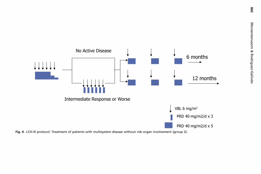
6 monthsNo Active Disease
Intermediate Response or Worse
12 months
VBL 6 mg/m2
PRD 40 mg/m2/d x 3
PRD 40 mg/m2/d x 5Fig. 4. LCH-III protocol: Treatment of patients with multisystem disease without risk-organ involvement (group 2).
Monsereenusorn
&Rodrig
uez-G
alin
do
860
Treatment of Langerhans Cell Histiocytosis 861
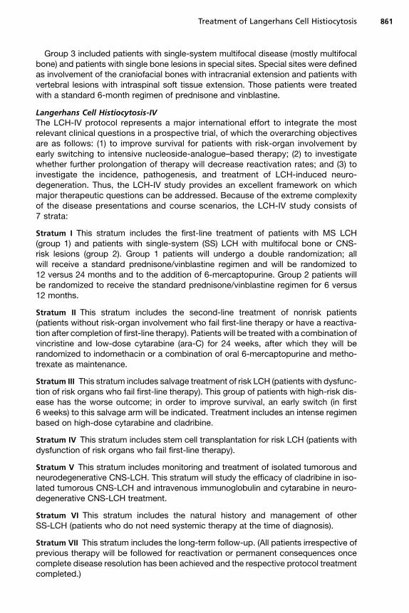
Group 3 included patients with single-system multifocal disease (mostly multifocalbone) and patients with single bone lesions in special sites. Special sites were definedas involvement of the craniofacial bones with intracranial extension and patients withvertebral lesions with intraspinal soft tissue extension. Those patients were treatedwith a standard 6-month regimen of prednisone and vinblastine.
Langerhans Cell Histiocytosis-IVThe LCH-IV protocol represents a major international effort to integrate the mostrelevant clinical questions in a prospective trial, of which the overarching objectivesare as follows: (1) to improve survival for patients with risk-organ involvement byearly switching to intensive nucleoside-analogue–based therapy; (2) to investigatewhether further prolongation of therapy will decrease reactivation rates; and (3) toinvestigate the incidence, pathogenesis, and treatment of LCH-induced neuro-degeneration. Thus, the LCH-IV study provides an excellent framework on whichmajor therapeutic questions can be addressed. Because of the extreme complexityof the disease presentations and course scenarios, the LCH-IV study consists of7 strata:
Stratum I This stratum includes the first-line treatment of patients with MS LCH(group 1) and patients with single-system (SS) LCH with multifocal bone or CNS-risk lesions (group 2). Group 1 patients will undergo a double randomization; allwill receive a standard prednisone/vinblastine regimen and will be randomized to12 versus 24 months and to the addition of 6-mercaptopurine. Group 2 patients willbe randomized to receive the standard prednisone/vinblastine regimen for 6 versus12 months.
Stratum II This stratum includes the second-line treatment of nonrisk patients(patients without risk-organ involvement who fail first-line therapy or have a reactiva-tion after completion of first-line therapy). Patients will be treated with a combination ofvincristine and low-dose cytarabine (ara-C) for 24 weeks, after which they will berandomized to indomethacin or a combination of oral 6-mercaptopurine and metho-trexate as maintenance.
Stratum III This stratum includes salvage treatment of risk LCH (patients with dysfunc-tion of risk organs who fail first-line therapy). This group of patients with high-risk dis-ease has the worse outcome; in order to improve survival, an early switch (in first6 weeks) to this salvage arm will be indicated. Treatment includes an intense regimenbased on high-dose cytarabine and cladribine.
Stratum IV This stratum includes stem cell transplantation for risk LCH (patients withdysfunction of risk organs who fail first-line therapy).
Stratum V This stratum includes monitoring and treatment of isolated tumorous andneurodegenerative CNS-LCH. This stratum will study the efficacy of cladribine in iso-lated tumorous CNS-LCH and intravenous immunoglobulin and cytarabine in neuro-degenerative CNS-LCH treatment.
Stratum VI This stratum includes the natural history and management of otherSS-LCH (patients who do not need systemic therapy at the time of diagnosis).
Stratum VII This stratum includes the long-term follow-up. (All patients irrespective ofprevious therapy will be followed for reactivation or permanent consequences oncecomplete disease resolution has been achieved and the respective protocol treatmentcompleted.)
Monsereenusorn & Rodriguez-Galindo862
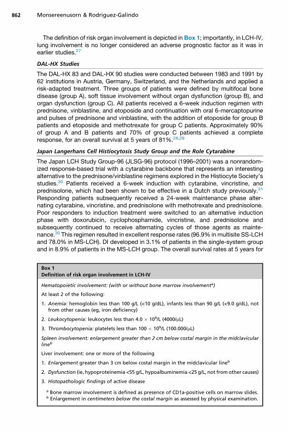
The definition of risk organ involvement is depicted in Box 1; importantly, in LCH-IV,lung involvement is no longer considered an adverse prognostic factor as it was inearlier studies.27
DAL-HX Studies
The DAL-HX 83 and DAL-HX 90 studies were conducted between 1983 and 1991 by62 institutions in Austria, Germany, Switzerland, and the Netherlands and applied arisk-adapted treatment. Three groups of patients were defined by multifocal bonedisease (group A), soft tissue involvement without organ dysfunction (group B), andorgan dysfunction (group C). All patients received a 6-week induction regimen withprednisone, vinblastine, and etoposide and continuation with oral 6-mercaptopurineand pulses of prednisone and vinblastine, with the addition of etoposide for group Bpatients and etoposide and methotrexate for group C patients. Approximately 90%of group A and B patients and 70% of group C patients achieved a completeresponse, for an overall survival at 5 years of 81%.28,29
Japan Langerhans Cell Histiocytosis Study Group and the Role Cytarabine
The Japan LCH Study Group-96 (JLSG-96) protocol (1996–2001) was a nonrandom-ized response-based trial with a cytarabine backbone that represents an interestingalternative to the prednisone/vinblastine regimens explored in the Histiocyte Society’sstudies.30 Patients received a 6-week induction with cytarabine, vincristine, andprednisolone, which had been shown to be effective in a Dutch study previously.31
Responding patients subsequently received a 24-week maintenance phase alter-nating cytarabine, vincristine, and prednisolone with methotrexate and prednisolone.Poor responders to induction treatment were switched to an alternative inductionphase with doxorubicin, cyclophosphamide, vincristine, and prednisolone andsubsequently continued to receive alternating cycles of those agents as mainte-nance.30 This regimen resulted in excellent response rates (96.9% in multisite SS-LCHand 78.0% in MS-LCH). DI developed in 3.1% of patients in the single-system groupand in 8.9% of patients in the MS-LCH group. The overall survival rates at 5 years for
Box 1
Definition of risk organ involvement in LCH-IV
Hematopoietic involvement: (with or without bone marrow involvementa)
At least 2 of the following:
1. Anemia: hemoglobin less than 100 g/L (<10 g/dL), infants less than 90 g/L (<9.0 g/dL), notfrom other causes (eg, iron deficiency)
2. Leukocytopenia: leukocytes less than 4.0 � 109/L (4000/mL)
3. Thrombocytopenia: platelets less than 100 � 109/L (100.000/mL)
Spleen involvement: enlargement greater than 2 cm below costal margin in the midclavicularlineb
Liver involvement: one or more of the following
1. Enlargement greater than 3 cm below costal margin in the midclavicular lineb
2. Dysfunction (ie, hypoproteinemia <55 g/L, hypoalbuminemia <25 g/L, not from other causes)
3. Histopathologic findings of active disease
a Bone marrow involvement is defined as presence of CD1a-positive cells on marrow slides.b Enlargement in centimeters below the costal margin as assessed by physical examination.
Treatment of Langerhans Cell Histiocytosis 863
the single-system and MS groups were 100% and 94.4% �3.2%, respectively; reac-tivation rates were 28.1% for single-system and 45.3% for MS.30 The good results ofthis cytarabine-based regimen have provided the rationale for the low-risk salvageregimen in LCH-IV and have prompted the proposal to consider cytarabine as agood alternative to vinblastine and prednisolone for front-line treatment of LCH.32
Because of the high reactivation rates in JLSG-96, the successor study JLSG-02was adapted to increase the cumulative dose of prednisolone during induction,to add cyclosporin A to the regimen for poor responders, and to increase theduration of continuation therapy with the addition of 6-mercaptopurine, vinblas-tine, prednisolone, and methotrexate for an additional 18 weeks, increasing thetotal duration of treatment to 12 months. Preliminary results showed an increasein the initial response rates and a decrease in disease reactivation for patientswith MS-LCH.33
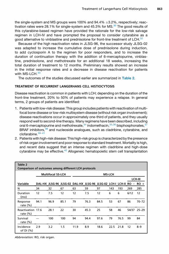
The outcomes of the studies discussed earlier are summarized in Table 2.
TREATMENT OF RECURRENT LANGERHANS CELL HISTIOCYTOSIS
Disease reactivation is common in patients with LCH; depending on the duration of thefront-line treatment, 20% to 50% of patients may experience a relapse. In generalterms, 2 groups of patients are identified:
1. Patientswith low-riskdisease: Thisgroup includespatientswith reactivationofmulti-focal bone disease or low-riskmultisystemdisease (without risk organ involvement);disease reactivations occur in approximately one-third of patients, and they usuallyrespondwell to second-line therapy.Many regimens havebeendescribed, includingoral 6-mercaptopurine and methotrexate,34 indomethacin,35,36 bisphosphonates,37
BRAF inhibitors,38 and nucleoside analogues, such as cladribine, cytarabine, andclofarabine.39–41
2. Patientswith high-risk disease: This high-risk group is characterizedby thepresenceof risk organ involvement and poor response to standard treatment.Mortality is high,and recent data suggest that an intense regimen with cladribine and high-dosecytarabine may be effective.42 Allogeneic hematopoietic stem cell transplantation
Table 2Comparison of outcomes among different LCH protocols
Variable
Multifocal SS-LCH MS-LCH
DAL-HX JLSG-96 JLSG-02 DAL-HX JLSG-96 JLSG-02 LCH-I LCH-II
LCH-III
RO L RO D
N 34 32 67 63 59 97 143 193 269 285
Duration(mo)
12 7.5 12 12 7.5 12 6 6 6/12 12
Responserate (%)
94.1 96.9 85.1 79 76.3 84.5 53 67 86 70–72
Reactivationrate (%)
17.6 28.1 22 30 45.3 25 58 46 54/37 25–29
Survivalrate (%)
— 100 100 94 94.4 97.6 79 76.5 99 84
Incidenceof DI (%)
2.9 3.2 1.5 11.9 8.9 18.6 22.5 21.8 12 8–9
Abbreviation: RO, risk organ.
Monsereenusorn & Rodriguez-Galindo864
has also beenproposed for those cases.43 BRAF inhibitorsmay also play a role in thetreatment of this group of patients.
2-Chlorodeoxyadenosine (Cladribine)
The analogue 2-chlorodeoxyadenosine (cladribine) is a purine analogue that inchildren has mainly been used in the treatment of acute myeloid leukemia, wherebyresponses of up to 59% are obtained as a single agent.44 Excellent clinical responses,with an overall response rate of 82%, were initially reported in adults with recurrentLCH.45 This finding prompted the study of this agent further in children with recurrentLCH. For patients with low-risk disease reactivation, cladribine (5 mg/m2/d � 5 days)has shown to induce responses in greater than 90% of the patients, althoughfurther reactivations still occur.39,46–48 One additional benefit of cladribine is its effecton CNS disease.49 A major problem with the use of cladribine is its limitation to a shorttreatment course; more than 4 or 6 courses of treatment are associated withprolonged myelosuppression, causing a clinical picture similar to myelodysplasia.For patients with high-risk disease, cladribine as a single agent has little effect,47
and a more intensive regimen is needed. The combination of higher dosages of cladri-bine (9 mg/m2/d � 5 days) with cytarabine (1 g/m2/d � 5 days) has induced completeresponses in patients with refractory disease.42,50 Although this treatment is effective,it is associated with high morbidity, with treatment-related mortality in excess of 20%.Recent data suggest that a lower-dosage regimen (cytarabine 100 mg/m2/d � 5 dayswith cladribine 5 mg/m2/d� 5 days) may also be effective in this group of patients withhigh-risk disease.51
Clofarabine
Clofarabine is a second-generation deoxyadenosine analogue with documented effi-cacy in the treatment of childrenwith relapsed acute lymphoblastic leukemia and acutemyeloid leukemia.52,53 Given the good results obtained with cladribine in the manage-ment of recurrent LCH, there could be a role for the use of this agent in this same pop-ulation. Clofarabine monotherapy at dosages of 25 to 30mg/m2/d� 5 days has shownto induce significant responses, including patients with MS-LCH with risk-organinvolvement and patients with refractory disease to cladribine and cytarabine.40,41,54
Moreover, clofarabine has demonstrated activity against juvenile xanthogranulomaand Rosai–Dorfman disease.41
Cytarabine
Cytarabine, alone or in combination with corticosteroids or vincristine, has shownefficacy against refractory LCH, particularly in cases with CNS disease.31,55 Dosagesof 100 to 150 mg/m2/d � 5 days have been typically used, and toxicity seems to below.55 Cytarabine at varying doses (100 mg to 1 g/m2/d � 5 days) has been used incombination with cladribine for the treatment of patients with refractory LCH withrisk-organ involvement (see earlier discussion).42,51
Bisphosphonates
Bisphosphonates are pyrophosphate analogues that inhibit the recruitment of osteo-clasts and reduce their activity and durability. Pamidronate has been used for thetreatment of bony lesions in patients without involvement of risk organs. Given at adosage of 1 mg/kg every 4 weeks for 6 doses, the 3-year progression-free survivalwas 56.3% �12.4%.37 The outcome was significantly better in children with a firstreactivation compared with second or subsequent reactivations. The results of this
Treatment of Langerhans Cell Histiocytosis 865
study underline the efficacy of bisphosphonates for the treatment of bony recurrentLCH, although prospective trials are needed to confirm the efficacy and safety ofbisphosphonates in this condition.56 Zoledronic acid has also shown to be effectivein adults with multifocal bone disease.57
Indomethacin
Prostaglandins have been implicated in the pathogenesis of the bone lesions in LCH.35
Treatment with indomethacin at dosages of 1 to 2 mg/kg/d given for a median of4 months has shown to induce responses and regression of bone lesions in morethan 90% of patients with symptomatic single-system bone lesions, either at diag-nosis or at the time of relapse.36 These findings provide the rationale for the use ofthis agent in the treatment of low-risk reactivations proposed in LCH-IV.
BRAF Inhibitors
The presence of the oncogenic BRAF V600E mutation in up to two-thirds of patients,as well as its association with an increased risk of reactivation, provides a very strongrationale for the use of BRAF inhibitors.58 The BRAF V600E gain-of-function mutationshave also been observed in a large proportion of patients with Erdheim-Chesterdisease (ECD).59 Vemurafenib has been reported to induce significant responses inpatients with multisystem and refractory ECD and LCH carrying the BRAF V600Emutation.38,60 Contrary to what has been observed with other malignancies, no drugresistance has been reported.60 Given the universal activation of the MAPK pathwayregardless of BRAF mutational status, the role of MEK inhibitors in the treatment ofLCH is currently being explored.
Imatinib Mesylate
Imatinibmesylate has shown an inhibitory effect on differentiation of CD341 progenitorcells intodendritic cells, thusproviding some rationale for its use in histiocytic disorders.Clinical and radiological responses have been observed in adult patients with LCH andECD either as first- or second-line treatment.61
Hematopoietic Stem Cell Transplantation
Despite progress made in the treatment of LCH, a significant proportion of patientswith risk-organ involvement fail to respond to standard or first-line salvage regimens.Hematopoietic stem cell transplantation may provide an effective alternative throughhigh-dose chemotherapy, replacement of stem cells, generation of a graft-versus-LCH effect, and immunomodulation. In one study, the 10-year overall survival forpatients with risk-organ involvement at diagnosis was 50%.62 Because of the previoustreatment-related effects, reduced intensity conditioning regimens are a preferableprocedure with low transplant-related morbidity and mortality.43
SPECIAL CONSIDERATIONSAdult Langerhans Cell Histiocytosis
The rarity of LCH in adults combined with the nonspecific and varied clinical presenta-tions typically result in missed and delayed diagnoses.63,64 Adult LCH usually presentsafter the fourth decade, and most patients have multisystem involvement at diagnosis.In 6% of the cases, patients have been diagnosed with other neoplastic diseases. Thelung is the organ most frequently involved; this presentation, either alone or as part ofmultisystem disease, is strongly associated with a smoking habit.65 In general, the clin-ical presentation and organs involved are similar to pediatric patients, with morefrequent involvement of the genitalia, particularly in women.66
Monsereenusorn & Rodriguez-Galindo866
Treatment of LCH in adults generally follows similar guidelines as pediatric LCHbut with some modifications.63 The more severe skin manifestations have shown torespond well to phototherapy67 and low-dose methotrexate.68 For patients requiringsystemic therapy, the adverse side effects associated with corticosteroid therapyand vinblastine may limit treatment compliance; for this reason, treatment with cytar-abine or cladribine is generally preferred,63,69 although BRAF inhibitors are beingincreasingly used in this population.38
Pulmonary Langerhans Cell Histiocytosis
Lung involvement in children with LCH usually presents in the context of multisystemdisease. In contrast, pulmonary involvement in adults usually occurs in isolation; it isstrongly associated with cigarette smoking, typically in the third to fifth decades oflife.19 The frequency of BRAF V600E mutations in lung lesions of LCH in adult patientsis lower (less than one-third of the cases), but the presence of the mutation seems tobe associated with the cumulative tobacco exposure.70 The clinical presentation ofpulmonary LCH is usually a nonspecific dry cough; the interval between the onsetof clinical symptoms and diagnosis varies but is approximately 6 months. The mostcommon finding on chest radiography is the presence of a reticulo-micronodularinfiltration, with cyst formation in more advanced cases. High-resolution computedtomography is required for better diagnosis; typical findings are the presence of small,often cavitated nodules and thin-wall cysts. Pulmonary function test usually showreduced carbon monoxide diffusing capacity in 70% to 90% of the patients. Thegold standard for diagnosis is lung biopsy and the identification of activated patho-genic LCH cells in loose granulomas. The natural history of pulmonary LCH is widelyvariable; smoking cessation is mandatory for improving, and up to 50% of the patientsexperience a favorable outcome either spontaneously or with a short course of ste-roids.19 Patients with suboptimal responses after smoking cessation and steroid ther-apy may benefit from treatment with cladribine.63,71 In cases of advanced disease,lung transplantation may be required.72
Central Nervous System Langerhans Cell Histiocytosis
CNS involvement may present in different forms and over a long period of time, rep-resenting a spectrum of diseases ranging from active infiltration by LCH to long-termeffects. The classic clinical presentation is diabetes insipidus18; however, with thewide use of advanced neuroimaging, CNS alterations in patients with LCH are morefrequent than previously thought.73 The risk factors for CNS-LCH include the presenceof CNS-risk lesions and extended disease activity.18 The clinical presentation of CNS-LCH depends on the site and type of CNS involvement. DI, the most common presen-tation, is the hallmark of hypothalamic pituitary involvement, which has been found in25% of all patients with LCH and in up to 50% of patients with MS-LCH.74,75 Growthhormone deficiency is the second most common endocrinopathy, and it occurs in upto 50% of patients with LCH with DI.76 Prolonged systemic therapy seems to be asso-ciated with a lower incidence of endocrinopathies; in the most recent studies, the inci-dence of DI in patients with MS-LCH is approximately 10%.26 Patients with tumorouslesions may present with headache, seizures, focal neurologic deficits, or signs andsymptoms of increased intracranial pressure. A not-yet-well-quantified proportion ofpatients with a history of LCH develop a progressive neurodegenerative disease.This risk is up to 10% in patients with DI.75 Some asymptomatic patients may havetypical MRI changes of radiologic neurodegeneration for years; however, othershave clinical neurodegeneration, including subtle tremor, abnormal reflexes, gaitdisturbance, motor spasticity, ataxia, dysarthria, dysphagia, behavioral changes,
Treatment of Langerhans Cell Histiocytosis 867
learning disorders, or psychiatric problems.18 The management of patients withCNS-LCH consists of a complete neurologic examination, neuropsychological testing,endocrine and ophthalmologic evaluation, and neuroimaging. Brain biopsy is onlyindicated for patients without an established diagnosis of LCH who have a negativediagnostic workup for extracranial LCH lesions.The most crucial investigative tool for CNS involvement by LCH is MRI. The classi-
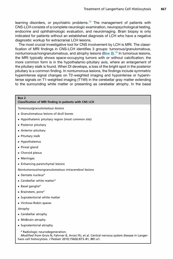
fication of MRI findings in CNS-LCH identifies 3 groups: tumorous/granulomatous,nontumorous/nongranulomatous, and atrophy lesions (Box 2).18 In tumorous lesions,the MRI typically shows space-occupying tumors with or without calcification; themore common form is in the hypothalamic-pituitary axis, where an enlargement ofthe pituitary stalk is found. When DI develops, a loss of the bright spot in the posteriorpituitary is a common finding. In nontumorous lesions, the findings include symmetrichyperintense signal changes on T2-weighted imaging and hypointense or hyperin-tense signals on T1-weighted imaging (T1WI) in the cerebellar gray matter extendingto the surrounding white matter or presenting as cerebellar atrophy. In the basal
Box 2
Classification of MRI finding in patients with CNS LCH
Tumorous/granulomatous lesions
� Granulomatous lesions of skull bones
� Hypothalamic pituitary region (most common site)
� Posterior pituitary
� Anterior pituitary
� Pituitary stalk
� Hypothalamus
� Pineal gland
� Choroid plexus
� Meninges
� Enhancing parenchymal lesions
Nontumorous/nongranulomatous intracerebral lesions
� Dentate nucleusa
� Cerebellar white mattera
� Basal gangliaa
� Brainstem, ponsa
� Supratentorial white matter
� Virchow-Robin spaces
Atrophy
� Cerebellar atrophy
� Midbrain atrophy
� Supratentorial atrophy
a Radiologic neurodegeneration.Modified from Grois N, Fahrner B, Arceci RJ, et al. Central nervous system disease in Langer-
hans cell histiocytosis. J Pediatr 2010;156(6):873–81, 881.e1.
Monsereenusorn & Rodriguez-Galindo868
ganglia, the MRI findings include hyperintense signals on T1WI usually involving theglobus pallidus. These findings are commonly referred to as radiological neurodegen-eration.18,77 Two other patterns of parenchymal white matter changes include thepresence of dilated Virchow-Robin spaces and a leukoencephalopathylike patternthat may involve the cerebellum, pons, and periventricular white matter.18
Cerebrospinal fluid studies are not generally recommended in all patients with LCHwith CNS symptoms; however, they should be considered in the context of investigationof biomarkers of neurodegeneration. The histopathology of CNS-LCH is notwell definedbecause of the lack of comprehensive studies and limited availability of tissue speci-mens. In general, 3 histopathologic patterns have been described: (1) circumscribedgranulomas with a variable presence of CD1a1 cells and a strong CD81 T-lymphocyticinfiltration; (2) neurodegenerative lesions with negative CD1a and a profound CD81 in-flammatory process, associated with neuronal and axonal degeneration and secondarymyelin loss; and (3) granulomas in infundibular tumors invading the hypothalamus withdiffuse infiltration of the surrounding CNS parenchyma by CD1a1 histiocytes.18
There are no standard guidelines for the treatment of CNS-LCH. For tumorouslesions and new-onset DI, treatment with a standard LCH regimen is indicated;vinblastine and prednisone or single-agent cladribine have shown to be effective.49,78
Treatment of neurodegenerative disease is less well defined. Improvement in theneurologic condition has been reported with the use of cytarabine55 and intravenousimmunoglobulin.79 Anecdotal responses or disease stabilization have also been docu-mented with infliximab80 and cis-retinoic acid.81 However, a better understanding ofthe pathophysiology of this syndrome is required to develop more rational treatments.
SUMMARY AND FUTURE DIRECTIONS
LCH is a neoplasm of myeloid origin characterized by activation of the MAPK/ERKpathway and with a wide spectrum of clinical presentations and variable outcomes.With current risk-adapted treatments, more than 80% of patients are cured. However,30% to 50% of patients experience disease recurrence; a significant number of sur-vivors develop neurodegeneration. Treatments aimed at achieving long-term cureswith low reactivation rates and that could prevent the development of neurodegener-ation need to be developed. Targeted therapies with BRAF or MEK inhibitors offer thepossibility of reframing the treatment of LCH in the near future.
REFERENCES
1. Badalian-Very G, Vergilio JA, Degar BA, et al. Recent advances in the under-standing of Langerhans cell histiocytosis. Br J Haematol 2012;156(2):163–72.
2. Allen CE, Li L, Peters TL, et al. Cell-Specific gene expression in Langerhans cellhistiocytosis lesions reveals a distinct profile compared with epidermal Langer-hans cells. J Immunol 2010;184(8):4557–67.
3. Chakraborty R, Hampton OA, Shen X, et al. Mutually exclusive recurrent somaticmutations in MAP2K1 and BRAF support a central role for ERK activation in LCHpathogenesis. Blood 2014;124(19):3007–15.
4. Nelson DS, Quispel W, Badalian-Very G, et al. Somatic activating ARAF mutationsin Langerhans cell histiocytosis. Blood 2014;123(20):3152–5.
5. Stalemark H, Laurencikas E, Karis J, et al. Incidence of Langerhans cell histiocy-tosis in children: a population-based study. Pediatr Blood Cancer 2008;51(1):76–81.
6. Bhatia S, Nesbit ME Jr, Egeler M, et al. Epidemiologic study of Langerhans cellhistiocytosis in children. J Pediatr 1997;130(5):774–84.
Treatment of Langerhans Cell Histiocytosis 869
7. Arico’ M, Scappaticci S, Danesio C. The genetics of Langerhans cell histiocyto-sis. In: Weitzman S, Egeler RM, editors. Histiocytic disorders of children andadults. Cambridge (United Kingdom): Cambridge University Press; 2005.p. 83–94.
8. Ribeiro KB, Degar B, Antoneli CBG, et al. Ethnicity, race, and socioeconomic sta-tus influence incidence of Langerhans cell histiocytosis. Pediatr Blood Cancer2015;62(6):982–7.
9. Venkatramani R, Rosenberg S, Indramohan G, et al. An exploratory epidemiolog-ical study of Langerhans cell histiocytosis. Pediatr Blood Cancer 2012;59(7):1324–6.
10. Hamre M, Hedberg J, Buckley J, et al. Langerhans cell histiocytosis: an explor-atory epidemiologic study of 177 cases. Med Pediatr Oncol 1997;28(2):92–7.
11. Akefeldt SO, FinnstromO,GavhedD, et al. Langerhans cell histiocytosis in childrenborn 1982-2005 after in vitro fertilization. Acta Paediatr 2012;101(11):1151–5.
12. Abla O, Egeler RM, Weitzman S. Langerhans cell histiocytosis: current conceptsand treatments. Cancer Treat Rev 2010;36(4):354–9.
13. Hyman DM, Diamond EL, Vibat CRT, et al. Prospective blinded study ofBRAFV600E mutation detection in cell-free DNA of patients with systemic histio-cytic disorders. Cancer Discov 2015;5(1):64–71.
14. Howarth D, Gilchrist G, Mullan B, et al. Langerhans cell histiocytosis: diagnosis,natural history, management, and outcome. Cancer 1999;85:2278–90.
15. Donadieu J, Egeler RM, Pritchard J. Langerhans cell histiocytosis: a clinicalupdate. In: Weitzman S, Egeler RM, editors. Histiocytic disorders of childrenand adults. Cambridge (United Kingdom): Cambridge University Press; 2005.p. 95–129.
16. Kilpatrick SE, Wenger DE, Gilchrist GS, et al. Langerhans’ cell histiocytosis(histiocytosis X) of bone. A clinicopathologic analysis of 263 pediatric and adultcases. Cancer 1995;76(12):2471–84.
17. Lau L, Krafchik B, Trebo MM, et al. Cutaneous Langerhans cell histiocytosis inchildren under one year. Pediatr Blood Cancer 2006;46(1):66–71.
18. Grois N, Fahrner B, Arceci RJ, et al. Central nervous system disease in Langer-hans cell histiocytosis. J Pediatr 2010;156(6):873–81, 881.e1.
19. Tazi A. Adult pulmonary Langerhans’ cell histiocytosis. Eur Respir J 2006;27(6):1272–85.
20. Galluzzo ML, Braier J, Rosenzweig SD, et al. Bone marrow findings at diagnosisin patients with multisystem Langerhans cell histiocytosis. Pediatr DevelopmentalPathol 2010;13(2):101–6.
21. Lahey ME. Histiocytosis X: an analysis of prognostic factors. J Pediatr 1975;87:184–9.
22. Komp DM, Herson J, Starling KA, et al. A staging system for histiocytosis X: aSouthwest Oncology Group study. Cancer 1981;47(4):798–800.
23. Gadner H, Grois N, Arico M, et al. A randomized trial of treatment for multisystemLangerhans’ cell histiocytosis. J Pediatr 2001;138:728–34.
24. Minkov M, Grois N, Heitger A, et al. Response to initial treatment of multisystemLangerhans cell histiocytosis: an important prognostic indicator. Med PediatrOncol 2002;39(6):581–5.
25. Gadner H, Grois N, P”tschger U, et al. Improved outcome in multisystem Lang-erhans cell histiocytosis is associated with therapy intensification. Blood 2008;111(5):2556–62.
26. Gadner H, Minkov M, Grois N, et al. Therapy prolongation improves outcome inmultisystem Langerhans cell histiocytosis. Blood 2013;121(25):5006–14.
Monsereenusorn & Rodriguez-Galindo870
27. Ronceray L, Potschger U, Janka G, et al, German Society for Pediatric Hematol-ogy and Oncology. Pulmonary involvement in pediatric-onset multisystem Lang-erhans cell histiocytosis: effect on course and outcome. J Pediatr 2012;161(1):129–33.e1–3.
28. Gadner H, Heitger A, Grois N, et al. Treatment strategy for disseminated Langer-hans cell histiocytosis. DAL HX-83 Study Group. Med Pediatr Oncol 1994;23(2):72–80.
29. Minkov M, Grois N, Heitger A, et al. Treatment of multisystem Langerhans cellhistiocytosis. Results of the DAL-HX 83 and DAL-HX 90 studies. Klin Padiatr2000;212(04):139–44.
30. Morimoto A, Ikushima S, Kinugawa N, et al. Improved outcome in the treatment ofpediatric multifocal Langerhans cell histiocytosis: results from the Japan Langer-hans Cell Histiocytosis Study Group-96 protocol study. Cancer 2006;107(3):613–9.
31. Egeler RM, de Kraker J, Voute PA. Cytosine-arabinoside, vincristine, and prednis-olone in the treatment of children with disseminated Langerhans cell histiocytosiswith organ dysfunction: experience at a single institution. Med Pediatr Oncol1993;21(4):265–70.
32. Simko SJ, McClain KL, Allen CE. Up-front therapy for LCH: is it time to test analternative to vinblastine/prednisone? Br J Haematol 2015;169(2):299–301.
33. Imashuku S, Kinugawa N, Matsuzaki A, et al. Langerhans cell histiocytosis withmultifocal bone lesions: comparative clinical features between single and multi-systems. Int J Hematol 2009;90(4):506–12.
34. Womer RB, Anunciato KR, Chehrenama M. Oral methotrexate and alternate-dayprednisone for low-risk Langerhans cell histiocytosis. Med Pediatr Oncol 1995;25:70–3.
35. Munn SE, Olliver L, Broadbent V, et al. Use of indomethacin in Langerhans cellhistiocytosis. Med Pediatr Oncol 1999;32(4):247–9.
36. Braier J, Rosso D, Pollono D, et al. Symptomatic bone Langerhans cell histiocy-tosis treated at diagnosis or after reactivation with indomethacin alone. J PediatrHematol Oncol 2014;36:280–4.
37. Morimoto A, Shioda Y, Imamura T, et al. Nationwide survey of bisphosphonatetherapy for children with reactivated Langerhans cell histiocytosis in Japan.Pediatr Blood Cancer 2011;56(1):110–5.
38. Haroche J, Cohen-Aubart F, Emile JF, et al. Dramatic efficacy of vemurafenib inboth multisystemic and refractory Erdheim-Chester disease and Langerhanscell histiocytosis harboring the BRAF V600E mutation. Blood 2013;121(9):1495–500.
39. Rodriguez-Galindo C, Kelly P, Jeng M, et al. Treatment of children with Langer-hans cell histiocytosis with 2-chlorodeoxyadenosine. Am J Hematol 2002;69(3):179–84.
40. Abraham A, Alsultan A, Jeng M, et al. Clofarabine salvage therapy for refractoryhigh-risk Langerhans cell histiocytosis. Pediatr Blood Cancer 2013;60(6):E19–22.
41. Simko SJ, Tran HD, Jones J, et al. Clofarabine salvage therapy in refractorymultifocal histiocytic disorders, including Langerhans cell histiocytosis, juvenilexanthogranuloma and Rosai–Dorfman disease. Pediatr Blood Cancer 2014;61(3):479–87.
42. Bernard F, Thomas C, Bertrand Y, et al. Multicentre pilot study of 2-chlorodeoxya-denosine and cytosine arabinoside combined chemotherapy in refractory Lang-erhans cell histiocytosis with hematological dysfunction. Eur J Cancer 2005;41(17):2682–9.
Treatment of Langerhans Cell Histiocytosis 871
43. Steiner M, Mathes-Martin S, Attarbaschi A, et al. Improved outcome of treatment-resistant high-risk Langerhans cell histiocytosis after allogeneic stem cell trans-plantation with reduced-intensity conditioning. Bone Marrow Transpl 2005;36(3):215–25.
44. Santana VM, Mirro J, Kearns C, et al. 2-chlorodeoxyadenosine produces a highrate of complete hematologic remission in relapsed acute myeloid leukemia.J Clin Oncol 1992;10:364–70.
45. Saven A, Burian C. Cladribine activity in adult Langerhans-cell histiocytosis.Blood 1999;93(12):4125–30.
46. Mottl H, Stary J, Chanova M, et al. Treatment of recurrent Langerhans cell histio-cytosis in children with 2-chlorodeoxyadenosine. Leuk Lymphoma 2006;47(9):1881–4.
47. Weitzman S, Wayne AS, Arceci R, et al. Nucleoside analogues in the therapy ofLangerhans cell histiocytosis: a survey of members of the Histiocyte Society andreview of the literature. Med Pediatr Oncol 1999;33:476–81.
48. Stine KC, Saylors RL, Saccente S, et al. Efficacy of continuous infusion 2-CDA(cladribine) in pediatric patients with Langerhans cell histiocytosis. Pediatr BloodCancer 2004;43(1):81–4.
49. Dhall G, Finlay JL, Dunkel IJ, et al. Analysis of outcome for patients with masslesions of the central nervous system due to Langerhans cell histiocytosis treatedwith 2-chlorodeoxyadenosine. Pediatr Blood Cancer 2008;50(1):72–9.
50. Choi EK, Park SR, Lee JH, et al. Induction of apoptosis by carboplatin and hyper-thermia alone or combined in WERI human retinoblastoma cells. Int J Hyperther-mia 2003;19(4):431–43.
51. Rosso DA, Amaral D, Latella A, et al. Reduced doses of cladribine and cytarabineregimen was effective and well tolerated in patients with refractory-risk multi-system Langerhans cell histiocytosis. Br J Haematol 2015. [Epub ahead of print].
52. Jeha S, Kantarjian H. Clofarabine for the treatment of acute lymphoblastic leuke-mia. Expert Rev Anticancer Ther 2007;7(2):113–8.
53. Moore AS, Kearns PR, Knapper S, et al. Novel therapies for children with acutemyeloid leukaemia. Leukemia 2013;27(7):1451–60.
54. Rodriguez-Galindo C, Jeng M, Khuu P, et al. Clofarabine in refractory Langerhanscell histiocytosis. Pediatr Blood Cancer 2008;51(5):703–6.
55. Allen CE, Flores R, Rauch R, et al. Neurodegenerative central nervous systemLangerhans cell histiocytosis and coincident hydrocephalus treated with vincris-tine/cytosine arabinoside. Pediatr Blood Cancer 2010;54(3):416–23.
56. Tsuda H, Yamasaki H, Tsuji T. Resolution of bone lysis in Langerhans cell histio-cytosis by bisphosphonate therapy. Br J Haematol 2011;154(3):287.
57. Sivendran S, Harvey H, Lipton A, et al. Treatment of Langerhans cell histiocytosisbone lesions with zoledronic acid: a case series. Int J Hematol 2011;93(6):782–6.
58. Berres ML, Lim KP, Peters T, et al. BRAF-V600E expression in precursor versusdifferentiated dendritic cells defines clinically distinct LCH risk groups. J ExpMed 2014;211(4):669–83.
59. Haroche J, Charlotte F, Arnaud L, et al. High prevalence of BRAF V600E muta-tions in Erdheim-Chester disease but not in other non-Langerhans cell histiocyto-ses. Blood 2012;120(13):2700–3.
60. Haroche J, Cohen-Aubart F, Emile JF, et al. Reproducible and sustained efficacyof targeted therapy with vemurafenib in patients with BRAF(V600E)-mutatedErdheim-Chester disease. J Clin Oncol 2015;33(5):411–8.
61. Janku F, Amin HM, Yang D, et al. Response of histiocytoses to imatinib mesylate:fire to ashes. J Clin Oncol 2010;28(31):e633–6.
Monsereenusorn & Rodriguez-Galindo872
62. Kudo K, Ohga S, Morimoto A, et al. Improved outcome of refractory Langerhanscell histiocytosis in children with hematopoietic stem cell transplantation in Japan.Bone Marrow Transpl 2010;45(5):901–6.
63. Girschikofsky M, Arico M, Castillo D, et al. Management of adult patients withLangerhans cell histiocytosis: recommendations from an expert panel on behalfof Euro-Histio-Net. Orphanet J Rare Dis 2013;8:72.
64. Pierro J, Vaiselbuh SR. Adult Langerhans cell histiocytosis as a diagnostic pitfall.J Clin Oncol 2014. [Epub ahead of print].
65. Arico M, Girschikofsky M, Genereau T, et al. Langerhans cell histiocytosis inadults. Report from the international registry of the Histiocyte Society. Eur J Can-cer 2003;39:2341–8.
66. Arico’ M, Juli ED, Genereau T, et al. Special aspects of Langerhans cell histiocy-tosis in adult. In: Weitzman S, Egeler RM, editors. Histiocytic disorders of childrenand adults. Cambridge (United Kingdom): Cambridge University Press; 2005.p. 174–86.
67. Imafuku S, Shibata S, Tashiro A, et al. Cutaneous Langerhans cell histiocytosis inan elderly man successfully treated with narrowband ultraviolet B. Br J Dermatol2007;157(6):1277–9.
68. Steen AE, Steen KH, Bauer R, et al. Successful treatment of cutaneous Langer-hans cell histiocytosis with low-dose methotrexate. Br J Dermatol 2001;145(1):137–40.
69. Minami M, Shima T, Kato K, et al. Successful treatment of adult Langerhans cellhistiocytosis with intensified chemotherapy. Int J Hematol 2015. [Epub ahead ofprint].
70. Roden AC, Hu X, Kip S, et al. BRAF V600E expression in Langerhans cell histio-cytosis: clinical and immunohistochemical study on 25 pulmonary and 54 extrap-ulmonary cases. Am J Surg Pathol 2014;38(4):548–51.
71. Lorillon G, Bergeron A, Detourmignies L, et al. Cladribine is effective againstcystic pulmonary Langerhans cell histiocytosis. Am J Respir Crit Care Med2012;186(9):930–2.
72. Dauriat G, Mal H, Thabut G, et al. Lung transplantation for pulmonary Langer-hans’ cell histiocytosis: a multicenter analysis. Transplantation 2006;81(5):746–50.
73. Haupt R, Minkov M, Astigarraga I, et al. Langerhans cell histiocytosis (LCH):guidelines for diagnosis, clinical work-up, and treatment for patients till the ageof 18 years. Pediatr Blood Cancer 2013;60(2):175–84.
74. Haupt R, Nanduri V, Calevo MG, et al. Permanent consequences in Langerhanscell histiocytosis patients: a pilot study from the Histiocyte Society-Late EffectsStudy Group. Pediatr Blood Cancer 2004;42(5):438–44.
75. Donadieu J, Rolon M-A, Thomas C, et al. Endocrine involvement in pediatric-onset Langerhans’ cell histiocytosis: a population-based study. J Pediatr 2004;144(3):344–50.
76. Donadieu J, Rolon M-A, Pion I, et al. Incidence of growth hormone deficiencyin pediatric-onset Langerhans cell histiocytosis: efficacy and safety of growthhormone treatment. J Clin Endocrinol Metab 2004;89(2):604–9.
77. Prosch H, Grois N, Wnorowski M, et al. Long-term MR imaging course of neuro-degenerative Langerhans cell histiocytosis. AJNR Am J Neuroradiol 2007;28(6):1022–8.
78. Ng Wing Tin S, Martin-Duverneuil N, Idbaih A, et al. Efficacy of vinblastine incentral nervous system Langerhans cell histiocytosis: a nationwide retrospectivestudy. Orphanet J Rare Dis 2011;6(1):83.
Treatment of Langerhans Cell Histiocytosis 873
79. Imashuku S, Shioda Y, Kobayashi R, et al. Neurodegenerative central nervoussystem disease as late sequelae of Langerhans cell histiocytosis. Report fromthe Japan LCH Study Group. Haematologica 2008;93:615–8.
80. Chohan G, Barnett Y, Gibson J, et al. Langerhans cell histiocytosis with refractorycentral nervous system involvement responsive to infliximab. J Neurol NeurosurgPsychiatry 2012;83(5):573–5.
81. Idbaih A, Donadieu J, Barthez M, et al. Retinoic acid therapy in “degenerative-like” neuro-Langerhans cell histiocytosis: a prospective pilot study. Pediatr BloodCancer 2004;43:55–8.
本文献由“学霸图书馆-文献云下载”收集自网络,仅供学习交流使用。
学霸图书馆(www.xuebalib.com)是一个“整合众多图书馆数据库资源,
提供一站式文献检索和下载服务”的24 小时在线不限IP
图书馆。
图书馆致力于便利、促进学习与科研,提供最强文献下载服务。
图书馆导航:
图书馆首页 文献云下载 图书馆入口 外文数据库大全 疑难文献辅助工具