Embed Size (px)
Citation preview
Case ReportClinical Pathway in the Treatment of Nocardial Brain Abscessesfollowing Systemic Infections
Yun-Cong Zheng,1 Tse-Lun Wang,2 Jee-Ching Hsu,3 Yung-Hsing Hsu,1 Wen-Hsing Hsu,1
Chih-Liang Wang,4 Aij-Lie Kwan,2 and Chih-Lung Lin1
1 Department of Neurosurgery, Chang Gung Memorial Hospital and University, Taoyuan, Taiwan2Department of Neurosurgery, Kaohsiung Medical University, Kaohsiung, Taiwan3Department of Anesthesiology, Chang Gung Memorial Hospital and University, Taoyuan, Taiwan4Department of Chest, Chang Gung Memorial Hospital and University, Taoyuan, Taiwan
Correspondence should be addressed to Chih-Lung Lin; [email protected]
Received 25 February 2014; Revised 22 May 2014; Accepted 13 July 2014; Published 28 August 2014
Academic Editor: Isabella Laura Simone
Copyright © 2014 Yun-Cong Zheng et al. This is an open access article distributed under the Creative Commons AttributionLicense, which permits unrestricted use, distribution, and reproduction in any medium, provided the original work is properlycited.
Nocardial infections are commonly encountered in patients with immunocompromised states. Cerebral nocardiosis is anuncommon clinical entity, representing only 2% of all cerebral abscesses. It has a higher mortality rate, especially for multiplecerebral lesions in immunocompromised hosts following systemic infections. However, an optimal treatment policy to deal withthese immunocompromised patients in Asia is still lacking.We retrospectively reviewed the subjects with nocardial brain abscessesfrom 2001 to 2011 at our medical center. All of them had multiple brain abscesses, underlying with immunocompromised statefollowing systemic infections. All cases were under steroid control due to their comorbidities for more than six months. Thecomorbidities and misdiagnosis often lead to poor prognosis. The change in the environments of the microorganisms caused byimmunosuppressive agents and multiple antibiotic uses may play an important role in this critical disorder. Aggressive craniotomyshould be performed in time to avoid grievous neurological outcomes. Our conclusion is that early diagnosis and appropriateantibiotic uses should be implemented promptly, and aggressive craniotomy should be performed for nocardial brain abscesses insubjects with systemic infections under an immunocompromised status.
1. Introduction
Cerebral nocardiosis is an uncommon clinical entity, rep-resenting only 2% of all cerebral abscesses [1, 2]. Currentliterature exists in the form of anecdotal reports, smallcase series, and retrospective studies, lacking prospectivestudies. An optimal treatment protocol for the managementof nocardial brain abscesses has not been established yet. Fewof the associated articles reported deal with cases in EastAsia. We will share our experiences at our institute in Taiwanand compare it to related articles to develop a treatmentpolicy that includes operation and medical methods tomanage nocardial brain abscesses in immunocompromisedpatients.
2. Patients and Methods
We retrospectively traced the patients with nocardial brainabscesses from 2001 to 2011 at our institute. The informa-tion about the character of the abscesses, location, symp-toms/signs, treatments, and clinical outcomes was obtainedby reviewing the charts and radiological reports. Threepatients were identified according to the laboratory culturesbased on the bacteria species obtained from the cerebralabscesses. Of these three patients, one was male and twowere female. All of them had an immunocompromised statusand concurrent steroid use. The clinical course, radiologicalfindings, and the management of the nocardial abscesses inthese cases are summarized in Table 1.
Hindawi Publishing CorporationCase Reports in Neurological MedicineVolume 2014, Article ID 584934, 8 pageshttp://dx.doi.org/10.1155/2014/584934
2 Case Reports in Neurological Medicine
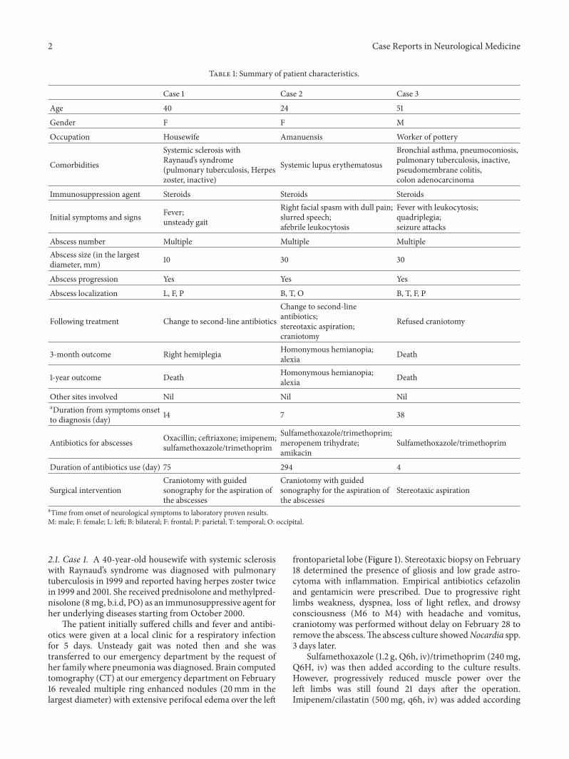
Table 1: Summary of patient characteristics.
Case 1 Case 2 Case 3
Age 40 24 51
Gender F F M
Occupation Housewife Amanuensis Worker of pottery
Comorbidities
Systemic sclerosis withRaynaud’s syndrome(pulmonary tuberculosis, Herpeszoster, inactive)
Systemic lupus erythematosus
Bronchial asthma, pneumoconiosis,pulmonary tuberculosis, inactive,pseudomembrane colitis,colon adenocarcinoma
Immunosuppression agent Steroids Steroids Steroids
Initial symptoms and signs Fever;unsteady gait
Right facial spasm with dull pain;slurred speech;afebrile leukocytosis
Fever with leukocytosis;quadriplegia;seizure attacks
Abscess number Multiple Multiple MultipleAbscess size (in the largestdiameter, mm) 10 30 30
Abscess progression Yes Yes Yes
Abscess localization L, F, P B, T, O B, T, F, P
Following treatment Change to second-line antibiotics
Change to second-lineantibiotics;stereotaxic aspiration;craniotomy
Refused craniotomy
3-month outcome Right hemiplegia Homonymous hemianopia;alexia Death
1-year outcome Death Homonymous hemianopia;alexia Death
Other sites involved Nil Nil NilaDuration from symptoms onsetto diagnosis (day) 14 7 38
Antibiotics for abscesses Oxacillin; ceftriaxone; imipenem;sulfamethoxazole/trimethoprim
Sulfamethoxazole/trimethoprim;meropenem trihydrate;amikacin
Sulfamethoxazole/trimethoprim
Duration of antibiotics use (day) 75 294 4
Surgical interventionCraniotomy with guidedsonography for the aspiration ofthe abscesses
Craniotomy with guidedsonography for the aspiration ofthe abscesses
Stereotaxic aspiration
aTime from onset of neurological symptoms to laboratory proven results.M: male; F: female; L: left; B: bilateral; F: frontal; P: parietal; T: temporal; O: occipital.
2.1. Case 1. A 40-year-old housewife with systemic sclerosiswith Raynaud’s syndrome was diagnosed with pulmonarytuberculosis in 1999 and reported having herpes zoster twicein 1999 and 2001. She received prednisolone andmethylpred-nisolone (8mg, b.i.d, PO) as an immunosuppressive agent forher underlying diseases starting from October 2000.
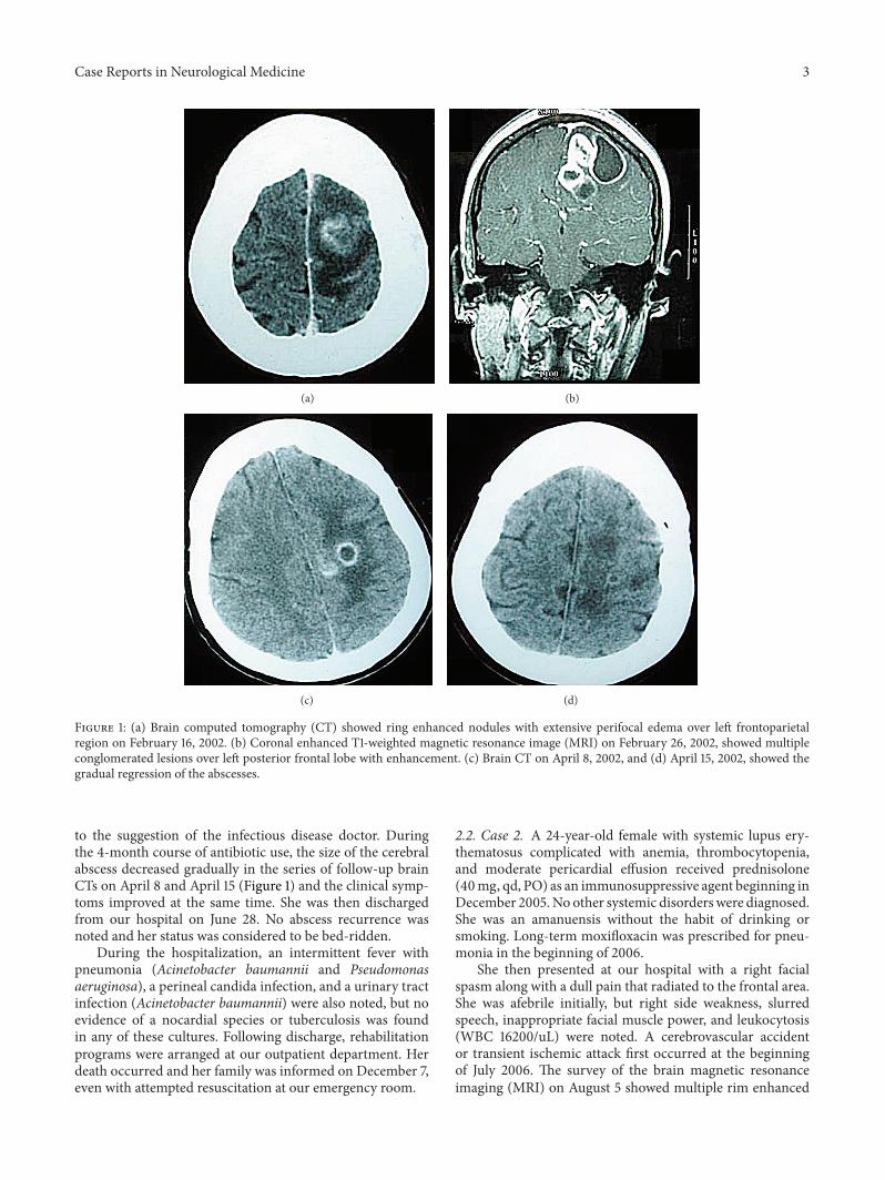
The patient initially suffered chills and fever and antibi-otics were given at a local clinic for a respiratory infectionfor 5 days. Unsteady gait was noted then and she wastransferred to our emergency department by the request ofher family where pneumonia was diagnosed. Brain computedtomography (CT) at our emergency department on February16 revealed multiple ring enhanced nodules (20mm in thelargest diameter) with extensive perifocal edema over the left
frontoparietal lobe (Figure 1). Stereotaxic biopsy on February18 determined the presence of gliosis and low grade astro-cytoma with inflammation. Empirical antibiotics cefazolinand gentamicin were prescribed. Due to progressive rightlimbs weakness, dyspnea, loss of light reflex, and drowsyconsciousness (M6 to M4) with headache and vomitus,craniotomy was performed without delay on February 28 toremove the abscess.The abscess culture showedNocardia spp.3 days later.
Sulfamethoxazole (1.2 g, Q6h, iv)/trimethoprim (240mg,Q6H, iv) was then added according to the culture results.However, progressively reduced muscle power over theleft limbs was still found 21 days after the operation.Imipenem/cilastatin (500mg, q6h, iv) was added according
Case Reports in Neurological Medicine 3
(a) (b)
(c) (d)
Figure 1: (a) Brain computed tomography (CT) showed ring enhanced nodules with extensive perifocal edema over left frontoparietalregion on February 16, 2002. (b) Coronal enhanced T1-weighted magnetic resonance image (MRI) on February 26, 2002, showed multipleconglomerated lesions over left posterior frontal lobe with enhancement. (c) Brain CT on April 8, 2002, and (d) April 15, 2002, showed thegradual regression of the abscesses.
to the suggestion of the infectious disease doctor. Duringthe 4-month course of antibiotic use, the size of the cerebralabscess decreased gradually in the series of follow-up brainCTs on April 8 and April 15 (Figure 1) and the clinical symp-toms improved at the same time. She was then dischargedfrom our hospital on June 28. No abscess recurrence wasnoted and her status was considered to be bed-ridden.
During the hospitalization, an intermittent fever withpneumonia (Acinetobacter baumannii and Pseudomonasaeruginosa), a perineal candida infection, and a urinary tractinfection (Acinetobacter baumannii) were also noted, but noevidence of a nocardial species or tuberculosis was foundin any of these cultures. Following discharge, rehabilitationprograms were arranged at our outpatient department. Herdeath occurred and her family was informed on December 7,even with attempted resuscitation at our emergency room.
2.2. Case 2. A 24-year-old female with systemic lupus ery-thematosus complicated with anemia, thrombocytopenia,and moderate pericardial effusion received prednisolone(40mg, qd, PO) as an immunosuppressive agent beginning inDecember 2005.No other systemic disorderswere diagnosed.She was an amanuensis without the habit of drinking orsmoking. Long-term moxifloxacin was prescribed for pneu-monia in the beginning of 2006.
She then presented at our hospital with a right facialspasm along with a dull pain that radiated to the frontal area.She was afebrile initially, but right side weakness, slurredspeech, inappropriate facial muscle power, and leukocytosis(WBC 16200/uL) were noted. A cerebrovascular accidentor transient ischemic attack first occurred at the beginningof July 2006. The survey of the brain magnetic resonanceimaging (MRI) on August 5 showed multiple rim enhanced
4 Case Reports in Neurological Medicine
lesions over the left temporal and bilateral occipital regionswith diffusion restriction and perifocal edema indicative ofmultiple brain abscesses (35mm in the largest diameter)(Figure 2). Therefore a small, left craniotomy for aspirationwas performed on August 7. The thick capsule walls werepreserved and the content yielded 20mL yellowish-greenpus where nocardial growth was cultured. Sulfamethoxazole(1.2 g, q6h)/trimethoprim (240mg, Q6H) was given accord-ing to the laboratory data.
After antibiotic use, the MRI on August 24 (Figure 2)still revealed progression of the abscesses in the bilateralparietal regions and the left lateral ventricle with devel-oping hydrocephalus. Sulfamethoxazole/trimethoprim waschanged to meropenem trihydrate (500mg, q8h) on August29 and amikacin (450mg, Q12h) on August 31 in accor-dance with the suggestions of the infectious disease depart-ment. Stereotaxic aspiration with the assistance of CT wasarranged on September 1 for the bilateral occipital abscessand the pathology report still revealed Nocardia spp. Signs ofincreased intracranial pressure (IICP) were noticed after thebiopsy and an emergency brain CT on September 3 showedleft brain swelling with subfalcine and uncal herniations.Therefore, craniotomy guided by sonography was used toremove the hematomas and excise of the left mesial temporalabscesses on September 4. We continued the meropenemand amikacin administration and she was transferred tothe rheumatic department ward on September 19 with aprojected smooth recovery. Then sulfamethoxazole (800mg,b.i.d, PO)/trimethoprim (160mg, b.i.d, PO) was added onceagain on September 21 and it became the only antibiotic afterNovember 14.
The brain MRI on October 26 (Figure 2) showed that theprevious residual brain abscesses and ventriculitis decreasedin size. After 5 months of hospitalization with antibioticcontrol, there was no recurrence of abscesses, but homony-mous hemianopia and alexia were noted during follow-up. The patient lost her job due to the above neurologicaldeficits after discharge on January 7, 2007. Sulfamethox-azole/trimethoprim was used until May 29. In 2008, shereceived right and then left total hip replacement due toavascular necrosis which was considered a complication oflong-term steroid use.
2.3. Case 3. A 51-year-old male from a rural town hadhistory of bronchial asthma and pneumoconiosis for 4 years.He received prednisolone (10mg, qd, PO) to control theobstructive lung related asthma starting in August 1999. Heinitially presented to our institute with dyspnea which wassuspected to be an asthma attack in February 2002. Due torespiratory failure, he was intubated with a mechanical venti-lator used in our intensive care unit where systemic steroidand a bronchodilator were added to treat the underlyingrespiratory disease. A fever episode which was consideredE. coli pneumonia related was noted on February 15, socefazolin (1gm, q8h, iv) was shifted to cefuroxime (750mg,q8h, iv), and then amikacin (400mg, q12h, iv) was addedon February 20. Tracheostomy was performed on February21 due to poor condition and inability to wean the patient
off the ventilator. A colonoscopy performed on February26 to investigate bloody stool revealed pseudomembranecolitis and polyps where adenocarcinoma was identified bybiopsy. Cefuroxime and amikacin were discontinued due tono significant pneumonia lesion.
InMarch 2002, steroidmyopathy or sedative agent relatedquadriplegia and seizure attackswere noted.However,muscleweakness that persisted after the adjustment from systemicsteroid to oral prednisolone and leukocytosis without feverwas noted on March 18. Metronidazole was added for pseu-domembranous colitis from to March 15 to March 29. Thecontrast enhanced brain CT on April 2 revealed multiplesolitary brain abscesses over the right temporal and bilateralfrontal area, but colon cancer with brain metastasis couldnot be ruled out (see Figure 3). Due to reoccurrence ofseizure attacks on April 4 even with an adequate dilantinlevel and an IICP sign, stereotaxic aspiration for the rightfrontal abscesses assisted by CT was performed on April8. Vancomycin (1 g, q12h, iv) and ceftriaxone (2 gm, q12h,iv) were temporarily used afterwards. Pus culture of theabscesses resulted in Nocardia species and sulfamethoxazole(800mg, q6h, iv)/trimethoprim (160mg, q6h, iv) was usedto replace vancomycin 3 days later, but the clinical conditionworsened rapidly. The follow-up brain CT found severebrain edema, even with a mannitol and dexamethasonedrip. Tachycardia was noted and complete electrocardiogramshowed paroxysmal supraventricular tachycardia (PSVT).We considered the IICP sign to be related to brain herniationand advised craniotomy for him, but his family refusedfurther surgical intervention. Intermittent apnea was foundin the afternoon of April 12 and severe brain edemawith brainherniation was identified. By the request of the family, thepatient was discharged and then died of brainstem failure.
3. Discussion
Members of the genus Nocardia are gram-positive, bacillary,and branching bacteria who belong to the family Nocar-diaceae though the branching rods of the partially acid-fastactinomycete were often misclassified as fungi in the past [3–5]. They are ubiquitous in the environment, living in soil,organic matter, and water [6]. The organism enters the bodyvia inhalation or direct percutaneous inoculation. Nocardialinfections often have a hematogenous route of infection froma primary lung focus [7]. If subsequent hematogenous dis-semination occurs, it may lead to the infection of any organ,with a particular predilection for the central nervous system(CNS) [6]. Nocardial infections are commonly encounteredin patients with immunocompromised states which includehuman immune deficiency virus (HIV) infection, alcoholabuse, alveolar proteins, diabetes mellitus, neoplasias, andorgan transplants [7].
Nocardial brain abscesses are extremely rare; only 3cases have been documented at our institution over thepast 10 years. All three cases were immunocompromisedpatients undergoing steroid control for more than half ayear. Nocardia asteroides in the Nocardia genus is the mostfrequent cause of nocardial disease in humans and accounts
Case Reports in Neurological Medicine 5
(a) (b) (c)
(d) (e) (f)
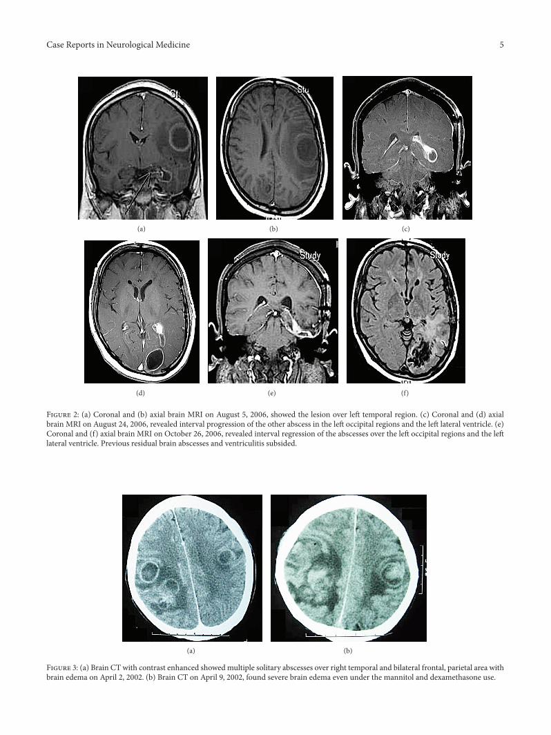
Figure 2: (a) Coronal and (b) axial brain MRI on August 5, 2006, showed the lesion over left temporal region. (c) Coronal and (d) axialbrain MRI on August 24, 2006, revealed interval progression of the other abscess in the left occipital regions and the left lateral ventricle. (e)Coronal and (f) axial brain MRI on October 26, 2006, revealed interval regression of the abscesses over the left occipital regions and the leftlateral ventricle. Previous residual brain abscesses and ventriculitis subsided.
(a) (b)
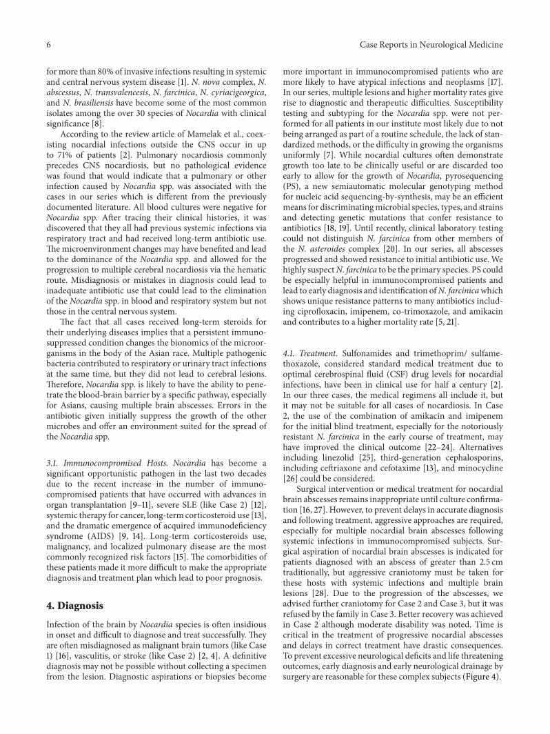
Figure 3: (a) Brain CT with contrast enhanced showedmultiple solitary abscesses over right temporal and bilateral frontal, parietal area withbrain edema on April 2, 2002. (b) Brain CT on April 9, 2002, found severe brain edema even under the mannitol and dexamethasone use.
6 Case Reports in Neurological Medicine
formore than 80% of invasive infections resulting in systemicand central nervous system disease [1]. N. nova complex, N.abscessus, N. transvalencesis, N. farcinica, N. cyriacigeorgica,and N. brasiliensis have become some of the most commonisolates among the over 30 species of Nocardia with clinicalsignificance [8].
According to the review article of Mamelak et al., coex-isting nocardial infections outside the CNS occur in upto 71% of patients [2]. Pulmonary nocardiosis commonlyprecedes CNS nocardiosis, but no pathological evidencewas found that would indicate that a pulmonary or otherinfection caused by Nocardia spp. was associated with thecases in our series which is different from the previouslydocumented literature. All blood cultures were negative forNocardia spp. After tracing their clinical histories, it wasdiscovered that they all had previous systemic infections viarespiratory tract and had received long-term antibiotic use.The microenvironment changes may have benefited and leadto the dominance of the Nocardia spp. and allowed for theprogression to multiple cerebral nocardiosis via the hematicroute. Misdiagnosis or mistakes in diagnosis could lead toinadequate antibiotic use that could lead to the eliminationof the Nocardia spp. in blood and respiratory system but notthose in the central nervous system.
The fact that all cases received long-term steroids fortheir underlying diseases implies that a persistent immuno-suppressed condition changes the bionomics of the microor-ganisms in the body of the Asian race. Multiple pathogenicbacteria contributed to respiratory or urinary tract infectionsat the same time, but they did not lead to cerebral lesions.Therefore, Nocardia spp. is likely to have the ability to pene-trate the blood-brain barrier by a specific pathway, especiallyfor Asians, causing multiple brain abscesses. Errors in theantibiotic given initially suppress the growth of the othermicrobes and offer an environment suited for the spread ofthe Nocardia spp.
3.1. Immunocompromised Hosts. Nocardia has become asignificant opportunistic pathogen in the last two decadesdue to the recent increase in the number of immuno-compromised patients that have occurred with advances inorgan transplantation [9–11], severe SLE (like Case 2) [12],systemic therapy for cancer, long-term corticosteroid use [13],and the dramatic emergence of acquired immunodeficiencysyndrome (AIDS) [9, 14]. Long-term corticosteroids use,malignancy, and localized pulmonary disease are the mostcommonly recognized risk factors [15]. The comorbidities ofthese patients made it more difficult to make the appropriatediagnosis and treatment plan which lead to poor prognosis.
4. Diagnosis
Infection of the brain by Nocardia species is often insidiousin onset and difficult to diagnose and treat successfully. Theyare often misdiagnosed as malignant brain tumors (like Case1) [16], vasculitis, or stroke (like Case 2) [2, 4]. A definitivediagnosis may not be possible without collecting a specimenfrom the lesion. Diagnostic aspirations or biopsies become
more important in immunocompromised patients who aremore likely to have atypical infections and neoplasms [17].In our series, multiple lesions and higher mortality rates giverise to diagnostic and therapeutic difficulties. Susceptibilitytesting and subtyping for the Nocardia spp. were not per-formed for all patients in our institute most likely due to notbeing arranged as part of a routine schedule, the lack of stan-dardized methods, or the difficulty in growing the organismsuniformly [7]. While nocardial cultures often demonstrategrowth too late to be clinically useful or are discarded tooearly to allow for the growth of Nocardia, pyrosequencing(PS), a new semiautomatic molecular genotyping methodfor nucleic acid sequencing-by-synthesis, may be an efficientmeans for discriminatingmicrobial species, types, and strainsand detecting genetic mutations that confer resistance toantibiotics [18, 19]. Until recently, clinical laboratory testingcould not distinguish N. farcinica from other members ofthe N. asteroides complex [20]. In our series, all abscessesprogressed and showed resistance to initial antibiotic use.Wehighly suspectN. farcinica to be the primary species. PS couldbe especially helpful in immunocompromised patients andlead to early diagnosis and identification ofN. farcinicawhichshows unique resistance patterns to many antibiotics includ-ing ciprofloxacin, imipenem, co-trimoxazole, and amikacinand contributes to a higher mortality rate [5, 21].
4.1. Treatment. Sulfonamides and trimethoprim/ sulfame-thoxazole, considered standard medical treatment due tooptimal cerebrospinal fluid (CSF) drug levels for nocardialinfections, have been in clinical use for half a century [2].In our three cases, the medical regimens all include it, butit may not be suitable for all cases of nocardiosis. In Case2, the use of the combination of amikacin and imipenemfor the initial blind treatment, especially for the notoriouslyresistant N. farcinica in the early course of treatment, mayhave improved the clinical outcome [22–24]. Alternativesincluding linezolid [25], third-generation cephalosporins,including ceftriaxone and cefotaxime [13], and minocycline[26] could be considered.
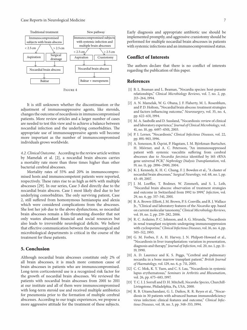
Surgical intervention or medical treatment for nocardialbrain abscesses remains inappropriate until culture confirma-tion [16, 27]. However, to prevent delays in accurate diagnosisand following treatment, aggressive approaches are required,especially for multiple nocardial brain abscesses followingsystemic infections in immunocompromised subjects. Sur-gical aspiration of nocardial brain abscesses is indicated forpatients diagnosed with an abscess of greater than 2.5 cmtraditionally, but aggressive craniotomy must be taken forthese hosts with systemic infections and multiple brainlesions [28]. Due to the progression of the abscesses, weadvised further craniotomy for Case 2 and Case 3, but it wasrefused by the family in Case 3. Better recovery was achievedin Case 2 although moderate disability was noted. Time iscritical in the treatment of progressive nocardial abscessesand delays in correct treatment have drastic consequences.To prevent excessive neurological deficits and life threateningoutcomes, early diagnosis and early neurological drainage bysurgery are reasonable for these complex subjects (Figure 4).
Case Reports in Neurological Medicine 7
Immunocompromised subjects with brain abscess
Aspiration Surgical drainage
Immunocompromised subjects with systemic infection and
multiple brain abscesses
Aspiration Craniotomy
Nocardial brain abscess
Baktar
Nocardial brain abscess
Traditional treatment New pathway
Baktar + meropenem
< 2.5 cm > 2.5 cm< 2.5 cm > 2.5 cm
Figure 4
It is still unknown whether the discontinuation or theadjustment of immunosuppressive agents, like steroids,changes the outcome of nocardiosis in immunocompromisedpatients. More review articles and a larger number of casesare needed to test this view and to achieve a balance betweennocardial infection and the underlying comorbidities. Theappropriate use of immunosuppressive agents will becomemore important as the number of immunocompromisedindividuals grows worldwide.
4.2. Clinical Outcome. According to the review article writtenby Mamelak et al. [2], a nocardial brain abscess carriesa mortality rate more than three times higher than otherbacterial cerebral abscesses.
Mortality rates of 55% and 20% in immunocompro-mised hosts and immunocompetent patients were reported,respectively. These rates rise to as high as 66% with multipleabscesses [29]. In our series, Case 3 died directly due to thenocardial brain abscess. Case 1 most likely died due to herunderlying comorbidities. The only surviving patient, Case2, still suffered from homonymous hemianopia and alexiawhich were considered complications from the abscesses.She lost her job due to the above dysfunctions, so nocardialbrain abscesses remain a life-threatening disorder that notonly wastes abundant financial and social resources butalso leads to irreversible neurological deficits. We believethat effective communication between the neurosurgical andmicrobiological departments is critical in the course of thetreatment for these patients.
5. Conclusion
Although nocardial brain abscesses constitute only 2% ofall brain abscesses, it is much more common cause ofbrain abscesses in patients who are immunocompromised.Long-term corticosteroid use is a recognized risk factor forthe growth of nocardial brain abscesses. We reviewed thepatients with nocardial brain abscesses from 2001 to 2011at our institute and all of them were immunocompromisedwith long-term steroid use and received multiple antibioticsfor pneumonia prior to the formation of multiple cerebralabscesses. According to our tragic experiences, we propose amore aggressive attitude for the treatment of these subjects.
Early diagnosis and appropriate antibiotic use should beimplemented promptly, and aggressive craniotomy should beperformed for multiple nocardial brain abscesses in patientswith systemic infections and an immunocompromised status.
Conflict of Interests
The authors declare that there is no conflict of interestsregarding the publication of this paper.
References
[1] B. L. Beaman and L. Beaman, “Nocardia species: host-parasiterelationships,” Clinical Microbiology Reviews, vol. 7, no. 2, pp.213–264, 1994.
[2] A. N. Mamelak, W. G. Obana, J. F. Flaherty, M. L. Rosenblum,and P. D. Holtom, “Nocardial brain abscess: treatment strategiesand factors influencing outcome,” Neurosurgery, vol. 35, no. 4,pp. 622–631, 1994.
[3] M. A. Saubolle and D. Sussland, “Nocardiosis: review of clinicaland laboratory experience,” Journal of ClinicalMicrobiology, vol.41, no. 10, pp. 4497–4501, 2003.
[4] P. I. Lerner, “Nocardiosis,” Clinical Infectious Diseases, vol. 22,pp. 891–903, 1996.
[5] A. Sonesson, B. Oqvist, P. Hagstam, I. M. Bjorkman-Burtscher,H. Miorner, and A. C. Petersson, “An immunosuppressedpatient with systemic vasculitis suffering from cerebralabscesses due to Nocardia farcinica identified by 16S rRNAgene universal PCR,” Nephrology Dialysis Transplantation, vol.19, no. 11, pp. 2896–2900, 2004.
[6] K. J. Kennedy, K. H. C. Chung, F. J. Bowden et al., “A cluster ofnocardial brain abscesses,” Surgical Neurology, vol. 68, no. 1, pp.43–49, 2007.
[7] J. M. Loeffler, T. Bodmer, W. Zimmerli, and S. L. Leib,“Nocardial brain abscess: observation of treatment strategiesand outcome in Switzerland from 1992 to 1999,” Infection, vol.29, no. 6, pp. 337–341, 2001.
[8] B. A. Brown-Elliott, J. M. Brown, P. S. Conville, and R. J.WallaceJr., “Clinical and laboratory features of the Nocardia spp. basedon currentmolecular taxonomy,”ClinicalMicrobiology Reviews,vol. 19, no. 2, pp. 259–282, 2006.
[9] R. C. Arduino, P. C. Johnson, and A. G. Miranda, “Nocardiosisin renal transplant recipients undergoing immunosuppressionwith cyclosporine,”Clinical Infectious Diseases, vol. 16, no. 4, pp.505–512, 1993.
[10] G. M. Forbes, F. A. H. Harvey, J. N. Philpott-Howard et al.,“Nocardiosis in liver transplantation: variation in presentation,diagnosis and therapy,” Journal of Infection, vol. 20, no. 1, pp. 11–19, 1990.
[11] A. D. Laurence and K. S. Peggs, “Cerebral and pulmonarynocardia in a bone marrow transplant patient,” British Journalof Haematology, vol. 129, no. 6, p. 711, 2005.
[12] C. C. Mok, K. Y. Yuen, and C. S. Lau, “Nocardiosis in systemiclupus erythematosus,” Seminars in Arthritis and Rheumatism,vol. 26, pp. 675–683, 1997.
[13] T. C. I. J. Sorrell andD. H.Mitchell,Nocardia Species, Churchill-Livingstone, Philadelphia, Pa, USA, 2000.
[14] R. B. Uttamchandani, G. L. Daikos, R. R. Reyes et al., “Nocar-diosis in 30 patients with advanced human immunodeficiencyvirus infection: clinical features and outcome,” Clinical Infec-tious Diseases, vol. 18, no. 3, pp. 348–353, 1994.
8 Case Reports in Neurological Medicine
[15] C. Kilincer, M. K. Hamamcioglu, O. Simsek et al., “Nocardialbrain abscess: review of clinical management,” Journal of Clini-cal Neuroscience, vol. 13, no. 4, pp. 481–485, 2006.
[16] A. Menku, A. Kurtsoy, B. Tucer, O. Yildiz, H. Akdemir, and R.Bayston, “Nocardia brain abscess mimicking brain tumour inimmunocompetent patients: report of two cases and review ofthe literature,” Acta Neurochirurgica, vol. 146, no. 4, pp. 411–414,2004.
[17] J. M. Embil, W. C. Halliday, and A. Nath, “Primary cerebellarT-cell lymphoma with acquired immunodeficiency syndrome,”Journal of NeuroVirology, vol. 3, no. 3, pp. 229–232, 1997.
[18] M. Ronaghi, M. Uhlen, and P. Nyren, “A sequencing methodbased on real-time pyrophosphate,” Science, vol. 281, no. 5375,pp. 363–365, 1998.
[19] M. Ronaghi and E. Elahi, “Pyrosequencing for microbial typ-ing,” Journal of Chromatography B: Analytical Technologies in theBiomedical and Life Sciences, vol. 782, no. 1-2, pp. 67–72, 2002.
[20] R. J.Wallace Jr., M. Tsukamura, B. A. Brown et al., “Cefotaxime-resistant Nocardia asteroides strains are isolates of the contro-versial speciesNocardia farcinica,” Journal of Clinical Microbiol-ogy, vol. 28, no. 12, pp. 2726–2732, 1990.
[21] C. A. Iannotti, G. S. Hall, G. W. Procop, M. J. Tuohy, S. M.Staugaitis, and R. J. Weil, “Solitary Nocardia farcinica brainabscess in an immunocompetent adult mimicking metastaticbrain tumor: rapid diagnosis by pyrosequencing and successfultreatment,” Surgical Neurology, vol. 72, no. 1, pp. 74–79, 2009.
[22] P. Boiron, F. Provost, G. Chevrier, and B. Dupont, “Review ofnocardial infections in France 1987 to 1990,” European Journalof Clinical Microbiology and Infectious Diseases, vol. 11, no. 8, pp.709–714, 1992.
[23] A. Krone, K. P. Schaal, A. Brawanski, and B. Schuknecht,“Noncardial cerebral abscess cured with imipenem/amikacinand enucleation,” Neurosurgical Review, vol. 12, no. 4, pp. 333–340, 1989.
[24] C. Farina, P. Boiron, A. Goglio et al., “Human nocardiosisin Northern Italy from 1982 to 1992,” Scandinavian Journal ofInfectious Diseases, vol. 27, no. 1, pp. 23–27, 1995.
[25] E. H. Moylett, S. E. Pacheco, B. A. Brown-Elliott et al., “Clinicalexperience with linezolid for the treatment of Nocardia infec-tion,” Clinical Infectious Diseases, vol. 36, no. 3, pp. 313–318,2003.
[26] R. J. Wallace Jr., L. C. Steele, G. Sumter, and J. M. Smith,“Antimicrobial susceptibility patterns of Nocardia asteroides,”Antimicrobial Agents and Chemotherapy, vol. 32, no. 12, pp.1776–1779, 1988.
[27] I. G. Fleetwood, J. M. Embil, and I. B. Ross, “Nocardia aster-oides cerebral abscess in immunocompetent hosts: report ofthree cases and review of surgical recommendations,” SurgicalNeurology, vol. 53, no. 6, pp. 605–610, 2000.
[28] G. Y. F. Lee, R. T. Daniel, B. P. Brophy et al., “Surgical treatmentof nocardial brain abscesses,” Neurosurgery, vol. 51, no. 3, pp.668–672, 2002.

Submit your manuscripts athttp://www.hindawi.com
Stem CellsInternational
Hindawi Publishing Corporationhttp://www.hindawi.com Volume 2014
Hindawi Publishing Corporationhttp://www.hindawi.com Volume 2014
MEDIATORSINFLAMMATION
of
Hindawi Publishing Corporationhttp://www.hindawi.com Volume 2014
Behavioural Neurology
EndocrinologyInternational Journal of
Hindawi Publishing Corporationhttp://www.hindawi.com Volume 2014
Hindawi Publishing Corporationhttp://www.hindawi.com Volume 2014
Disease Markers
Hindawi Publishing Corporationhttp://www.hindawi.com Volume 2014
BioMed Research International
OncologyJournal of
Hindawi Publishing Corporationhttp://www.hindawi.com Volume 2014
Hindawi Publishing Corporationhttp://www.hindawi.com Volume 2014
Oxidative Medicine and Cellular Longevity
Hindawi Publishing Corporationhttp://www.hindawi.com Volume 2014
PPAR Research
The Scientific World JournalHindawi Publishing Corporation http://www.hindawi.com Volume 2014
Immunology ResearchHindawi Publishing Corporationhttp://www.hindawi.com Volume 2014
Journal of
ObesityJournal of
Hindawi Publishing Corporationhttp://www.hindawi.com Volume 2014
Hindawi Publishing Corporationhttp://www.hindawi.com Volume 2014
Computational and Mathematical Methods in Medicine
OphthalmologyJournal of
Hindawi Publishing Corporationhttp://www.hindawi.com Volume 2014
Diabetes ResearchJournal of
Hindawi Publishing Corporationhttp://www.hindawi.com Volume 2014
Hindawi Publishing Corporationhttp://www.hindawi.com Volume 2014
Research and TreatmentAIDS
Hindawi Publishing Corporationhttp://www.hindawi.com Volume 2014
Gastroenterology Research and Practice
Hindawi Publishing Corporationhttp://www.hindawi.com Volume 2014
Parkinson’s Disease
Evidence-Based Complementary and Alternative Medicine
Volume 2014Hindawi Publishing Corporationhttp://www.hindawi.com