Embed Size (px)
Citation preview
Clinical Positron Emission Tomography/Magnetic Resonance Imaging ApplicationsGustav K. von Schulthess, MD, PhD, MD hon., Felix Pierre Kuhn, MD, MS,Philipp Kaufmann, MD, and Patrick Veit-Haibach, MD
Although clinical positron emission tomography (PET)/computed tomography (CT) applicationswere obvious and have completely replaced PET in oncology, clinical applications of PET/magnetic resonance (MR) are currently not clearly defined. This is due to the lack of clinicaldata, which is mainly because PET/MR technology is not clinically mature at this point. Openissues are technical and concern ease of obtaining PET attenuation correction maps, dealingwith, for example, MR surface coil metal in the PET field-of-view and appropriate workflowsleading to a cost-effective examination. All issues can be circumvented by using a shuttle-connected PET/CT-MR system, but the penalty is that simultaneous PET and MR imaging arenot possible and potential motion between examinations may occur. Clinically, some systemsinstalled worldwide start to have a reasonable bulk of clinical data. Preliminary results suggestthat in oncology, PET/MR may have advantages over PET/CT in head and neck imaging. In liverimaging, more PET-positive lesions are seen on MR than on CT, but that does not mean thatPET/MR is superior to PET/CT. Possibly in some settings where a contrast-enhanced PET/CTis needed to be diagnostic, PET/MR can be done without contrast media. Although PET/CT hasvirtually no role in brain imaging, this may be an important domain for PET/MR, particularly indementia imaging. The role of PET/MR in the heart is as yet undefined, and much research willhave to be done to elucidate this role. At this point, it is also not clear where the simultaneityafforded by a fully integrated PET/MR is really needed. Sequential data acquisition even onseparate systems and consecutive software image fusion may well be appropriate. With theincreasing installed base of systems, clinical data will be forthcoming and define more clearlywhere there is clinical value in PET/MR at an affordable price.
Semin Nucl Med 43:3-10 © 2013 Elsevier Inc. All rights reserved.ntPgidoPi
Positron emission tomography (PET)/magnetic resonance(MR) has been used in experimental setups for almost 20
years.1 The long time it has taken until this imaging modality hasstarted to enter the clinical arena has to do with technical andpractical hurdles that the clinical applications of PET/MR arefacing. Technically, implementation of PET/MR faces 3 majorproblems.
� First, the photomultiplier-based PET scanners cur-rently used do not work within or near the magneticenvironment of an MR scanner,
� Second, metallic objects such as surface coils used to getbest MR image quality interfere with the gamma raysfrom PET, causing unwanted attenuation, and
Department of Medical Radiology, University Hospital, CH-8091 Zurich,Switzerland.
Address reprint requests to Gustav K. von Schulthess, Department ofMedical Radiology, University Hospital, CH-8091, Zurich, Switzer-
tland. E-mail: [email protected]
0001-2998/13/$-see front matter © 2013 Elsevier Inc. All rights reserved.http://dx.doi.org/10.1053/j.semnuclmed.2012.08.005
� Third, MR data, unlike those acquired using computedtomography (CT), are not readily usable for attenuationcorrection (AC).2-4 This limits quantification of PETdata, which is particularly problematic when using PETin therapy response monitoring.
Clinically, PET/MR does not arrive into a “blue ocean,” butinto an environment where PET/CT has proven its clinicalcapabilities in thousands of publications and millions of clin-ical examinations worldwide.5-8 Because of the relative tech-
ical ease of producing clinically effective PET/CT scanners,he focus shifted away from PET/MR, and in 2001, effectiveET/CT scanners were introduced for clinical use. The inte-ration of PET and CT has resulted in much data that give usnsights into the need for clinical integrated imaging. Theata have also helped identify the strengths and weaknessesf PET/CT, the latter being hoped to be partly overcome byET/MR. In contrast, the technical challenges in implement-
ng PET/MR clearly demonstrate its major shortcomings. In-
egrated imaging with PET/CT is highly synergistic: the sum3

pviPbPsi
4 G.K. von Schulthess et al
of PET and CT is more than its parts. PET/CT provides clin-ically critical anatomic correlation, resulting in increased sen-sitivity and specificity, AC, and an increase in productivity atonce. Whether such synergy can be attained with MR re-mains to be demonstrated.
Problem 1:Photomultipliers Unfit forthe Magnetic Environment in MRCurrent photomultiplier (PMT) detectors in PET scanners donot function within or near MR scanners. Hence, new detec-tors have to be designed, such as avalanche photodiode orsilicon PMT–based PET detector systems. This is discussedelsewhere in this issue. With such detector systems, full in-tegration and simultaneous image acquisition of PET and MRdata are possible; such systems are called simultaneous sys-tems.9 Currently, only Siemens provides such systems. Alter-natively, PET and MR scanners are placed far enough apartthat the magnetic field does not interfere with the PMTs. Insuch systems, a shuttle and a transfer board is required totransfer patient from PET(/CT) to MR scanner or vice versawhen hardware coregistration of images is to be achieved.Such systems are called sequential systems.9 All PET/CT (andsingle-photon emission computed tomography [SPECT]/CT)systems are sequential integrated systems, in which hardwareimage registration is achieved by the patient system tableserving as shuttle transfer system. Sequential PET/MR sys-tems have been implemented by GE Healthcare and Philipseither as 2-room PET/CT-MR (GE Healthcare) or as 1-roomPET-MR (Philips Medical Systems). For completeness sake, ithas to be mentioned that in many situations, software fusionof images achieves excellent results, particularly in the brainand heart. Systems not connected by hardware are calledseparate systems.9
Problem 2: Interference ofMetallic Structures Used inMR with the Gamma Rays of PETFor best MR image quality, surface coil arrangements areneeded. In extended body imaging, this is achieved witharray coils. Coils contain various metal parts such as themetallic conductors and the amplifier electronics. Thesecause major artifacts in the PET images and have to be cor-rected for. As will be seen, this problem can be circumventedin sequential PET/MR systems. Both development of newdetectors and modification of surface coils to reduce theirmetal content are major tasks, which currently are beingaddressed by the system manufacturers and are tasks that fewacademic institutions can address successfully.
Problem 3: AC ofPET Data by MR DataAs MR does not provide an attenuation map for ionizing
radiation, PET data cannot be subject to AC, which makesthem qualitative images only. Somehow, MR data have to beacquired such that they can be readily transformed into“quasi” CT data and from there into attenuation maps. Themajor challenge in using MR data for AC of PET data isrelated to the inability of conventional MR pulse sequences tounequivocally show bone. Various studies have demon-strated that omission of bone and its replacement by softtissues in attenuation maps lead to erroneous standard up-take values (SUVs) in the brain and in the bones and theirvicinity, which differ by as much as 25% from the correctvalues. This is the current approach taken obtaining bodydata in integrated systems without CT.2,3,10 Various ap-
roaches to overcome this problem are currently under in-estigation and are subject of another contribution in thisssue. Although for simultaneous and 1-room sequentialET/MR systems, the inability to unequivocally visualizeone poses a serious problem regarding clinical utility, theET/CT-MR sequential system avoids this problem, as inuch systems, CT data are available from the PET/CT exam-nation.
Using a PET/CT-MR toAvoid Technical Issues andFocus on Clinical Data AcquisitionThe PET/CT-MR configuration thus has major advantages atthis point to address the key issue of clinical PET/MR,namely, to demonstrate the clinical value of this technologyrelative to PET/CT. As in this setup, PET and MR scanners donot interfere, the best state-of-the-art PMT-based PET and3-T MR scanners can be connected by a shuttle transfer sys-tem and used to acquire clinical data. AC can be done withthe CT of the PET/CT scanner, avoiding the use of the stillflawed ways of obtaining AC maps from MR data for correct-ing PET. This is a unique feature of PET/CT-MR systems.Artifacts introduced by MR coils can also be avoided. If thesurface coils are arranged in such a way around the patientthat he/she is “inserted” into and “removed” from them whenhe/she is moved into and out of the MR scanner, no coils areon the patient within the PET(/CT) scanner. This is similar tomoving a hand into and out of a glove, and we have termedthis the “glove approach” (Fig. 1).
Consequently, the PET/CT-MR 2-room system approachhas the unique property to provide hardware-coregistereddata sets of PET, CT, and MR, thereby permitting to performclinical studies, in which PET/CT vs PET-MR data can besubject to blind comparative evaluation. PET-MR configura-tions—whether fully integrated or shuttle connected—pro-vide “only” PET and MR data sets; thus, for comparison, theyhave to undergo a PET/CT first and then a PET/MR or viceversa. This results in a PET(1)/CT data set from one systemand a PET(2)/MR data set from a second system. As PET(1)and PET(2) scanners differ from each other in make and areacquired at least 30 minutes apart, resulting in some changesin, for example, fluorodeoxyglucose (FDG) tissue distribu-tion and related alterations in SUV, a “clean” comparison of
PET/CT and PET/MR data is difficult, as differences observed
Clinical integrated PET/MR applications 5
in PET/CT vs PET/MR data may not only be due to the 2technologies used, but could also be due to the differentmake of the PET scanners or a difference in FDG distributionat the 2 differing imaging times.
Development of aClinical Workflow for PET/MRAlthough the PET workflow for various clinical imaging stud-ies is usually straightforward, in MR, “anything goes,” that is,the variety of pulse sequences to be used on a patient isvirtually unlimited. In clinical PET/CT, the dominant work-flow is that of oncologic staging in which 6-10 typically15-cm axial FOVs are acquired in 2- to 4-minute steps, lead-ing to a partial- or full-body examination. In the brain and theheart, single field-of-view scans are coupled—depending onneed—with a dynamic data acquisition, which takes 10-60minutes. In a fully integrated PET/MR scanner, this PET dataacquisition strategy has to be matched: “the agile horse ofMR” has to be in tune with “the slowly trotting PET.” Hence,PET calls the “shot,” and the technology is appropriatelycalled PET/MR. If MR is used for imaging sequences muchbeyond the time it takes to acquire the PET data, a PET/MRscanner ends up being mostly an MR scanner, and imagingcost becomes prohibitive on an integrated system. A rule of
Figure 1 Patient lying on table with built-in posterior coil is slippedinto head coil, and anterior torso coil is placed on patient. All theseoperations do not interfere with the patients’ position, therebymaintaining coordinate registration between positron emission to-mography (PET), computed tomography (CT), and magnetic reso-nance (MR) data. (Color version of figure is available online.)
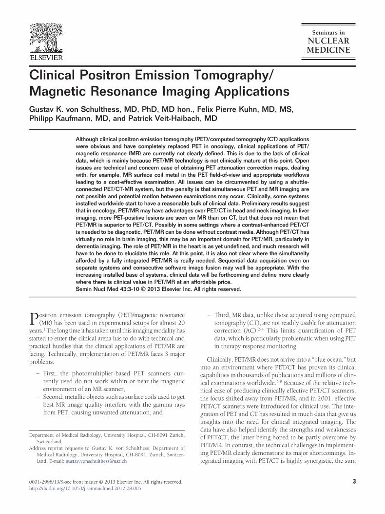
Figure 2 Sixty-five year old male status post rectal cancContrast-enhanced PET/CT. (C) Non–contrast-enhancetrast-enhanced T1 fat-sat MR. One of the fluorodeoxyglucontrast-enhanced CT (A, white arrow). On MR and PET
non–contrast-enhanced T1-weighted image as well as in PET/thumb is that data acquisition on an integrated system usingonly 1 of the 2 systems should not exceed the sum of thepatient upload and download times onto the imaging table,that is, 10-15 minutes.9 The development of much of a work-flow for integrated PET/MR can already be done using shut-tle-connected systems by choosing an MR data acquisitionprotocol adhering to a prescription in which the table istranslated every 2-4 minutes by 15-25 cm.
Oncology—Partial-/Whole-Body WorkflowIn PET/CT, usually a low-dose CT without contrast media(CM) is acquired, followed by the PET scan and—if re-quired—by a dedicated full-organ or region-focused CM-enhanced CT. It is unlikely that an oncological PET/MRworkflow will look much different. Hence, the goal in a si-multaneous PET/MR must be to acquire axial MR data match-ing or surpassing in quality the low-dose CT data during thetime the PET scanner acquires PET data in a given bed posi-tion. In other words, one needs to develop a “low-dose MRprotocol.” The axial PET scanner FOVs in the existing orevolving fully integrated PET/MR scanners of the major ven-dors will be 25 cm. Thus, MR data acquisition will need toacquire an adequate axial slice number of approximately 50slices in 2-4 minutes. Currently, MR can accomplish this taskin �1 minute using Dixon-type in- and out-of-phase images(Fig. 2). Compared with low-dose CT images, these imagesprovide a superior soft-tissue contrast, and therefore are suit-able for imaging the body from head to pelvic floor, withsubstantial quality limitations in the lung. For lung imaging,current pulse sequences provide clearly poorer images thanCT. Much work is currently being done on ultrashort andzero-echo-time MR imaging.11,12 Such MR pulse sequenceswith their low sensitivity to tissue susceptibility changes—sorelevant in the lung—might prove better than those currentlyavailable. As such sequences should also be able to imagebone with its very short T2 relaxation times, they may alsoprovide MR data, which will be useful to segment bone forAC maps. In addition and as stated previously in the text, aclinical PET/MR workflow may contain some additional or-gan-focused MR data acquisition at the end of the examina-tion, not lasting more than 15 minutes.
new liver metastases. (A) Contrast-enhanced CT. (B)at-saturation (fat-sat) MR. (D) PET/MR with non–con-FDG)-positive liver metastasis is almost invisible on the
and D), the FDG-positive lesion can be detected on the
er andd T1 fcose (/MR (C
MR.

CelltsaadalMc
Cwloml
aaiasmHoMrhaea
Mel
contra
6 G.K. von Schulthess et al
Brain and Heart ImagingFor static brain and heart imaging, the same considerationsprobably apply, with the MR data acquisition not exceedingthat of the PET data acquisition by more than 15 minutes. Incontrast, dynamic/static PET organ data acquisition can take30-50 minutes and will allow for much more MR data cap-ture. The length of dynamic examinations will prove costlyowing to the high cost of a fully integrated PET/MR scanner.
Clinical Data and PotentialUses of PET/MR in OncologyVarious oncological indications and concepts are being dis-cussed in the literature and also evaluated in our department.Firstly, one has to differentiate between whole-body imaging(head to upper thighs or to feet) or partial-body/organ imag-ing, where PET/magnetic resonance imaging (MRI) will beonly done in a specific anatomical area. We will discuss somemajor examples, with no entitlement to completeness.
One indication always mentioned as “natural” for PET/MRIis the visualization of liver lesions (primary and metastatic).Based on the relatively high liver 18F-FDG-activity in PET/
T, the detection of focal liver lesions with only slightlylevated FDG activity can be impaired, and the intrinsicallyower soft-tissue contrast of CT compared with MRI in theiver is well known. Contrast media in CT can increase de-ection rates for liver lesions, but generally MRI is still con-idered superior. However, there are only few studies evalu-ting contrast-enhanced (CE) PET/CT compared with MRI,nd a large meta-analysis did not find statistically significantifferences in lesion detection.13 Initial comparison of datacquired in our PET/CT-MRI system shows no difference inesion detection when comparing CE PET/CT and non-CE
RI. Lesion conspicuity was significantly better on the MRIomponent when compared with the CE CT component14
(Fig. 2). Furthermore, comparisons of standard low-dosePET/CT and non-CE PET/MRI showed significantly betterresults concerning lesion conspicuity in the liver.15
It is obvious that primary liver malignancies will also profit
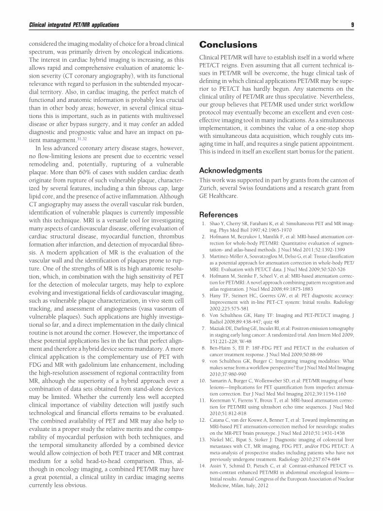
Figure 3 Fourty-six year old male with (A) Contrast-eenhanced T1 fat-sat MR. (D) PET/MR with contrast-enhas an FDG-positive Rouvière lymph node (white arrow)lymph node) in (A) and (B) are inferior compared with
from PET/MRI imaging. For example, hepatocellular carcino-
mas are always evaluated by CE MRI. In contrast, large trialshave indicated that detection and characterization can besignificantly improved when imaged with 18F-choline PET/
T.16 Thus, the combination of other tracers than 18F-FDGith MRI imaging may improve imaging diagnostics in liver
esions beyond current stand-alone PET/CT and MRI, andverall PET/MRI—even partially without CM—might beore accurate in the diagnosis and characterization of liver
esions.Another indication requiring only regional extent imaging
nd expected advantages of PET/MRI over PET/CT is headnd neck cancer. In these tumors, surrounding soft-tissuenfiltration is important for local staging as well as surgicalnd radiotherapy planning (Fig. 3). Because of its higheroft-tissue contrast, MRI has become the most significantorphologic modality for imaging head and neck cancer.owever, PET/CT has proven to be superior for the detectionf lymph node metastases in head and neck cancer.17 BecauseRI and PET/CT are currently used for primary staging and
estaging in many of these patients, a combined PET/MRIere is likely resulting in a one-stop shop. PET/MRI will haven additive value in patients with metallic dental implants, asxisting and new MRI sequences can minimize metal dentalrtifacts, which can impair the diagnosis on CT significantly.
Examples and potential indications for whole-body PET/RI are manifold. Several publications already compared, for
xample, PET/CT vs diffusion weighed imaging (DWI) inymphoma patients.18 Most of the studies currently availablegenerally found satisfying agreement when evaluating thediagnostic accuracy of those 2 methods. MRI publicationsalso indicated that the DWI-MRI might be sufficient for fol-low-up on this patient population, especially when evaluat-ing the apparent diffusion coefficient (ADC) values. How-ever, at this point, there is no established routine clinicalstaging and therapy follow-up protocol using DWI-MRI inthis setting. Thus, in PET/MRI, FDG uptake data (SUV) andtumor lesion glycolysis as well as the diffusion data and ADCvalues can be combined and correlated. A conceivable futureapproach for primary staging and for follow-up in this patient
d CT. (B) Contrast-enhanced PET/CT. (C) Contrast-T1 fat-sat MR. A large right-sided tongue cancer as wellctable. However, the lesion conspicuity (especially thest-enhanced MRI and PET/MRI.
nhanceancedis dete
group would be to have PET/MRI for primary staging and

danpHlba
oaaiad
dradt
Clinical integrated PET/MR applications 7
only DWI-MRI imaging for follow-up. This could substan-tially reduce radiation dose from imaging, but first, it has tobe demonstrated that the high sensitivity of DWI coupledwith relatively low specificity will result in an accurateenough examination.
Staging of bronchial carcinoma might also become an im-portant indication for PET/MRI. Currently, CT provides un-paralleled image quality and tissue contrast in the lung and isworldwide the method of choice for lung nodule detectionand evaluation of parenchymal texture. MR is not even closein matching this ability of CT in lung imaging, even withbreath-hold gradient recalled echo sequences or gating.However, patients with the suspicion of lung cancer referredto PET/CT or PET/MRI have already undergone almost al-ways a chest CT. Thus, with a chest CT available, it might beless important that MR within the context of a PET/MR ex-amination provides accuracy comparable with CT if the restof the information gained by PET/MR is superior to that ofPET/CT. There are initial promising results for PET/MRI atleast for bronchial carcinoma detection,19 but CT in PET/CTcan certainly be no match to brain imaging afforded by PET/MR, and MR is recommended for staging in all patients withstage II tumors.20 In fact, in this setting, it probably makes no
ifference if a PET/CT and an MR or a CT and a PET/MR arecquired, as all modalities are needed for proper staging. It isoteworthy that our current PET/CT-MRI setting is able torovide all those staging steps within one imaging procedure.ere, the CT from PET/CT can be used for state-of-the-art
ung imaging, the PET/CT provides the “standard” whole-ody staging, and MRI can be performed in the brain and/orny other area of interest (eg, liver).
Overall, we do see several advantages in PET/MRI imaging inncologic indications in imaging of body regions/organs as wells in partial-/whole-body imaging. Currently, the PET/CT-MRIpproach offers the highest flexibility to provide complete stag-ng concepts. To define where PET/MRI can be used to betterdvantage compared with PET/CT, PET/MRI will have to beefined in sizable patient series for various disease entities.
PotentialApplications in NeurologyIn neuroimaging, morphologic images are often a prerequi-site for processing and analysis of functional data. Further-more, data from PET and MR are often complementary, andas a consequence, the integration of the information fromboth modalities yields the most accurate diagnosis. This canand has been achieved by software image fusion. Hence, anintegrated PET/MR is often not mandatory in clinical mul-tiparametric neuroimaging. Strictly speaking, simultaneousPET and MR imaging is only necessary if the visualized func-tional processes are time dependent in PET and MR. Exam-ples are functional neuropsychiatric examinations measuringtask-induced changes of blood flow and oxygen consump-tion rate or the measurement of the arterial input function byMRI for its use in compartment modeling in quantitative PET
studies. The rare physiological necessity of simultaneous lPET/MR in clinical routine does not preclude the usefulnessof an integrated scanner. To read PET and MR images andintegrate the complementary information of the 2 modalities,both scans should take place at roughly the same time, at least onthe same day. This is often impossible because of having 2 sep-arate scanners in separate imaging divisions (neuroradiology/nuclear medicine) leads to scheduling conflicts. It is disagree-able for the patient to have 2 separate appointments andlaborious for the referring physician to coordinate 2 examina-tions. Furthermore, to promote combined PET/MR neuroimag-ing, the multiplatform image acquisitions have to be perceivedas 1 examination. In clinical routine, it is desirable to provideonly 1 medical report for the referring physician at the end.Hence, integrated PET/MRI has a good potential to become apowerful tool for quantitative imaging and objective decisionmaking as a one-stop-shop examination of the brain.
On the technical side, the integration of PET and MR,rather than PET and CT, allows for a dose reduction to thepatient by omitting the CT scan. In PET imaging, the CT is inmost cases only used for AC, which in the future probablycan also be accomplished by MR. For implementing auto-mated image analysis tools to quantify PET data, it is helpfulto have MR images always from the same MR device, as thisincreases the robustness of the processing algorithm. Nowa-days, much of quantitative image analysis is executed bytransforming the MR images into stereotactic space using theMontreal Neurological Institute/International Consortium ofBrain Mapping standard brain template. In a second step, thiscalculated transformation matrix is then applied to the PETimages. Using atlases or other predefined regions of interestallows reproducible and user-independent quantitative dataanalysis. Automated quantitative analysis of brain patholo-gies is of major importance for the standardization of treat-ment and therapy assessment. Furthermore, partial volumecorrection of the PET data based on MR images might in-crease the exactness of radiotracer quantification. Hence, al-though often there is no absolute need for an integratedPET/MR scanner, there are important arguments why such anintegrated system may improve data quality in brain imaging.
Multimodal PET/MR neuroimaging has various interestingapplications, that is, in neuro-oncology, neurodegeneration,or epilepsy.
Neuro-OncologyIn vivo visualization of molecular targets influences clinicaldecision making and outcome. Brain tumors are often heter-ogeneous, and stereotactic biopsies of the highest tumorgrade is crucial for further treatment planning.21 It is knownthat hypoxic areas in tumor tissue are more resistant to ra-diotherapy, and that the local microenvironment stronglyinfluences the effectiveness of chemotherapy.22 Therefore, to
etermine the most effective therapy, it is important to takeelevant imaging biomarkers of PET and MR into consider-tion, that is, cell proliferation, metabolic activity, cellularensity, cell death, growth factor expression, vascular struc-ure, neoangiogenesis, oxygenation status, pH, and drug de-
ivery.
ttdfsmdp w
itns
qp
8 G.K. von Schulthess et al
Neurodegenerative DiseaseIn Alzheimer’s disease, an early and accurate detection ofamyloid plaques by PET is of major importance, as there isevidence that these deposits provoke the regional neurode-generation. For the evaluation of disease progression andtherapy response, integrating morphologic and functionalinformation from PET and MR is mandatory. Interesting bio-markers are, for instance, the metabolic activity measured inPET23 and the functional connectivity as measured by rest-ing-state functional MRI.24 Morphologic images are essentialo exclude other causes of dementia like multi-infarct demen-ia or normal-pressure hydrocephalus (Fig. 4). In movementisorders, morphologic imaging does not allow for early dif-erentiation of Parkinson disease from atypical parkinsonianyndromes. However, the addition of functional studies per-its to recognize degeneration patterns specific to Parkinsonisease, multiple system atrophy, progressive supranuclearalsy, and corticobasal degeneration.25 Once again, to evalu-
ate the effectiveness of treatment, quantitative imaging is cru-cial.
EpilepsyPatients with refractory epilepsy despite medical treatmentmay undergo surgery to resect the epileptogenic focus. Beforesurgery, it has to be verified that the suspected focus indeedis causing the epilepsy. Hence, for optimal therapy outcome,it is required to combine the available localization tech-niques, that is, morphologic MRI, functional MRI, PET, andEEG.26
Clinical Data and PotentialUses of PET/MR in CardiologyNuclear myocardial perfusion imaging (MPI) offers well-es-tablished tools for the assessment of coronary artery disease,
Figure 4 (A) Pittsburgh compound B is a specific radiotracand atrophy. In Alzheimer’s disease, FDG is typically redand temporal cortex. (C) Vascular dementia. The diagnothe National Institute of Neurological Disorders anl’Enseignement en Neurosciences criteria.
and a large body of literature underlines the strong prognos-
tic value of nuclear MPI. Although superiority of PET overSPECT MPI has been suggested for accuracy and can be ex-pected regarding study duration, staff radiation exposure,reduced patient dose, improved patient comfort, and poten-tially economic benefit, in reality, PET still plays a limitedrole in daily clinical routine for MPI compared with SPECT.This may be due on the one hand to the lack of solid data tosupport a superior accuracy of PET over SPECT and on theother hand to the limited availability of PET for cardiac stud-ies. Although with the widespread use of oncology PET scan-ning, the availability of PET scanners has dramatically in-creased, this was not yet paralleled by a substantial rise incardiac PET applications. The main drawback has been thelack of a flow tracer for PET MPI, with a half-life long enoughto allow for shipment to satellite PET centers without cyclo-tron. This may change with the advent of new 18F-tracerssuch as 18F-flurpiridaz,27 and clinical phase 3 trials are under
ay, but results are not yet available.Although PET is the most reliable noninvasive tool for
dentification of myocardial viability in ischemic dysfunc-ional left ventricles and the only method validated in a prog-ostic interventional outcome study meeting all criteria ofolid comparative effectiveness research,28 its clinical role has
remained modest for several reasons. One reason is that thereare competing radiation-free methods in the hands of cardi-ologists, such as echocardiography and MRI, which allowidentification of viability. Another reason—potentially witheven more impact—is recent controversial results of the Sur-gical Treatment for Ischemic Heart Failure trial,29 which have
uestioned the clinical benefits of viability assessment onatient outcome in ischemic left ventricular dysfunction.30
It is in this context that the potential importance of a se-quential cardiac PET and MR scanning or a combinedPET/MR imaging tool for cardiac application has to be dis-cussed. Several years ago, the introduction of hybrid imaging
�-amyloid plaques. (B) FDG visualizes hypometabolismn the posterior cingulate gyrus, as well as in the parietalased on clinical and radiologic findings summarized inke/Association Internationale pour la Recherche et
er foruced isis is bd Stro
systems with multislice CT and PET devices, which is now
Clinical integrated PET/MR applications 9
considered the imaging modality of choice for a broad clinicalspectrum, was primarily driven by oncological indications.The interest in cardiac hybrid imaging is increasing, as thisallows rapid and comprehensive evaluation of anatomic le-sion severity (CT coronary angiography), with its functionalrelevance with regard to perfusion in the subtended myocar-dial territory. Also, in cardiac imaging, the perfect match offunctional and anatomic information is probably less crucialthan in other body areas; however, in several clinical situa-tions this is important, such as in patients with multivesseldisease or after bypass surgery, and it may confer an addeddiagnostic and prognostic value and have an impact on pa-tient management.31,32
In less advanced coronary artery disease stages, however,no flow-limiting lesions are present due to eccentric vesselremodeling and, potentially, rupturing of a vulnerableplaque. More than 60% of cases with sudden cardiac deathoriginate from rupture of such vulnerable plaque, character-ized by several features, including a thin fibrous cap, largelipid core, and the presence of active inflammation. AlthoughCT angiography may assess the overall vascular risk burden,identification of vulnerable plaques is currently impossiblewith this technique. MRI is a versatile tool for investigatingmany aspects of cardiovascular disease, offering evaluation ofcardiac structural disease, myocardial function, thrombusformation after infarction, and detection of myocardial fibro-sis. A modern application of MR is the evaluation of thevascular wall and the identification of plaques prone to rup-ture. One of the strengths of MR is its high anatomic resolu-tion, which, in combination with the high sensitivity of PETfor the detection of molecular targets, may help to exploreevolving and investigational fields of cardiovascular imaging,such as vulnerable plaque characterization, in vivo stem celltracking, and assessment of angiogenesis (vasa vasorum ofvulnerable plaques). Such applications are highly investiga-tional so far, and a direct implementation in the daily clinicalroutine is not around the corner. However, the importance ofthese potential applications lies in the fact that perfect align-ment and therefore a hybrid device seems mandatory. A moreclinical application is the complementary use of PET withFDG and MR with gadolinium late enhancement, includingthe high-resolution assessment of regional contractility fromMR, although the superiority of a hybrid approach over acombination of data sets obtained from stand-alone devicesmay be limited. Whether the currently less well acceptedclinical importance of viability detection will justify suchtechnological and financial efforts remains to be evaluated.The combined availability of PET and MR may also help toevaluate in a proper study the relative merits and the compa-rability of myocardial perfusion with both techniques, andthe temporal simultaneity afforded by a combined devicewould allow coinjection of both PET tracer and MR contrastmedium for a solid head-to-head comparison. Thus, al-though in oncology imaging, a combined PET/MR may havea great potential, a clinical utility in cardiac imaging seems
currently less obvious.ConclusionsClinical PET/MR will have to establish itself in a world wherePET/CT reigns. Even assuming that all current technical is-sues in PET/MR will be overcome, the huge clinical task ofdefining in which clinical applications PET/MR may be supe-rior to PET/CT has hardly begun. Any statements on theclinical utility of PET/MR are thus speculative. Nevertheless,our group believes that PET/MR used under strict workflowprotocol may eventually become an excellent and even cost-effective imaging tool in many indications. As a simultaneousimplementation, it combines the value of a one-stop shopwith simultaneous data acquisition, which roughly cuts im-aging time in half, and requires a single patient appointment.This is indeed in itself an excellent start bonus for the patient.
AcknowledgmentsThis work was supported in part by grants from the canton ofZurich, several Swiss foundations and a research grant fromGE Healthcare.
References1. Shao Y, Cherry SR, Farahani K, et al: Simultaneous PET and MR imag-
ing. Phys Med Biol 1997;42:1965-19702. Hofmann M, Bezrukov I, Mantlik F, et al: MRI-based attenuation cor-
rection for whole-body PET/MRI: Quantitative evaluation of segmen-tation- and atlas-based methods. J Nucl Med 2011;52:1392-1399
3. Martinez-Möller A, Souvatzoglou M, Delso G, et al: Tissue classificationas a potential approach for attenuation correction in whole-body PET/MRI: Evaluation with PET/CT data. J Nucl Med 2009;50:520-526
4. Hofmann M, Steinke F, Scheel V, et al: MRI-based attenuation correc-tion for PET/MRI: A novel approach combining pattern recognition andatlas registration. J Nucl Med 2008;49:1875-1883
5. Hany TF, Steinert HC, Goerres GW, et al: PET diagnostic accuracy:Improvement with in-line PET-CT system: Initial results. Radiology2002;225:575-581
6. Von Schulthess GK, Hany TF: Imaging and PET-PET/CT imaging. JRadiol 2008;89:438-447; quiz 48
7. Maziak DE, Darling GE, Inculet RI, et al: Positron emission tomographyin staging early lung cancer: A randomized trial. Ann Intern Med 2009;151:221-228; W-48
8. Ben-Haim S, Ell P: 18F-FDG PET and PET/CT in the evaluation ofcancer treatment response. J Nucl Med 2009;50:88-99
9. von Schulthess GK, Burger C: Integrating imaging modalities: Whatmakes sense from a workflow perspective? Eur J Nucl Med Mol Imaging2010;37:980-990
10. Samarin A, Burger C, Wollenweber SD, et al: PET/MR imaging of bonelesions—Implications for PET quantification from imperfect attenua-tion correction. Eur J Nucl Med Mol Imaging 2012;39:1154-1160
11. Keereman V, Fierens Y, Broux T, et al: MRI-based attenuation correc-tion for PET/MRI using ultrashort echo time sequences. J Nucl Med2010;51:812-818
12. Catana C, van der Kouwe A, Benner T, et al: Toward implementing anMRI-based PET attenuation-correction method for neurologic studieson the MR-PET brain prototype. J Nucl Med 2010;51:1431-1438
13. Niekel MC, Bipat S, Stoker J: Diagnostic imaging of colorectal livermetastases with CT, MR imaging, FDG PET, and/or FDG PET/CT: Ameta-analysis of prospective studies including patients who have notpreviously undergone treatment. Radiology 2010;257:674-684
14. Assiri Y, Schmid D, Pietsch C, et al: Contrast-enhanced PET/CT vs.non-contrast enhanced PET/MRI in abdominal oncological lesions—Initial results. Annual Congress of the European Association of Nuclear
Medicine, Milan, Italy, 2012
2
2
2
2
10 G.K. von Schulthess et al
15. Crook DW, Mader C, Kuhn FP, et al: Diagnostic performance ofPET/MR versus PET/CT in the abdomen. Presented at: European Con-gress of Radiology, Vienna, 2012
16. Talbot JN, Fartoux L, Balogova S, et al: Detection of hepatocellularcarcinoma with PET/CT: A prospective comparison of 18F-fluorocho-line and 18F-FDG in patients with cirrhosis or chronic liver disease.J Nucl Med 2010;51:1699-1706
17. Hustinx R, Lucignani G: PET/CT in head and neck cancer: An update.Eur J Nucl Med Mol Imaging 2010;37:645-651
18. van Ufford HM, Kwee TC, Beek FJ, et al: Newly diagnosed lymphoma:Initial results with whole-body T1-weighted, STIR, and diffusion-weighted MRI compared with 18F-FDG PET/CT. Am J Roentgenol2011;196:662-669
19. Schwenzer NF, Schraml C, Müller M, et al: Pulmonary lesion assess-ment: Comparison of whole-body hybrid MR/PET and PET/CT imag-ing—Pilot study. Radiology 2012;264:551-558
20. NCCN. Clinical Practice Guidelines in Oncology. NCCN. Available at:http://www.nccn.org/professionals/physician_gls/f_guidelines.asp. Ac-cessed October 12, 2012
1. Asselin MC, O’Connor JP, Boellaard R, et al: Quantifying heterogeneityin human tumours using MRI and PET. Eur J Cancer 2012;48:447-455
2. O’Connor JP, Jackson A, Asselin MC, et al: Quantitative imaging bio-markers in the clinical development of targeted therapeutics: Currentand future perspectives. Lancet Oncol 2008;9:766-776
3. Ossenkoppele R, Tolboom N, Foster-Dingley JC, et al: Longitudinalimaging of Alzheimer pathology using [11C]PIB, [18F]FDDNP and[18F]FDG PET. Eur J Nucl Med Mol Imaging 2012;39:990-1000
4. Kenny ER, Blamire AM, Firbank MJ, et al: Functional connectivity in
cortical regions in dementia with Lewy bodies and Alzheimer’s disease.Brain 2012;135:569-581
25. Teune LK, Bartels AL, de Jong BM, et al: Typical cerebral metabolicpatterns in neurodegenerative brain diseases. Mov Disord 2010;25:2395-2404
26. Duncan J: The current status of neuroimaging for epilepsy. Curr OpinNeurol 2009;22:179-184
27. Yu M, Nekolla SG, Schwaiger M, et al: The next generation of cardiacpositron emission tomography imaging agents: Discovery of flurpiridazF-18 for detection of coronary disease. Semin Nucl Med 2011;41:305-313
28. Beanlands RS, Nichol G, Huszti E, et al: F-18-fluorodeoxyglucose pos-itron emission tomography imaging-assisted management of patientswith severe left ventricular dysfunction and suspected coronary dis-ease: A randomized, controlled trial (PARR-2). J Am Coll Cardiol 2007;50:2002-2012
29. Bonow RO, Maurer G, Lee KL, et al: Myocardial viability and survival inischemic left ventricular dysfunction. N Engl J Med 2011;364:1617-1625
30. Cortigiani L, Bigi R, Sicari R: Is viability still viable after the STICH trial?Eur Heart J Cardiovasc Imaging 2012;13:219-226
31. Pazhenkottil AP, Nkoulou RN, Ghadri JR, et al: Prognostic value ofcardiac hybrid imaging integrating single-photon emission computedtomography with coronary computed tomography angiography. EurHeart J 2011;32:1465-1471
32. Pazhenkottil AP, Nkoulou RN, Ghadri JR, et al: Impact of cardiac hy-brid single-photon emission computed tomography/computed tomog-raphy imaging on choice of treatment strategy in coronary artery dis-
ease. Eur Heart J 2011;32:2824-2829