Embed Size (px)
Citation preview

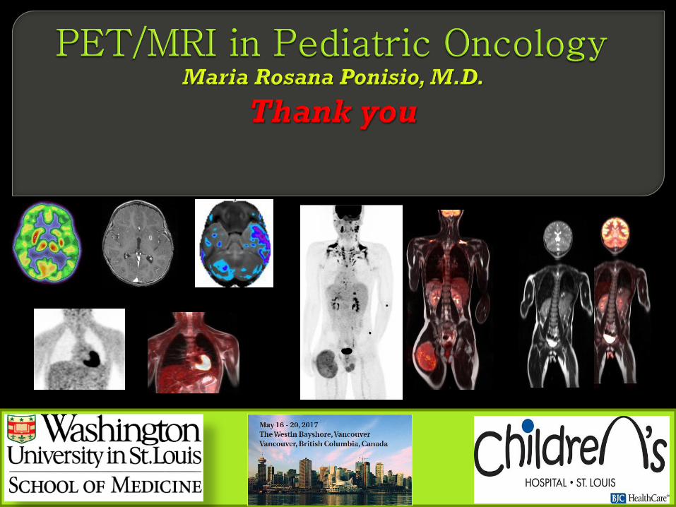
May 20, 2017PET/MRI Session

No Financial Disclosures
Ponisio, Maria Rosana MDWashington University in St. Louis

McConathy Jonathan, M.D, PhD, Nuclear Medicine
Dehdashti Farrok, M.D, Nuclear Medicine
Geetika Khanna, M.D, Pediatric Radiology
Karen Gauvain, M.D, Pediatric Oncology
Ponisio, Maria Rosana MDWashington University in St. Louis
Select the BEST statement regarding the advantages of PET/MR:
1. Lower cumulative dose of ionizing radiation with removal of the
CT component
2. Allows for combined metabolic, functional and anatomical
imaging
3. Increases the safety profile for vulnerable patients reducing
radiation and anesthesia requirements.
4. All of the above
May 20, 2017PET/MRI
Ponisio, Maria Rosana MDWashington University in St. Louis

Select the CORRECT answer regarding simultaneous PET/MR:
1. MR based PET attenuation correction is not needed for PET/MR
2. There is no correlation between standardized uptake values
(SUVs) on PET/CT and PET/MR
3. PET/MR has lower cost and shorter scan times then PET/CT
4. PET/MR provides superior evaluation of the lung parenchyma
over PET/CT
5. None of above.
May 20, 2017PET/MRI Session
Ponisio, Maria Rosana MDWashington University in St. Louis
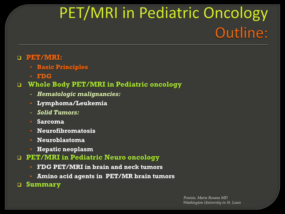
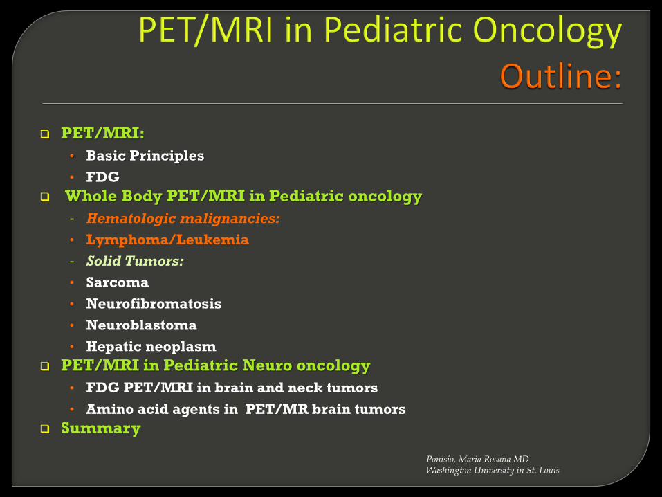
PET/MRI:
• Basic Principles
• FDG
Whole Body PET/MRI in Pediatric oncology
- Hematologic malignancies:
• Lymphoma/Leukemia
- Solid Tumors:
• Sarcoma
• Neurofibromatosis
• Neuroblastoma
• Hepatic neoplasm
PET/MRI in Pediatric Neuro oncology
• FDG PET/MRI in brain and neck tumors
• Amino acid agents in PET/MR brain tumors
Summary
Ponisio, Maria Rosana MDWashington University in St. Louis
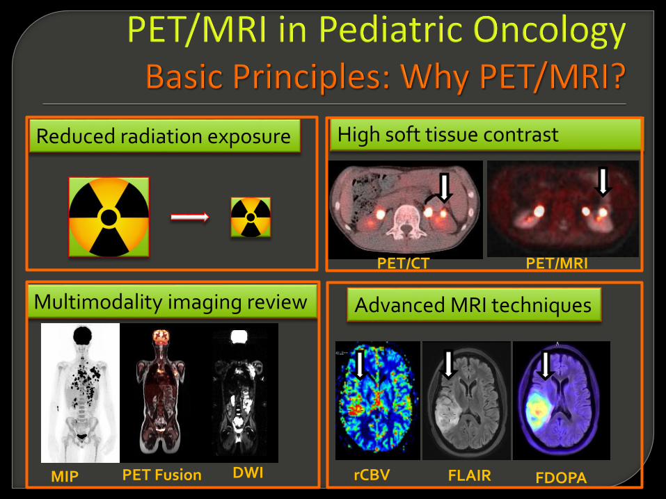
Reduced radiation exposure High soft tissue contrast
PET/CT PET/MRI
Multimodality imaging review
DWIPET Fusion
Advanced MRI techniques
rCBV FLAIR FDOPAMIP

Sequential Scanner Simultaneous PET/MRI
PET and MRI
PET/MRI
Types
Radiology: Volume 267: Number 1—April 2013
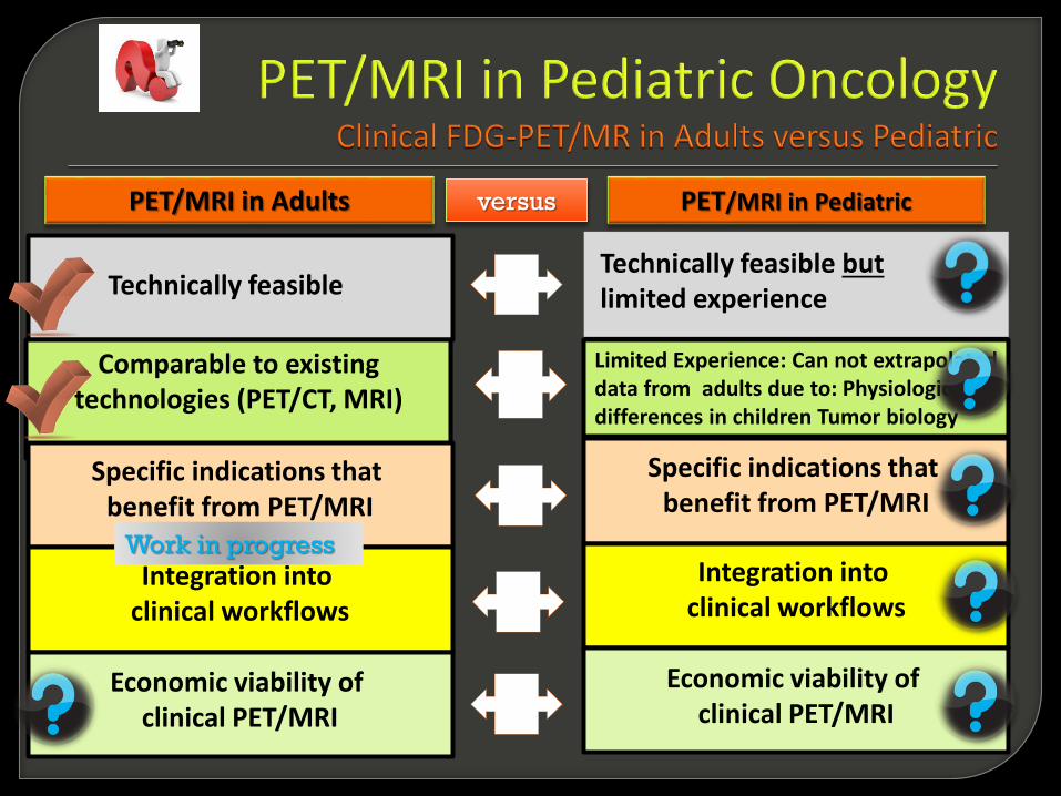
Technically feasible
Comparable to existing technologies (PET/CT, MRI)
Specific indications that benefit from PET/MRI
Integration into clinical workflows
Economic viability of clinical PET/MRI
Technically feasible butlimited experience
Limited Experience: Can not extrapolated data from adults due to: Physiologic differences in children Tumor biology
Specific indications that benefit from PET/MRI
Integration into clinical workflows
Economic viability of clinical PET/MRI
PET/MRI in Adults PET/MRI in Pediatricversus
Work in progress
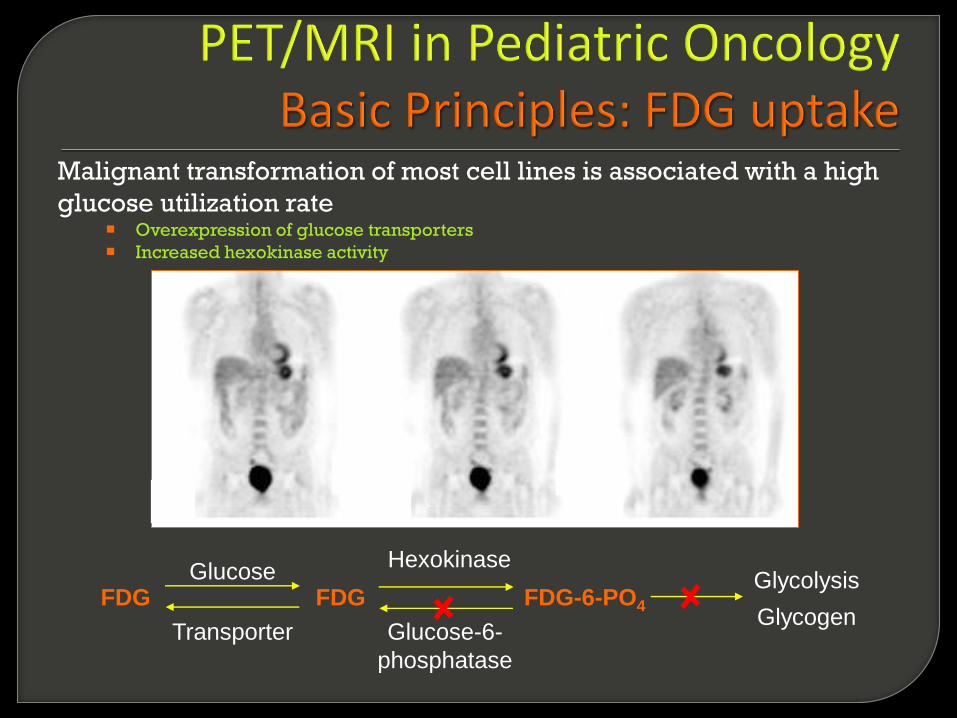
Glucose
Transporter
Hexokinase
Glucose-6-
phosphatase
GlycogenFDG FDG FDG-6-PO4
Glycolysis
Malignant transformation of most cell lines is associated with a high
glucose utilization rate Overexpression of glucose transporters
Increased hexokinase activity

PET/MRI:
• Basic Principles
• FDG
Whole Body PET/MRI in Pediatric oncology
- Hematologic malignancies:
• Lymphoma/Leukemia
- Solid Tumors:
• Sarcoma
• Neurofibromatosis
• Neuroblastoma
• Hepatic neoplasm
PET/MRI in Pediatric Neuro oncology
• FDG PET/MRI in brain and neck tumors
• Amino acid agents in PET/MR brain tumors
Summary
Ponisio, Maria Rosana MDWashington University in St. Louis

FDG-PET/CT Initial Staging
14-year-old girl with classical
Hodgkin lymphoma
PET/CT Follow-up
Treatment monitoring during
chemotherapy (ABVD).
PET/CTCTPET
Initial Staging
Follow up
Ponisio, Maria Rosana ,MDWashington University in St. Louis
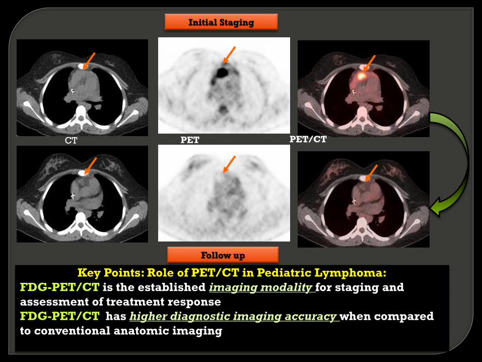
Key Points: Role of PET/CT in Pediatric Lymphoma:
FDG-PET/CT is the established imaging modality for staging and
assessment of treatment response
FDG-PET/CT has higher diagnostic imaging accuracy when compared
to conventional anatomic imaging
PET/CTPET
Initial Staging
Follow up
CT
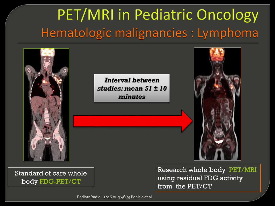
Standard of care whole
body FDG-PET/CT
Research whole body PET/MRI
using residual FDG activity
from the PET/CT
Interval between
studies: mean 51 ± 10
minutes
Pediatr Radiol. 2016 Aug;46(9) Ponisio at al.
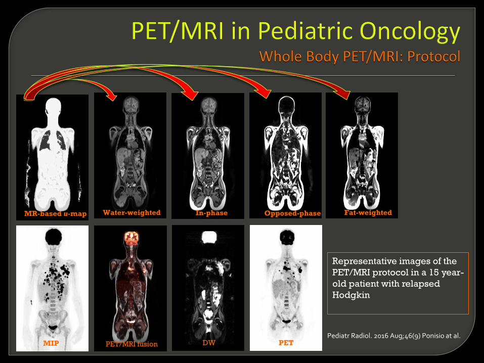
MR-based u-map Water-weighted Opposed-phase Fat-weightedIn-phase
MIP PET/MRI fusion DW PET
Representative images of the
PET/MRI protocol in a 15 year-
old patient with relapsed
Hodgkin
Pediatr Radiol. 2016 Aug;46(9) Ponisio at al.

Fused PET/MRI
DWI
T2 HASTEFDG-PET/MR MIPFDG-PET/CT MIP
Pediatr Radiol. 2016 Aug;46(9) Ponisio at al.

Correlation plots of PET/CT and PET/MRI from all selected normal organs: A and
FDG-avid lesions: B with the corresponding correlation coefficients (r2)Ponisio et al, 2016 Ped Radiology
A B
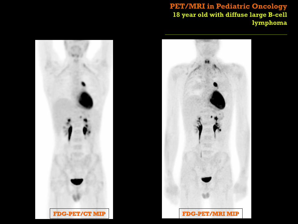
FDG-PET/MRI MIPFDG-PET/CT MIP
PET/CT PET/MRI
PET/MRI
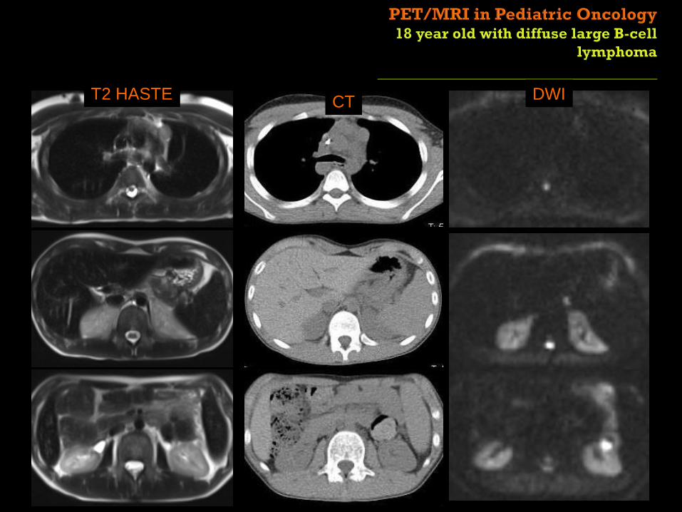
T2 HASTE DWICT
PET/CT PET/MRI
PET/MRI
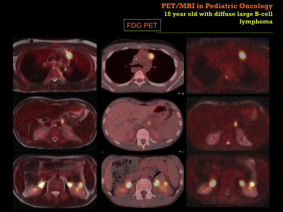
FDG PET
A
DC
B
A B
PET/CT
PET/MRI
PET/CT
PET/MRI
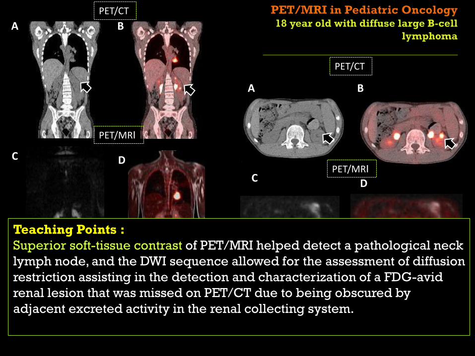
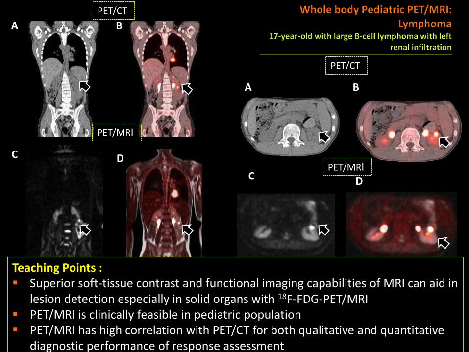
PET/CT and PET/MRI in the same patient perfomed in the same day
Restricted diffusion on DWI (C) and the excellent PET/MRI co-registration allowed identification of the renal lesion and differentiation from excreted FDG in the collecting system
C D
Teaching Points :
Superior soft-tissue contrast of PET/MRI helped detect a pathological neck
lymph node, and the DWI sequence allowed for the assessment of diffusion
restriction assisting in the detection and characterization of a FDG-avid
renal lesion that was missed on PET/CT due to being obscured by
adjacent excreted activity in the renal collecting system.

Sagittal mmap PET/MRI fusion Attenuated PET
Please note lack of artifacts on non-attenuated PET images
Key Points:
The µmap as well as the non-attenuation -corrected data should always
be inspected in addition to the attenuation corrected data
µ-map Fused PET-HASTE AC PET NAC-PET

Breathing artifact which simulated a lung base nodule on PET/MRI images
PET/CT perform the same day is normal
Attenuated PET PET/MRI fusion
Key Points:
Errors in attenuation correction within the lung can be cause by
respiratory misalignment of PET and MR data due to the difference in
duration of two examinations resulting in mismatch of PET emission
and MR attenuating data

Purpose:
Feasibility in pediatric patients
Dosimetry
Images quality
Diagnostic performance (Quantitative analysis):
SUV mean measures on normal organs
Mean Standardized Uptake Value (SUVmean) measured on FDG-avid lesions
Conclusions:
Pediatric FDG- PET/MRI is feasible
Decreases radiation exposure (39%)
Good image quality
High correlation with PET/CT (r2=0.88)
Very high correlation with PET/CT (r2=0.94)
Pediatr Radiol. 2016 Aug;46(9) Ponisio at al.
A
DC
B
A B
PET/CT
PET/MRI
PET/CT
PET/MRI
PET/CT and PET/MRI in the same patient perfomed in the same day
Restricted diffusion on DWI (C) and the excellent PET/MRI co-registration allowed identification of the renal lesion and differentiation from excreted FDG in the collecting system
C D
Teaching Points : Superior soft-tissue contrast and functional imaging capabilities of MRI can aid in
lesion detection especially in solid organs with 18F-FDG-PET/MRI PET/MRI is clinically feasible in pediatric population PET/MRI has high correlation with PET/CT for both qualitative and quantitative
diagnostic performance of response assessment

PET/MRI:
• Basic Principles
• FDG
Whole Body PET/MRI in Pediatric oncology
- Hematologic malignancies:
• Lymphoma/Leukemia
- Solid Tumors:
• Sarcoma
• Neurofibromatosis
• Neuroblastoma
• Hepatic neoplasm
PET/MRI in Pediatric Neuro oncology
• FDG PET/MRI in brain and neck tumors
• Amino acid agents in PET/MR brain tumors
Summary
Ponisio, Maria Rosana MDWashington University in St. Louis

FDG-PET/MRI MIP

T2 FS DWI ADCFDG PET

D
C
D
Preoperative CT
Dedicated abdominal FDG-PET/MRI
A
B
T2 FS DWI ADC

MRI PET/CT

PET/CT PET/MRI
PET/MRI
T2 HASTE DWICT

PET/CT PET/MRI
PET/MRI
T2 HASTE DWICT
FUSED PET/MRI

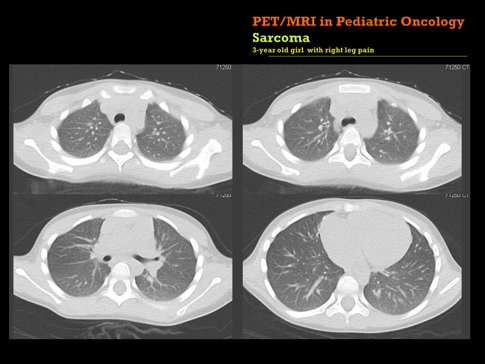
Whole body FDG PET/MRIDedicated chest FDG PET/MRI
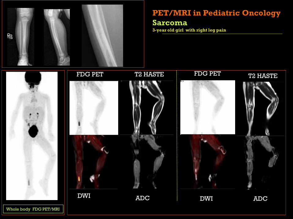
Whole body FDG PET/MRI
DWI ADC
T2 HASTE
DWIADC
FDG PET FDG PET T2 HASTE

T1 TSE T1 FS T1 FS with contrast
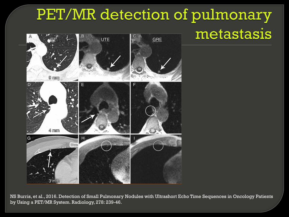
NS Burris, et al., 2016. Detection of Small Pulmonary Nodules with Ultrashort Echo Time Sequences in Oncology Patients
by Using a PET/MR System. Radiology, 278: 239-46.

T1 T1 +C T2
T1 T1 +C T2

PET/MRI:
• Basic Principles
• FDG
Whole Body PET/MRI in Pediatric oncology
- Hematologic malignancies:
• Lymphoma/Leukemia
- Solid Tumors:
• Sarcoma
• Neurofibromatosis
• Neuroblastoma
• Hepatic neoplasm
PET/MRI in Pediatric Neuro oncology
• FDG PET/MRI in brain and neck tumors
• Amino acid agents in PET/MR brain tumors
Summary
Ponisio, Maria Rosana MDWashington University in St. Louis

FDG-PET has limited utility in brain tumors
• primarily for distinguishing radiation necrosis from recurrent
tumor
Amino acid-PET is well-established in adult neuro-
oncology Targets system L amino acid transport
Crosses the intact blood-brain barrier (BBB) therefore
visualizes both enhancing and non-enhancing tumors
Can visualize the entire tumor volume
[18F]FDG [18F]FDOPA

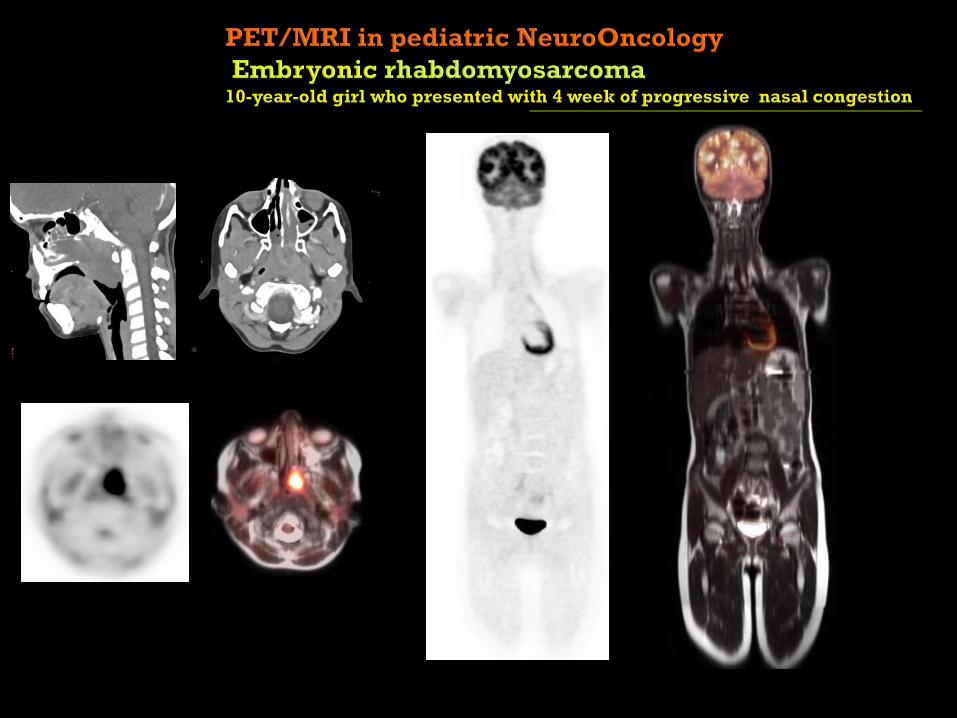
8-year-old with a right thalamic grade IV small cell astrocytoma
FDOPA PET/MRI prior to starting bevacizumab therapy
FDOPA PET/MRI after 4 weeks of bevacizumab therapy
T1 with contrastFDOPA FDOPA/FLAIR
A
B
Karen Gauvain at al.
Neuro-Oncology
Practice , in press
2017

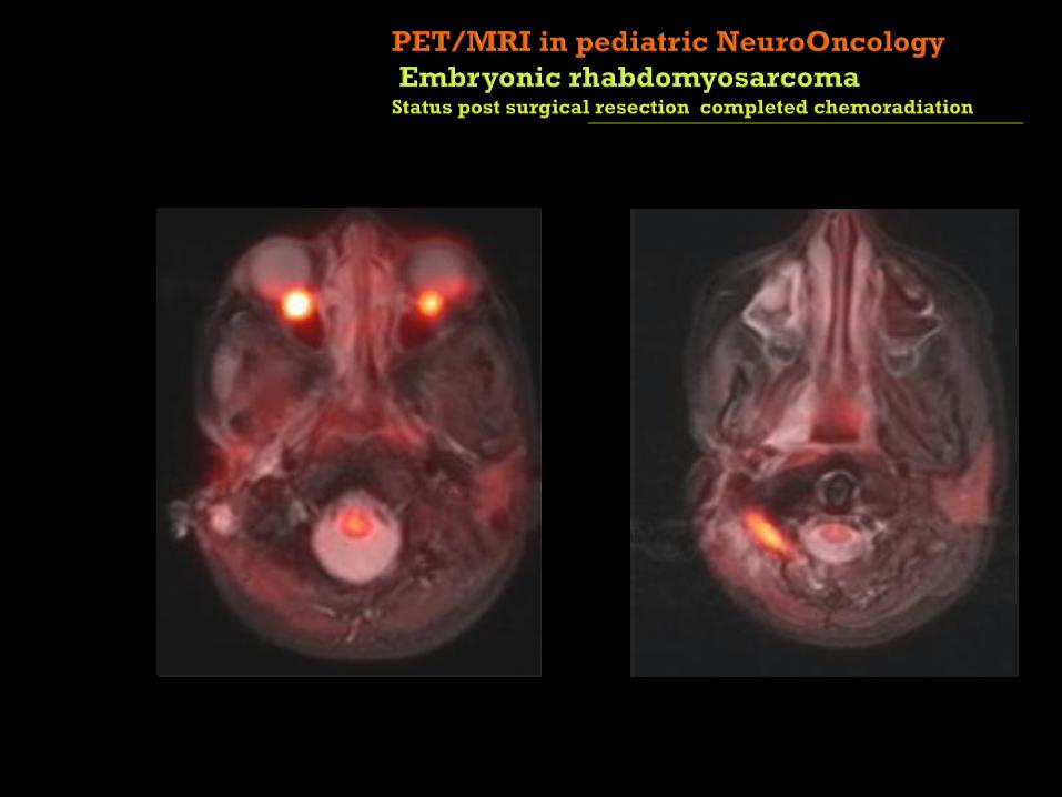
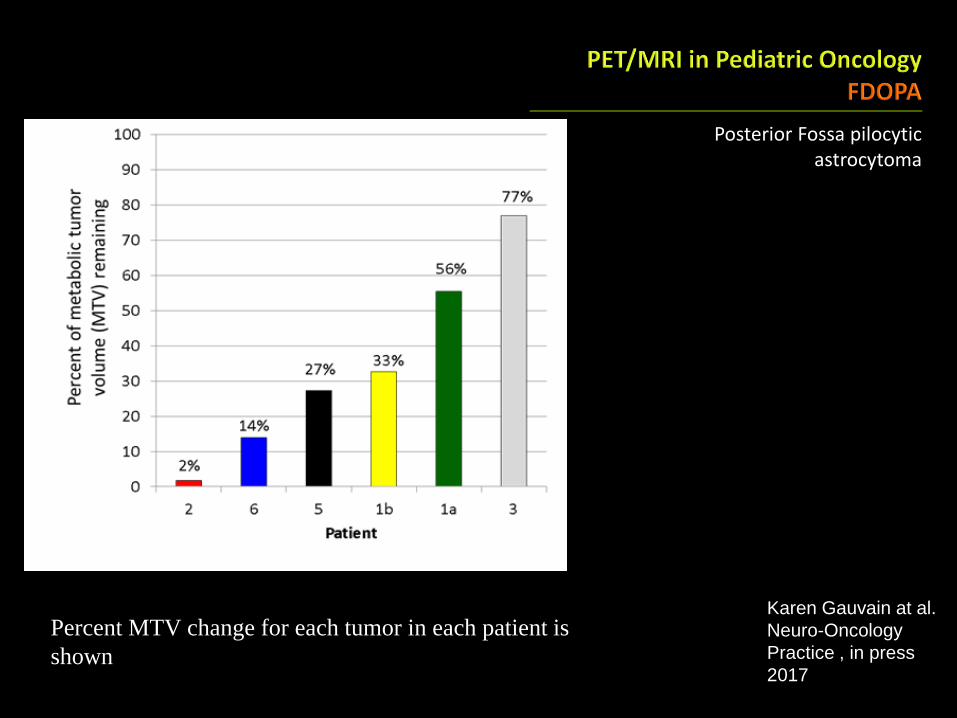
Posterior Fossa pilocyticastrocytoma
FDOPA PET/MRI prior to starting bevacizumab therapy
T1 with contrastFDOPA FDOPA/FLAIR
FDOPA PET/MRI after 4 weeks of bevacizumab therapy
Karen Gauvain at al.
Neuro-Oncology
Practice , in press
2017
Posterior Fossa pilocyticastrocytoma
Karen Gauvain at al.
Neuro-Oncology
Practice , in press
2017
Percent MTV change for each tumor in each patient is
shown
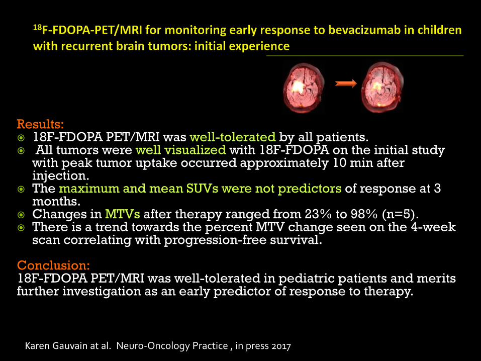
Results: 18F-FDOPA PET/MRI was well-tolerated by all patients. All tumors were well visualized with 18F-FDOPA on the initial study
with peak tumor uptake occurred approximately 10 min after injection.
The maximum and mean SUVs were not predictors of response at 3 months.
Changes in MTVs after therapy ranged from 23% to 98% (n=5). There is a trend towards the percent MTV change seen on the 4-week
scan correlating with progression-free survival.
Conclusion:18F-FDOPA PET/MRI was well-tolerated in pediatric patients and merits further investigation as an early predictor of response to therapy.
Karen Gauvain at al. Neuro-Oncology Practice , in press 2017
PET/MRI is expected to have an emerging role in pediatric
diagnostic imaging:
Lower radiation exposure than of PET/CT with similar
performance
Potential reductions in total scan time, repeated examinations,
and repeated sedation or anesthesia for certain indications
Introduction of new tracers may expand the use of PET/MRI
Simultaneous acquisition of anatomic and functional data, and
development of multi-parametric applications will improve
diagnostic performance
However, further investigation is needed