Embed Size (px)
Citation preview
Clinical StudyClinical Usefulness of Monitoring Expression Levels of CCL24(Eotaxin-2) mRNA on the Ocular Surface in Patients with VernalKeratoconjunctivitis and Atopic Keratoconjunctivitis
Yukiko Shiraki, Jun Shoji, and Noriko Inada
Division of Ophthalmology, Department of Visual Sciences, Nihon University School of Medicine, Tokyo, Japan
Correspondence should be addressed to Noriko Inada; [email protected]
Received 29 April 2016; Revised 2 August 2016; Accepted 18 August 2016
Academic Editor: Magnus Ivarsson
Copyright © 2016 Yukiko Shiraki et al.This is an open access article distributed under the Creative Commons Attribution License,which permits unrestricted use, distribution, and reproduction in any medium, provided the original work is properly cited.
Purpose. This study aimed to evaluate the clinical efficacy of using expression levels of CCL24 (eotaxin-2) mRNA on the ocularsurface as a biomarker in patients with vernal keratoconjunctivitis (VKC) and atopic keratoconjunctivitis (AKC).Methods. Eighteenpatients with VKC or AKC (VKC/AKC group) and 12 control subjects (control group) were enrolled in this study. The VKC/AKCclinical score was determined by objective findings in patients by using the 5-5-5 exacerbation grading scale. All subjects underwentmodified impression cytology and specimenswere obtained from the upper tarsal conjunctiva. Expression levels ofCCL24 (eotaxin-2) mRNA on the ocular surface were determined using real-time reverse transcription polymerase chain reaction. Results. TheVKC group was divided into two subgroups, depending on the clinical score: the active stage subgroup with 100 points or moreof clinical scores and the stable stage subgroup with 100 points or less. CCL24 (eotaxin-2) mRNA expression levels in the activeVKC/AKC stage subgroup were significantly higher than those in the stable VKC/AKC subgroup and the control group. Clinicalscores correlated significantly with CCL24 (eotaxin-2) mRNA expression levels in the VKC group. Conclusions. CCL24 (eotaxin-2)mRNA expression levels on the ocular surface are a useful biomarker for clinical severity of VKC/AKC.
1. Introduction
Allergic conjunctival diseases (ACD) are conjunctival inflam-matory disorders, characterized by an immediate hypersensi-tivity reaction associated with antigen-specific IgE antibodies[1]. Patients with ACD are usually categorized into thefollowing types based on clinical criteria: seasonal allergicconjunctivitis (SAC), perennial allergic conjunctivitis (PAC),vernal keratoconjunctivitis (VKC), atopic keratoconjunctivi-tis (AKC), and giant papillary conjunctivitis (GPC) [2].SAC and PAC are characterized by conjunctival hyperemia,conjunctival edema, and papillary hyperplasia of tarsal con-junctiva and are classified as mild ACD. In contrast, VKC ischaracterized by the development of proliferative lesions ofthe conjunctiva, including giant papillary proliferation of thetarsal conjunctiva and gelatinous cell infiltration of the limbalconjunctiva. VKC also complicates corneal disorders, such asshield ulcer and punctate corneal keratitis, and is classifiedas a severe ACD, because the visual prognosis may be poor.AKC usually develops in older patients with atopic dermatitis
and also complicates severe ocular surface diseases, includinggiant papillary conjunctivitis, shield ulcer, and dry eye.
In shield ulcers of patients with VKC and AKC, deposi-tions of major basic protein and eosinophil cationic protein(ECP), which comprise specific granules of eosinophils, havereportedly been observed histologically in the corneal ulcera-tive lesions [3, 4]. Furthermore, ECP levels in the tears ofVKCand AKC patients are reportedly increased, in comparisonwith those in controls [5–7]. Therefore, the pathophysiolog-ical characteristics of these severe ACD, including VKC andAKC, include eosinophilic inflammation in the conjunctiva.
Eotaxin is a member of the CC chemokine family andis divided into three subfamilies, namely, CCL11/eotaxin-1,CCL24/eotaxin-2, and CCL26/eotaxin-3. Eotaxin-1, eotaxin-2, and eotaxin-3 interact with the CC chemokine recep-tor 3 (CCR3). Representative inflammatory cells express-ing CCR3 on their cell surfaces are eosinophils, type-2helper T cells (Th2), and basophils. IL-13, which is a Th2-derived cytokine, induces eotaxin in vitro through activation
Hindawi Publishing CorporationJournal of OphthalmologyVolume 2016, Article ID 3573142, 5 pageshttp://dx.doi.org/10.1155/2016/3573142
2 Journal of Ophthalmology
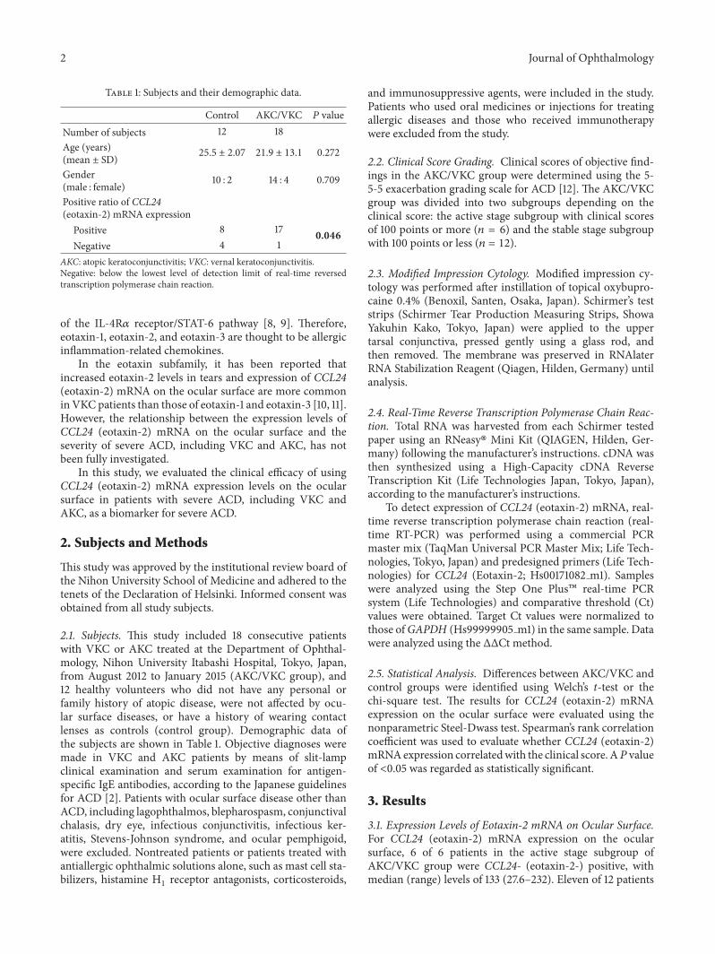
Table 1: Subjects and their demographic data.
Control AKC/VKC 𝑃 valueNumber of subjects 12 18Age (years)(mean ± SD) 25.5 ± 2.07 21.9 ± 13.1 0.272
Gender(male : female) 10 : 2 14 : 4 0.709
Positive ratio of CCL24(eotaxin-2) mRNA expression
Positive 8 17 0.046Negative 4 1
AKC: atopic keratoconjunctivitis; VKC: vernal keratoconjunctivitis.Negative: below the lowest level of detection limit of real-time reversedtranscription polymerase chain reaction.
of the IL-4R𝛼 receptor/STAT-6 pathway [8, 9]. Therefore,eotaxin-1, eotaxin-2, and eotaxin-3 are thought to be allergicinflammation-related chemokines.
In the eotaxin subfamily, it has been reported thatincreased eotaxin-2 levels in tears and expression of CCL24(eotaxin-2) mRNA on the ocular surface are more commoninVKC patients than those of eotaxin-1 and eotaxin-3 [10, 11].However, the relationship between the expression levels ofCCL24 (eotaxin-2) mRNA on the ocular surface and theseverity of severe ACD, including VKC and AKC, has notbeen fully investigated.
In this study, we evaluated the clinical efficacy of usingCCL24 (eotaxin-2) mRNA expression levels on the ocularsurface in patients with severe ACD, including VKC andAKC, as a biomarker for severe ACD.
2. Subjects and Methods
This study was approved by the institutional review board ofthe Nihon University School of Medicine and adhered to thetenets of the Declaration of Helsinki. Informed consent wasobtained from all study subjects.
2.1. Subjects. This study included 18 consecutive patientswith VKC or AKC treated at the Department of Ophthal-mology, Nihon University Itabashi Hospital, Tokyo, Japan,from August 2012 to January 2015 (AKC/VKC group), and12 healthy volunteers who did not have any personal orfamily history of atopic disease, were not affected by ocu-lar surface diseases, or have a history of wearing contactlenses as controls (control group). Demographic data ofthe subjects are shown in Table 1. Objective diagnoses weremade in VKC and AKC patients by means of slit-lampclinical examination and serum examination for antigen-specific IgE antibodies, according to the Japanese guidelinesfor ACD [2]. Patients with ocular surface disease other thanACD, including lagophthalmos, blepharospasm, conjunctivalchalasis, dry eye, infectious conjunctivitis, infectious ker-atitis, Stevens-Johnson syndrome, and ocular pemphigoid,were excluded. Nontreated patients or patients treated withantiallergic ophthalmic solutions alone, such as mast cell sta-bilizers, histamine H
1receptor antagonists, corticosteroids,
and immunosuppressive agents, were included in the study.Patients who used oral medicines or injections for treatingallergic diseases and those who received immunotherapywere excluded from the study.
2.2. Clinical Score Grading. Clinical scores of objective find-ings in the AKC/VKC group were determined using the 5-5-5 exacerbation grading scale for ACD [12]. The AKC/VKCgroup was divided into two subgroups depending on theclinical score: the active stage subgroup with clinical scoresof 100 points or more (𝑛 = 6) and the stable stage subgroupwith 100 points or less (𝑛 = 12).
2.3. Modified Impression Cytology. Modified impression cy-tology was performed after instillation of topical oxybupro-caine 0.4% (Benoxil, Santen, Osaka, Japan). Schirmer’s teststrips (Schirmer Tear Production Measuring Strips, ShowaYakuhin Kako, Tokyo, Japan) were applied to the uppertarsal conjunctiva, pressed gently using a glass rod, andthen removed. The membrane was preserved in RNAlaterRNA Stabilization Reagent (Qiagen, Hilden, Germany) untilanalysis.
2.4. Real-Time Reverse Transcription Polymerase Chain Reac-tion. Total RNA was harvested from each Schirmer testedpaper using an RNeasy� Mini Kit (QIAGEN, Hilden, Ger-many) following the manufacturer’s instructions. cDNA wasthen synthesized using a High-Capacity cDNA ReverseTranscription Kit (Life Technologies Japan, Tokyo, Japan),according to the manufacturer’s instructions.
To detect expression of CCL24 (eotaxin-2) mRNA, real-time reverse transcription polymerase chain reaction (real-time RT-PCR) was performed using a commercial PCRmaster mix (TaqMan Universal PCR Master Mix; Life Tech-nologies, Tokyo, Japan) and predesigned primers (Life Tech-nologies) for CCL24 (Eotaxin-2; Hs00171082 m1). Sampleswere analyzed using the Step One Plus� real-time PCRsystem (Life Technologies) and comparative threshold (Ct)values were obtained. Target Ct values were normalized tothose ofGAPDH (Hs99999905 m1) in the same sample. Datawere analyzed using the ΔΔCt method.
2.5. Statistical Analysis. Differences between AKC/VKC andcontrol groups were identified using Welch’s t-test or thechi-square test. The results for CCL24 (eotaxin-2) mRNAexpression on the ocular surface were evaluated using thenonparametric Steel-Dwass test. Spearman’s rank correlationcoefficient was used to evaluate whether CCL24 (eotaxin-2)mRNAexpression correlatedwith the clinical score. A𝑃 valueof <0.05 was regarded as statistically significant.
3. Results
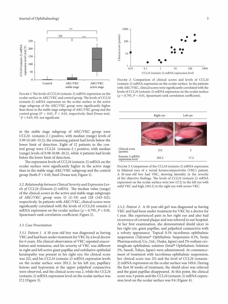
3.1. Expression Levels of Eotaxin-2 mRNA on Ocular Surface.For CCL24 (eotaxin-2) mRNA expression on the ocularsurface, 6 of 6 patients in the active stage subgroup ofAKC/VKC group were CCL24- (eotaxin-2-) positive, withmedian (range) levels of 133 (27.6–232). Eleven of 12 patients
Journal of Ophthalmology 3
Control AKC/VKCstable stage
AKC/VKCactive stage
CCL2
4 (e
otax
in-2
) mRN
A
NS
0
50
100
150
200
250
expr
essio
n le
vel (ΔΔ
CT)
∗∗
∗∗
Figure 1: The levels of CCL24 (eotaxin-2) mRNA expression on theocular surface in AKC/VKC and control group.The levels of CCL24(eotaxin-2) mRNA expression on the ocular surface in the activestage subgroup of the AKC/VKC group were significantly higherthan those in the stable stage subgroup of AKC/VKC group and thecontrol group (𝑃 < 0.01, 𝑃 < 0.01, respectively, Steel-Dwass test).∗𝑝 < 0.05; NS: not significant.
in the stable stage subgroup of AKC/VKC group wereCCL24- (eotaxin-2-) positive, with median (range) levels of5.99 (0.140–33.2); the remaining patient had levels below thelower limit of detection. Eight of 12 patients in the con-trol group were CCL24- (eotaxin-2-) positive, with median(range) levels of 0.98 (0.08–20.2), while 4 patients had levelsbelow the lower limit of detection.
The expression levels ofCCL24 (eotaxin-2) mRNA on theocular surface were significantly higher in the active stagethan in the stable stage AKC/VKC subgroup and the controlgroup (both P < 0.01; Steel-Dwass test; Figure 1).
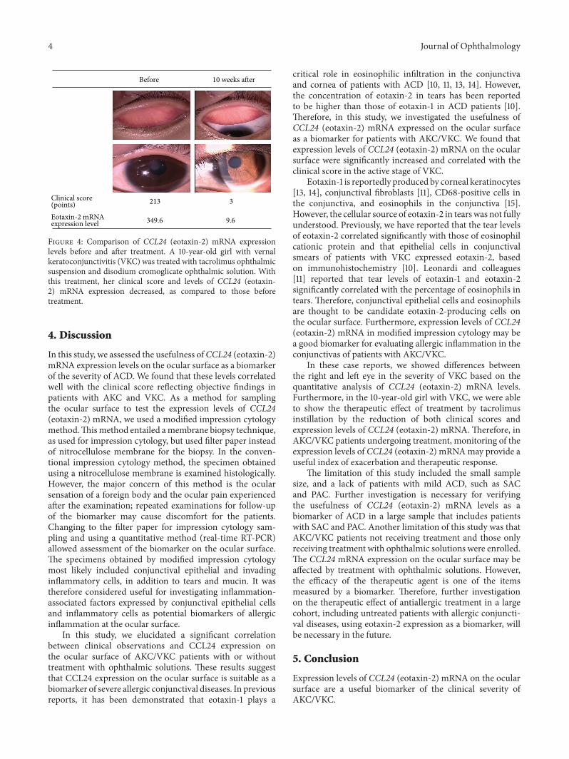
3.2. Relationship betweenClinical Severity and Expression Lev-els of CCL24 (Eotaxin-2) mRNA. The median value (range)of the clinical scores in the active and stable stage subgroupsof AKC/VKC group were 13 (2–33) and 128 (343–112),respectively. In patients with AKC/VKC, clinical scores weresignificantly correlated with the levels of (CCL24) eotaxin-2mRNA expression on the ocular surface (𝜌 = 0.795, P < 0.01,Spearman’s rank correlation coefficient; Figure 2).
3.3. Case Presentation
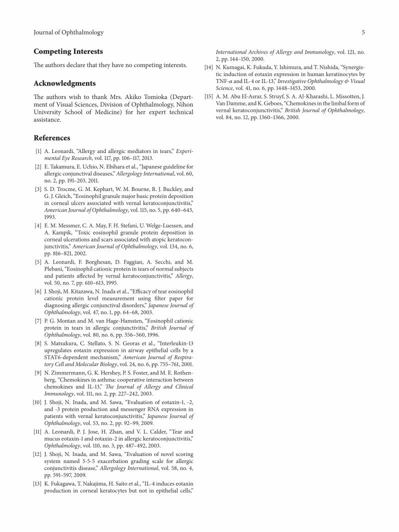
3.3.1. Patient 1. A 10-year-old boy was diagnosed as havingVKCand had been under treatment forVKCby a local doctorfor 6 years. His clinical observation of VKC repeated exacer-bation and remission, and his severity of VKC was differentin right and left active giant papillae and exfoliative epithelialkeratopathy was present in his right eye; his clinical scorewas 212, and his CCL24 (eotaxin-2) mRNA expression levelson the ocular surface were 203.2. In his left eye, papillarylesions and hyperemia at the upper palpebral conjunctivawere observed, and the clinical score was 2, while the CCL24(eotaxin-2) mRNA expression level on the ocular surface was17.2 (Figure 3).
0.01 0.1 1 10 100 1000CCL24 (eotaxin-2) mRNA expression level
1
10
100
1000
Clin
ical
scor
e (po
ints)
Figure 2: Comparison of clinical scores and levels of CCL24(eotaxin-2) mRNA expression on the ocular surface. In the patientswithAKC/VKC, clinical scores were significantly correlatedwith thelevels of CCL24 (eotaxin-2) mRNA expression on the ocular surface(𝜌 = 0.795, 𝑃 < 0.01, Spearman’s rank correlation coefficient).
212 2Clinical score(points)
203.2 17.2Eotaxin-2 mRNAexpression level
Right eye Left eye
Figure 3: Comparison of the CCL24 (eotaxin-2) mRNA expressionin bilateral eyes of a vernal keratoconjunctivitis (VKC) patient.A 10-year-old boy had VKC, showing laterality in the severityof the objective findings. The levels of CCL24 (eotaxin-2) mRNAexpression on the ocular surface were low (17.2) in the left eye withmild VKC and high (203.2) in the right eye with severe VKC.
3.3.2. Patient 2. A 10-year-old girl was diagnosed as havingVKC and had been under treatment for VKC by a doctor for1 year. She experienced pain in her right eye and also hadrecurrence of corneal plaque andwas referred to our hospital.At her first examination, she demonstrated shield ulcer inher right eye, giant papillae, and palpebral conjunctiva witha velvety appearance. Topical 0.1% tacrolimus ophthalmicsuspension (Talymus� Ophthalmic Suspension 0.1%, SenjuPharmaceutical, Co., Ltd., Osaka, Japan) and 2% sodium cro-moglicate ophthalmic solution (Intal� Ophthalmic Solution2%, Sanofi, Tokyo, Japan) were administered. At commence-ment of treatment with tacrolimus ophthalmic suspension,her clinical score was 213 and the level of CCL24 (eotaxin-2) mRNA expression on the ocular surface was 349.6. Duringthe first 10 weeks of treatment, the shield ulcer was relievedand the giant papillae disappeared. At this point, the clinicalscore was 3 points and the CCL24 (eotaxin-2) mRNA expres-sion level on the ocular surface was 9.6 (Figure 4).
4 Journal of Ophthalmology
213 3Clinical score(points)
349.6 9.6Eotaxin-2 mRNAexpression level
Before 10 weeks after
Figure 4: Comparison of CCL24 (eotaxin-2) mRNA expressionlevels before and after treatment. A 10-year-old girl with vernalkeratoconjunctivitis (VKC) was treated with tacrolimus ophthalmicsuspension and disodium cromoglicate ophthalmic solution. Withthis treatment, her clinical score and levels of CCL24 (eotaxin-2) mRNA expression decreased, as compared to those beforetreatment.
4. Discussion
In this study, we assessed the usefulness ofCCL24 (eotaxin-2)mRNA expression levels on the ocular surface as a biomarkerof the severity of ACD. We found that these levels correlatedwell with the clinical score reflecting objective findings inpatients with AKC and VKC. As a method for samplingthe ocular surface to test the expression levels of CCL24(eotaxin-2) mRNA, we used a modified impression cytologymethod.Thismethod entailed amembrane biopsy technique,as used for impression cytology, but used filter paper insteadof nitrocellulose membrane for the biopsy. In the conven-tional impression cytology method, the specimen obtainedusing a nitrocellulose membrane is examined histologically.However, the major concern of this method is the ocularsensation of a foreign body and the ocular pain experiencedafter the examination; repeated examinations for follow-upof the biomarker may cause discomfort for the patients.Changing to the filter paper for impression cytology sam-pling and using a quantitative method (real-time RT-PCR)allowed assessment of the biomarker on the ocular surface.The specimens obtained by modified impression cytologymost likely included conjunctival epithelial and invadinginflammatory cells, in addition to tears and mucin. It wastherefore considered useful for investigating inflammation-associated factors expressed by conjunctival epithelial cellsand inflammatory cells as potential biomarkers of allergicinflammation at the ocular surface.
In this study, we elucidated a significant correlationbetween clinical observations and CCL24 expression onthe ocular surface of AKC/VKC patients with or withouttreatment with ophthalmic solutions. These results suggestthat CCL24 expression on the ocular surface is suitable as abiomarker of severe allergic conjunctival diseases. In previousreports, it has been demonstrated that eotaxin-1 plays a
critical role in eosinophilic infiltration in the conjunctivaand cornea of patients with ACD [10, 11, 13, 14]. However,the concentration of eotaxin-2 in tears has been reportedto be higher than those of eotaxin-1 in ACD patients [10].Therefore, in this study, we investigated the usefulness ofCCL24 (eotaxin-2) mRNA expressed on the ocular surfaceas a biomarker for patients with AKC/VKC. We found thatexpression levels of CCL24 (eotaxin-2) mRNA on the ocularsurface were significantly increased and correlated with theclinical score in the active stage of VKC.
Eotaxin-1 is reportedly produced by corneal keratinocytes[13, 14], conjunctival fibroblasts [11], CD68-positive cells inthe conjunctiva, and eosinophils in the conjunctiva [15].However, the cellular source of eotaxin-2 in tearswas not fullyunderstood. Previously, we have reported that the tear levelsof eotaxin-2 correlated significantly with those of eosinophilcationic protein and that epithelial cells in conjunctivalsmears of patients with VKC expressed eotaxin-2, basedon immunohistochemistry [10]. Leonardi and colleagues[11] reported that tear levels of eotaxin-1 and eotaxin-2significantly correlated with the percentage of eosinophils intears. Therefore, conjunctival epithelial cells and eosinophilsare thought to be candidate eotaxin-2-producing cells onthe ocular surface. Furthermore, expression levels of CCL24(eotaxin-2) mRNA in modified impression cytology may bea good biomarker for evaluating allergic inflammation in theconjunctivas of patients with AKC/VKC.
In these case reports, we showed differences betweenthe right and left eye in the severity of VKC based on thequantitative analysis of CCL24 (eotaxin-2) mRNA levels.Furthermore, in the 10-year-old girl with VKC, we were ableto show the therapeutic effect of treatment by tacrolimusinstillation by the reduction of both clinical scores andexpression levels of CCL24 (eotaxin-2) mRNA. Therefore, inAKC/VKC patients undergoing treatment, monitoring of theexpression levels of CCL24 (eotaxin-2) mRNAmay provide auseful index of exacerbation and therapeutic response.
The limitation of this study included the small samplesize, and a lack of patients with mild ACD, such as SACand PAC. Further investigation is necessary for verifyingthe usefulness of CCL24 (eotaxin-2) mRNA levels as abiomarker of ACD in a large sample that includes patientswith SAC and PAC. Another limitation of this study was thatAKC/VKC patients not receiving treatment and those onlyreceiving treatment with ophthalmic solutions were enrolled.The CCL24 mRNA expression on the ocular surface may beaffected by treatment with ophthalmic solutions. However,the efficacy of the therapeutic agent is one of the itemsmeasured by a biomarker. Therefore, further investigationon the therapeutic effect of antiallergic treatment in a largecohort, including untreated patients with allergic conjuncti-val diseases, using eotaxin-2 expression as a biomarker, willbe necessary in the future.
5. Conclusion
Expression levels of CCL24 (eotaxin-2) mRNA on the ocularsurface are a useful biomarker of the clinical severity ofAKC/VKC.
Journal of Ophthalmology 5
Competing Interests
The authors declare that they have no competing interests.
Acknowledgments
The authors wish to thank Mrs. Akiko Tomioka (Depart-ment of Visual Sciences, Division of Ophthalmology, NihonUniversity School of Medicine) for her expert technicalassistance.
References
[1] A. Leonardi, “Allergy and allergic mediators in tears,” Experi-mental Eye Research, vol. 117, pp. 106–117, 2013.
[2] E. Takamura, E. Uchio, N. Ebihara et al., “Japanese guideline forallergic conjunctival diseases,”Allergology International, vol. 60,no. 2, pp. 191–203, 2011.
[3] S. D. Trocme, G. M. Kephart, W. M. Bourne, R. J. Buckley, andG. J. Gleich, “Eosinophil granulemajor basic protein depositionin corneal ulcers associated with vernal keratoconjunctivitis,”American Journal of Ophthalmology, vol. 115, no. 5, pp. 640–643,1993.
[4] E. M. Messmer, C. A. May, F. H. Stefani, U. Welge-Luessen, andA. Kampik, “Toxic eosinophil granule protein deposition incorneal ulcerations and scars associated with atopic keratocon-junctivitis,” American Journal of Ophthalmology, vol. 134, no. 6,pp. 816–821, 2002.
[5] A. Leonardi, F. Borghesan, D. Faggian, A. Secchi, and M.Plebani, “Eosinophil cationic protein in tears of normal subjectsand patients affected by vernal keratoconjunctivitis,” Allergy,vol. 50, no. 7, pp. 610–613, 1995.
[6] J. Shoji, M. Kitazawa, N. Inada et al., “Efficacy of tear eosinophilcationic protein level measurement using filter paper fordiagnosing allergic conjunctival disorders,” Japanese Journal ofOphthalmology, vol. 47, no. 1, pp. 64–68, 2003.
[7] P. G. Montan and M. van Hage-Hamsten, “Eosinophil cationicprotein in tears in allergic conjunctivitis,” British Journal ofOphthalmology, vol. 80, no. 6, pp. 556–560, 1996.
[8] S. Matsukura, C. Stellato, S. N. Georas et al., “Interleukin-13upregulates eotaxin expression in airway epithelial cells by aSTAT6-dependent mechanism,” American Journal of Respira-tory Cell andMolecular Biology, vol. 24, no. 6, pp. 755–761, 2001.
[9] N. Zimmermann, G. K. Hershey, P. S. Foster, andM. E. Rothen-berg, “Chemokines in asthma: cooperative interaction betweenchemokines and IL-13,” The Journal of Allergy and ClinicalImmunology, vol. 111, no. 2, pp. 227–242, 2003.
[10] J. Shoji, N. Inada, and M. Sawa, “Evaluation of eotaxin-1, -2,and -3 protein production and messenger RNA expression inpatients with vernal keratoconjunctivitis,” Japanese Journal ofOphthalmology, vol. 53, no. 2, pp. 92–99, 2009.
[11] A. Leonardi, P. J. Jose, H. Zhan, and V. L. Calder, “Tear andmucus eotaxin-1 and eotaxin-2 in allergic keratoconjunctivitis,”Ophthalmology, vol. 110, no. 3, pp. 487–492, 2003.
[12] J. Shoji, N. Inada, and M. Sawa, “Evaluation of novel scoringsystem named 5-5-5 exacerbation grading scale for allergicconjunctivitis disease,” Allergology International, vol. 58, no. 4,pp. 591–597, 2009.
[13] K. Fukagawa, T. Nakajima, H. Saito et al., “IL-4 induces eotaxinproduction in corneal keratocytes but not in epithelial cells,”
International Archives of Allergy and Immunology, vol. 121, no.2, pp. 144–150, 2000.
[14] N. Kumagai, K. Fukuda, Y. Ishimura, and T. Nishida, “Synergis-tic induction of eotaxin expression in human keratinocytes byTNF-𝛼 and IL-4 or IL-13,” Investigative Ophthalmology &VisualScience, vol. 41, no. 6, pp. 1448–1453, 2000.
[15] A. M. Abu El-Asrar, S. Struyf, S. A. Al-Kharashi, L. Missotten, J.VanDamme, andK.Geboes, “Chemokines in the limbal formofvernal keratoconjunctivitis,” British Journal of Ophthalmology,vol. 84, no. 12, pp. 1360–1366, 2000.

Submit your manuscripts athttp://www.hindawi.com
Stem CellsInternational
Hindawi Publishing Corporationhttp://www.hindawi.com Volume 2014
Hindawi Publishing Corporationhttp://www.hindawi.com Volume 2014
MEDIATORSINFLAMMATION
of
Hindawi Publishing Corporationhttp://www.hindawi.com Volume 2014
Behavioural Neurology
EndocrinologyInternational Journal of
Hindawi Publishing Corporationhttp://www.hindawi.com Volume 2014
Hindawi Publishing Corporationhttp://www.hindawi.com Volume 2014
Disease Markers
Hindawi Publishing Corporationhttp://www.hindawi.com Volume 2014
BioMed Research International
OncologyJournal of
Hindawi Publishing Corporationhttp://www.hindawi.com Volume 2014
Hindawi Publishing Corporationhttp://www.hindawi.com Volume 2014
Oxidative Medicine and Cellular Longevity
Hindawi Publishing Corporationhttp://www.hindawi.com Volume 2014
PPAR Research
The Scientific World JournalHindawi Publishing Corporation http://www.hindawi.com Volume 2014
Immunology ResearchHindawi Publishing Corporationhttp://www.hindawi.com Volume 2014
Journal of
ObesityJournal of
Hindawi Publishing Corporationhttp://www.hindawi.com Volume 2014
Hindawi Publishing Corporationhttp://www.hindawi.com Volume 2014
Computational and Mathematical Methods in Medicine
OphthalmologyJournal of
Hindawi Publishing Corporationhttp://www.hindawi.com Volume 2014
Diabetes ResearchJournal of
Hindawi Publishing Corporationhttp://www.hindawi.com Volume 2014
Hindawi Publishing Corporationhttp://www.hindawi.com Volume 2014
Research and TreatmentAIDS
Hindawi Publishing Corporationhttp://www.hindawi.com Volume 2014
Gastroenterology Research and Practice
Hindawi Publishing Corporationhttp://www.hindawi.com Volume 2014
Parkinson’s Disease
Evidence-Based Complementary and Alternative Medicine
Volume 2014Hindawi Publishing Corporationhttp://www.hindawi.com