Embed Size (px)
Citation preview
Clinical StudyRefractive and Quality of Vision Outcomes withToric IOL Implantation in Low Astigmatism
Eduardo Scaldini Buscacio,1,2 Lia Florim Patrão,2 and Haroldo Vieira de Moraes Jr.1
1Federal University of Rio de Janeiro, Rio de Janeiro, RJ, Brazil2Hospital de Olhos Niteroi, Rio de Janeiro, RJ, Brazil
Correspondence should be addressed to Eduardo Scaldini Buscacio; [email protected]
Received 14 September 2016; Revised 14 November 2016; Accepted 23 November 2016
Academic Editor: Antonio Benito
Copyright © 2016 Eduardo Scaldini Buscacio et al. This is an open access article distributed under the Creative CommonsAttribution License, which permits unrestricted use, distribution, and reproduction in any medium, provided the original work isproperly cited.
Purpose. To evaluate the refractive and the quality of vision outcomes of toric IOL implantation in patients with low astigmatism.Design. Prospective study of single-arm. Methods. Patients with corneal astigmatism range from 0,75D to 1,5D and cataract thatunderwent cataract surgerywith toric IOL.Themeasurementswere performedpreoperatively and 6weeks after the surgery. Patientswere evaluated for visual acuity with and without correction, contrast sensitivity, static and dynamic refraction, and quality of lifequestionnaire. Pre- and postoperative values were compared and their variations were evaluated for linear correlation. Results. 21eyes of 21 patients. Postoperativemean uncorrected visual acuity was 0.80±0.19, and the best corrected visual acuity was 0.97±0.15.𝑝 < 0.001 compared to preoperative values.The average postoperative refractive cylinder was −0.34±0.39.The questionnaire’s totalvalue before and after surgery was, respectively, 43.20 ± 15.76 and 79.70 ± 10.11 (𝑝 < 0.001). The correlation coefficients betweenthe values of the questionnaire variation and the UCVA, BCVA, and CS variation were, respectively, 0.548 (𝑝 = 0.005), 0.508(𝑝 = 0.009), and 0.409 (𝑝 = 0.033). Conclusion. Patients with low astigmatism who underwent phacoemulsification with toric IOLimplantation experienced significant decrease in refractive astigmatism and improvement in their quality of life.
1. Introduction
Freedom from glasses is an increasingly important objectivein cataract surgery. Current biometry techniques are precisein the correction of spherical refractive errors (myopia andhyperopia). However, a failure to correct refractive errorsassociated with astigmatism during cataract surgery maycompromise the patient’s ability to be free of glasses.
The prevalence of corneal astigmatism is 95% in thepopulation. Recent studies on different ethnic groups haveconfirmed that among cataract patients; approximately 60%present a prevalence of corneal astigmatism lower than 1.5diopters (D) and greater than 0.75D [1, 2].
Among the treatment options for astigmatism, the toricintraocular lens (IOL) implantation is considered to be themost effective. Despite the excellent refractive results for toriclenses, which provide a visual acuity greater than 20/40 in
more than 80% of cases and freedom from glasses in approx-imately 70% of cases [3], residual refractive astigmatism andcomplaints regarding vision still persist after surgery.
Questionnaires have been developed to evaluate visualquality. One important instrument is the 25-item VisualFunctioning Questionnaire (VFQ-25) produced by theNational Eye Institute (NEI) of the United States. The ques-tionnaire measures the influence of low vision and visualsymptoms on specific areas of overall health, such as theemotional state and social functioning [4].
The objective of this study was to evaluate the refractiveand the quality of vision outcomes of toric IOL implantationin patients with low astigmatism.
2. Methods
2.1. Study Design and Sample Selection. A single-arm,blinded, prospective study was performed. It included
Hindawi Publishing CorporationJournal of OphthalmologyVolume 2016, Article ID 5424713, 8 pageshttp://dx.doi.org/10.1155/2016/5424713
2 Journal of Ophthalmology
patients with cataracts who repeatedly visited a public hospi-tal and underwent phacoemulsification, preoperative testing,and postoperative testing to measure improvements in dis-tance best corrected (BCVA) and uncorrected (UCVA) visualacuity and contrast sensitivity (CS). The cataract classifica-tion, demographic and epidemiological data, and the visualquality questionnaire were performed.
The inclusion criteria were cataract diagnosis confirmedby biomicroscopy, corneal astigmatism range from 0.75D to1.5D, and no previous ocular surgeries or ocular diseasesthat could compromise vision. The exclusion criteria wereincomplete postoperative follow-up and intraoperative andpostoperative complications like posterior capsule ruptureand IOL misalignment.
Ethics committee approval for the study protocol wasobtained. All patients signed an informed consent frombefore preoperative examinations.
The participants were evaluated before and after pha-coemulsification. The following data were used in the study:
(a) UCVA and BCVA.
(b) CS.
(c) Refraction and spherical equivalent (SE).
(d) Score on the visual quality questionnaire.
The eye with the lower visual acuity was chosen in eachpatient.
2.2. Sample Size. This study was planned to identify a dif-ference between questionnaire domains of at least 20 points,a level of significance of 5%, and 80% power. The standarddeviation of a previous study was found to be 20 points, andthis was therefore considered the minimal difference for thepresent study. The sample size was calculated after consider-ing quantitative differences in the group before and after theintervention; accordingly, a minimum of 21 group partici-pants were determined [5].
2.3. The Procedure. The same surgeon (E.S.B.) performed allsurgeries. The surgery performed was phacoemulsificationwith a toric IOL implantation while the patient was undertopical anesthesia. The main incision was a 2.5mm clearcorneal incision and it was made at 120∘.
2.4. IOL Specification. Rayner T Flex� IOLs were used in thisstudy.This is an aspheric toric IOL, the IOL edges are squared,and its spherical aberration is neutral. Rayner T Flex has anoptic body diameter of 5.75 or 6.25mm and an overall lengthof 12 or 12.5mm. The estimated A-constant is 118.9 and thespherical power ranges from +6 to +30D and the cylinderpower ranges from +1 to +6D.
2.5. IOL Calculation. The data obtained by the IOL Master�500 were inserted into the supplier’s website (RaynerCalcula-tor�), and the intraocular lens that came closest to emmetro-pia was chosen. The surgically induced astigmatism was setat 0.3D on the axis where the main incision was made.
2.6. Ophthalmologic Evaluation. The ophthalmologic eval-uation was performed by a specialist (L.F.P.) and includedthe patient’s clinical history and the following exams: BCVA,UCVA, CS, tonometry, corneal topography, and retinal eval-uation.
Biomicroscopy was performed to characterize cataractsby type (cortical, nuclear, or posterior subcapsular) and bygrade (1 to 6) according to the international lens opacityclassification system (LOCS III) [6].
The participants were evaluated on the first and seventhpostoperative days, as well as 6 weeks after surgery.
2.7. IOL Stability. The rotational stability of the IOL wasevaluated 6 weeks postoperatively at the slit-lamp.
2.8. Improved Corrected andUncorrected Distance Visual Acu-ity. Visual acuity at 6 meters was measured using the Snellenchart; values were obtained up to the decimal notation.
2.9. Contrast Sensitivity. This was measured using the Pelli–Robson chart. As recommended in the chart, the test wasperformed in a roomwith uniform lighting, and the table pre-sented amean luminance of 85 cd/m2.This situationwas con-sidered photopic, with an acceptable range of 60 to 120 cd/m2.This value was tested before the exam using a Gossen-Starlitemeter. The patient was seated 1 meter from the table, thusallowing legibility of the optotypes, with a spatial frequencyof approximately 1 cycle per degree (CPD) at this testing dis-tance.The CS value considered was that which correspondedto the last group of three letters in which the patient was ableto read at least two correctly.
2.10. Corneal Astigmatism. Corneal astigmatism was evalu-ated using the IOL Master 500 device and 6 central pointsthat were 2.5mm in diameter.
2.11. Refractive Value. Autorefraction was performed usingtheHuvitzHRK-7000� devicewith threemeasurements fromwhich the mean value was calculated. This value was testedsubjectively using the Bausch & Lomb� refractor.
2.12. Spherical Equivalent Calculation. The SE was calculatedas the sum of the value of the spherical degree and half of thecylindrical degree.
2.13. Questionnaire. The NEI VFQ-25 was used. The ques-tionnaire is composed of 25 questions that evaluate variousdomains of quality of life and visual functioning, as outlinedin Table 1 [4].
The patients themselves completed the questionnaires;they were aided by the specialist when questions arose.
Each question corresponds to a numerical value estab-lished so that a score of 100 represents the best conditionsand a score of 0 reflects the worst conditions.Themeans werecalculated according to the following equation: the mean isdefined as the sum of the numerical values of each questionwithin a given domain divided by the number of itemsevaluated in that domain [4].
Journal of Ophthalmology 3
Table 1: Domains considered in the NEI VFQ-25. The table reportsthe domains considered in the VFQ-25, the number of itemsconsidered in each domain, and their respective questions.
Domain Number ofitems
Questions thatassessed the domain
General health 1 1General vision 1 2Ocular pain 2 4 and 19Near activities 3 5, 6, and 7Distance activities 3 8, 9, and 14Vision specificSocial functioning 2 11 and 13Mental health 4 3, 21, 22, and 25Role difficulties 2 17 and 18Dependency 3 20, 23, and 24Driving 3 15c, 16, and 16aColor vision 1 12Peripheral vision 1 10
2.14. Statistical Analyses. The SPSS program, version 23.0 forMac (SPSS Inc., Chicago, Illinois, USA), was used in thestatistical analyses. The normality of the sample was eval-uated using the Kolmogorov-Smirnov test. Noncontinuousvariables were expressed as frequency, and the continuousvariables were expressed as means and standard deviations.The variables were statistically analyzed using the paired 𝑡-test when the values were continuous and presented normaldistribution and using the Wilcoxon signed-rank test whenthey did not present normal distribution. The Pearson cor-relation coefficient (for normal distribution variables) andthe Spearman correlation coefficient (for variables that didnot present normal distribution) were used to compare VFQwith visual acuity, CS, and refractive values. Results wereconsidered significant when 𝑝 < 0.05.
3. Results
3.1. Clinical and Demographic Characteristics. A total of 21eyes from 21 patients were studied.
The mean age was 68.9 ± 10 (SD) years and age rangedfrom 50 to 87 years. Of these participants, 13 (61.9%) werefemale and 8 (38.1) were male. Fifteen participants (71.4%)had their right eye operated and 6 participants (28.6%) hadtheir left eye operated.
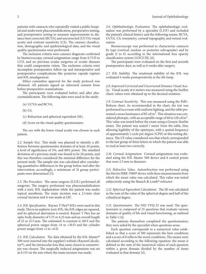
The results of cataract type as classified by the LOCS IIIwere shown in Figures 1 and 2.
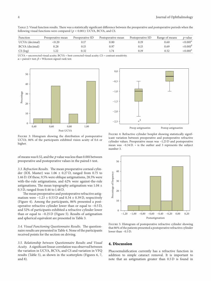
3.2. Visual Acuity Results. As shown in Table 2, the meanpreoperative UCVA was 0.2 ± 0.17 (SD) and average postop-erative UCVA was 0.80 ± 0.19 (SD); the range of means was0.60, and the𝑝 valuewas less than 0.001 between preoperativeand postoperative values in the paired 𝑡-test. With regard topostoperative UCVA, 47% of patients exhibited visual acuitybetter than or equal to 20/25, and 86% presented visual acuitybetter than or equal to 20/30 (Figure 3).
Perc
enta
ge
80
60
40
20
0
LOCS III type
Nuclear Cortical Posteriorsubcapsular
Figure 1: Histogram classifying the type of cataract according to theLOCS III system; this figure shows that 15 (71.4%) were nuclear, 1(4.8%) was cortical, and 5 (23.8%) were posterior subcapsular.
Perc
enta
ge
40
30
20
10
0
LOCS III grade1,00 2,00 3,00 4,00 5,00 6,00
Figure 2: Histogram classifying the type of cataract according tothe LOCS III system; this figure shows that 1 (4.8%) was of grade 1,1 (4.8%) was of grade 2, 3 (14.3%) were of grade 3, 5 (23.8%) were ofgrade 4, 8 (38.1%) were of grade 5, and 3 (14.3%) of were grade 6.
The mean preoperative and postoperative BCVA were0.28 ± 0.15 (SD) and 0.97 ± 0.15 (SD) respectively, range ofmeans was 0.69, and the 𝑝 value was less than 0.001 betweenpreoperative and postoperative values in the Wilcoxonsigned-rank test. With regard to postoperative BCVA, 100%of patients presented visual acuity that was better than orequal to 20/25.
The mean BCVA in the contralateral eye was 0.28 ± 0.25(SD). When the paired 𝑡-test was applied, no significantdifference in BCVA was found between the eye included inthe study and the contralateral eye (𝑝 = 0.1053).
The mean preoperative and postoperative photopic CSwere 1.22±0.32 (SD) and 1.74±0.19 (SD), respectively, range
4 Journal of Ophthalmology
Table 2: Visual function results. There was a statistically significant difference between the preoperative and postoperative periods when thefollowing visual functions were compared (𝑝 < 0.001): UCVA, BCVA, and CS.
Function Preoperative mean Preoperative SD Postoperative mean Postoperative SD Range of means 𝑝 valueUCVA (decimal) <0.20 0.17 0.80 0.19 0.60 <0.001𝛼
BCVA (decimal) 0.28 0.15 0.97 0.15 0.69 <0.001𝛽
CS (log) 1.22 0.32 1.74 0.19 0.52 <0.001𝛼
UCVA = uncorrected visual acuity; BCVA = best-corrected visual acuity; CS = contrast sensitivity.𝛼 = paired 𝑡-test; 𝛽 =Wilcoxon signed-rank test.
Perc
enta
ge
50
40
30
20
10
0
Post-UCVA0,40 0,60 0,80 1,00
Figure 3: Histogram showing the distribution of postoperativeUCVA: 86% of the participants exhibited vision acuity of 0.6 orhigher.
ofmeanswas 0.52, and the𝑝 valuewas less than 0.001 betweenpreoperative and postoperative values in the paired 𝑡-test.
3.3. Refraction Results. Themean preoperative corneal cylin-der (IOL Master) was 1.06 ± 0.27D, ranged from 0.75 to1.46D. Of these, 9.5% were oblique astigmatisms, 28.5% werewith-the-rule astigmatisms, and 62% were against-the-ruleastigmatisms. The mean topography astigmatism was 1.04 ±0.3D, ranged from 0.46 to 1.48D.
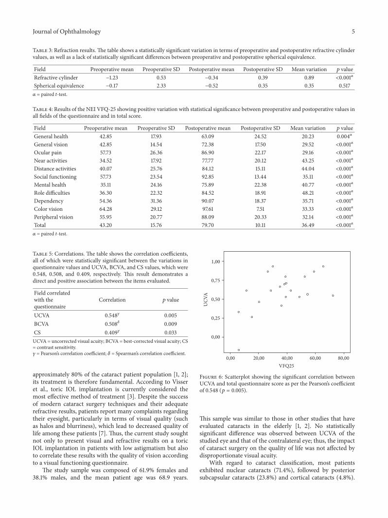
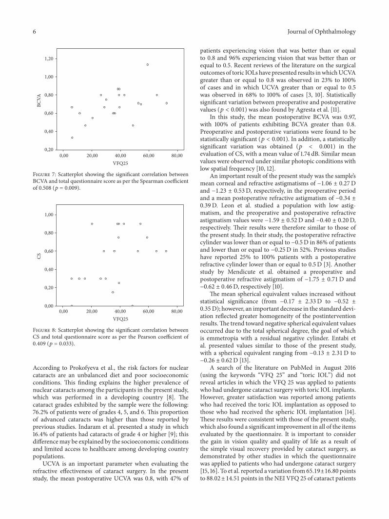
Themean preoperative and postoperative refractive astig-matism were −1.23 ± 0.53D and 0.34 ± 0.39D, respectively(Figure 4). Among the participants, 86% presented a post-operative refractive cylinder lower than or equal to −0.5D,and 52% of participants exhibited a refractive cylinder lowerthan or equal to −0.25D (Figure 5). Results of astigmatismand spherical equivalent are presented in Table 3.
3.4. Visual Functioning Questionnaire Results. The question-naire results are presented in Table 4. None of the participantsreceived points for the section on driving.
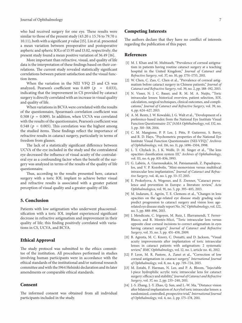
3.5. Relationship between Questionnaire Results and VisualAcuity. Asignificant linear correlationwas observed betweenthe variation in UCVA, BCVA, and CS and variation in VFQresults (Table 5), as shown in the scatterplots (Figures 6, 7,and 8).
0,0
−0,5
−1,0
−1,5
−2,0
3−2,5
Preop astigmastim Postop astigmastim
Figure 4: Refractive cylinder boxplot showing statistically signif-icant variation between preoperative and postoperative refractivecylinder values. Preoperative mean was −1.23D and postoperativemean was −0.34D. ∘ is the outlier and 3 represents the subjectnumber 3.
Perc
enta
ge o
f fre
quen
cy
50
40
30
20
10
0
Postastigmatism−1,20 −1,00 −0,80 −0,60 −0,40 −0,20 0,00 0,20
Figure 5: Histogram of postoperative refractive cylinder showingthat 86%of the patients presented a postoperative refractive cylinderlower than −0.5D.
4. Discussion
Phacoemulsification currently has a refractive function inaddition to simple cataract removal. It is important tonote that an astigmatism greater than 0.5D is found in
Journal of Ophthalmology 5
Table 3: Refraction results. The table shows a statistically significant variation in terms of preoperative and postoperative refractive cylindervalues, as well as a lack of statistically significant differences between preoperative and postoperative spherical equivalence.
Field Preoperative mean Preoperative SD Postoperative mean Postoperative SD Mean variation 𝑝 valueRefractive cylinder −1.23 0.53 −0.34 0.39 0.89 <0.001𝛼
Spherical equivalence −0.17 2.33 −0.52 0.35 0.35 0.517𝛼 = paired 𝑡-test.
Table 4: Results of the NEI VFQ-25 showing positive variation with statistical significance between preoperative and postoperative values inall fields of the questionnaire and in total score.
Field Preoperative mean Preoperative SD Postoperative mean Postoperative SD Mean variation 𝑝 valueGeneral health 42.85 17.93 63.09 24.52 20.23 0.004𝛼
General vision 42.85 14.54 72.38 17.50 29.52 <0.001𝛼
Ocular pain 57.73 26.36 86.90 22.17 29.16 <0.001𝛼
Near activities 34.52 17.92 77.77 20.12 43.25 <0.001𝛼
Distance activities 40.07 25.76 84.12 15.11 44.04 <0.001𝛼
Social functioning 57.73 23.54 92.85 13.44 35.11 <0.001𝛼
Mental health 35.11 24.16 75.89 22.38 40.77 <0.001𝛼
Role difficulties 36.30 22.32 84.52 18.91 48.21 <0.001𝛼
Dependency 54.36 31.36 90.07 18.37 35.71 <0.001𝛼
Color vision 64.28 29.12 97.61 7.51 33.33 <0.001𝛼
Peripheral vision 55.95 20.77 88.09 20.33 32.14 <0.001𝛼
Total 43.20 15.76 79.70 10.11 36.49 <0.001𝛼
𝛼 = paired 𝑡-test.
Table 5: Correlations. The table shows the correlation coefficients,all of which were statistically significant between the variations inquestionnaire values and UCVA, BCVA, and CS values, which were0.548, 0.508, and 0.409, respectively. This result demonstrates adirect and positive association between the items evaluated.
Field correlatedwith thequestionnaire
Correlation 𝑝 value
UCVA 0.548𝛾 0.005BCVA 0.508𝛿 0.009CS 0.409𝛾 0.033UCVA = uncorrected visual acuity; BCVA = best-corrected visual acuity; CS= contrast sensitivity.𝛾 = Pearson’s correlation coefficient; 𝛿 = Spearman’s correlation coefficient.
approximately 80% of the cataract patient population [1, 2];its treatment is therefore fundamental. According to Visseret al., toric IOL implantation is currently considered themost effective method of treatment [3]. Despite the successof modern cataract surgery techniques and their adequaterefractive results, patients report many complaints regardingtheir eyesight, particularly in terms of visual quality (suchas halos and blurriness), which lead to decreased quality oflife among these patients [7]. Thus, the current study soughtnot only to present visual and refractive results on a toricIOL implantation in patients with low astigmatism but alsoto correlate these results with the quality of vision accordingto a visual functioning questionnaire.
The study sample was composed of 61.9% females and38.1% males, and the mean patient age was 68.9 years.
UCV
A
1,00
0,75
0,50
0,25
0,00
VFQ25
0,00 20,00 40,00 60,00 80,00
Figure 6: Scatterplot showing the significant correlation betweenUCVA and total questionnaire score as per the Pearson’s coefficientof 0.548 (𝑝 = 0.005).
This sample was similar to those in other studies that haveevaluated cataracts in the elderly [1, 2]. No statisticallysignificant difference was observed between UCVA of thestudied eye and that of the contralateral eye; thus, the impactof cataract surgery on the quality of life was not affected bydisproportionate visual acuity.
With regard to cataract classification, most patientsexhibited nuclear cataracts (71.4%), followed by posteriorsubcapsular cataracts (23.8%) and cortical cataracts (4.8%).
6 Journal of OphthalmologyBC
VA
1,20
1,00
0,80
0,60
0,40
0,20
VFQ25
0,00 20,00 40,00 60,00 80,00
Figure 7: Scatterplot showing the significant correlation betweenBCVA and total questionnaire score as per the Spearman coefficientof 0.508 (𝑝 = 0.009).
CS
1,00
0,80
0,60
0,40
0,20
0,00
VFQ25
0,00 20,00 40,00 60,00 80,00
Figure 8: Scatterplot showing the significant correlation betweenCS and total questionnaire score as per the Pearson coefficient of0.409 (𝑝 = 0.033).
According to Prokofyeva et al., the risk factors for nuclearcataracts are an unbalanced diet and poor socioeconomicconditions. This finding explains the higher prevalence ofnuclear cataracts among the participants in the present study,which was performed in a developing country [8]. Thecataract grades exhibited by the sample were the following:76.2% of patients were of grades 4, 5, and 6. This proportionof advanced cataracts was higher than those reported byprevious studies. Indaram et al. presented a study in which16.4% of patients had cataracts of grade 4 or higher [9]; thisdifferencemay be explained by the socioeconomic conditionsand limited access to healthcare among developing countrypopulations.
UCVA is an important parameter when evaluating therefractive effectiveness of cataract surgery. In the presentstudy, the mean postoperative UCVA was 0.8, with 47% of
patients experiencing vision that was better than or equalto 0.8 and 96% experiencing vision that was better than orequal to 0.5. Recent reviews of the literature on the surgicaloutcomes of toric IOLs have presented results inwhichUCVAgreater than or equal to 0.8 was observed in 23% to 100%of cases and in which UCVA greater than or equal to 0.5was observed in 68% to 100% of cases [3, 10]. Statisticallysignificant variation between preoperative and postoperativevalues (𝑝 < 0.001) was also found by Agresta et al. [11].
In this study, the mean postoperative BCVA was 0.97,with 100% of patients exhibiting BCVA greater than 0.8.Preoperative and postoperative variations were found to bestatistically significant (𝑝 < 0.001). In addition, a statisticallysignificant variation was obtained (𝑝 < 0.001) in theevaluation of CS, with a mean value of 1.74 dB. Similar meanvalues were observed under similar photopic conditions withlow spatial frequency [10, 12].
An important result of the present study was the sample’smean corneal and refractive astigmatisms of −1.06 ± 0.27Dand −1.23 ± 0.53D, respectively, in the preoperative periodand a mean postoperative refractive astigmatism of −0.34 ±0.39D. Leon et al. studied a population with low astig-matism, and the preoperative and postoperative refractiveastigmatism values were −1.59 ± 0.52D and −0.40 ± 0.20D,respectively. Their results were therefore similar to those ofthe present study. In their study, the postoperative refractivecylinder was lower than or equal to −0.5D in 86% of patientsand lower than or equal to −0.25D in 52%. Previous studieshave reported 25% to 100% patients with a postoperativerefractive cylinder lower than or equal to 0.5D [3]. Anotherstudy by Mendicute et al. obtained a preoperative andpostoperative refractive astigmatism of −1.75 ± 0.71D and−0.62 ± 0.46D, respectively [10].
The mean spherical equivalent values increased withoutstatistical significance (from −0.17 ± 2.33D to −0.52 ±0.35D); however, an important decrease in the standard devi-ation reflected greater homogeneity of the postinterventionresults.The trend toward negative spherical equivalent valuesoccurred due to the total spherical degree, the goal of whichis emmetropia with a residual negative cylinder. Entabi etal. presented values similar to those of the present study,with a spherical equivalent ranging from −0.13 ± 2.31D to−0.26 ± 0.62D [13].
A search of the literature on PubMed in August 2016(using the keywords “VFQ 25” and “toric IOL”) did notreveal articles in which the VFQ 25 was applied to patientswho had undergone cataract surgery with toric IOL implants.However, greater satisfaction was reported among patientswho had received the toric IOL implantation as opposed tothose who had received the spheric IOL implantation [14].These results were consistent with those of the present study,which also found a significant improvement in all of the itemsevaluated by the questionnaire. It is important to considerthe gain in vision quality and quality of life as a result ofthe simple visual recovery provided by cataract surgery, asdemonstrated by other studies in which the questionnairewas applied to patients who had undergone cataract surgery[15, 16]. To et al. reported a variation from 65.19±16.80 pointsto 88.02±14.51 points in the NEI VFQ 25 of cataract patients
Journal of Ophthalmology 7
who had received surgery for one eye. These results weresimilar to those of the present study (43.20 ± 15.76 to 79.70 ±10.11), bothwith a significant𝑝 value [15]. Lin et al. presenteda mean variation between preoperative and postoperativeaspheric and spheric IOLs of 13.95 and 13.02, respectively; thepresent study found a mean positive variation of 36.49 [16].
More important than refractive, visual, and quality of lifedata is the interpretation of these findings based on their cor-relations. The current study revealed statistically significantcorrelations between patient satisfaction and the visual func-tion items.
When the variation in the NEI VFQ 25 and CS wasanalyzed, Pearson’s coefficient was 0.409 (𝑝 = 0.033),indicating that the improvement in CS provided by cataractsurgery is directly correlatedwith an increase in visual qualityand quality of life.
When variations in BCVAwere correlatedwith the resultsof the questionnaire, Spearman’s correlation coefficient was0.508 (𝑝 = 0.009). In addition, when UCVA was correlatedwith the results of the questionnaire, Pearson’s coefficient was0.548 (𝑝 = 0.005). This correlation was the highest amongthe studied items. These findings reflect the importance ofrefractive results in cataract surgery, particularly in terms offreedom from glasses.
The lack of a statistically significant difference betweenUCVA of the eye included in the study and the contralateraleye decreased the influence of visual acuity of the contralat-eral eye as a confounding factor when the benefit of the sur-gery was analyzed in terms of the results of the quality of lifequestionnaire.
Thus, according to the results presented here, cataractsurgery with a toric IOL implant to achieve better visualand refractive results is associated with a greater patientperception of visual quality and a greater quality of life.
5. Conclusion
Patients with low astigmatism who underwent phacoemul-sification with a toric IOL implant experienced significantdecrease in refractive astigmatism and improvement in theirquality of life; this finding positively correlated with varia-tions in CS, UCVA, and BCVA.
Ethical Approval
The study protocol was submitted to the ethics commit-tee of the institution. All procedures performed in studiesinvolving human participants were in accordance with theethical standards of the institutional and/or national researchcommittee andwith the 1964Helsinki declaration and its lateramendments or comparable ethical standards.
Consent
The informed consent was obtained from all individualparticipants included in the study.
Competing Interests
The authors declare that they have no conflict of interestsregarding the publication of this paper.
References
[1] M. I. Khan and M. Muhtaseb, “Prevalence of corneal astigma-tism in patients having routine cataract surgery at a teachinghospital in the United Kingdom,” Journal of Cataract andRefractive Surgery, vol. 37, no. 10, pp. 1751–1755, 2011.
[2] W. Chen, C. Zuo, C. Chen et al., “Prevalence of corneal astig-matism before cataract surgery in Chinese patients,” Journal ofCataract and Refractive Surgery, vol. 39, no. 2, pp. 188–192, 2013.
[3] N. Visser, N. J. C. Bauer, and R. M. M. A. Nuijts, “Toricintraocular lenses: historical overview, patient selection, IOLcalculation, surgical techniques, clinical outcomes, and compli-cations,” Journal of Cataract and Refractive Surgery, vol. 39, no.4, pp. 624–627, 2013.
[4] A. M. Rentz, J. W. Kowalski, J. G. Walt et al., “Development of apreference-based index from the National Eye Institute VisualFunctionQuestionnaire-25,” JAMAOphthalmology, vol. 132, no.3, pp. 310–318, 2014.
[5] C. M. Mangione, P. P. Lee, J. Pitts, P. Gutierrez, S. Berry,and R. D. Hays, “Psychometric properties of the National EyeInstitute Visual Function Questionnaire (NEI-VFQ),” Archivesof Ophthalmology, vol. 116, no. 11, pp. 1496–1504, 1998.
[6] L. T. Chylack Jr., J. K. Wolfe, D. M. Singer et al., “The lensopacities classification system III,” Archives of Ophthalmology,vol. 111, no. 6, pp. 831–836, 1993.
[7] G. Labiris, A. Giarmoukakis, M. Patsiamanidi, Z. Papadopou-los, and V. P. Kozobolis, “Mini-monovision versus multifocalintraocular lens implantation,” Journal of Cataract and Refrac-tive Surgery, vol. 41, no. 1, pp. 53–57, 2015.
[8] E. Prokofyeva, A. Wegener, and E. Zrenner, “Cataract preva-lence and prevention in Europe: a literature review,” ActaOphthalmologica, vol. 91, no. 5, pp. 395–405, 2013.
[9] M. Indaram, E. Agron, T. E. Clemons et al., “Changes in lensopacities on the age-related eye disease study grading scalepredict progression to cataract surgery and vision loss: age-related eye disease study reportNo. 34,”Ophthalmology, vol. 122,no. 5, pp. 888–896, 2015.
[10] J. Mendicute, C. Irigoyen, M. Ruiz, I. Illarramendi, T. Ferrer-Blasco, and R. Montes-Mico, “Toric intraocular lens versusopposite clear corneal incisions to correct astigmatism in eyeshaving cataract surgery,” Journal of Cataract and RefractiveSurgery, vol. 35, no. 3, pp. 451–458, 2009.
[11] B. Agresta, M. C. Knorz, C. Donatti, and D. Jackson, “Visualacuity improvements after implantation of toric intraocularlenses in cataract patients with astigmatism: 2 systematicreview,” BMC Ophthalmology, vol. 12, no. 1, article no. 41, 2012.
[12] P. Leon, M. R. Pastore, A. Zanei et al., “Correction of lowcorneal astigmatism in cataract surgery,” International Journalof Ophthalmology, vol. 8, no. 4, pp. 719–724, 2015.
[13] M. Entabi, F. Harman, N. Lee, and P. A. Bloom, “Injectable1-piece hydrophilic acrylic toric intraocular lens for cataractsurgery: efficacy and stability,” Journal of Cataract andRefractiveSurgery, vol. 37, no. 2, pp. 235–240, 2011.
[14] J.-S. Zhang, J.-Y. Zhao, Q. Sun, and L.-W. Ma, “Distance visionafter bilateral implantation of AcrySof toric intraocular lenses: arandomized, controlled, prospective trial,” International Journalof Ophthalmology, vol. 4, no. 2, pp. 175–178, 2011.

8 Journal of Ophthalmology
[15] K. G. To, L. B. Meuleners, M. L. Fraser et al., “The impact ofcataract surgery on vision-related quality of life for bilateralcataract patients in Ho Chi Minh City, Vietnam: A ProspectiveStudy,” Health and Quality of Life Outcomes, vol. 12, article 16,2014.
[16] I.-C. Lin, I.-J. Wang, M.-S. Lei, L. L.-K. Lin, and F.-R. Hu,“Improvements in vision-related quality of life with AcrySof IQSN60WF aspherical intraocular lenses,” Journal of Cataract andRefractive Surgery, vol. 34, no. 8, pp. 1312–1317, 2008.

Submit your manuscripts athttp://www.hindawi.com
Stem CellsInternational
Hindawi Publishing Corporationhttp://www.hindawi.com Volume 2014
Hindawi Publishing Corporationhttp://www.hindawi.com Volume 2014
MEDIATORSINFLAMMATION
of
Hindawi Publishing Corporationhttp://www.hindawi.com Volume 2014
Behavioural Neurology
EndocrinologyInternational Journal of
Hindawi Publishing Corporationhttp://www.hindawi.com Volume 2014
Hindawi Publishing Corporationhttp://www.hindawi.com Volume 2014
Disease Markers
Hindawi Publishing Corporationhttp://www.hindawi.com Volume 2014
BioMed Research International
OncologyJournal of
Hindawi Publishing Corporationhttp://www.hindawi.com Volume 2014
Hindawi Publishing Corporationhttp://www.hindawi.com Volume 2014
Oxidative Medicine and Cellular Longevity
Hindawi Publishing Corporationhttp://www.hindawi.com Volume 2014
PPAR Research
The Scientific World JournalHindawi Publishing Corporation http://www.hindawi.com Volume 2014
Immunology ResearchHindawi Publishing Corporationhttp://www.hindawi.com Volume 2014
Journal of
ObesityJournal of
Hindawi Publishing Corporationhttp://www.hindawi.com Volume 2014
Hindawi Publishing Corporationhttp://www.hindawi.com Volume 2014
Computational and Mathematical Methods in Medicine
OphthalmologyJournal of
Hindawi Publishing Corporationhttp://www.hindawi.com Volume 2014
Diabetes ResearchJournal of
Hindawi Publishing Corporationhttp://www.hindawi.com Volume 2014
Hindawi Publishing Corporationhttp://www.hindawi.com Volume 2014
Research and TreatmentAIDS
Hindawi Publishing Corporationhttp://www.hindawi.com Volume 2014
Gastroenterology Research and Practice
Hindawi Publishing Corporationhttp://www.hindawi.com Volume 2014
Parkinson’s Disease
Evidence-Based Complementary and Alternative Medicine
Volume 2014Hindawi Publishing Corporationhttp://www.hindawi.com


![Clinical Outcomes After Cataract Surgery With a New ...lentech.com.co/archivos/estudios/Clinical Outcomes... · Journal of Refractive Surgery ÊUÊ6 °ÊÎÓ]Ê °ÊÇ]ÊÓä£È](https://img.pdfslide.net/doc/110x75/5fa0346ee061442e0d6c2fd0/clinical-outcomes-after-cataract-surgery-with-a-new-outcomes-journal-of.jpg)