Embed Size (px)
Citation preview

How to Improve your Refractive Cataract Surgery Outcomes by Skilful Interpretation of Corneal Mapping
Arthur Cummings FRCSEd Wellington Eye Clinic, Dublin, Ireland
Course IC-16 ESCRS Copenhagen 10th September 2016
Consultant for Alcon / WaveLight/TearLab
AIMS of Course
• Help manage refractive expectations of cataract surgery patients
• Help with managing toric IOL’s
• Help with LRI’s, OCCI’s, effect of incision size and architecture
• Help with selecting multifocal IOL candidates
Why address astigmatism?
• Astigmatism is the KEY factor for success with multifocal IOL’s
• Correcting astigmatism provides better UCVA, BCVA for distance
and near
• Glasses that may be required are lighter, cheaper and easier to
wear / get used to
What is “Refractive Cataract?”
• The intended outcome is emmetropia
• The intended outcome has addressed astigmatism
• The intended outcome may have addressed presbyopia too depending
on patient wishes (multifocal IOL, monovision)
• The patient is free of glasses for at least distance vision (monofocal,
emmetropia) or completely free of glasses
Devices
• Placido disk (in relative detail)
• Scheimpflug (in relative detail)
• Cassini (Introduction)
Topolyzer (Keratograph)
• Placido disk
• Tear film reflections
• Central scotoma where camera is situated
• Auto-capture, very repeatable
Oculyzer (Pentacam)
• Scheimpflug camera
• Captures scatter so does not see tear film but
corneal surface
• No central scotoma
• Auto-capture, very repeatable
Diagnostic Applications
• Screening for IOL’s (toric, multifocal)
• Screening for corneal health
• Screening for AC parameters

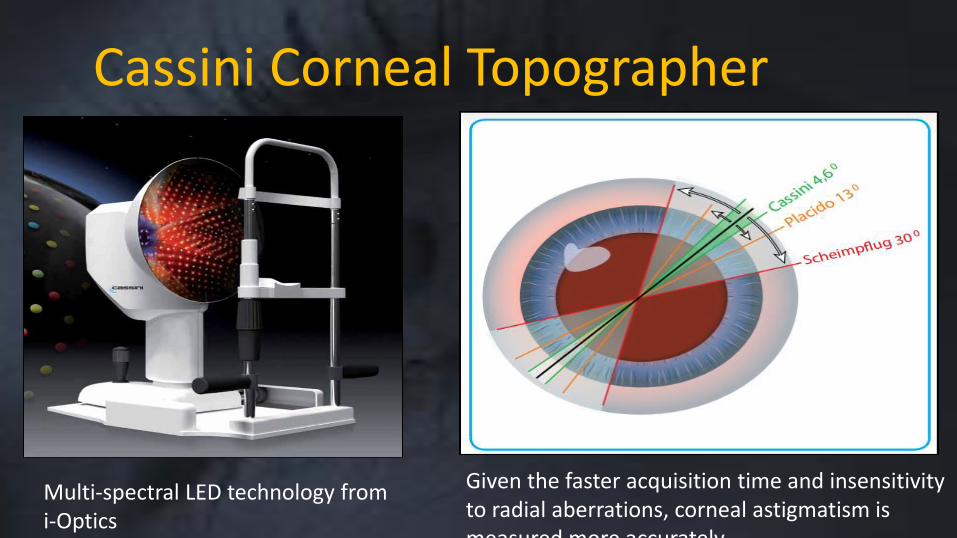
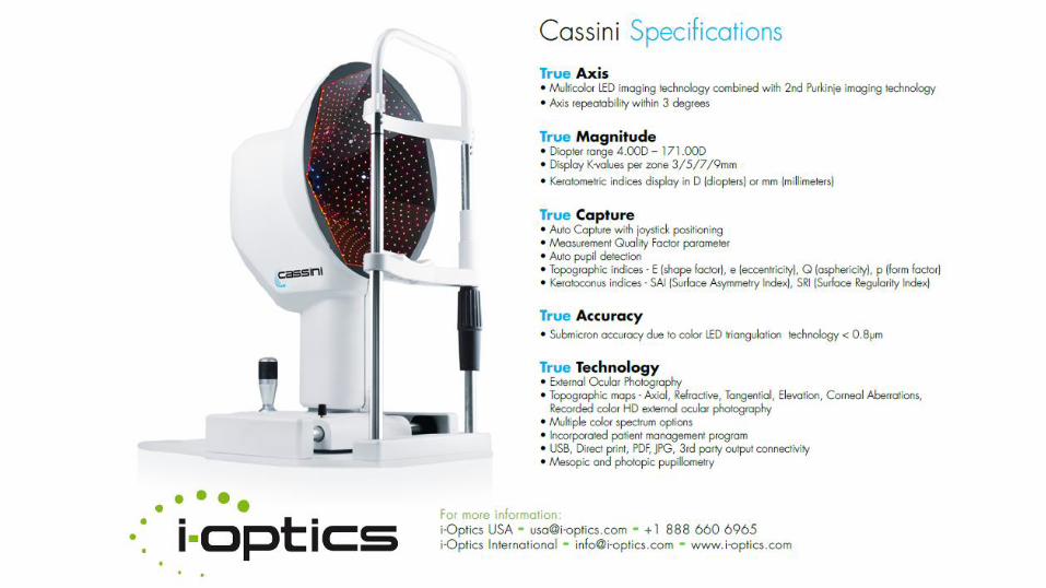
Cassini Corneal Topographer
Multi-spectral LED technology from i-Optics
Given the faster acquisition time and insensitivity to radial aberrations, corneal astigmatism is measured more accurately
Cassini and Toric IOL’s
Arthur Cummings
Wellington Eye Clinic
How does the iTrace work?
• Simultaneous corneal topography and whole eye
wavefront mapping
• Refraction
• Can separate corneal from intra-ocular optics
• Can therefore help manage post-op toric IOL’s
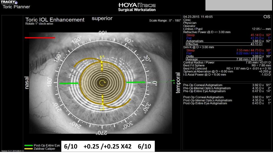
6/10 +0.25 /+0.25 X42 6/10
Summary
• Value of adding posterior corneal data is understood
• What about the geometry and geography of the
crystalline lens and the final position and orientation of
the IOL?
• Mirricon from ClearSight may have more answers?
Pre-Operative
• IOL Calculations
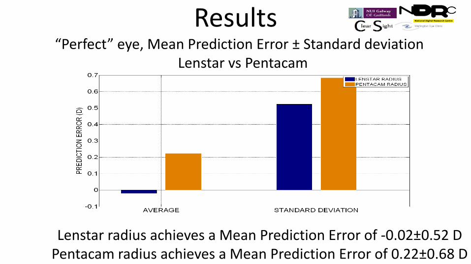
Results
Lenstar radius achieves a Mean Prediction Error of -0.02±0.52 D Pentacam radius achieves a Mean Prediction Error of 0.22±0.68 D
“Perfect” eye, Mean Prediction Error ± Standard deviation Lenstar vs Pentacam
Pre-Operative
• IOL calculations
• IOL type
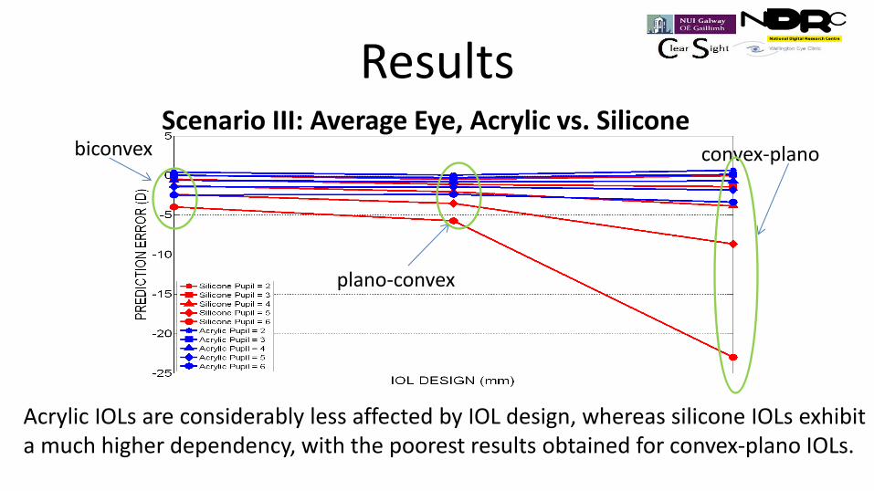
Results Scenario III: Average Eye, Acrylic vs. Silicone
Acrylic IOLs are considerably less affected by IOL design, whereas silicone IOLs exhibit a much higher dependency, with the poorest results obtained for convex-plano IOLs.
biconvex
plano-convex
convex-plano
Pre-Operative
• IOL calculations
• IOL type
• Incision type: Scleral, limbal, corneal
Pre-Operative
• IOL calculations
• IOL type
• Incision type
• Incision shape: 3 step, 2-step, straight-in
Pre-Operative
• IOL calculations
• IOL type
• Incision type: Scleral, limbal, corneal
• Incision shape: 3 step, 2-step, straight-in
• Incision size: <2mm, 2.2mm, 2.5mm, 2.8mm, >2.8mm
Pre-Operative
• IOL calculations
• IOL type
• Incision type: Scleral, limbal, corneal
• Incision shape: 3 step, 2-step, straight-in
• Incision size: <2mm, 2.2mm, 2.5mm, 2.8mm, >2.8mm
• Incision location: Superior, Temporal, on axis
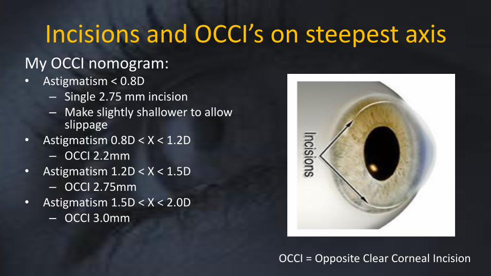
Incisions and OCCI’s on steepest axis My OCCI nomogram: • Astigmatism < 0.8D
– Single 2.75 mm incision – Make slightly shallower to allow
slippage • Astigmatism 0.8D < X < 1.2D
– OCCI 2.2mm • Astigmatism 1.2D < X < 1.5D
– OCCI 2.75mm • Astigmatism 1.5D < X < 2.0D
– OCCI 3.0mm
OCCI = Opposite Clear Corneal Incision
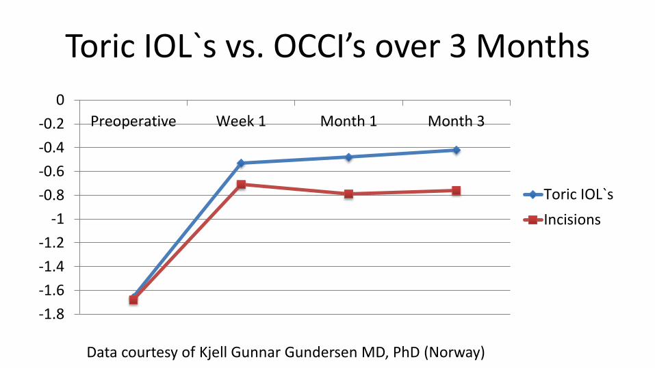
Toric IOL`s vs. OCCI’s over 3 Months
-1.8
-1.6
-1.4
-1.2
-1
-0.8
-0.6
-0.4
-0.2
0
Preoperative Week 1 Month 1 Month 3
Toric IOL`s
Incisions
Data courtesy of Kjell Gunnar Gundersen MD, PhD (Norway)

Superior Cataract Incision
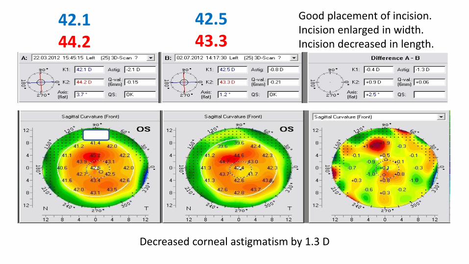
42.1 44.2
42.5 43.3
Decreased corneal astigmatism by 1.3 D
Good placement of incision. Incision enlarged in width. Incision decreased in length.
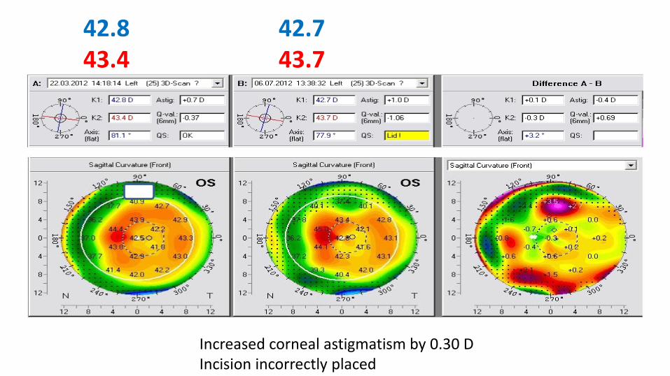
42.8 43.4
42.7 43.7
Increased corneal astigmatism by 0.30 D Incision incorrectly placed
Temporal Cataract Incision
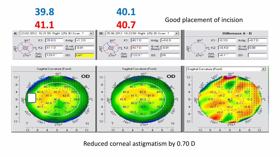
39.8 41.1
40.1 40.7
Reduced corneal astigmatism by 0.70 D
Good placement of incision
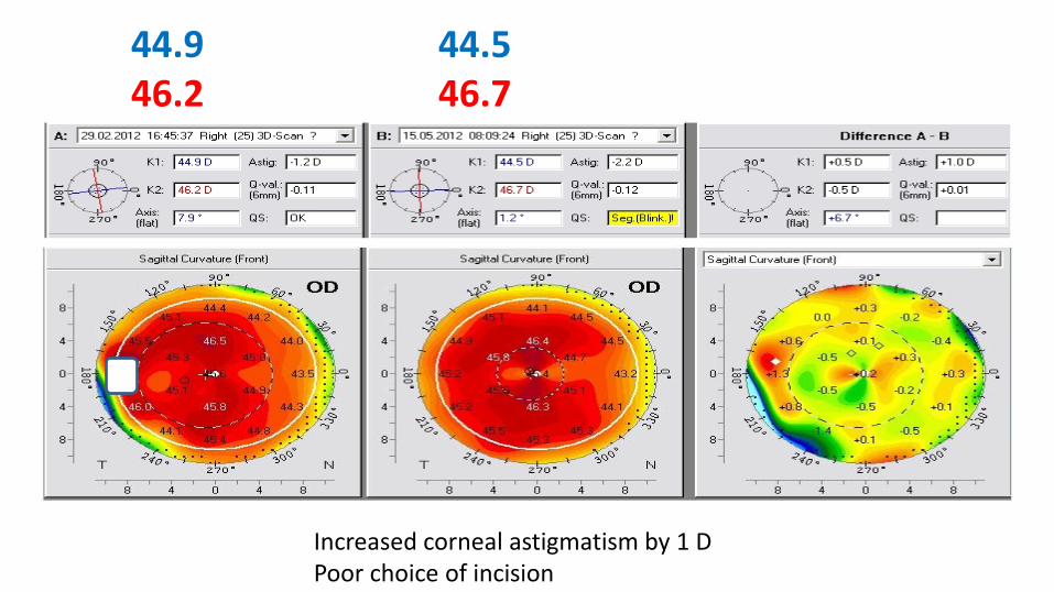
44.9 46.2
44.5 46.7
Increased corneal astigmatism by 1 D Poor choice of incision

Cataract On-Axis Incision
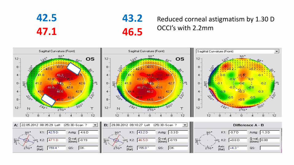
42.5 47.1
43.2 46.5
Reduced corneal astigmatism by 1.30 D OCCI’s with 2.2mm
Pre-Operative
• IOL calculations
• IOL type
• Incision type: Scleral, limbal, corneal
• Incision shape: 3 step, 2-step, straight-in
• Incision size: <2mm, 2.2mm, 2.5mm, 2.8mm, >2.8mm
• Incision location: Superior, Temporal, on axis
Shallow Anterior Chambers
• Mostly hyperopes
• Mostly shorter eyes
• 3 critical values
– AC volume < 100 mm3
– ACD < 2.1mm
– AC angle < 26 degrees
Post-Operative
• Detecting tight sutures
• Detecting wound gape
• Detecting irregular astigmatism
• Guiding suture removal with the Pentacam

Thank You for Your Attention