Embed Size (px)
Citation preview
Hindawi Publishing CorporationInternational Journal of Vascular MedicineVolume 2012, Article ID 536392, 7 pagesdoi:10.1155/2012/536392
Clinical Study
Urgent Carotid Surgery: Is It Still out of Debate?
C. Battocchio, C. Fantozzi, L. Rizzo, F. Persiani, S. Raffa, and M. Taurino
Azienda Ospedaliera Sant’Andrea, Facolta di Medicina e Psicologia, Sapienza-Universita di Roma, 00189 Roma, Italy
Correspondence should be addressed to F. Persiani, [email protected]
Received 24 October 2011; Revised 8 January 2012; Accepted 9 January 2012
Academic Editor: Mark Morasch
Copyright © 2012 C. Battocchio et al. This is an open access article distributed under the Creative Commons Attribution License,which permits unrestricted use, distribution, and reproduction in any medium, provided the original work is properly cited.
Patients with symptomatic tight carotid stenosis have an increased short-time risk of stroke and an increased long-term riskof ischaemic vascular events compared with the general population. The aim of this study is to assess the safety, efficacy, andlimitations of urgent CEA or CAS, in patients with carotid stenosis greater than 70% and clinically characterized by recurrentTIA or brain damage following a stroke (<2.5 cm). This study involved 28 patients divided into two groups. Group A consisted ofsixteen patients who had undergone CEA, and group B consisted of twelve patients who had undergone CAS. Primary endpointswere mortality, neurological morbidity (by NIHSS) and postoperative hemorrhagic cerebral conversion, at 30 days. Ten patients(62.5%) of group A experienced an improvement in their initial neurological deficit while in 4 cases (26%) the deficit remainedstable. Two cases of neurologic mortality are presented. At 1 month, 9 patients (75%) of group B experienced an improvement intheir initial neurological deficit while 3 patients (25%) had a neurological impairment. Urgent or deferred surgical or endovasculartreatment have a satisfactory outcome considering the profile in very high-risk patient population. Otherwise in selected patientsCEA seems to be preferred to CAS.
1. Introduction
Ipsilateral >50% carotid stenosis is found in about 10% ofcarotid territory ischaemic strokes and in about 15% of TIAs(transitory ischemic attack), and is associated with a partic-ularly high risk of recurrent stroke [1–3], both in the acutephase and long term [4]. Recent studies showed that 4−20%of TIA patients will have a stroke within 90 days after a TIA,half within the first 2 days [5, 6]. Reanalysis of controlledtrials of CEA (carotid endarterectomy) indicated that surgeryconferred the greatest benefit when performed in the first2 weeks following the index symptoms, perhaps as early as48 h after the index event [7, 8]. Delaying intervention quiteprobably means that patients are better selected and thiscould guarantee better early outcomes, but this delay canalso result in an interval stroke rate of 9–15% [9]. In thepast few years CAS (carotid artery stenting) has emerged asa treatment alternative to CEA. A recent study found thatearly CAS might be associated with an increased risk forstroke and death [10]. In fact CAS, in the acute stage, remainschallenging because of the limited therapeutic window andrisk of hyperperfusion syndrome or cerebral hemorrhagicinfarction after revascularization [11]. The real goal of early
treatment is to stop the plaque embolization from a vulnera-ble lesion at the carotid bifurcation [12]. Another importantquestion is: is there a patient at higher risk of neurologicalimpairment in the short term? The ABCD2 score is aprognostic score that we, retrospectively, used to validateour practice [13]. This retrospective study investigates thesafety, efficacy, and limitations of urgent CEA or CAS forsymptomatic patients with recent first or recurrent TIA orminor stroke, bearing severe carotid artery stenosis.
2. Methods
This was a retrospective review of two groups of patients:group A, with patients treated with open surgery, and groupB, with patients treated with endovascular technique. In ourinstitution the guidelines of treatment do not consider BMT,so a control group is not available. All patients were observedin emergency department (ED) by a neurologist and avascular surgeon. Cerebral CT scan or MRI was performed inall patients before and after the treatment. If CT scan or MRIruled out hemorrhagic events, intravenous heparin therapywas administered immediately. Demographic data, including

2 International Journal of Vascular Medicine
Table 1: Comparison of clinical characteristics and results of clinical workup between patients in surgical group and those in endovasculargroup.
ParameterSurgical group
(n = 16)Endovascular group
(n = 12)
Sex
Female 5 3
Male 11 9
Age (y)
Mean 70 75
Vascular risk factors
Hypertension 7 (43) 5 (41)
Current cigarette smoking 8 (50) 7 (58)
Diabetes mellitus 6 (37) 8 (66)
Hypercholesterolemia 9 (56) 6 (50)
Heart disease 3 (18) 5 (41)
History of amaurosis fugax, retinal infarct, transient ischemic attack, or stroke 4 (25) 3 (25)
Neurological status during treatment
Asymptomatic (TIA) 7 (44) 3 (25)
Symptomatic stable (minor stroke) 3 (18) 5 (41)
Symptomatic stable (major stroke) 6 (37) 4 (33)
Median NIHSS score 7.4 4.4
Time from onset of symptoms to treatment
Emergency (<24 h) 13 (81) 11 (91)
Urgency (<7 days) 3 (18) 1 (9)
Early neurological results
Improvements (>1 NIHSS) 10 (63) 9 (75)
Stable 4 (25) —
Impairment (>1 NIHSS) 2 (12.5) 3 (25)
Early death 2 (12.5) —
New ischemic lesions p.o. — 3 (25)
Cerebral hemorrhage 2 (12.5) —
Note: numbers in parentheses are percentages.
age, sex, symptom details, risk factors, medications, andoperative data were collected for all patients. The NationalInstitutes of Health Stroke Scale (NIHSS) was used forneurological assessment; the score was recorded routinelybefore, 24 h, and 3 days after treatment. Ecocolor Doppler(ECD) was performed in all patients to evaluate the carotidbifurcation (type of plaque and haemodynamics of thelesion). Anatomy of aortic arch and supra-aortic trunks werealso evaluated with angio-CT o MRI in all cases. All patientsunderwent transcranial Doppler (TCD) before surgery toevaluate the presence and frequency of microembolic signal(MES), to detect the middle cerebral artery (MCA) patency,and to test the intraoperative clamping tolerance.
The emergency treatment was performed in patients withvery tight stenosis and clinically characterized by recurrentTIA or stroke with little cerebral damage (<2.5 cm). Forpatients with different clinical picture, we had the possibilityto keep time to prepare them in the best way for surgery.
Only one patient was undergoing preoperativefibrinolytic therapy and then she died of haemorrhagic
postoperative cerebral infarction. All patients received in-travenous heparin before carotid intervention.
Patients were excluded if cerebral ischemic lesions weregreater than 2.5 cm, if there was loss of consciousness, if therewere signs of intracranial hemorrhage, and if the patients orfamily did not give informed consent.
Group A. Sixteen patients who had undergone CEA afterTIA, minor or major stroke, from October 2004 to August2010, were identified. Clinical characteristics of this groupof patients are shown in Table 1. In 13 patients a patchangioplasty, in 2 cases a direct suture of arteriotomy, and in1 case an eversion CEA were performed.
A Pruitt Inahara shunt has been used in 8 patients. Forinterventions performed under general anaesthesia, it hasbeen placed when, during clamping, the DTC signal showeda reduction of 2/3 of middle cerebral artery flow, while forinterventions performed under local anaesthesia, it has beenused when, during clamping, a clinical deficit appeared. In

International Journal of Vascular Medicine 3
Table 2: Nicolaides plaque classification.
Plaque Morphologic characterization
Type 1 Uniformly echolucent plaque
Type 2Predominantly echolucent plaques with less than 50%echogenic areas
Type 3Predominantly echogenic plaques with less than 50%echolucent areas
Type 4 Uniformly echogenic plaques
Type 5Could not be classified (heavy calcification and acousticshadows)
suitable patients CEA was undertaken on the next availableoperating room, with a preference for local anaesthesia.
Group B. Twelve patients who had undergone CAS after TIAminor or major stroke, from September 2004 to February2011, were identified. Clinical characteristics of this groupof patients are shown in Table 1. The cerebral protection wasperformed in all cases with distal embolic protection device.In 8 cases the CAS was performed with femoral access, in4 patients a cervical cutdown was used. In suitable patientsCAS was undertaken on the next available operating roomby vascular surgeon, in local anaesthesia.
CEA contraindications were high-risk patients, hostileneck, and high bifurcation. CAS contraindications weredifficult access anatomy, very tight stenosis, and soft or verycalcified lesions (Table 2) [14].
In absence of contraindications for CAS or CEA the op-erative technique was chosen according to surgeon’s prefer-ence.
All patient were observed in critical care unit after oper-ation.
Retrospectively we applied ABCD2 score [13] to validateor not our therapeutic strategy (the appendix).
Patients were followed up for 30 days after their clinicalpresentation. Stroke, TIAs, deaths, and hospitalization forcardiovascular events were identified for all patients. Theprimary outcome was recurrent TIA or minor stroke orstroke occurring within 30 days of TIA or minor stroke pre-sentation. Secondary outcome were cardiovascular events,requiring hospitalization, and death.
3. Statistical Analysis
Patients were divided for analysis into subgroups accordingto perioperative characteristics and were compared withrespect to the occurrence of postoperative neurologic eventsor NIHSS score variation (≥1).
Statistical analysis was performed with chi-square testand Mann-Whitney test.
4. Results
From February 2003 to February 2011 we treated “early” 28patients with carotid disease, for a total of 16 TEA (57%)and 12 CAS (43%). A summary of the clinical results for allpatients group are given in Table 1. The mean age was 70years (min 60, max 80) in group A and 75 years (min 70,
Table 3
NIHSS
surgical endovascular P value∗
At admission 7.4 4.4 <0.0001∗
Chi-square test for trend.
max 80) in group B. The interval from symptom onset toarrival at the emergency department did not differ betweengroups. The prevalence of common vascular risk factors wasalso similar between two groups. All patients had mild tomoderate neurologic deficit: median NIHSS score of 7.4 ingroup A and 4.4 in group B (Table 3). This is the onlysignificant difference between the two groups. At admission,early signs of cerebral ischemia were present at CT or MRimaging in 10 (62.5%) of 16 patients in group A and in 7(58%) of 12 patients of group B. The mean grade of stenosiswas 77.5% (from 70 to 85) for group A and 75% (from 70 to80) for group B. Type of plaque at ECD was I in 5 cases, II in5 cases, III in 3 cases, IV in 1 cases, and V in 2 cases in groupA. Type of plaque at ECD was I in 1 case, II in 3 cases, III in 7cases, IV in 1 cases, and V in 0 cases in group B. TCD showedrelevant MES in 4 patients of group A and in all patients ofthe group B. TCD depicted 16 patients with possible clampischemia (8 in group A and 9 in group B). The CT or MRimaging revealed 4 cases of contraindication to endovascularfemoral procedure (Leriche, tortuosity, and hostile arch).
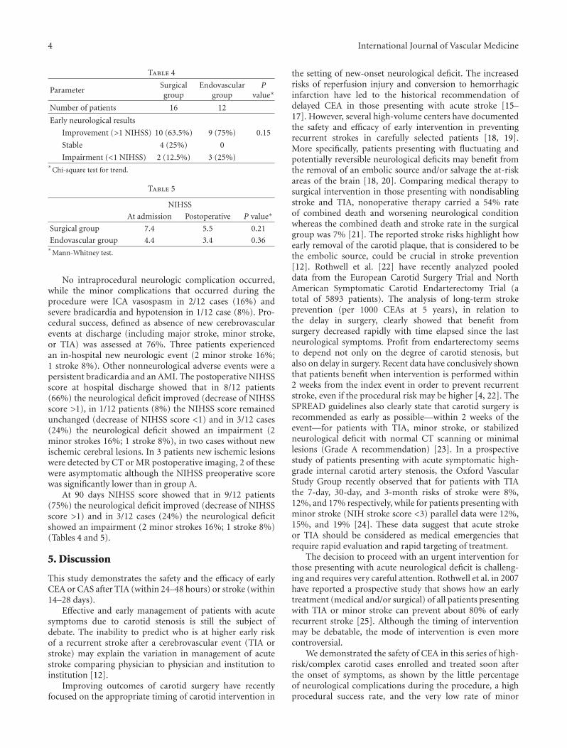
Group A. Successful CEA was achieved in all cases (100%).No intraprocedural neurologic complication occurred inall but two. Procedural success, defined as absence of newcerebrovascular events at discharge (including major stroke,minor stroke, or TIA) was assessed at 87.45%. One femaleand one male patient with crescendo TIA experienced an in-hospital new neurologic event: stroke due to hemorrhagictransformation of ischemic lesion. A Pruitt Inahara shunthas been used in one patient. These patients died one monthafter coma. Other nonneurological adverse event was oneacute myocardial infarction (AMI). An haematoma drainagein the postoperative period was never necessary. The post-operative NIHSS score at hospital discharge showed thatin 10/16 patients (62.5%) the neurological deficit improved(decrease of NIHSS score >1), in 4/16 patients (25%) theNIHSS score remained unchanged (decrease of NIHSS score<1), and in 2 cases (12.5%) the neurological deficit showedan impairment (coma). At 30 days NIHSS score showed thatin 11/16 patients (68.7%) the neurological deficit improved(decrease of NIHSS score >1), in 4/16 patients (25%) theNIHSS score remained unchanged (decrease of NIHSS score<1), and in 2 cases (12.5%) the neurological deficit showedan impairment (coma) (Tables 4 and 5).
No new ischemic lesions were detected by CT or MRpostoperative imaging although the NIHSS preoperativescore was significantly higher than in group B.
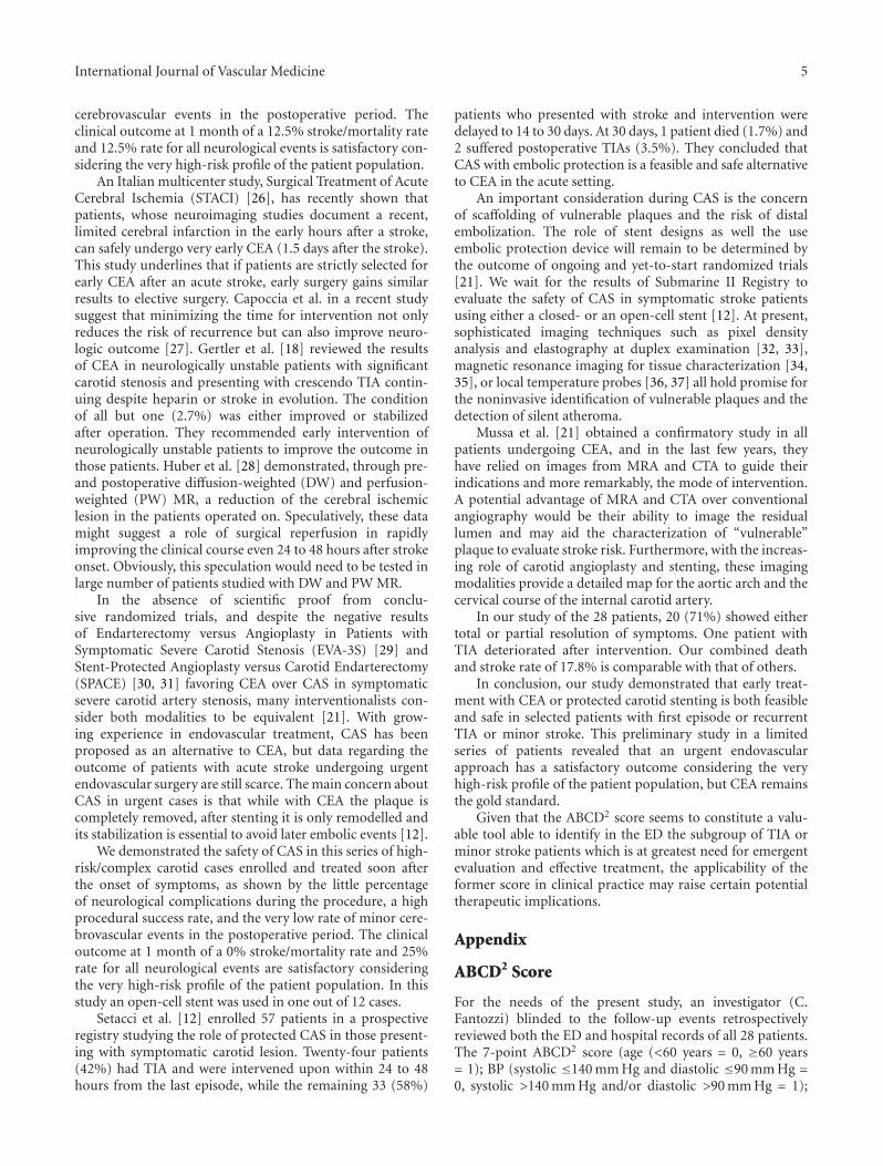
Group B. Successful CAS was achieved in all cases (100%).In 8 cases a Boston Scientific Carotid Wallstent, in 3 cases anInvatec Cristallo Ideale stent, and in 1 case an EV3 ProtegeRX carotid stent were implanted.
4 International Journal of Vascular Medicine
Table 4
ParameterSurgicalgroup
Endovasculargroup
Pvalue∗
Number of patients 16 12
Early neurological results
Improvement (>1 NIHSS) 10 (63.5%) 9 (75%) 0.15
Stable 4 (25%) 0
Impairment (<1 NIHSS) 2 (12.5%) 3 (25%)∗
Chi-square test for trend.
Table 5
NIHSS
At admission Postoperative P value∗
Surgical group 7.4 5.5 0.21
Endovascular group 4.4 3.4 0.36∗
Mann-Whitney test.
No intraprocedural neurologic complication occurred,while the minor complications that occurred during theprocedure were ICA vasospasm in 2/12 cases (16%) andsevere bradicardia and hypotension in 1/12 case (8%). Pro-cedural success, defined as absence of new cerebrovascularevents at discharge (including major stroke, minor stroke,or TIA) was assessed at 76%. Three patients experiencedan in-hospital new neurologic event (2 minor stroke 16%;1 stroke 8%). Other nonneurological adverse events were apersistent bradicardia and an AMI. The postoperative NIHSSscore at hospital discharge showed that in 8/12 patients(66%) the neurological deficit improved (decrease of NIHSSscore >1), in 1/12 patients (8%) the NIHSS score remainedunchanged (decrease of NIHSS score <1) and in 3/12 cases(24%) the neurological deficit showed an impairment (2minor strokes 16%; 1 stroke 8%), in two cases without newischemic cerebral lesions. In 3 patients new ischemic lesionswere detected by CT or MR postoperative imaging, 2 of thesewere asymptomatic although the NIHSS preoperative scorewas significantly lower than in group A.
At 90 days NIHSS score showed that in 9/12 patients(75%) the neurological deficit improved (decrease of NIHSSscore >1) and in 3/12 cases (24%) the neurological deficitshowed an impairment (2 minor strokes 16%; 1 stroke 8%)(Tables 4 and 5).
5. Discussion
This study demonstrates the safety and the efficacy of earlyCEA or CAS after TIA (within 24–48 hours) or stroke (within14–28 days).
Effective and early management of patients with acutesymptoms due to carotid stenosis is still the subject ofdebate. The inability to predict who is at higher early riskof a recurrent stroke after a cerebrovascular event (TIA orstroke) may explain the variation in management of acutestroke comparing physician to physician and institution toinstitution [12].
Improving outcomes of carotid surgery have recentlyfocused on the appropriate timing of carotid intervention in
the setting of new-onset neurological deficit. The increasedrisks of reperfusion injury and conversion to hemorrhagicinfarction have led to the historical recommendation ofdelayed CEA in those presenting with acute stroke [15–17]. However, several high-volume centers have documentedthe safety and efficacy of early intervention in preventingrecurrent strokes in carefully selected patients [18, 19].More specifically, patients presenting with fluctuating andpotentially reversible neurological deficits may benefit fromthe removal of an embolic source and/or salvage the at-riskareas of the brain [18, 20]. Comparing medical therapy tosurgical intervention in those presenting with nondisablingstroke and TIA, nonoperative therapy carried a 54% rateof combined death and worsening neurological conditionwhereas the combined death and stroke rate in the surgicalgroup was 7% [21]. The reported stroke risks highlight howearly removal of the carotid plaque, that is considered to bethe embolic source, could be crucial in stroke prevention[12]. Rothwell et al. [22] have recently analyzed pooleddata from the European Carotid Surgery Trial and NorthAmerican Symptomatic Carotid Endarterectomy Trial (atotal of 5893 patients). The analysis of long-term strokeprevention (per 1000 CEAs at 5 years), in relation tothe delay in surgery, clearly showed that benefit fromsurgery decreased rapidly with time elapsed since the lastneurological symptoms. Profit from endarterectomy seemsto depend not only on the degree of carotid stenosis, butalso on delay in surgery. Recent data have conclusively shownthat patients benefit when intervention is performed within2 weeks from the index event in order to prevent recurrentstroke, even if the procedural risk may be higher [4, 22]. TheSPREAD guidelines also clearly state that carotid surgery isrecommended as early as possible—within 2 weeks of theevent—for patients with TIA, minor stroke, or stabilizedneurological deficit with normal CT scanning or minimallesions (Grade A recommendation) [23]. In a prospectivestudy of patients presenting with acute symptomatic high-grade internal carotid artery stenosis, the Oxford VascularStudy Group recently observed that for patients with TIAthe 7-day, 30-day, and 3-month risks of stroke were 8%,12%, and 17% respectively, while for patients presenting withminor stroke (NIH stroke score <3) parallel data were 12%,15%, and 19% [24]. These data suggest that acute strokeor TIA should be considered as medical emergencies thatrequire rapid evaluation and rapid targeting of treatment.
The decision to proceed with an urgent intervention forthose presenting with acute neurological deficit is challeng-ing and requires very careful attention. Rothwell et al. in 2007have reported a prospective study that shows how an earlytreatment (medical and/or surgical) of all patients presentingwith TIA or minor stroke can prevent about 80% of earlyrecurrent stroke [25]. Although the timing of interventionmay be debatable, the mode of intervention is even morecontroversial.
We demonstrated the safety of CEA in this series of high-risk/complex carotid cases enrolled and treated soon afterthe onset of symptoms, as shown by the little percentageof neurological complications during the procedure, a highprocedural success rate, and the very low rate of minor
International Journal of Vascular Medicine 5
cerebrovascular events in the postoperative period. Theclinical outcome at 1 month of a 12.5% stroke/mortality rateand 12.5% rate for all neurological events is satisfactory con-sidering the very high-risk profile of the patient population.
An Italian multicenter study, Surgical Treatment of AcuteCerebral Ischemia (STACI) [26], has recently shown thatpatients, whose neuroimaging studies document a recent,limited cerebral infarction in the early hours after a stroke,can safely undergo very early CEA (1.5 days after the stroke).This study underlines that if patients are strictly selected forearly CEA after an acute stroke, early surgery gains similarresults to elective surgery. Capoccia et al. in a recent studysuggest that minimizing the time for intervention not onlyreduces the risk of recurrence but can also improve neuro-logic outcome [27]. Gertler et al. [18] reviewed the resultsof CEA in neurologically unstable patients with significantcarotid stenosis and presenting with crescendo TIA contin-uing despite heparin or stroke in evolution. The conditionof all but one (2.7%) was either improved or stabilizedafter operation. They recommended early intervention ofneurologically unstable patients to improve the outcome inthose patients. Huber et al. [28] demonstrated, through pre-and postoperative diffusion-weighted (DW) and perfusion-weighted (PW) MR, a reduction of the cerebral ischemiclesion in the patients operated on. Speculatively, these datamight suggest a role of surgical reperfusion in rapidlyimproving the clinical course even 24 to 48 hours after strokeonset. Obviously, this speculation would need to be tested inlarge number of patients studied with DW and PW MR.
In the absence of scientific proof from conclu-sive randomized trials, and despite the negative resultsof Endarterectomy versus Angioplasty in Patients withSymptomatic Severe Carotid Stenosis (EVA-3S) [29] andStent-Protected Angioplasty versus Carotid Endarterectomy(SPACE) [30, 31] favoring CEA over CAS in symptomaticsevere carotid artery stenosis, many interventionalists con-sider both modalities to be equivalent [21]. With grow-ing experience in endovascular treatment, CAS has beenproposed as an alternative to CEA, but data regarding theoutcome of patients with acute stroke undergoing urgentendovascular surgery are still scarce. The main concern aboutCAS in urgent cases is that while with CEA the plaque iscompletely removed, after stenting it is only remodelled andits stabilization is essential to avoid later embolic events [12].
We demonstrated the safety of CAS in this series of high-risk/complex carotid cases enrolled and treated soon afterthe onset of symptoms, as shown by the little percentageof neurological complications during the procedure, a highprocedural success rate, and the very low rate of minor cere-brovascular events in the postoperative period. The clinicaloutcome at 1 month of a 0% stroke/mortality rate and 25%rate for all neurological events are satisfactory consideringthe very high-risk profile of the patient population. In thisstudy an open-cell stent was used in one out of 12 cases.
Setacci et al. [12] enrolled 57 patients in a prospectiveregistry studying the role of protected CAS in those present-ing with symptomatic carotid lesion. Twenty-four patients(42%) had TIA and were intervened upon within 24 to 48hours from the last episode, while the remaining 33 (58%)
patients who presented with stroke and intervention weredelayed to 14 to 30 days. At 30 days, 1 patient died (1.7%) and2 suffered postoperative TIAs (3.5%). They concluded thatCAS with embolic protection is a feasible and safe alternativeto CEA in the acute setting.
An important consideration during CAS is the concernof scaffolding of vulnerable plaques and the risk of distalembolization. The role of stent designs as well the useembolic protection device will remain to be determined bythe outcome of ongoing and yet-to-start randomized trials[21]. We wait for the results of Submarine II Registry toevaluate the safety of CAS in symptomatic stroke patientsusing either a closed- or an open-cell stent [12]. At present,sophisticated imaging techniques such as pixel densityanalysis and elastography at duplex examination [32, 33],magnetic resonance imaging for tissue characterization [34,35], or local temperature probes [36, 37] all hold promise forthe noninvasive identification of vulnerable plaques and thedetection of silent atheroma.
Mussa et al. [21] obtained a confirmatory study in allpatients undergoing CEA, and in the last few years, theyhave relied on images from MRA and CTA to guide theirindications and more remarkably, the mode of intervention.A potential advantage of MRA and CTA over conventionalangiography would be their ability to image the residuallumen and may aid the characterization of “vulnerable”plaque to evaluate stroke risk. Furthermore, with the increas-ing role of carotid angioplasty and stenting, these imagingmodalities provide a detailed map for the aortic arch and thecervical course of the internal carotid artery.
In our study of the 28 patients, 20 (71%) showed eithertotal or partial resolution of symptoms. One patient withTIA deteriorated after intervention. Our combined deathand stroke rate of 17.8% is comparable with that of others.
In conclusion, our study demonstrated that early treat-ment with CEA or protected carotid stenting is both feasibleand safe in selected patients with first episode or recurrentTIA or minor stroke. This preliminary study in a limitedseries of patients revealed that an urgent endovascularapproach has a satisfactory outcome considering the veryhigh-risk profile of the patient population, but CEA remainsthe gold standard.
Given that the ABCD2 score seems to constitute a valu-able tool able to identify in the ED the subgroup of TIA orminor stroke patients which is at greatest need for emergentevaluation and effective treatment, the applicability of theformer score in clinical practice may raise certain potentialtherapeutic implications.
Appendix
ABCD2 Score
For the needs of the present study, an investigator (C.Fantozzi) blinded to the follow-up events retrospectivelyreviewed both the ED and hospital records of all 28 patients.The 7-point ABCD2 score (age (<60 years = 0, ≥60 years= 1); BP (systolic ≤140 mm Hg and diastolic ≤90 mm Hg =0, systolic >140 mm Hg and/or diastolic >90 mm Hg = 1);

6 International Journal of Vascular Medicine
clinical features (unilateral weakness = 2, speech disturbancewithout weakness = 1, other symptom = 0); durationof symptoms (<10 minutes = 0, 10 to 59 minutes = 1,≥60 minutes = 2) and diabetes [1] was computed in 20cases. Clinical features were categorized as motor weakness(focal, usually unilateral, weakness of one or more of face,arm, hand, or leg) versus speech disturbance (defined asdysarthria or dysphasia or both) versus all other symptoms(numbness, change in vision, dizziness or vertigo, and gaitdisturbance) according to the OCSP definition of the ABCD2
score. Seven patients with unavailable BP recordings (n = 2)or duration of TIA symptoms (n = 5) at the ED records wereexcluded from further evaluation.
References
[1] P. M. Rothwell, “Medical and surgical management of symp-tomatic carotid stenosis,” International Journal of Stroke, vol.1, no. 3, pp. 140–149, 2006.
[2] J. K. Lovett, A. J. Coull, and P. M. Rothwell, “Early risk ofrecurrence by subtype of ischemic stroke in population-basedincidence studies,” Neurology, vol. 62, no. 4, pp. 569–573, 2004.
[3] J. F. Fairhead, Z. Mehta, and P. M. Rothwell, “Population-based study of delays in carotid imaging and surgery and therisk of recurrent stroke,” Neurology, vol. 65, no. 3, pp. 371–375,2005.
[4] A. R. Naylor, “Delay may reduce procedural risk, but atwhat price to the patient?” European Journal of Vascular andEndovascular Surgery, vol. 35, no. 4, pp. 383–391, 2008.
[5] D. Kleindorfer, P. Panagos, A. Pancioli et al., “Incidenceand short-term prognosis of transient ischemic attack in apopulation-based study,” Stroke, vol. 36, no. 4, pp. 720–723,2005.
[6] M. Daffertshoter, O. Mielke, A. Pullwitt, M. Felsenstein, andM. Hennerici, “Transient ischemic attacks are more than”ministrokes”,” Stroke, vol. 35, no. 11, pp. 2453–2458, 2004.
[7] A. P. Gasecki, M. Eliasziw, and M. B. Pritz, “Timing of carotidendarterectomy after stroke,” Stroke, vol. 29, no. 12, pp. 2667–2668, 1998.
[8] J. E. Crozier, J. Reid, G. H. Welch, K. W. Muir, and W. P. Stuart,“Early carotid endarterectomy following thrombolysis in thehyperacute treatment of stroke,” British Journal of Surgery, vol.98, no. 2, pp. 235–238, 2011.
[9] P. S. K. Paty, R. C. Darling III, P. J. Feustel et al., “Early carotidendarterectomy after acute stroke,” Journal of Vascular Surgery,vol. 39, no. 1, pp. 148–154, 2004.
[10] R. Topakian, A. M. Strasak, M. Sonnberger et al., “Timing ofstenting of symptomatic carotid stenosis is predictive of 30-day outcome,” European Journal of Neurology, vol. 14, no. 6,pp. 672–678, 2007.
[11] K. Imai, T. Mori, H. Izumoto, M. Watanabe, and K. Majima,“Emergency carotid artery stent placement in patients withacute ischemic stroke,” American Journal of Neuroradiology,vol. 26, no. 5, pp. 1249–1258, 2005.
[12] C. Setacci, G. de Donato, E. Chisci et al., “Deferred urgencycarotid artery stenting in symptomatic patients: clinicallessons and biomarker patterns from a prospective registry,”European Journal of Vascular and Endovascular Surgery, vol. 35,no. 6, pp. 644–651, 2008.
[13] S. C. Johnston, P. M. Rothwell, M. N. Nguyen-Huynh et al.,“Validation and refinement of scores to predict very early
stroke risk after transient ischaemic attack,” The Lancet, vol.369, no. 9558, pp. 283–292, 2007.
[14] G. Geroulakos, J. Domjan, A. Nicolaides et al., “Ultrasoniccarotid artery plaque structure and the risk of cerebralinfarction on computed tomography,” Journal of VascularSurgery, vol. 20, no. 2, pp. 263–266, 1994.
[15] W. F. Blaisdell, R. H. Clauss, J. G. Galbraith, A. M. Imparato,and E. J. Wylie, “Joint study of extracranial arterial occlusion.IV. A review of surgical considerations,” Journal of theAmerican Medical Association, vol. 209, no. 12, pp. 1889–1895,1969.
[16] M. E. Bruetman, W. S. Fields, E. S. Crawford, and M. E.Debakey, “Cerebral hemorrhage in carotid artery surgery,”Archives of neurology, vol. 9, pp. 458–467, 1963.
[17] E. J. Wylie, M. F. Hein, and J. E. Adams, “Intracranialhemorrhage following surgical revascularization for treatmentof acute strokes,” Journal of neurosurgery, vol. 21, pp. 212–215,1964.
[18] J. P. Gertler, J. D. Blankensteijn, D. C. Brewster et al., “Carotidendarterectomy for unstable and compelling neurologic con-ditions: do results justify an aggressive approach?” Journal ofVascular Surgery, vol. 19, no. 1, pp. 32–42, 1994.
[19] C. Schneider, K. Johansen, R. Konigstein, C. Metzner, and W.Oettinger, “Emergency carotid thromboendarterectomy: safeand effective,” World Journal of Surgery, vol. 23, no. 11, pp.1163–1167, 1999.
[20] “Beneficial effect of carotid endarterectomy in symptomaticpatients with high-grade carotid stenosis. North Americansymptomatic carotid endarterectomy trial collaborators,” TheNew England Journal of Medicine, vol. 325, no. 7, pp. 445–453,1991.
[21] F. F. Mussa, N. Aaronson, P. J. Lamparello et al., “Outcomeof carotid endarterectomy for acute neurological deficit,”Vascular and Endovascular Surgery, vol. 43, no. 4, pp. 364–369,2009.
[22] P. M. Rothwell, M. Eliasziw, S. A. Gutnikov, C. P. Warlow,and H. J. M. Barnett, “Endarterectomy for symptomaticcarotid stenosis in relation to clinical subgroups and timingof surgery,” The Lancet, vol. 363, no. 9413, pp. 915–924, 2004.
[23] SPREAD, “Stroke prevention and educational awareness dif-fusion,” 2007, http://www.spread.it/.
[24] A. J. Coull, J. K. Lovett, and P. M. Rothwell, “Populationbased study of early risk of stroke after transient ischaemicattack or minor stroke: implications for public education andorganisation of services,” British Medical Journal, vol. 328, no.7435, pp. 326–328, 2004.
[25] P. M. Rothwell, M. F. Giles, A. Chandratheva et al., “Earlyuse of existing preventive strategies for stroke (EXPRESS)study. Effect of urgent treatment of transient ischaemic attackand minor stroke on early recurrent stroke (EXPRESS study):a prospective population-based sequential comparison,” TheLancet, vol. 370, no. 9596, pp. 1432–1442, 2007.
[26] E. Sbarigia, D. Toni, F. Speziale, M. C. Acconcia, and P.Fiorani, “Early carotid endarterectomy after ischemic stroke:the results of a prospective multicenter Italian study,” EuropeanJournal of Vascular and Endovascular Surgery, vol. 32, no. 3, pp.229–235, 2006.
[27] L. Capoccia, E. Sbarigia, F. Speziale, D. Toni, and P. Fiorani,“Urgent carotid endarterectomy to prevent recurrence andimprove neurologic outcome in mild-to-moderate acute neu-rologic events,” Journal of Vascular Surgery, vol. 53, no. 3, pp.622–628, 2011.
[28] R. Huber, B. T. Muler, R. J. Seitz, M. Siebler, U. Modder, and W.Sandmann, “Carotid surgery in acute symptomatic patients,”

International Journal of Vascular Medicine 7
European Journal of Vascular and Endovascular Surgery, vol. 25,no. 1, pp. 60–67, 2003.
[29] J. L. Mas, G. Chatellier, B. Beyssen et al., “Endarterectomyversus stenting in patients with symptomatic severe carotidstenosis,” The New England Journal of Medicine, vol. 355, no.16, pp. 1660–1671, 2006.
[30] H. H. Eckstein, P. Ringleb, J. R. Allenberg et al., “Results ofthe stent-protected angioplasty versus carotid endarterectomy(SPACE) study to treat symptomatic stenoses at 2 years:a multinational, prospective, randomised trial,” The LancetNeurology, vol. 7, no. 10, pp. 893–902, 2008.
[31] SPACE Collaborative Group, P. A. Ringleb, J. Allenberg etal., “30 day results from the SPACE trial of stent-protectedangioplasty versus carotid endarterectomy in symptomaticpatients: a randomised non-inferiority trial,” The Lancet, vol.368, no. 9543, pp. 1239–1247, 2006.
[32] B. K. Lal, R. W. Hobson II, P. J. Pappas et al., “Pixel distributionanalysis of B-mode ultrasound scan images predicts histologicfeatures of atherosclerotic carotid plaques,” Journal of VascularSurgery, vol. 35, no. 6, pp. 1210–1217, 2002.
[33] E. I. Cespedes, C. L. de Korte, A. F. van der Steen, C. von Birge-len, and C. T. Lancee, “Intravascular elastography: principlesand potentials,” Seminars in Interventional Cardiology, vol. 2,no. 1, pp. 55–62, 1997.
[34] M. Shinnar, J. T. Fallon, S. Wehrli et al., “The diagnosticaccuracy of ex vivo MRI for human atherosclerotic plaquecharacterization,” Arteriosclerosis, Thrombosis, and VascularBiology, vol. 19, no. 11, pp. 2756–2761, 1999.
[35] M. Taurino, C. Battocchio, C. Maggiore et al., “Color flowdoppler versus magnetic resonance angiography for preopera-tive evaluation of the extracranial carotid vessels: comparativeand operative findings,” Italian Journal of Vascular andEndovascular Surgery, vol. 12, no. 3, pp. 91–99, 2005.
[36] W. Casscells, B. Hathorn, M. David et al., “Thermal detectionof cellular infiltrates in living atherosclerotic plaques: possibleimplications for plaque rupture and thrombosis,” The Lancet,vol. 347, no. 9013, pp. 1447–1449, 1996.
[37] S. Verheye, G. R. De Meyer, G. Van Langenhove, M. W.Knaapen, and M. M. Kockx, “In vivo temperature hetero-geneity of atherosclerotic plaques is determined by plaquecomposition,” Circulation, vol. 105, no. 13, pp. 1596–1601,2002.

Submit your manuscripts athttp://www.hindawi.com
Stem CellsInternational
Hindawi Publishing Corporationhttp://www.hindawi.com Volume 2014
Hindawi Publishing Corporationhttp://www.hindawi.com Volume 2014
MEDIATORSINFLAMMATION
of
Hindawi Publishing Corporationhttp://www.hindawi.com Volume 2014
Behavioural Neurology
EndocrinologyInternational Journal of
Hindawi Publishing Corporationhttp://www.hindawi.com Volume 2014
Hindawi Publishing Corporationhttp://www.hindawi.com Volume 2014
Disease Markers
Hindawi Publishing Corporationhttp://www.hindawi.com Volume 2014
BioMed Research International
OncologyJournal of
Hindawi Publishing Corporationhttp://www.hindawi.com Volume 2014
Hindawi Publishing Corporationhttp://www.hindawi.com Volume 2014
Oxidative Medicine and Cellular Longevity
Hindawi Publishing Corporationhttp://www.hindawi.com Volume 2014
PPAR Research
The Scientific World JournalHindawi Publishing Corporation http://www.hindawi.com Volume 2014
Immunology ResearchHindawi Publishing Corporationhttp://www.hindawi.com Volume 2014
Journal of
ObesityJournal of
Hindawi Publishing Corporationhttp://www.hindawi.com Volume 2014
Hindawi Publishing Corporationhttp://www.hindawi.com Volume 2014
Computational and Mathematical Methods in Medicine
OphthalmologyJournal of
Hindawi Publishing Corporationhttp://www.hindawi.com Volume 2014
Diabetes ResearchJournal of
Hindawi Publishing Corporationhttp://www.hindawi.com Volume 2014
Hindawi Publishing Corporationhttp://www.hindawi.com Volume 2014
Research and TreatmentAIDS
Hindawi Publishing Corporationhttp://www.hindawi.com Volume 2014
Gastroenterology Research and Practice
Hindawi Publishing Corporationhttp://www.hindawi.com Volume 2014
Parkinson’s Disease
Evidence-Based Complementary and Alternative Medicine
Volume 2014Hindawi Publishing Corporationhttp://www.hindawi.com