Embed Size (px)
Citation preview
JOURNAL OF CLINICAL MICROBIOLOGY, July 2009, p. 2097–2105 Vol. 47, No. 70095-1137/09/$08.00�0 doi:10.1128/JCM.01486-08Copyright © 2009, American Society for Microbiology. All Rights Reserved.
Clonal Complexes and Diversity of Exotoxin Gene Profiles inMethicillin-Resistant and Methicillin-Susceptible
Staphylococcus aureus Isolates from Patientsin a Spanish Hospital�
M. A. Argudín,1 M. C. Mendoza,1 F. J. Mendez,1,2 M. C. Martín,3 B. Guerra,4 and M. R. Rodicio1*Area de Microbiología, Departamento de Biología Funcional, and Instituto Universitario de Biotecnología de Asturias, Universidad de
Oviedo, Oviedo, Asturias, Spain1; Hospital Universitario Central de Asturias, Oviedo, Spain2; Instituto de Productos Lacteos deAsturias, Villaviciosa, Asturias, Spain3; and Federal Institute for Risk Assessment, Berlin, Germany4
Received 2 August 2008/Returned for modification 2 October 2008/Accepted 13 May 2009
Molecular epidemiology studies have allowed the identification of the methicillin (meticillin)-resistant (MRSA)and methicillin-susceptible (MSSA) clonal complexes (CCs) and clones of Staphylococcus aureus circulating in aSpanish hospital recently. Of 81 isolates tested, 32.1% were MRSA. Most of them carried staphylococcal cassettechromosome mec (SCCmec) IVc (88.5%) and belonged to CC5 (88.5%; multilocus sequence typing types ST125[mainly associated with spa type t067], ST5, and ST228). A higher diversity was found among MSSA isolates(67.9%). Eighty percent shared the genetic background of major MRSA lineages (CC5 [38.2%; ST125 and ST5],CC30 [25.5%; ST30], CC45 [14.5%; ST45 and ST47], and CC8 [1.8%; ST8]), but CC12, CC15, CC51, and CC59 werealso detected. Many exotoxin genes were present in each of the 81 isolates, independent of whether they wereinvolved in sepsis (11 to 22) or other types of infections (13 to 21), and they appeared in 73 combinations. Therelevant data are that (i) all isolates were positive for hemolysin and leukotoxin genes (98.8% for lukED and 25.9%for lukPV); (ii) all contained an enterotoxin gene cluster (egc with or without seu), frequently with one or more genesencoding classical enterotoxins; (iii) about half were positive for tst and 95% were positive for exfoliatin-encodinggenes (eta, etb, and/or etd); and (iv) the four agr groups were detected, with agrII (55.6%) and agrIII (23.5%) beingthe most frequent. Taken together, results of the present study suggest a frequent acquisition and/or loss of exotoxingenes, which may be mediated by efficient intralineage transfer of mobile genetic elements and exotoxin genestherein and by eventual breakage of interlineage barriers.
Staphylococcus aureus is both a commensal bacterium and anextremely versatile pathogen that causes a wide range of dis-eases in humans, including superficial, deep-seated, and sys-temic infections, as well as a variety of toxemic syndromes,such as toxic shock syndrome (TSS), staphylococcal scalded-skin syndrome (SSSS), and staphylococcal food poisoning (36).S. aureus produces a wide range of virulence factors that me-diate host colonization, invasion of damaged skin and mucosa,dissemination through the body, and evasion of host defensemechanisms (8, 12). Relevant among them are a variety ofexotoxins that comprise �-, �-, �-, and �-hemolysins, leukotox-ins (the classical LukS-PV–LukF-PV Panton-Valentine leuko-cidin [LukPV], LukE-LukD [LukED] and LukM-LukF�-PV[LukM]), exfoliative toxins, and pyrogenic toxin superantigens,such as the staphylococcal TSS toxin (TSST-1, first referred toas SEF) and staphylococcal enterotoxins (SEs) (14, 29, 43).Five major serological types of SEs, SEA through SEE (knownas classical enterotoxins, encoded by the sea to see genes,respectively) have been initially identified. However, new typesof SEs and their coding genes (seg through seu) were laterreported. Several SE genes (seg, sei, sem, sen, and seo) are partof an operon termed the enterotoxin gene cluster (egc), of
which a variant that contains seu instead of the two pseudo-genes present in the originally described egc between sei andsen has been identified (27, 33). Both TSST-1 and SEs arepotent activators of T-cell populations, leading to massive pro-liferation and uncontrolled release of proinflammatory cyto-kines (14). Expression of most virulence factors in S. aureus isunder the control of the agr (accessory gene regulator) locus,which encodes a two-component signaling pathway and itsactivating ligand, a bacterial-density-sensing peptide termedthe autoinducing peptide (37). S. aureus strains can be subdi-vided into four major agr groups, based on polymorphisms inthe amino acid sequence of the autoinducing peptide and othercomponents of the system (26, 28). Within a given group, eachstrain produces a peptide that can activate the agr response inother members of the group, whereas the autoinducing pep-tides produced by different groups are usually mutually inhib-itory (26).
Apart from having pathogenic versatility, S. aureus can adaptrapidly to the selective pressure of antibiotics, with the emer-gence and spread of methicillin (meticillin)-resistant S. aureus(MRSA) isolates being a relevant example. Resistance tomethicillin and other beta-lactam antibiotics is caused by themecA gene, situated on a mobile genetic element, the staphy-lococcal cassette chromosome mec (SCCmec), which consistsof the mec gene complex, the ccr gene complex, and the “junk-yard” regions. Based on the variability of the differently com-bined components, several types of SCCmec and several vari-
* Corresponding author. Mailing address: Area de Microbiología,Departamento de Biología Funcional, Facultad de Medicina, Ju-lian Clavería 6, 33006 Oviedo, Spain. Phone: 34985103562. Fax:34985103148. E-mail: [email protected].
� Published ahead of print on 20 May 2009.
2097
on March 15, 2018 by guest
http://jcm.asm
.org/D
ownloaded from

ants of the types have been distinguished (21, 23, 24, 25, 30, 31,38, 45, 51).
In the present work, the techniques most commonly appliedin epidemiological studies of S. aureus were used to identify theprevalent and sporadic MRSA and methicillin-susceptible S.aureus (MSSA) clones that have been causing disease in aSpanish hospital (the Hospital Universitario Central de Astu-rias [HUCA]) over a recent time period (2005 to 2006). Thesemethods included pulsed-field gel electrophoresis (PFGE) ofSmaI-digested genomic DNA (SmaI PFGE), S. aureus proteinA gene (spa) typing, multilocus sequence typing (MLST) anal-ysis, and SCCmec typing of MRSA (4, 9, 40, 42, 50, 52). Therisk for human health posed by the accumulation of virulencegenes in S. aureus (34) along with the potential application ofsuch genes for subtyping prompted the assessment of the vir-ulence gene repertoire of the HUCA isolates, with regard tothe agr group and 30 exotoxin-encoding genes.
MATERIALS AND METHODS
S. aureus isolates. Eighty-one S. aureus isolates were analyzed in this study.Each was collected from a different patient attending the HUCA from December2005 to December 2006. These isolates were identified in the hospital by use ofstandard procedures. Briefly, suspected colonies of S. aureus obtained in primarycultures were tested for agglutination with Pastorex Staph-plus (Bio-Rad Labo-ratories SA, Alcobendas, Madrid, Spain) and for thermonuclease (on DNase testagar; Biomedics, Madrid, Spain) and coagulase production (coagulase plasma;Becton Dickinson, San Agustín de Gualdalix, Madrid, Spain). Isolates thatproved to be positive for the three tests were also evaluated for susceptibility orresistance to methicillin. S. aureus isolates were recovered from suppurativesamples from spontaneous infections (5 isolates from patients with conjunctivitis,abscess in the hand, umbilical pyogenic granuloma, parotiditis, or infected as-cites), surgical wounds (29 isolates associated with hand or leg amputations, hip,knee, or valvular prostheses, varicose veins, cellulitis, arthrodesis, osteomyelitis,Morton’s neuroma, thoracotomy, tracheotomy, pleuritis [in two patients withesophageal cancer], or colon infection [in a patient with colon cancer]), urine (1isolate from a patient with postcatheter urinary tract infection), tracheobronchialaspirates (11 isolates from patients with staphylococcal pneumonia, who haverequired intubation or assisted ventilation after cerebrovascular accident, braininjury, or polytraumatism), skin exudates (2 isolates from children with SSSS, 1also involved in sepsis), and blood (33 isolates from patients who developedsepsis, most of them with severe underlying conditions, such as leukemia, stom-ach cancer, liver transplantation, cerebral vascular accident, thrombosis, aorticaneurysm, chronic renal insufficiency subjected to hemodialysis or peritonealdialysis, osteomyelitis, pneumonia, and/or the presence of inserted catheters).
Macrorestriction-PFGE analysis. Whole DNA from each S. aureus isolate wasanalyzed by SmaI PFGE using a CHEF-DRIII SYS220/240 (Bio-Rad Labora-tories, S.A., Madrid, Spain) system and the consensus protocol of the EuropeanHARMONY group (42). The resulting profiles were analyzed visually, and thepresence or absence of fragments larger than ca. 20 kb was recorded. Thoseshowing one or more mismatching bands were considered different and labeledwith an “S” followed by a number. Genetic similarity between profiles wasdetermined by the unweighted-pair method with arithmetic averages and Jac-card’s coefficient, using the software program MVSP version 3.1 (MultivariateStatistics Package for PCs; RockWare Inc.). NCTC 8325 was used as a referencestrain for PFGE.
spa typing and MLST. spa typing and MLST were performed as described byShopsin et al. (50) and Enright et al. (9), respectively. PCR products weresequenced by Qiagen (Hilden, Germany). spa types were identified with thesoftware Ridom Staphtype (Ridom GmbH), and sequence types (STs) wereassigned by using the MLST website (http://www.mlst.net). Isolates were groupedinto a single clonal cluster (CC) when five of the seven housekeeping genes usedin the MLST scheme were identical (9).
SCCmec typing and virulence gene content. SCCmec was typed by the multi-plex PCR strategy reported by Zhang et al. (60). Primers for the exotoxin genesand the agr group have been described previously (3, 15, 16, 39, 41, 55). PCRassays were performed at least twice for each exotoxin gene and isolate, andpositive and negative controls were always included. A dendrogram of similarityshowing the clustering of the isolates according to virulence gene profiles was
constructed by using Jaccard’s coefficient of similarity (BioNumerics version 5.1;Applied Maths).
Statistical methods. Statistical comparison between isolates giving positiveand negative test results was performed with the �2 test, using SPSS software(version 15.0; SPSS, Chicago, IL). Differences between groups were consideredstatistically significant if P values were �0.05. The discrimination index (DI; i.e.,the probability that two unrelated isolates would be assigned to different SmaI orvirulence profiles) was calculated using Simpson’s index of diversity (53).
RESULTS
Genotypic typing of the isolates. For a detailed assessmentof the epidemiology of S. aureus circulating in the HUCA, the81 isolates selected for the present study were subjected toSmaI PFGE, and representative subsets were also typed by spatyping and MLST.
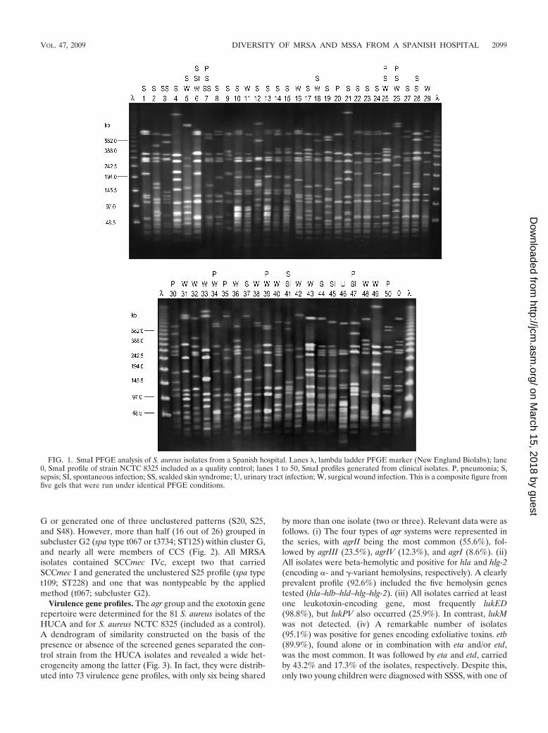
SmaI PFGE yielded a DI of 0.98 and identified 50 profiles(S1 to S50; Fig. 1), with only 15 including more than one isolate(two up to seven). On the basis of the coefficient of similarityof Jaccard, a dendrogram was constructed (Fig. 2). At a cutoffpoint of 0.64, two major clusters (B and G), five minor clusters(A, C, D, E, and F), and several ungrouped branches wererevealed. Cluster G, which accounted for 43.2% of the isolates,could be in turn separated into subclusters G1 and G2. Thir-teen isolates of cluster G were selected for spa typing (Table1). Four of them, all belonging to G1, were assigned to t002,another two were assigned to t3734, and seven were assigned tot067, and these last nine were included in G2. MLST per-formed on one or two isolates of each spa type associated t002with ST5, while the t067 and t3734 isolates were associatedwith ST125 (which differs from ST5 by only one base pair in theamplified region of the yqiL gene [46]). Interestingly, repre-sentative isolates of cluster C and the single S20 isolate thatgenerated the unclustered branch closest to cluster G were alsoof spa type t067 and ST125, whereas the next branch includedthree S25 isolates that were of spa type t109 and ST228 (atwo-locus variant of ST5, with 1- and 2-bp differences in the tpiand yqiL amplified regions, respectively [48]). ST5, ST125, andST228 belong to the same CC, namely, CC5, which clearlypredominated in the HUCA.
With regard to the remaining SmaI PFGE clusters, the fol-lowing observations were made. (i) Representative isolates ofcluster B showed spa types t012 (four out of five tested) andt021 (the remaining one), and one isolate of each type provedto be of ST30 within CC30. (ii) Isolates belonging to cluster Dwere of spa types t282 and t1618 (one and two out of threetested, respectively), and one of the latter isolates was of ST45.(iii) Isolates in cluster A were of spa types t081, t383, andt1494, and the t383 isolate was of ST47. Both ST45 and ST47(which differs from ST45 by one change in the amplified regionof aroE [9]) belong to CC45, although the correspondingPFGE profiles grouped separately in the dendrogram. (iv) Twospa types and one ST were associated with clusters E (t547 andt084; both ST15) and F (t160 and t1381; ST12). Finally, theS32, S12, and S19 unclustered SmaI PFGE profiles were t024,t159, and nontypeable, respectively. The predicted ST for t024is ST8 (CC8), t159 could be of ST121 or ST427 (CC51), andthe last type was experimentally assigned to ST59 (CC59).
Incidence of MRSA and SCCmec typing. Of the isolatesunder study, 55 were MSSA (67.9%) and 26 were MRSA(32.1%). The latter belonged to SmaI PFGE clusters A, C, and
2098 ARGUDIN ET AL. J. CLIN. MICROBIOL.
on March 15, 2018 by guest
http://jcm.asm
.org/D
ownloaded from
G or generated one of three unclustered patterns (S20, S25,and S48). However, more than half (16 out of 26) grouped insubcluster G2 (spa type t067 or t3734; ST125) within cluster G,and nearly all were members of CC5 (Fig. 2). All MRSAisolates contained SCCmec IVc, except two that carriedSCCmec I and generated the unclustered S25 profile (spa typet109; ST228) and one that was nontypeable by the appliedmethod (t067; subcluster G2).
Virulence gene profiles. The agr group and the exotoxin generepertoire were determined for the 81 S. aureus isolates of theHUCA and for S. aureus NCTC 8325 (included as a control).A dendrogram of similarity constructed on the basis of thepresence or absence of the screened genes separated the con-trol strain from the HUCA isolates and revealed a wide het-erogeneity among the latter (Fig. 3). In fact, they were distrib-uted into 73 virulence gene profiles, with only six being shared
by more than one isolate (two or three). Relevant data were asfollows. (i) The four types of agr systems were represented inthe series, with agrII being the most common (55.6%), fol-lowed by agrIII (23.5%), agrIV (12.3%), and agrI (8.6%). (ii)All isolates were beta-hemolytic and positive for hla and hlg-2(encoding �- and �-variant hemolysins, respectively). A clearlyprevalent profile (92.6%) included the five hemolysin genestested (hla–hlb–hld–hlg–hlg-2). (iii) All isolates carried at leastone leukotoxin-encoding gene, most frequently lukED(98.8%), but lukPV also occurred (25.9%). In contrast, lukMwas not detected. (iv) A remarkable number of isolates(95.1%) was positive for genes encoding exfoliative toxins. etb(89.9%), found alone or in combination with eta and/or etd,was the most common. It was followed by eta and etd, carriedby 43.2% and 17.3% of the isolates, respectively. Despite this,only two young children were diagnosed with SSSS, with one of
FIG. 1. SmaI PFGE analysis of S. aureus isolates from a Spanish hospital. Lanes , lambda ladder PFGE marker (New England Biolabs); lane0, SmaI profile of strain NCTC 8325 included as a quality control; lanes 1 to 50, SmaI profiles generated from clinical isolates. P, pneumonia; S,sepsis; SI, spontaneous infection; SS, scalded skin syndrome; U, urinary tract infection; W, surgical wound infection. This is a composite figure fromfive gels that were run under identical PFGE conditions.
VOL. 47, 2009 DIVERSITY OF MRSA AND MSSA FROM A SPANISH HOSPITAL 2099
on March 15, 2018 by guest
http://jcm.asm
.org/D
ownloaded from

the responsible isolates being positive for etb, eta, and agrIVand the other being positive for etb and agrII. (v) About half ofthe isolates (51.9%) carried tst, although none of the patientssuffered from TSS. (vi) All isolates contained an egc or anegc-like cluster (70.4% and 29.6%, respectively), frequentlytogether with other SE genes, including those encoding classi-cal enterotoxins (92.6%). Of the latter, sec, sea, seb, and sedwere present in 80.2%, 55.6%, 27.2%, and 24.7% of the iso-lates, respectively. Each of these genes appeared either alone(in a few cases) or with one or two of the other in differentcombinations. All HUCA isolates were negative for see. Thesed, sej, and ser genes carried by plasmids were present in24.7% of the isolates, with the three coinciding in 17.3% ofthem. Among other SE genes, sep was the most frequent(24.7%), but seh, sek, sel, and seq were also represented(12.3%, 2.5%, 2.5%, and 3.7%, respectively). Interestingly,typing of the isolates on the basis of the virulence gene profileyielded a DI close to 1 (0.997).
Overall, a very high number of exotoxin genes was present in
TABLE 1. Relationships between CC, MLST type, spa type, andPFGE clustera
CC MLST type(s) (n)b spa type(s) (n)c PFGEclusterd
CC5 ST5 (2) t002 (4) G1ST125 (2) t067 (7), t3734 (2) G2ST125 t067 (4) CST228 t109 (3) –(ST125) t067 –
CC8 (ST8) t024 –CC12 ST12 t160, t1381 FCC15 ST15 (2) t084 (2), t547 ECC30 ST30 (2) t012 (4), t021 BCC45 ST45 t282, t1618 (2) D
ST47 t081 (2), t383, t1494 ACC51 (ST121, ST427) t159 –CC59 ST59 NT –
a The numbers of isolates (n) are listed in parentheses if the number is morethan one.
b Determined or presumptive (according to the spa type; in parentheses) STsof representative isolates are shown.
c NT, nontypeable.d –, unclustered PFGE profile.
FIG. 2. Dendrogram showing the relatedness between SmaI macrorestriction fragment profiles generated from the S. aureus clinical isolatesand the control strain (NCTC 8325). At a Jaccard coefficient of similarity (J) of 0.64, seven clusters (labeled A to G, the latter with subclusters G1and G2) and several unclustered branches were detected. Footnote a, determined multilocus sequence types of representative isolates; footnoteb, MSSA and MRSA. n, number of isolates.
2100 ARGUDIN ET AL. J. CLIN. MICROBIOL.
on March 15, 2018 by guest
http://jcm.asm
.org/D
ownloaded from

FIG. 3. Dendrogram showing the relatedness between virulence gene profiles (for 30 exotoxin genes and the agr group) of the S. aureus isolatestested. At a Jaccard coefficient of similarity (J) of ca. 0.63, two main clusters, termed V1 and V2, and three subclusters within V2 (V2a, V2b, andV2c) are indicated. The type of infection caused by each isolate, the SmaI PFGE cluster to which it belongs, the SCCmec type (I, IVc, or nottypeable [nt]) of MRSA, and the spa type as well as the MLST type of representative isolates, are shown at the right of the dendrogram.
VOL. 47, 2009 DIVERSITY OF MRSA AND MSSA FROM A SPANISH HOSPITAL 2101
on March 15, 2018 by guest
http://jcm.asm
.org/D
ownloaded from
each of the HUCA isolates, independently of whether theywere involved in spontaneous infections (13 up to 16), surgicalwound infections (13 up to 21), pneumonia (13 up to 19),sepsis (11 up to 22), or SSSS (followed or not followed bysepsis, 14 and 18, respectively). Among the screened genes,only sep was significantly more frequent in isolates associatedwith sepsis (P 0.032), and seb was more frequent in thosethat caused other infections (P 0.032). Moreover, agrII andsep were more common in MRSA isolates than in MSSAisolates (P values of 0.029 and 0.048, respectively), while agrIIIand tst were more common in MSSA isolates (P values of 0.021and 0.032, respectively).
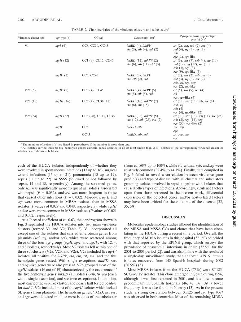
At a Jaccard coefficient of ca. 0.63, the dendrogram shown inFig. 3 separated the HUCA isolates into two main virulenceclusters (termed V1 and V2; Table 2). V1 incorporated allexcept one of the isolates that carried enterotoxin genes fromplasmids (sed, sej, and/or ser), which were scattered amongthree of the four agr groups (agrII, agrI, and agrIV, with 12, 4,and 3 isolates, respectively). Most V2 isolates fell within one ofthree subclusters (V2a, V2b, and V2c). V2a included five agrIVisolates, all positive for lukPV, eta, etb, tst, sea, and the fivehemolysin genes tested. With single exceptions, lukED, sec,and egc-like genes were also present. V2b grouped most of theagrIII isolates (16 out of 19) characterized by the occurrence ofthe five hemolysin genes, lukED (all isolates), etb, tst, sea (eachwith a single exception), and sec (two exceptions). In addition,most carried the egc-like cluster, and nearly half tested positivefor lukPV. V2c included most of the agrII isolates which lackedSE genes from plasmids. The hemolysin genes, lukED, etb, sec,and egc were detected in all or most isolates of the subcluster
(from ca. 80% up to 100%), while eta, tst, sea, seb, and sep wererelatively common (32.4% to 44.1%). Finally, data compiled inFig. 3 failed to reveal a correlation between virulence geneprofile(s) and type of disease, with all clusters and subclustersgrouping isolates involved in sepsis together with isolates thatcaused other types of infections. Accordingly, virulence factorsapart from those screened in the present work, differentialexpression of the detected genes, and/or host-related factorsmay have been critical for the outcome of the disease (32,35, 58).
DISCUSSION
Molecular epidemiology studies allowed the identification ofthe MRSA and MSSA CCs and clones that have been circu-lating in the HUCA during a recent time period. Overall, thefrequency of MRSA isolates in this hospital (32.1%) coincidedwith that reported by the EPINE group, which surveys theprevalence of nosocomial infections in Spain (32.5% for the2001-to-2003 period [2]), and was also in line with the results ofa single-day surveillance study that analyzed 439 S. aureusisolates recovered from 143 Spanish hospitals during 2002(30.5%) (5).
Most MRSA isolates from the HUCA (73%) were ST125-SCCmec IV isolates. This clone emerged in Spain during 1996,although it was first reported in 2001, and has now becomepredominant in Spanish hospitals (46, 47, 56). At a lowerfrequency, it was also found in Norway (13). As in the presentstudy, a strong correlation between ST125 and spa type t067was observed in both countries. Most of the remaining MRSA
TABLE 2. Characteristics of the virulence clusters and subclustersa
Virulence cluster (n) agr type (n) CC (n) Cytotoxin(s) (n)b Pyrogenic toxin superantigengene(s) (n)b
V1 agrI (4) CC8, CC30, CC45 lukED (4), lukPV tst (2), sea, seb (2), sec (4)eta (3), etb (4), etd (2) sed (4), sej (3), ser (3)
sehegc (3), egc-like
agrII (12) CC5 (9), CC15, CC45 lukED (12), lukPV (2) tst (3), sea (7), seb (4), sec (10)eta (6), etb (11), etd (3) sed (12), sej (12), ser (10)
seh (3), sep (3)egc (9), egc-like (3)
agrIV (3) CC5, CC45 lukED (3), lukPV tst (2), sea (2), seb, sec (3)eta, etb (2), etd sed (3), sej (3), ser (2)
sek, sel, sep, seqegc (2), egc-like
V2a (5) agrIV (5) CC5 (4), CC45 lukED (4), lukPV (5) tst (5), sea (5), sec (4)eta (5), etb (5), etd sel
egc, egc-like (4)V2b (16) agrIII (16) CC5 (4), CC30 (11) lukED (16), lukPV (7) tst (15), sea (15), seb, sec (14)
eta (6), etb (15) sed, sejseh (4)egc (4), egc-like (12)
V2c (34) agrII (32) CC5 (28), CC15, CC45 lukED (32), lukPV (5) tst (10), sea (13), seb (11), sec (25)eta (12), etb (28), etd (2) seh (2), sep (14), seq
egc (30), egc-like (2)agrIV CC5 lukED, etb sec, sep
egcagrI CC45 lukED, etb, etd tst, sea, sec
egc
a The numbers of isolates (n) are listed in parentheses if the number is more than one.b All isolates carried three to five hemolysin genes; exotoxin genes detected in all or most (more than 75%) isolates of the corresponding virulence cluster or
subcluster are shown in boldface.
2102 ARGUDIN ET AL. J. CLIN. MICROBIOL.
on March 15, 2018 by guest
http://jcm.asm
.org/D
ownloaded from

isolates from the HUCA were also positive for SCCmec IV butbelonged to other STs (i.e., ST5, ST228, or ST47); two carriedSCCmec I and were of ST228, while one was not typeable bythe applied method (60). The ST5-SCCmec IV (pediatricclone) (49) and ST228-SCCmec I (southern German clone)(48) clones are major hospital-acquired MRSA clones, widelydistributed worldwide (7, 46, 56). In summary, typing of MRSAisolates from the HUCA revealed that nearly all belonged toCC5 (88.5%), one of the most diversified lineages of MRSA(48), and carried SCCmec IV (also 88.5%), specificallySCCmec IVc, which in Spain accounted only for 24% of theMRSA isolates, whereas subtype IVa was the most frequent(73.3%) (47).
As previously reported by other authors (7, 10, 52), a higherdiversity of CCs was found among the MSSA isolates of theHUCA, which accounted for 67.9% of the analyzed isolates.Of them, 80.0% had a genetic background common to majorMRSA lineages, namely, CC5 (38.2%), CC30 (25.5%), CC45(14.5%), and CC8 (1.8%) (7, 17, 48). As was the case forMRSA, MSSA isolates with a CC5 genetic background mostlybelonged to SmaI PFGE subclusters G1 and G2 and were ofeither ST5 or ST125; CC30 isolates fell into SmaI PFGE clus-ter B and were of ST30; and ST47 and ST45, both members ofCC45, were separated by SmaI PFGE into clusters A and D.Four successful MSSA lineages different from the majorMRSA clones, namely, CC12, CC15 (PFGE clusters F and E),CC51, and CC59 (each with one sporadic isolate), were alsodetected. Isolates of these four lineages have been observed inboth nosocomial and community settings in different countries(1, 18, 19, 44, 54, 57). Finally, it is worth noting that, like inother studies (4, 20), a good concordance between typing bySmaI PFGE, spa type, and MLST type was found for theHUCA isolates. In fact, most isolates with the same MLST hadsimilar SmaI profiles and, hence, grouped within the samePFGE cluster. However, several SmaI PFGE clusters and/orbranches could be associated with a certain CC and even witha certain ST, consistent with the higher discrimination of theformer method.
With regard to the virulence gene repertoires, the high num-ber of exotoxin genes carried by each of the 81 isolates ana-lyzed in the present work (from 11 up to 22) is remarkable. AllHUCA isolates harbored genes encoding four or five hemo-lysins, one or two leukocidins, and five up to 13 enterotoxins.All tested positive for egc with or without seu, and most werepositive for one or more genes encoding classical enterotoxins,including high percentages of sec and sea. Moreover, all exceptfour isolates contained at least one exfoliatin gene (eta, etb,and/or etd), whereas tst was present in half of them, and theleukotoxin gene lukPV was present in about one-fourth. InSpanish hospitals, the incidence of MSSA isolates carryinglukPV has increased from 0% in 2002 up to 36.4% in 2006 to2007 (5, 47). In the present study, 66.7% of the lukPV-positiveisolates were MSSA, but 33.3% were MRSA, and most be-longed to the prevalent CC5. Of the 30 virulence genesscreened, only lukM and see were absent from the series. Thosethat were detected appeared in many different combinations,with a total of 73 virulence profiles displayed by 81 isolates. Awide diversity of virulence gene combinations has also beenreported for S. aureus isolates from England, Germany, andFrance (6, 22, 35).
Overall, the high number of exotoxin genes carried by eachisolate and the rather indiscriminate distribution of thesegenes among different agr groups and genomic backgrounds,together with the striking diversity of exotoxin gene combina-tions, are consistent with a frequent acquisition and/or loss ofthe screened genes. It is widely accepted that mobile geneticelements carrying exotoxin genes can efficiently spread withinlineages (35). In addition, they may be eventually capable ofbreaking interlineage barriers. An important barrier is the SauItype I restriction-modification system, which appears to be oneof the major mechanisms underlying the clonal structure of S.aureus (11, 59). Nevertheless, a mobile genetic element canenter a CC from which it was originally excluded by means ofa SauI restriction mutant. Once in such a mutant, the elementmight become modified at the specific sequences recognized bythe hsdS1 and hsdS2 products characteristic of this particularlineage. Afterwards, its transfer to other members of the samelineage would be possible. In any case, the accumulation ofexotoxin genes within S. aureus isolates is a matter of concern,since it may result in enhanced pathogenicity under appropri-ate circumstances (34). In the HUCA, the epidemiologicalsurveillance of these potential “superbugs” could be facilitated,at least in the near future, by these findings with regard to theremarkable diversity of exotoxin gene combinations, whichnearly differentiated at the strain level.
ACKNOWLEDGMENTS
This work was supported by project FIS-06-PI052489 from the Span-ish Ministry of Science and Innovation and projects BfR 45-004 and45-005 of the Federal Institute for Risk Assessment (BfR), Germany.M. A. Argudín is the recipient of grant FPU AP-2004-3641 from theMinistry of Science and Innovation, Spain, cofunded by the EuropeanSocial Fund. Part of the work was performed during a short stay ofM. A. Argudín at the Department of Biological Safety of BfR, sup-ported by the same grant.
We are grateful to Ana Fleites and Marta Landero from theHospital Universitario Central de Asturias for supplying S. aureusisolates, to J. Hammerl and S. Hertwig (BfR) for their advice on spatyping and MLST, and to the Department of Biological Safety fortheir hospitality.
REFERENCES
1. Aires de Sousa, M., T. Conceicao, C. Simas, and H. de Lencastre. 2005.Comparison of genetic backgrounds of methicillin-resistant and -susceptibleStaphylococcus aureus isolates from Portuguese hospitals and the commu-nity. J. Clin. Microbiol. 43:5150–5157.
2. Asensio, A., R. Canton, J. Vaque, J. Rossello, F. Calbo, J. Garcia-Caballero,V. Dominguez, A. Hernandez, A. Trilla, and EPINE Working Group. 2006.Nosocomial and community-acquired methicillin-resistant Staphylococcusaureus infections in hospitalized patients (Spain, 1993–2003). J. Hosp. Infect.63:465–471.
3. Becker, K., A. W. Friedrich, G. Lubritz, M. Weilert, G. Peters, and C. VonEiff. 2003. Prevalence of genes encoding pyrogenic toxin superantigens andexfoliative toxins among strains of Staphylococcus aureus isolated from bloodand nasal specimens. J. Clin. Microbiol. 41:1434–1439.
4. Cookson, B. D., D. A. Robinson, A. B. Monk, S. Murchan, A. Deplano, R. deRyck, M. J. Struelens, C. Scheel, V. Fussing, S. Salmenlinna, J. Vuopio-Varkila, C. Cuny, W. Witte, P. T. Tassios, N. J. Legakis, W. van Leeuwen, A.van Belkum, A. Vindel, J. Garaizar, S. Haeggman, B. Olsson-Liljequist, U.Ransjo, M. Muller-Premru, W. Hryniewicz, A. Rossney, B. O’Connell, B. D.Short, J. Thomas, S. O’Hanlon, and M. C. Enright. 2007. Evaluation ofmolecular typing methods in characterizing a European collection of epi-demic methicillin-resistant Staphylococcus aureus strains: the HARMONYcollection. J. Clin. Microbiol. 45:1830–1837.
5. Cuevas, O., E. Cercenado, E. Bouza, C. Castellares, P. Trincado, R. Cabrera,A. Vindel, and Spanish Group for the Study of Staphylococcus. 2007. Molec-ular epidemiology of methicillin-resistant Staphylococcus aureus in Spain: amulticentre prevalence study (2002). Clin. Microbiol. Infect. 13:250–256.
6. Dauwalder, O., G. Lina, G. Durand, M. Bes, H. Meugnier, V. Jarlier, B.
VOL. 47, 2009 DIVERSITY OF MRSA AND MSSA FROM A SPANISH HOSPITAL 2103
on March 15, 2018 by guest
http://jcm.asm
.org/D
ownloaded from

Coignard, F. Vandenesch, J. Etiene, and F. Laurent. 2008. Epidemiology ofinvasive methicillin-resistant Staphylococcus aureus clones collected inFrance in 2006 and 2007. J. Clin. Microbiol. 46:3454–3458.
7. Deurenberg, R. H., and E. E. Stobbering. 2008. The evolution of Staphylo-coccus aureus. Infect. Genet. Evol. 8:747–763.
8. Dinges, M. M., P. M. Orwin, and M. Schlievert. 2000. Exotoxins of Staphy-lococcus aureus. Clin. Microbiol. Rev. 13:16–34.
9. Enright, M. C., N. P. Day, C. E. Davis, S. J. Peacock, and B. G. Spratt. 2000.Multilocus sequence typing for characterization of methicillin-resistant andmethicillin-susceptible clones of Staphylococcus aureus. J. Clin. Microbiol.38:1008–1015.
10. Faria, N. A., J. A. Carrico, D. C. Oliveira, M. Ramirez, and H. de Lencastre.2008. Analysis of typing methods for epidemiological surveillance of bothmethicillin-resistant and methicillin-susceptible Staphylococcus aureusstrains. J. Clin. Microbiol. 46:136–144.
11. Feng, Y., C.-J. Chen, L.-H. Su, S. Hu, J. Yu, and C.-H. Chiu. 2008. Evolutionand pathogenesis of Staphylococcus aureus: lessons learned from genotypingand comparative genomics. FEMS Microbiol. Rev. 32:23–37.
12. Ferry, T., T. Perpoint, F. Vandenesch, and J. Etienne. 2005. Virulencedeterminants in Staphylococcus aureus and their involvement in clinical syn-dromes. Curr. Infect. Dis. Rep. 7:420–428.
13. Fossum, A. E., and G. Butcholm. 2006. Increased incidence of methicillin-resistant Staphylococcus aureus ST80, novel ST125 and SCCmecIV in thesouth-eastern part of Norway during a 12-year period. Clin. Microbiol. In-fect. 12:627–633.
14. Fraser, J. D., and T. Proft. 2008. The bacterial superantigen and superanti-gen-like proteins. Inmunol. Rev. 225:226–243.
15. Fueyo, J. M., M. C. Mendoza, M. A. Alvarez, and M. C. Martin. 2005.Relationships between toxin gene content and genetic background in nasalcarried isolates of Staphylococcus aureus from Asturias, Spain. FEMS Mi-crobiol. Lett. 243:447–454.
16. Fueyo, J. M., M. C. Mendoza, M. R. Rodicio, J. Muniz, M. A. Alvarez, andM. C. Martin. 2005. Cytotoxin and pyrogenic toxin superantigen gene pro-files of Staphylococcus aureus associated with subclinical mastitis in dairycows and relationships with macrorestriction genomic profiles. J. Clin. Mi-crobiol. 43:1278–1284.
17. Gomes, A. R., H. Westh, and H. de Lencastre. 2006. Origins and evolution ofmethicillin-resistant Staphylococcus aureus clonal lineages. Antimicrob.Agents Chemother. 50:3237–3244.
18. Grundmann, H., S. Hori, M. C. Enright, C. Webster, A. Tami, E. J. Feil, andT. Pitt. 2002. Determining the genetic structure of the natural population ofStaphylococcus aureus: a comparison of multilocus sequence typing withpulsed-field gel electrophoresis, randomly amplified polymorphic DNA anal-ysis, and phage typing. J. Clin. Microbiol. 40:4544–4546.
19. Hallin, M., O. Denis, A. Deplano, R. De Mendonca, R. De Ryck, S. Rottiers,and M. J. Struelens. 2007. Genetic relatedness between methicillin-suscep-tible and methicillin-resistant Staphylococcus aureus: results of a nationalsurvey. J. Antimicrob. Chemother. 59:465–472.
20. Hallin, M., A. Deplano, O. Denis, R. De Mendonca, R. De Ryck, and M. J.Strudels. 2007. Validation of pulsed-field gel electrophoresis and spa typingfor long-term, nationwide epidemiological surveillance studies of Staphylo-coccus aureus infections. J. Clin. Microbiol. 45:127–133.
21. Hisata, K., K. Kuwahara-Arai, M. Yamanoto, T. Ito, Y. Nakatomi, L. Cui, T.Baba, M. Terasawa, C. Sotozono, S. Kinoshita, Y. Yamashiro, and K. Hira-matsu. 2005. Dissemination of methicillin-resistant staphylococci amonghealthy Japanese children. J. Clin. Microbiol. 43:3364–3372.
22. Holtfreter, S., D. Grumann, M. Schmudde, H. T. T. Nguyen, P. Eichler, B.Strommenger, K. Kopron, J. Kotola, S. Giedrys-Kalemba, I. Steinmetz, W.White, and B. M. Broker. 2007. Clonal distribution of superantigen genes inclinical Staphylococcus aureus isolates. J. Clin. Microbiol. 45:2669–2680.
23. Ito, T., Y. Katayama, and K. Hiramatsu. 1999. Cloning and nucleotidesequence determination of the entire mec DNA of pre-methicillin-resistantStaphylococcus aureus N315. Antimicrob. Agents Chemother. 43:1449–1458.
24. Ito, T., Y. Katayama, K. Asada, N. Mori, K. Tsutsumimoto, C. Tiensasitorn,and K. Hiramatsu. 2001. Structural comparison of three types of staphylo-coccal cassette chromosome mec integrated in the chromosome of MRSAstrains in the world. Antimicrob. Agents Chemother. 45:1323–1336.
25. Ito, T., X. X. Ma, F. Takeuchi, K. Okuma, H. Yuzawa, and K. Hiramatsu.2004. Novel type V staphylococcal cassette chromosome mec driven by anovel cassette chromosome recombinase, ccrC. Antimicrob. Agents Che-mother. 48:2637–2651.
26. Jarraud, S., G. J. Lyon, A. M. Figueiredo, G. Lina, F. Vandenesch, J.Etienne, T. W. Muir, and R. P. Novick. 2000. Exfoliatin-producing strainsdefine a fourth agr specificity group in Staphylococcus aureus. J. Bacteriol.182:6517–6522.
27. Jarraud, S., M. A. Peyrat, A. Lim, A. Tristan, M. Bes, C. Mougel, J. Etienne,F. Vandenesch, M. Bonneville, and G. Lina. 2001. egc, a highly prevalentoperon of enterotoxin gene, forms a putative nursery of superantigens inStaphylococcus aureus. J. Immunol. 166:669–677.
28. Ji, G., R. Beavis, and R. P. Novick. 1997. Bacterial interference caused byautoinducing peptide variants. Science 276:2027–2030.
29. Kaneko, J., and Y. Kamio. 2004. Bacterial two-component and hetero-hep-
tameric pore-forming cytolytic toxins: structures, pore-forming mechanism,and organization of the genes. Biosci. Biotechnol. Biochem. 68:981–1003.
30. Katayama, Y., T. Ito, and K. Hiramatsu. 2000. A new class of geneticelement, staphylococcal cassette chromosome mec, encodes methicillin re-sistance in Staphylococcus aureus. Antimicrob. Agents Chemother. 44:1549–1555.
31. Kwon, N. H., K. T. Park, J. S. Moon, W. K. Jung, S. H. Kim, J. M. Kim, S. K.Hong, H. C. Koo, Y. S. Joo, and Y. H. Park. 2005. Staphylococcal cassettechromosome mec (SCCmec) characterization and molecular analysis formethicillin-resistant Staphylococcus aureus and novel SCCmec subtype IVgisolated from bovine milk in Korea. J. Antimicrob. Chemother. 56:624–632.
32. Labandeira-Rey, M., F. Couzon, S. Boisset, E. L. Brown, M. Bes, Y. Benito,E. M. Barbu, V. Vazquez, M. Hook, J. Etienne, F. Vandenesch, and M. G.Bowden. 2007. Staphylococcus aureus Panton-Valentine leukocidin causesnecrotizing pneumonia. Science 315:1130–1133.
33. Letertre, C., S. Perelle, F. Dilasser, and P. Fach. 2003. Identification of a newputative enterotoxin SEU encoded by the egc cluster of Staphylococcus au-reus. J. Appl. Microbiol. 95:38–43.
34. Lindsay, J. A., and M. T. G. Holden. 2004. Staphylococcus aureus: superbug,super genome? Trends Microbiol. 12:378–385.
35. Lindsay, J. A., C. E. Moore, N. P. Day, S. J. Peacock, A. A. Witney, R. A.Stabler, S. E. Husain, P. D. Butcher, and J. Hinds. 2006. Microarrays revealthat each of the ten dominant lineages of Staphylococcus aureus has a uniquecombination of surface-associated and regulatory genes. J. Bacteriol. 188:669–676.
36. Lowy, F. D. 1998. Staphylococcus aureus infections. N. Engl. J. Med. 339:520–532.
37. Lyon, G. J., and R. P. Novick. 2004. Peptide signaling in Staphylococcusaureus and other Gram-positive bacteria. Peptides 25:1389–1403.
38. Ma, X. X., T. Ito, C. Tiensasitorn, M. Jamklang, P. Chongtrakool, S. Boyle-Vavra, R. S. Daum, and K. Hiramatsu. 2002. Novel type of staphylococcalcassette chromosome mec identified in community-acquired methicillin-re-sistant Staphylococcus aureus strains. Antimicrob. Agents Chemother. 46:1147–1152.
39. McClure, J. A., J. M. Conly, V. Lau, S. Elsayed, T. Louie, W. Hutchins, andK. Zhang. 2006. Novel multiplex PCR assay for detection of the staphylo-coccal virulence marker Panton-Valentine leukocidin genes and simulta-neous discrimination of methicillin-susceptible from -resistant staphylococci.J. Clin. Microbiol. 44:1141–1144.
40. McDougal, L. K., C. D. Steward, G. E. Killgore, J. M. Chaitram, S. K.McAllister, and F. C. Tenover. 2003. Pulsed-field gel electrophoresis typingof oxacillin-resistant Staphylococcus aureus isolates from the United States:establishing a national database. J. Clin. Microbiol. 41:5113–5120.
41. Moore, P. C. L., and J. A. Lindsay. 2001. Genetic variation among hospitalisolates of methicillin-sensitive Staphylococcus aureus: evidence of horizontaltransfer of virulence genes. J. Clin. Microbiol. 39:2760–2767.
42. Murchan, S., M. E. Kaufmann, A. Deplano, R. de Ryck, M. Struelens, C. E.Zinn, V. Fussing, S. Salmenlinna, J. Vuopio-Varkila, N. El Solh, C. Cuny, W.Witte, P. T. Tassios, N. Legakis, W. van Leeuwen, A. van Belkum, A. Vindel,I. Laconcha, J. Garaizar, S. Haeggman, B. Olsson-Liljequist, U. Ransjo, G.Coombes, and B. Cookson. 2003. Harmonization of pulsed-field gel electro-phoresis protocols for epidemiological typing of strains methicillin-resistantStaphylococcus aureus: a single approach developed by consensus in 10 Eu-ropean laboratories and its application for tracing the spread of relatedstrains. J. Clin. Microbiol. 41:1574–1585.
43. Nishifuji, K., M. Sugai, and M. Amagai. 2008. Staphylococcal exfoliativetoxins: “molecular scissors” of bacteria that attack the cutaneous defensebarrier in mammals. J. Dermatol. Sci. 49:21–31.
44. Nulens, E., E. E. Stobberingh, H. van Dessel, S. Sebastian, F. H. van Tiel,P. B. Beisser, and R. H. Deurenberg. 2008. Molecular characterization ofStaphylococcus aureus bloodstream isolates collected in a Dutch universityhospital between 1999 and 2006. J. Clin. Microbiol. 46:2438–2441.
45. Oliveira, D. C., A. Tomasz, and H. de Lencastre. 2001. The evolution ofpandemic clones of methicillin-resistant Staphylococcus aureus: identificationof two ancestral genetic backgrounds and the associated mec elements.Microb. Drug Resist. 7:349–361.
46. Perez-Roth, E., F. Lorenzo-Díaz, N. Batista, A. Moreno, and S. Mendez-Alvarez. 2004. Tracking methicillin-resistant Staphylococcus aureus clonesduring a 5-year period (1998 to 2002) in a Spanish hospital. J. Clin. Micro-biol. 42:4649–4656.
47. Perez-Vazquez, M., A. Vindel, C. Marcos, J. Oteo, O. Cuevas, P. Trincado, V.Bautista, H. Grundmann, J. Campos, et al. 2009. Spread of invasive SpanishStaphylococcus aureus spa-type t067 associated with a high prevalence ofaminoglycoside-modifying enzyme ant(4�)-Ia and the efflux pump genesmrsA/mrsB. J. Antimicrob. Chem. 63:21–31.
48. Robinson, D. A., and M. C. Enright. 2003. Evolutionary models of theemergence of methicillin-resistant Staphylococcus aureus. Antimicrob.Agents Chemother. 47:3926–3934.
49. Sa-Leao, R., I. Santos Sanches, D. Dias, I. Peres, R. M. Barros, and H. deLencastre. 1999. Detection of an archaic clone of Staphylococcus aureus withlow-level resistance to methicillin in a pediatric hospital in Portugal and in
2104 ARGUDIN ET AL. J. CLIN. MICROBIOL.
on March 15, 2018 by guest
http://jcm.asm
.org/D
ownloaded from
international samples: relics of a formerly widely disseminated strain?J. Clin. Microbiol. 37:1913–1920.
50. Shopsin, B., M. Gomez, S. O. Montgomery, D. H. Smith, M. Waddington,D. E. Dodge, D. A. Bost, M. Riehman, S. Naidich, and B. N. Kreiswirth. 1999.Evaluation of protein A gene polymorphic region DNA sequencing fortyping of Staphylococcus aureus strains. J. Clin. Microbiol. 37:3556–3563.
51. Shore, A., A. S. Rossney, C. T. Keane, M. C. Enright, and D. C. Coleman.2005. Seven novel variants of the staphylococcal chromosomal cassette mecin methicillin-resistant Staphylococcus aureus isolates from Ireland. Antimi-crob. Agents Chemother. 49:2070–2083.
52. Strommenger, B., C. Braulke, D. Heuck, C. Schmidt, B. Pasemann, U.Nubel, and W. Witte. 2008. spa typing of Staphylococcus aureus as a frontlinetool in epidemiological typing. J. Clin. Microbiol. 46:574–581.
53. Struelens, M. J., A. Bauernfeind, A. Van Belkum, D. Blanc, B. D. Cookson,L. Dijkshoorn, N. El Solh, J. Etienne, J. Garaizar, P. Gerner-Smidh, N.Legakis, H. de Lencastre, M. H. Nicolas, T. L. Pitt, U. Romling, V. Rosdahl,and W. Witte. 1996. Consensus guidelines for appropriate use and evaluationof microbial epidemiologic typing systems. Clin. Microbiol. Infect. 2:2–11.
54. Takano, T., W. Higuchi, T. Otsuka, T. Baranovich, S. Enany, K. Saito, H.Isobe, S. Dohmae, K. Ozaki, M. Takano, Y. Iwao, M. Shibuya, T. Okubo, S.Yabe, D. Shi, I. Reva, L.-J. Teng, and T. Yamamoto. 2008. Novel character-istics of community-acquired methicillin-resistant Staphylococcus aureusstrains belonging to multilocus sequence type 59 in Taiwan. Antimicrob.Agents Chemother. 52:837–845.
55. Tenover, F. C., L. K. McDougal, R. V. Goering, G. Killgore, S. J. Projan, J. B.
Patel, and P. M. Dunman. 2006. Characterization of a strain of community-associated methicillin-resistant Staphylococcus aureus widely disseminated inthe United States. J. Clin. Microbiol. 44:108–118.
56. Vindel, A., P. Trincado, E. Gomez, R. Cabrera, T. Boquete, C. Sola, S.Valdezate, and J. A. Saez-Nieto. 2006. Prevalence and evolution of methi-cillin resistant Staphylococcus aureus in Spanish hospitals between 1996 and2002. J. Clin. Microbiol. 44:266–270.
57. Vivoni, A. M., B. A. Diep, A. C. de Gouveia Magalhaes, K. R. N. Santos, L. W.Riley, G. F. Sensabaugh, and B. Moreira. 2006. Clonal composition ofStaphylococcus aureus isolates at a Brazilian university hospital: identifica-tion of international circulating lineages. J. Clin. Microbiol. 44:1686–1691.
58. Voyich, J. M., K. R. Braughton, D. E. Sturdevant, A. R. Whitney, B. Saïd-Salim, S. F. Porcella, R. D. Long, D. W. Dorward, D. J. Gardner, B. N.Kreiswirth, J. M. Musser, and F. R. DeLeo. 2005. Insights into mechanismsused by Staphylococcus aureus to avoid destruction by human neutrophils.J. Immunol. 175:3907–3919.
59. Waldron, D. E., and J. A. Lindsay. 2006. SauI: a novel lineage-specific typeI restriction-modification system that blocks horizontal gene transfer intoStaphylococcus aureus and between S. aureus of different lineages. J. Bacte-riol. 188:5578–5585.
60. Zhang, K., J. A. McClure, E. Sameer, T. Louie, and J. M. Conly. 2005. Novelmultiple PCR assay for characterization and concomitant subtyping of staph-ylococcal cassette chromosome mec types I to IV in methicillin-resistantStaphylococcus aureus. J. Clin. Microbiol. 43:5026–5033.
VOL. 47, 2009 DIVERSITY OF MRSA AND MSSA FROM A SPANISH HOSPITAL 2105
on March 15, 2018 by guest
http://jcm.asm
.org/D
ownloaded from