Embed Size (px)
Citation preview

CNS: Anatomy and Physiology
Dr. Milind KothariProfessor of Neurology
Assoc Dean Student Affairs

Two Anatomical Divisions – Central nervous system (CNS)
• Brain• Spinal cord
– Peripheral nervous system (PNS)• All the neural tissue outside CNS• Afferent division (sensory input)• Efferent division (motor output)
– Somatic nervous system– Autonomic nervous system
General Organization of the nervous system

General Organization of the nervous system
Brain & spinal cord

Why is this so hard?
• Multiple anatomies• Embryological• Lobes• Vascular anatomy• Neurochemical anatomy
• 3 dimensional• Structures with multiple names • Sometimes left is right and sometimes left is
left

Principal CNS regions
Telencephalon
Diencephalon
Brainstem• Midbrain• Pons• Medulla
Spinal cord
Cerebellum
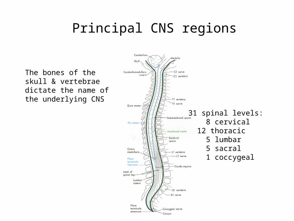
31 spinal levels: 8 cervical 12 thoracic 5 lumbar 5 sacral 1 coccygeal
The bones of the skull & vertebrae dictate the name of the underlying CNS
Principal CNS regions

Frontal Parietal
Temporal
Occipital
Principal CNS regions

These named structures provide critical reference points for learning region-specific function as well as localizing a diagnosis…
…they also permit a significant expansion of the cortical surface area.
The paired cerebral cortices are covered by named sulci and gyri.
Telencephalon

• Midbrain• Pons• Medulla
Brainstem

Diencephalon, Brainstem & Cerebellum

15

CNS Structures
• Lobes• Cortex• Ventricles• Subcortical structures• Brainstem• Spinal Cord

Central Nervous System– CNS: brain and spinal cord– Necessary for the maintenance of homeostasis– Contains 1011 neurons– Contains 1014 synapses– Responsible for everything we perceive, do, feel,
and think

Histology of neural tissue
Two types of neural cells in the nervous system:
Neurons - For processing, transfer, and storage of information
Neuroglia – For support, regulation & protection of neurons

Glial Cells– 90% of CNS composed of glia– Five types of glial cells
• Astrocyte—numerous functions• Ependymal cells—line cavities• Microglia—phagocytes• Oligodendrocytes—form myelin• Schwann cells (located in PNS)—form myelin

Glial Cells

Astrocytes– Development of neural connections– Possibly modulate synaptic activity– Remove neurotransmitter from synaptic cleft– Communicate to neurons through chemical
messengers– Protect neurons against toxic substances
and oxidative stress
Microglia– Protect CNS from foreign matter through
phagocytosis• Bacteria• Dead or injured cells
– Protect CNS from oxidative stress

CNS: Physical Support

Cerebrospinal Fluid (CSF)– Extracellular fluid of the CNS– Secreted by ependymal cells of the
choroid plexus• Circulates to subarachnoid space and ventricles• Reabsorbed by arachnoid villi
– Functions• Cushions brain • Maintains stable interstitial fluid environment

Cerebral Spinal Fluid

CSF Production– Total volume of CSF = 125–150 mL– Choroid plexus produces 400–500 mL/day– Recycled three times a day

Blood Supply to the CNS– CNS comprises 2% of body weight (3–4 pounds)
• Receives 15% of blood supply
– High metabolic rate• Brain uses 20% of oxygen consumed by body
at rest• Brain uses 50% of glucose consumed by body
at rest
– Depends on blood flow for energy

CNS: Gray and White Matter

White Matter in Brain
– Projection fibers• Cerebral cortex with lower levels of brain or spinal cord
– Association fibers • Connect two areas of cerebral cortex on same
side of brain
– Commissural fibers • Connect same cortical regions on two sides of brain
– Corpus callosum • Primary location of commissural fibers

CNS: Gray and White Matter

Brain

Midbrain
Spinal cordPons
Medulla oblongata
Forebrain
Cerebrum
Thalamus
Hypothalamus
Pituitary gland
Brainstem
Diencephalon
Cerebellum
Corpus callosum
(c) Midsagittal section
Brain: Midsagittal View
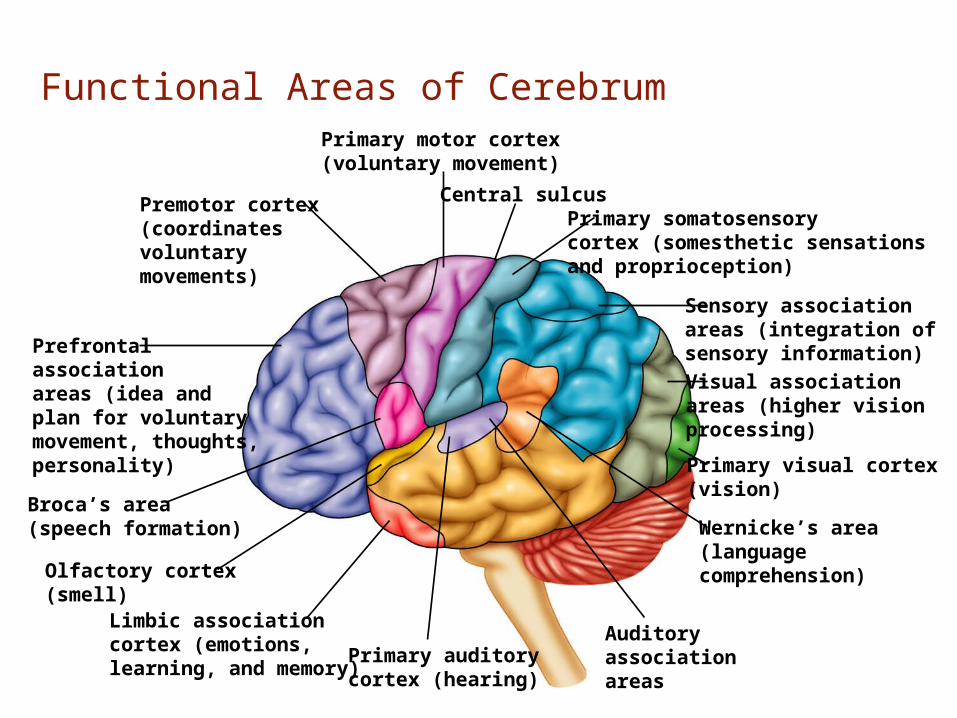
Premotor cortex(coordinatesvoluntarymovements)
Primary somatosensorycortex (somesthetic sensationsand proprioception)
Sensory associationareas (integration ofsensory information)
Primary motor cortex(voluntary movement)
Central sulcus
Prefrontalassociationareas (idea andplan for voluntarymovement, thoughts,personality)
Broca’s area(speech formation)
Limbic associationcortex (emotions,learning, and memory)
Olfactory cortex(smell)
Visual associationareas (higher visionprocessing)
Wernicke’s area(languagecomprehension)
Auditoryassociationareas
Primary auditorycortex (hearing)
Primary visual cortex(vision)
Functional Areas of Cerebrum

Topographical Organization: Motor

Topographical Organization: Sensory

Imaging
• CT• MRI

Advantages of CT
• cost• availability• decreased scan time• decreased sensitivity to patient motion• better visualization of acute blood• better visualization of bony abnormalities

Computerized Tomography
Brain window
Bone window

Advantages of MRI
• no ionizing radiation (peds & pregnant) • direct coronal, sagittal and transverse images• excellent contrast resolution• no interference from bony structures

Axial view

Coronal View

Sagittal view

Questions?