Embed Size (px)
Citation preview
Ramezani et al. / J Zhejiang Univ-Sci A (Appl Phys & Eng) 2020 21(11):859-875
859
Coaxial 3D bioprinting of organ prototyps from nutrients
delivery to vascularization*
Hamed RAMEZANI, Lu-yu ZHOU, Lei SHAO, Yong HE†‡ The State Key Laboratory of Fluid Power and Mechatronic Systems and Key Laboratory of 3D Printing Process and Equipment of
Zhejiang Province, School of Mechanical Engineering, Zhejiang University, Hangzhou 310027, China †E-mail: [email protected]
Received June 8, 2020; Revision accepted Sept. 4, 2020; Crosschecked Oct. 28, 2020
Abstract: Vascular networks inside organs provide the means for metabolic exchange and adequate nutrition. Similarly, vascular or nutrient networks are needed when building tissue constructs >500 μm in vitro due to the hydrogel compact pore size of bioinks. As the hydrogel used in bioinks is rather soft, it is a great challenge to reconstruct effective vascular networks. Recently, coaxial 3D bioprinting was developed to print tissue constructs directly using hollow hydrogel fibers, which can be treated as built-in microchannels for nutrient delivery. Furthermore, vascular networks could be printed directly through coaxial 3D bioprinting. This review summarizes recent advances in coaxial bioprinting for the fabrication of complex vascularized tissue constructs including methods, the effectiveness of varying strategies, and the use of sacrificial bioink. The limitations and challenges of coaxial 3D bioprinting are also summarized.
Key words: 3D bioprinting; Coaxial bioprinting; Vascularization; Bioink https://doi.org/10.1631/jzus.A2000261 CLC number: Q819
1 Why is coaxial 3D bioprinting needed?
Applications with 3D bioprinting to design and manufacture 3D cellular structures for use in trans-plantation therapies are emerging. The unique ad-vantage of this technology is its ability to build 3D structures with bioactive components, such as cells and biocompatible materials (Lee and Yeong, 2016; Mandrycky et al., 2016; He et al., 2019, 2020). The ultimate goal of 3D bioprinting is to produce func-tional living organs for regenerative medicine or or-gan prototypes for drug screening (Ng et al., 2019).
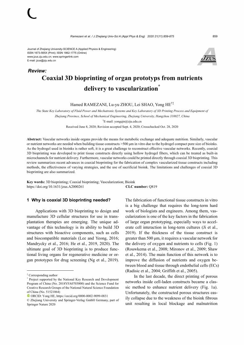
The fabrication of functional tissue constructs in vitro is a big challenge that requires the long-term hard work of biologists and engineers. Among them, vas-cularization is one of the key factors in the fabrication of large organ prototyping, especially ways to accel-erate cell interaction in long-term cultures (Ji et al., 2019). If the thickness of the tissue construct is greater than 500 μm, it requires a vascular network for the delivery of oxygen and nutrients to cells (Fig. 1) (Rouwkema et al., 2008; Mironov et al., 2009; Shaw et al., 2014). The main function of this network is to improve the diffusion of nutrients and oxygen be-tween blood and tissue through endothelial cells (ECs) (Radisic et al., 2004; Griffith et al., 2005).
In the last decade, the direct printing of porous networks inside cell-laden constructs became a clas-sic method to enhance nutrient delivery (Fig. 1a). Unfortunately, the constructed porous structures eas-ily collapse due to the weakness of the bioink fibrous unit resulting in local blockage and malnutrition
Journal of Zhejiang University-SCIENCE A (Applied Physics & Engineering)
ISSN 1673-565X (Print); ISSN 1862-1775 (Online)
www.jzus.zju.edu.cn; www.springerlink.com
E-mail: [email protected]
Review:
‡ Corresponding author
* Project supported by the National Key Research and Development Program of China (No. 2018YFA0703000) and the Science Fund for Creative Research Groups of the National Natural Science Foundation of China (No. 51521064)
ORCID: Yong HE, https://orcid.org/0000-0002-9099-0831 © Zhejiang University and Springer-Verlag GmbH Germany, part of Springer Nature 2020
Ramezani et al. / J Zhejiang Univ-Sci A (Appl Phys & Eng) 2020 21(11):859-875
860
(Kolesky et al., 2014; Murphy and Atala, 2014; Paulsen and Miller, 2015; Cornelissen et al., 2017; Datta et al., 2017; Hann et al., 2019; Xie et al., 2020a, 2020b). To overcome these obstacles, sacrificial bi-oprinting has been used for prototyping vascular or-gan structures. This technique embeds sacrificial ink into a hydrogel matrix, then removes the ink and seeds ECs via perfusing cell suspension into channels (Ji et al., 2019). Several studies have reported the use of sacrificial bioprinting for vascular prototyping. Miller et al. (2012) printed rigid filament networks of carbohydrate glass to be used as a cytocompatible template lined with living ECs. Bertassoni et al. (2014) constructed microchannel networks to vascu-larize tissue using a hydrogel construct from bi-oprinted agarose template fibers, while Lee et al. (2014) fabricated a perfused vascular channel within a collagen scaffold using a 3D bioprinting method.
Nonetheless, the difference in functional capac-ity between printed vascular tissue and natural tissue persists, particularly in fabricated biomimetic
multilayered vascular structures composed of multi-ple materials. Thus, sacrificial bioprinting has dis-advantages in vascular prototyping for the following reasons. It is a slow, multistep process due to the necessity to use a mold for creating the vessel-like channel. It is easy to seed ECs in simple and straight channel structures, however, it is unrealistic to achieve this in complex channels. Complex tissue/ organ constructs are limited in their ability to provide the environment favorable to cells that can promote a duplication of channel morphology and function re-quired of highly vascularized organs (Fig. 1b).
Coaxial bioprinting overcomes the above limi-tations by combining classical and sacrificial bi-oprinting to fabricate biomimetic hollow structures in a single-step process. This approach has a simple setup of complex vascular networks, and provides a feasible strategy for channel endothelialization. It creates complex constructs with vascular networks by co-culturing ECs with multiple cell types (Fig. 1c). Sacrificial inks play a critical role in maximizing the
Fig. 1 Requirements to fabricate vascular organ prototypes by coaxial bioprinting: (a) typical bioprinting; (b) sacrificialbioprinting; coaxial bioprinting (c)
Ramezani et al. / J Zhejiang Univ-Sci A (Appl Phys & Eng) 2020 21(11):859-875
861
advantages of coaxial bioprinting of vascular organs because of the inherent qualities of the ink. It allows for printability with short cross-link periods while retaining requisite mechanical strength. Furthermore, it provides biocompatibility for encapsulating ECs. It also facilitates cooperation with the shell ink for co-bioprinting. The coaxial bioprinting approach achieves nutrient and oxygen supply to cells present in the prototyping vascular structure. Ozbolat et al. (2014) experimented with the fabrication of micro-fluidic channels using alginate via the coaxial nozzle to print hollow tubes. A multi-arm bio-printer was designed to print filament structures and deposit cell spheroids between the filaments to create a hybrid structure that supports the cell spheroids in three di-mensions. The research group of the present review (Gao et al., 2015) developed a coaxial 3D bioprinting method to create hollow filament constructs. This technique includes the concurrent fabrication of both microchannels and scaffolds. The microchannel sys-tem involved allows nutrient delivery for cell growth, while bioink supports cell proliferation. Relevant achievements open up new avenues for vascular or-gan prototyping. Recently, we further developed a novel method to print cell-laden structures with vas-cularized channels directly via coaxial 3D bioprinting (Shao et al., 2020a).
This review focuses on the latest progress, and compares the efficacy of various techniques and bi-omaterials used in coaxial bioprinting for vascular organ prototyping. It attempts to elucidate topics such as: (1) factors that must be considered when bi-oprinting vascular organ prototyping; (2) a list of preferred biomaterials; (3) the principle of fabrication of endothelialized-channels including its potential mechanisms; (4) the most notable achievements stemming from this technology; (5) the next big challenges.
2 Potentially useful biomaterials for coaxial 3D bioprinting
Considering that bioink is a mixture of cells and
biomaterials, the biomaterials most suitable for use in coaxial 3D bioprinting should have the following characteristics (Murphy and Atala, 2014): (i) They can act as an extracellular matrix (ECM). (ii) Degra-
dation behavior could be controlled both in short- and long-term functions (Haycock, 2011). (iii) They have good printability performance, i.e. printed cells are able to maintain proper shapes. The shape fidelity of printed cells is affected by parameters such as sub-stance concentration, surface tension, and shear- thinning behavior. Shear-thinning reduces the shear force required for the flow of material extruded from the printer nozzle. Printing parameters, such as speed and nozzle size, also impact the final constructs (Kyle et al., 2017; Townsend et al., 2019). (iv) The me-chanical strength of a biomaterial must support the continued function of a construct using different crosslinks (Guvendiren et al., 2016). Until recently, this has been the feature absent from most bio-materials. (v) As alluded to above, an immediate crosslink ensures the formation of shape fidelity. Rapid crosslinking allows the biomaterial to retain structural integrity after deposition. It also prepares a suitable environment for the encapsulated cells (Pe-reira and Bártolo, 2015). An appropriate crosslinking mechanism leads to the structural development of an individual layer with strong mechanical properties for stability maintenance.
Hence, hydrogels are often perceived to be suitable biomaterials for the direct printing of vascu-lar constructs by coaxial bioprinting, because they have a structure similar to the ECM with desirable capabilities, such as of nutrient and oxygen diffusion (He et al., 2016; Blaeser et al., 2017; Spang and Christman, 2018). Hydrogels commonly used in co-axial bioprinting are made from natural or synthetic polymers including gelatin, collagen, alginate, and gelatin/methacrylate (GelMA) (Ng et al., 2019). Generally speaking, natural and synthetic polymers should be combined into bioink for better biocom-patibility, crosslinkability, mechanical and thermal properties, and printability.
2.1 Alginate
The properties that make alginate attractive for use as a biomaterial include non-immunogenicity, rapid crosslinkability, low toxicity, and good bio-degradability and biocompatibility (Axpe and Oyen, 2016). Alginate is a naturally derived polymer of β-mannuronic acid (M) and α-guluronic acid (G) (Pawar and Edgar, 2012). The physical properties of alginate hydrogels are determined by the ratio of M to
Ramezani et al. / J Zhejiang Univ-Sci A (Appl Phys & Eng) 2020 21(11):859-875
862
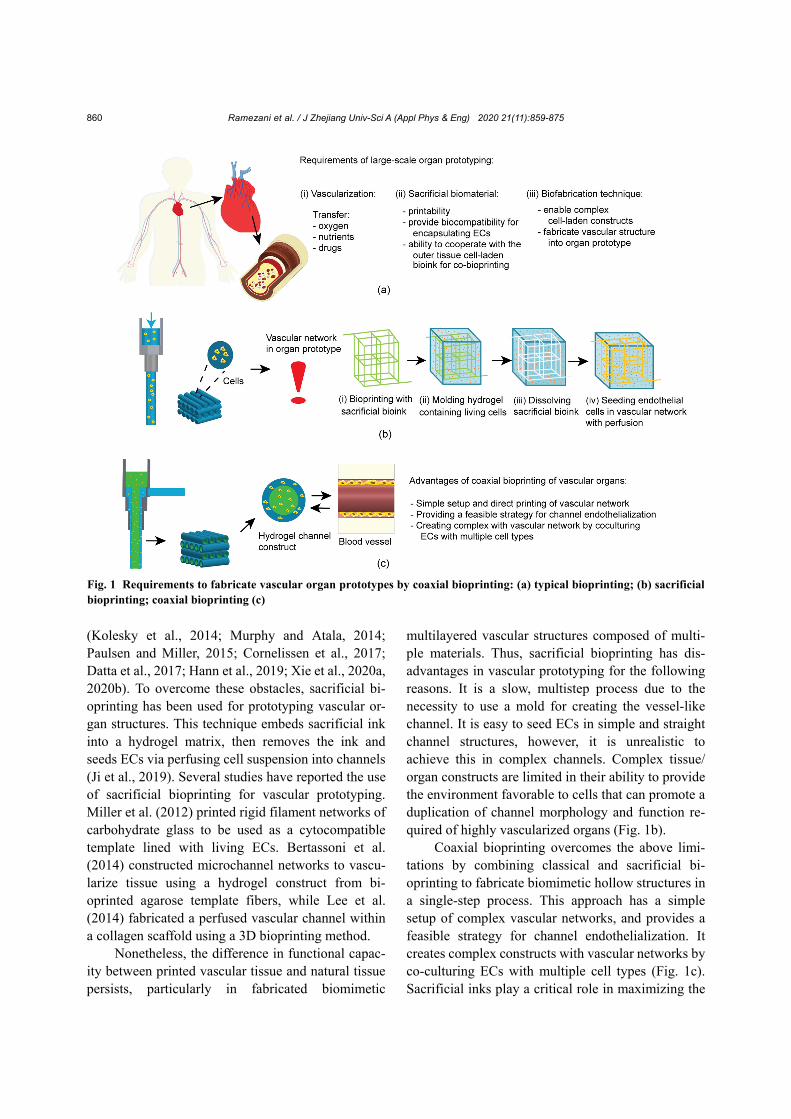
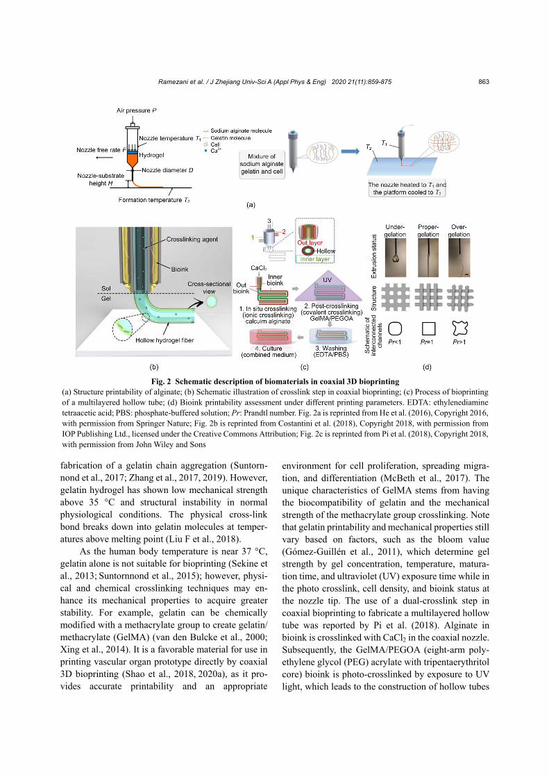
G component blocks. The larger the molecular weight of the blocks, the greater the strength of the alginate. Its physical properties tend to improve with its mo-lecular weight (George and Abraham, 2006). Alginate dissolves in water at ambient temperature to form a hydrogel via intermolecular crosslinking between divalent calcium (Ca2+) ions and G component blocks. The success of gelation depends on alginate concentration and its crosslinker. During 3D bi-oprinting, cells are embedded in alginate hydrogel. Higher concentrations of alginate restrict cell bioac-tivity, while lower concentrations reduce the me-chanical properties of the construct. Thus, the appro-priate alginate concentration must be used for effec-tive vascular organ prototyping. According to Park et al. (2017), an alginate hydrogel of 3% (in weight) with a low to high molecular weight ratio of 1:2 has good printability and provides a suitable environment for cell growth and proliferation. Although alginate has properties similar to those of extracellular matri-ces, it lacks bioactivity. The blend of alginate hydro-gel with other polymers, however, improves cellular activity and printing resolution. To this point, He et al. (2016) concurred regarding the importance of vis-cosity, air pressure, nozzle feed rate, and printing distance between nozzle and substrate, when they related to the successful use of alginate/gelatin printability. The best parameters to print high-quality scaffolds with diffusion and fusion and without damage cells were set in this manner (Fig. 2a). A mix of alginate/gelatin/collagen and human corneal epi-thelial cells (HCECs) can be incubated in a sodium medium to construct a cell-laden tissue with excellent cell viability, as demonstrated by Wu et al. (2016). When Chung et al. (2013) combined alginate with gelatin, they observed enhanced cell growth and im-proved mechanical properties on par with pre- crosslink alginate.
The crosslinking mechanism of bioink-based alginate is an important factor in the coaxial bi-oprinting method. The bioink to be used in coaxial bioprinting must have the proper viscosity to be ex-truded through the nozzle to rapidly stabilize, thus maintain a structure. The pre- and situ-crosslink principles are important in choosing the desirable method. The pre-crosslink approach uses high vis-cosity bioink for rapid extrusion (Aguado et al., 2012;
Unagolla and Jayasuriya, 2020). To achieve this, the flow properties of a solution should be controlled to ensure bio-printability. The number of crosslinking agents is important in material solutions to assure uniformity during extrusion (Hennink and van Nos-trum, 2012; Ouyang et al., 2017). For example, alginate-based bioinks are used in coaxial nozzle bioprinting due to their fast ionic crosslinking ability, which is determined by optimal concentrations of alginate and crosslinker (Onoe et al., 2013; Costantini et al., 2018). Therefore, alginate has the potential to fabricate microfibers and vascularized organs using the core/shell crosslink principle. In this method, the bioink-based alginate and crosslink solutions simul-taneously extrude through the nozzle, with the gela-tion mechanism occurring at the end of the process within the dispensing head. The bulk alginate is con-structed by bioink extruded through the outer nozzle, while the crosslinking solution is pumped from the sheath part of the inner nozzle resulting in an imme-diate crosslink for the fabrication of a hollow vascular- like structure (Costantini et al., 2018) (Fig. 2b).
2.2 Gelatin (GelMA)
Gelatin has been widely used as a preferred bi-oink in coaxial 3D bioprinting (Wang et al., 2017) due to its high biocompatibility, rapid biodegradability, non-immunogenicity, and printing fidelity (Yao et al., 2009; Wang et al., 2013). It is a water-soluble natural polymer obtained from the hydrolysis of the triple helix of collagen into single-strand molecules by chemical pre-treatment followed by heat treatment, whereby non-covalent bonds within collagen are broken and proteins are destabilized altering the helix structure, thus soluble gelatin is formed (Kuijpers et al., 2000; Gómez-Guillén et al., 2011; Liu F et al., 2018). The solid form of gelatin requires dissolution in phosphate-buffered saline or a cell culture medium to form a solution to prepare for printing. Different types of cells or bioactive agents can be used as bio-ink in the gelatin hydrogel (Madl et al., 2016). The behavior of the gelatin solution is dependent on temperature, pH, concentration, and crosslink mech-anism. In sol-gel transitions, for example, when the temperature of a gelatin solution drops below 35 °C, the viscosity increases, which changes the gelatin random coil to a coil helix structure leading to the
Ramezani et al. / J Zhejiang Univ-Sci A (Appl Phys & Eng) 2020 21(11):859-875
863
fabrication of a gelatin chain aggregation (Suntorn-nond et al., 2017; Zhang et al., 2017, 2019). However, gelatin hydrogel has shown low mechanical strength above 35 °C and structural instability in normal physiological conditions. The physical cross-link bond breaks down into gelatin molecules at temper-atures above melting point (Liu F et al., 2018).
As the human body temperature is near 37 °C, gelatin alone is not suitable for bioprinting (Sekine et al., 2013; Suntornnond et al., 2015); however, physi-cal and chemical crosslinking techniques may en-hance its mechanical properties to acquire greater stability. For example, gelatin can be chemically modified with a methacrylate group to create gelatin/ methacrylate (GelMA) (van den Bulcke et al., 2000; Xing et al., 2014). It is a favorable material for use in printing vascular organ prototype directly by coaxial 3D bioprinting (Shao et al., 2018, 2020a), as it pro-vides accurate printability and an appropriate
environment for cell proliferation, spreading migra-tion, and differentiation (McBeth et al., 2017). The unique characteristics of GelMA stems from having the biocompatibility of gelatin and the mechanical strength of the methacrylate group crosslinking. Note that gelatin printability and mechanical properties still vary based on factors, such as the bloom value (Gómez-Guillén et al., 2011), which determine gel strength by gel concentration, temperature, matura-tion time, and ultraviolet (UV) exposure time while in the photo crosslink, cell density, and bioink status at the nozzle tip. The use of a dual-crosslink step in coaxial bioprinting to fabricate a multilayered hollow tube was reported by Pi et al. (2018). Alginate in bioink is crosslinked with CaCl2 in the coaxial nozzle. Subsequently, the GelMA/PEGOA (eight-arm poly-ethylene glycol (PEG) acrylate with tripentaerythritol core) bioink is photo-crosslinked by exposure to UV light, which leads to the construction of hollow tubes
Fig. 2 Schematic description of biomaterials in coaxial 3D bioprinting (a) Structure printability of alginate; (b) Schematic illustration of crosslink step in coaxial bioprinting; (c) Process of bioprinting of a multilayered hollow tube; (d) Bioink printability assessment under different printing parameters. EDTA: ethylenediamine tetraacetic acid; PBS: phosphate-buffered solution; Pr: Prandtl number. Fig. 2a is reprinted from He et al. (2016), Copyright 2016, with permission from Springer Nature; Fig. 2b is reprinted from Costantini et al. (2018), Copyright 2018, with permission fromIOP Publishing Ltd., licensed under the Creative Commons Attribution; Fig. 2c is reprinted from Pi et al. (2018), Copyright 2018, with permission from John Wiley and Sons
Ramezani et al. / J Zhejiang Univ-Sci A (Appl Phys & Eng) 2020 21(11):859-875
864
(Fig. 2c). Ouyang et al. (2016) showed how the properties of gelatin/alginate bioink combine with specific printing parameters to affect the shape fidel-ity of a 3D construct. Bioink printability is evaluated using the status report of the bioink needle (under- gelation, proper-gelation, or over-gelation) to fine- tune parameters to achieve the best possible printing fidelity (Fig. 2d). If gelatin or GelMA proportions or concentration are increased, the viscosity and printa-bility of hybrid GelMA hydrogel is enhanced for use in bioprinting according to van den Bulcke et al. (2000). However, high concentrations of GelMA can reduce cell activity due to its highly cross-linked hydrogel network (Liu et al., 2017). The GelMA hy-drogel enables photopolymerization by the means of a water-soluble photoinitiator and UV-light. The UV exposure dramatically enhances mechanical strength and structural fidelity; the UV light crosslink can improve cell viability of the GelMA hydrogel by approximately 83% with a cell density of 1.5×10⁶ cells/mL (Liu WJ et al., 2018; Das and Basu, 2019).
Additionally, low concentration of GelMA is used appropriately for cell activity in coaxial 3D bi-oprinting, but it requires alginate to provide me-chanical support for the bioink (Liu et al., 2017; Ashammakhi et al., 2019). A type of PEG acrylate having a tripentaerythritol core was embedded into GelMA to enhance alginate hydrogel’s mechanical properties, cell viability, and stability in another study. It was demonstrated that GelMA enables the con-struction of the vascular channel at 37 °C, extending cell life and improving cell function by selecting the appropriate GelMA concentration and UV crosslinks (Ashammakhi et al., 2019). Consequently, alginate and gelatin/GelMA are widely used as biomaterial sources in the process of vascular organ prototyping (Table 1).
2.3 Collagen and other biomaterials
Collagen protein is a major component of the ECM—a key element in the structure of blood vessels. It is a biodegradable, biocompatible substance with low immunogenicity that can improve the adhesion and proliferation of cells on scaffolds (Abraham et al., 2008; Glowacki and Mizuno, 2008; Parenteau-Bareil et al., 2010; Nagel and Kelly, 2013). Collagen mol-ecules share structural similarities RGD (the tripep-
tide Arg-Gly-Asp consists of arginine, glycine, and aspartate peptide) with gelatin having three polypep-tide chains, such as glycine, alanine (Ala), proline, and hydroxyproline, which make up their triple helix structure (Persikov et al., 2005; Liu F et al., 2018). The acid solubility of collagen is affected by hydrogel pH and temperature, and results in limited applica-tions and difficulty with 3D printing. Pure collagen hydrogel demonstrates weak mechanical properties, low viscosity, and a rapid degradation rate (Helary et al., 2010; Liu F et al., 2018). Collagen needs to be embedded in other polymers, such as alginate, fibrin, agarose, and hyaluronic acid, to enhance the proper-ties and printability of collagen for use as bioink (Rücker et al., 2006; Nagel and Kelly, 2013).
For example, the combination of type I collagen with thermo-responsive agarose hydrogel increases the printability and mechanical properties of a pure collagen hydrogel, and achieves improved print con-tours of constructs and good cell viability after 21 d (Duarte Campos et al., 2016). The 3D-bioprinting of fresh collagen hydrogel to fabricate components of the human heart was described by Lee et al. (2019). The pH of collagen hydrogel can be controlled by embedding the collagen self-assembly in a buffered support material and adjusting filament resolution to 20 mm. In order to reduce gelation time, improve mechanical properties, and speed biodegradation, the mechanical properties of collagen hydrogel may be improved by increasing collagen concentration from 12 to 24 mg/mL and combining it with shear-thinning biomaterials such as chitosan hyaluronic acid and fibrin. Improved printability is achieved when colla-gen hydrogel is combined with other hydrogels.
3 Overview of the principal methods of co-axial 3D bioprinting
Coaxial 3D bioprinting has simplified the pro-
cess of directly printing vascular constructs for nu-trient delivery. The most commonly used method involves core-shell flows within a coaxial nozzle. In this approach, one or multiple materials in laminar flow can be used in parallel streams. The multiple phase filaments thus include multiple materials fab-ricated as fiber. These multiple phases have several capillaries connected in a coaxial form. During
Ramezani et al. / J Zhejiang Univ-Sci A (Appl Phys & Eng) 2020 21(11):859-875
865
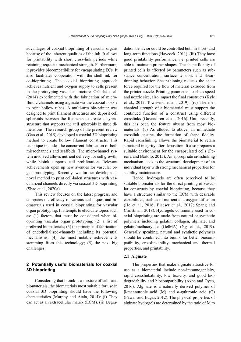
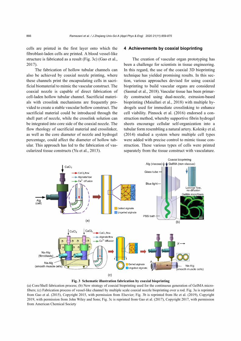
printing, for example, when two materials have been loaded and dispensed individually from inner and outer capillaries via a coaxial nozzle, the structure can be created by the dispensed materials. Therefore, two-phase filaments are achieved by these two mate-rials in coaxial distribution. The utilization of a co-axial nozzle in extrusion-based bioprinting increases the possibility of producing a hollow structure. The coaxial nozzle is fixed on the axis that moves along a pre-planned path. In this approach, if the calcium chloride solution is dispensed from the inner capillary, whereas the alginate solution is delivered from the outer capillary of the coaxial nozzle, the result is the construction of a hollow fiber. The material used in this method must have a rapid crosslinking mecha-nism to impede collapse within the nozzle (Fig. 3a) (Gao et al., 2015). If the bioink is pumped into the inner capillary and the crosslink agent solution to the outer capillary of the nozzle, a single-phase filament is printed. Furthermore, the size of the hollow fiber can be adjusted by controlling pressure (Colosi et al., 2016).
In a different method, the non-viscous GelMA solution was loaded into the internal needle, and a viscous solution containing sodium alginate to the external needle. Due to a low Reynolds number, these materials created laminar flow in the transparent
capillary channel. The crosslinking mechanism was the blue light created by the GelMA fiber as the standard product (Fig. 3b) (Shao et al., 2019). Mi-crofluidic bioprinting using a coaxial nozzle is an-other strategy to create micro-fibrous constructs, where GelMA/alginate is printed through a core/sheath coaxial nozzle. This coaxial nozzle, which is assembled in extrusion bioprinting, is stable and concentric, leading to a continuous generation of hollow microfibers. In this method, alginate can be crosslinked with CaCl2 and GelMA bioink in an al-ginate sheath with a form of in situ gelation, and photo-crosslinked with UV light. Printing can be improved if the bioink extrusion rate is matched with nozzle speed (Liu WJ et al., 2018).
In another approach, bioink is extruded by two coaxial nozzles to print a hollow filament in a rotating rod temple. As bioink from the outer needle contains alginate, a crosslink solution is extruded from the inner needle. The flow rate of both solutions is the same, resulting in a hollow filament twined over a rod. This hollow alginate filament is partially attached to the crosslink-loaded fibroblasts and smooth muscle cells via the use of the coaxial nozzle rolling process. Concurrently, ECs are seeded in the inner wall. In this formation, multilevel fluidic channels with multiple layers of cells are fabricated, whereby smooth muscle
Table 1 Biomaterials used in coaxial bioprinting
Bioink composition Cell type Crosslink mechanism Bioprinting technique
Reference
GelMA-Alginate HVECs, MDAMB-231, MCF7 breast cancer cells, NIH/3T3 mouse fibroblast
CaCl2 and UV crosslink Coaxial extrusion bioprinting
Liu WJ et al., 2018
GelMA-alginate, 4-arm PEGTA
HUVECs and MSCs Ca2+ ion covalent and UV photo crosslinking
Coaxial extrusion bioprinting
Jia et al., 2016
Alginate Bovine cartilage progenitor cells (CPCs)
2%–5% CaCl2 solution Coaxial nozzle in single arm robotic printer
Yu et al., 2013; Zhang et al., 2013
Alginate/ PEG-fibrinogen
HUVECs/iPSC-CMs CaCl2 and UV crosslink Coaxial extrusion bioprinting
Maiullari et al., 2018
Alginate/GelMA/ PEGTA
HUVECs/hMSCs Calcium ions Coaxial extrusion bioprinting
Wu et al., 2016
Alginate/GelMA/PEG HUVECs/HBdSMCs/HUCs/ hMSCs
In situ crosslink: CaCl2 post crosslink: UV exposure
Coaxial extrusion bioprinting
Pi et al., 2018
GelMA/Gelatin Osteoblast, human umbilical vein endothelial cells
Photo-crosslinking mechanism Coaxial extrusion bioprinting
Shao et al., 2020b
Note: PEGTA: poly(ethylene glycol)-tetra-acrylate; HUVECs: human umbilical vein endothelial cells; iPSC-CMs: induced pluripotent stem cell-derived cardiomyocytes; hMSCs: human mesenchymal stem cells; HBdSMCs: human bladder smooth muscle cells; HUCs: human urothelial cells
Ramezani et al. / J Zhejiang Univ-Sci A (Appl Phys & Eng) 2020 21(11):859-875
866
cells are printed in the first layer onto which the fibroblast-laden cells are printed. A blood vessel-like structure is fabricated as a result (Fig. 3c) (Gao et al., 2017).
The fabrication of hollow tubular channels can also be achieved by coaxial nozzle printing, where these channels print the encapsulating cells in sacri-ficial biomaterial to mimic the vascular construct. The coaxial nozzle is capable of direct fabrication of cell-laden hollow tubular channel. Sacrificial materi-als with crosslink mechanisms are frequently pro-vided to create a stable vascular hollow construct. The sacrificial material could be introduced through the shell part of nozzle, while the crosslink solution can be integrated into core side of the coaxial nozzle. The flow rheology of sacrificial material and crosslinker, as well as the core diameter of nozzle and hydrogel percentage, could affect the diameter of hollow tub-ular. This approach has led to the fabrication of vas-cularized tissue constructs (Yu et al., 2013).
4 Achievements by coaxial bioprinting
The creation of vascular organ prototyping has been a challenge for scientists in tissue engineering. In this regard, the use of the coaxial 3D bioprinting technique has yielded promising results. In this sec-tion, various approaches devised for using coaxial bioprinting to build vascular organs are considered (Sasmal et al., 2018). Vascular tissue has been primar-ily constructed using dual-nozzle, extrusion-based bioprinting (Maiullari et al., 2018) with multiple hy-drogels used for immediate crosslinking to enhance cell viability. Pinnock et al. (2016) endorsed a con-struction method, whereby supportive fibrin hydrogel sheets encourage cellular self-organization into a tubular form resembling a natural artery. Kolesky et al. (2014) studied a system where multiple cell types were added with precise control to mimic tissue con-struction. These various types of cells were printed separately from the tissue construct with vasculature.
Fig. 3 Schematic illustration fabrication by coaxial bioprinting (a) Core/Shell fabrication process; (b) New strategy of coaxial bioprinting used for the continuous generation of GelMA micro-fibers; (c) Fabrication process of vessel-like channel by multiple scale coaxial nozzle bioprinting over a rod. Fig. 3a is reprinted from Gao et al. (2015), Copyright 2015, with permission from Elsevier; Fig. 3b is reprinted from He et al. (2019), Copyright 2019, with permission from John Wiley and Sons; Fig. 3c is reprinted from Gao et al. (2017), Copyright 2017, with permission from American Chemical Society
Ramezani et al. / J Zhejiang Univ-Sci A (Appl Phys & Eng) 2020 21(11):859-875
867
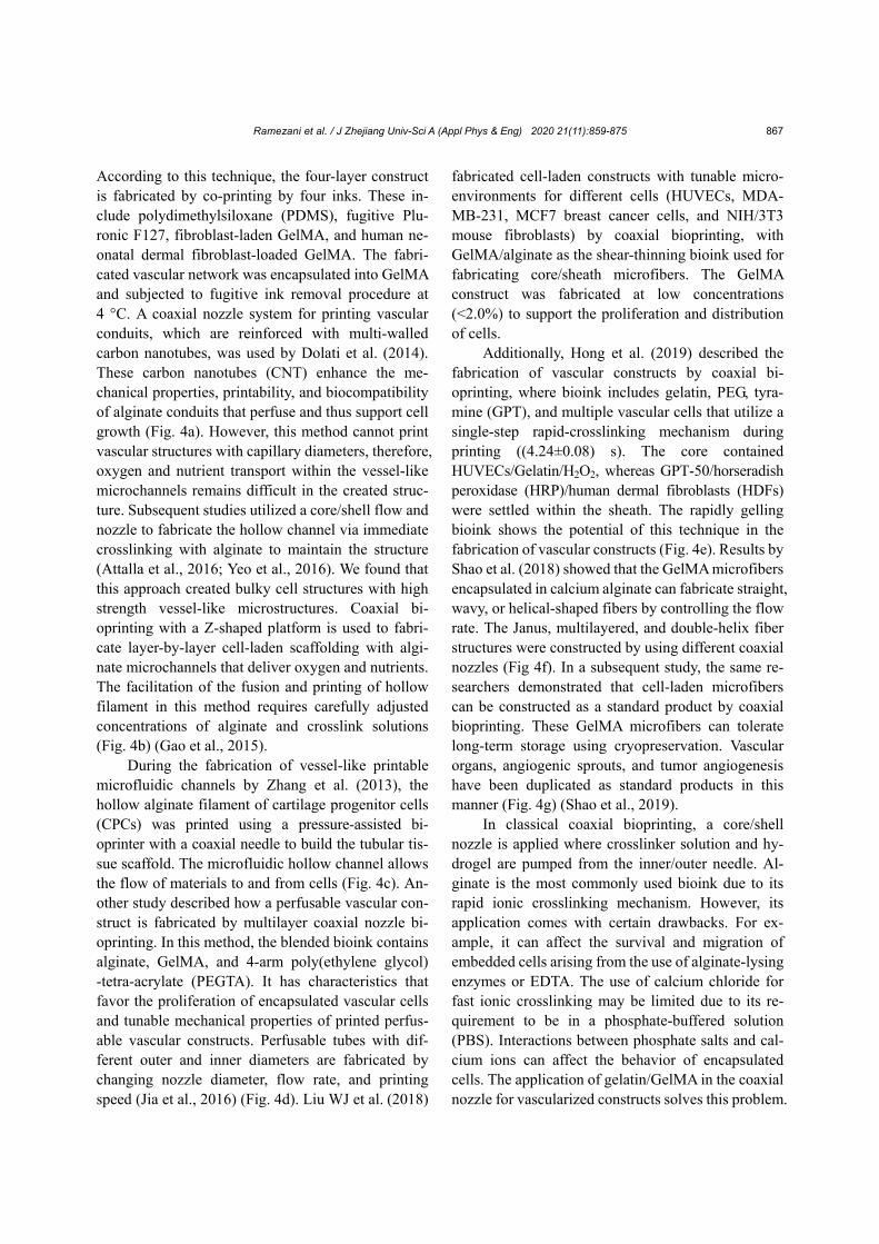
According to this technique, the four-layer construct is fabricated by co-printing by four inks. These in-clude polydimethylsiloxane (PDMS), fugitive Plu-ronic F127, fibroblast-laden GelMA, and human ne-onatal dermal fibroblast-loaded GelMA. The fabri-cated vascular network was encapsulated into GelMA and subjected to fugitive ink removal procedure at 4 °C. A coaxial nozzle system for printing vascular conduits, which are reinforced with multi-walled carbon nanotubes, was used by Dolati et al. (2014). These carbon nanotubes (CNT) enhance the me-chanical properties, printability, and biocompatibility of alginate conduits that perfuse and thus support cell growth (Fig. 4a). However, this method cannot print vascular structures with capillary diameters, therefore, oxygen and nutrient transport within the vessel-like microchannels remains difficult in the created struc-ture. Subsequent studies utilized a core/shell flow and nozzle to fabricate the hollow channel via immediate crosslinking with alginate to maintain the structure (Attalla et al., 2016; Yeo et al., 2016). We found that this approach created bulky cell structures with high strength vessel-like microstructures. Coaxial bi-oprinting with a Z-shaped platform is used to fabri-cate layer-by-layer cell-laden scaffolding with algi-nate microchannels that deliver oxygen and nutrients. The facilitation of the fusion and printing of hollow filament in this method requires carefully adjusted concentrations of alginate and crosslink solutions (Fig. 4b) (Gao et al., 2015).
During the fabrication of vessel-like printable microfluidic channels by Zhang et al. (2013), the hollow alginate filament of cartilage progenitor cells (CPCs) was printed using a pressure-assisted bi-oprinter with a coaxial needle to build the tubular tis-sue scaffold. The microfluidic hollow channel allows the flow of materials to and from cells (Fig. 4c). An-other study described how a perfusable vascular con-struct is fabricated by multilayer coaxial nozzle bi-oprinting. In this method, the blended bioink contains alginate, GelMA, and 4-arm poly(ethylene glycol) -tetra-acrylate (PEGTA). It has characteristics that favor the proliferation of encapsulated vascular cells and tunable mechanical properties of printed perfus-able vascular constructs. Perfusable tubes with dif-ferent outer and inner diameters are fabricated by changing nozzle diameter, flow rate, and printing speed (Jia et al., 2016) (Fig. 4d). Liu WJ et al. (2018)
fabricated cell-laden constructs with tunable micro-environments for different cells (HUVECs, MDA- MB-231, MCF7 breast cancer cells, and NIH/3T3 mouse fibroblasts) by coaxial bioprinting, with GelMA/alginate as the shear-thinning bioink used for fabricating core/sheath microfibers. The GelMA construct was fabricated at low concentrations (<2.0%) to support the proliferation and distribution of cells.
Additionally, Hong et al. (2019) described the fabrication of vascular constructs by coaxial bi-oprinting, where bioink includes gelatin, PEG, tyra-mine (GPT), and multiple vascular cells that utilize a single-step rapid-crosslinking mechanism during printing ((4.24±0.08) s). The core contained HUVECs/Gelatin/H2O2, whereas GPT-50/horseradish peroxidase (HRP)/human dermal fibroblasts (HDFs) were settled within the sheath. The rapidly gelling bioink shows the potential of this technique in the fabrication of vascular constructs (Fig. 4e). Results by Shao et al. (2018) showed that the GelMA microfibers encapsulated in calcium alginate can fabricate straight, wavy, or helical-shaped fibers by controlling the flow rate. The Janus, multilayered, and double-helix fiber structures were constructed by using different coaxial nozzles (Fig 4f). In a subsequent study, the same re-searchers demonstrated that cell-laden microfibers can be constructed as a standard product by coaxial bioprinting. These GelMA microfibers can tolerate long-term storage using cryopreservation. Vascular organs, angiogenic sprouts, and tumor angiogenesis have been duplicated as standard products in this manner (Fig. 4g) (Shao et al., 2019).
In classical coaxial bioprinting, a core/shell nozzle is applied where crosslinker solution and hy-drogel are pumped from the inner/outer needle. Al-ginate is the most commonly used bioink due to its rapid ionic crosslinking mechanism. However, its application comes with certain drawbacks. For ex-ample, it can affect the survival and migration of embedded cells arising from the use of alginate-lysing enzymes or EDTA. The use of calcium chloride for fast ionic crosslinking may be limited due to its re-quirement to be in a phosphate-buffered solution (PBS). Interactions between phosphate salts and cal-cium ions can affect the behavior of encapsulated cells. The application of gelatin/GelMA in the coaxial nozzle for vascularized constructs solves this problem.
Ramezani et al. / J Zhejiang Univ-Sci A (Appl Phys & Eng) 2020 21(11):859-875
868
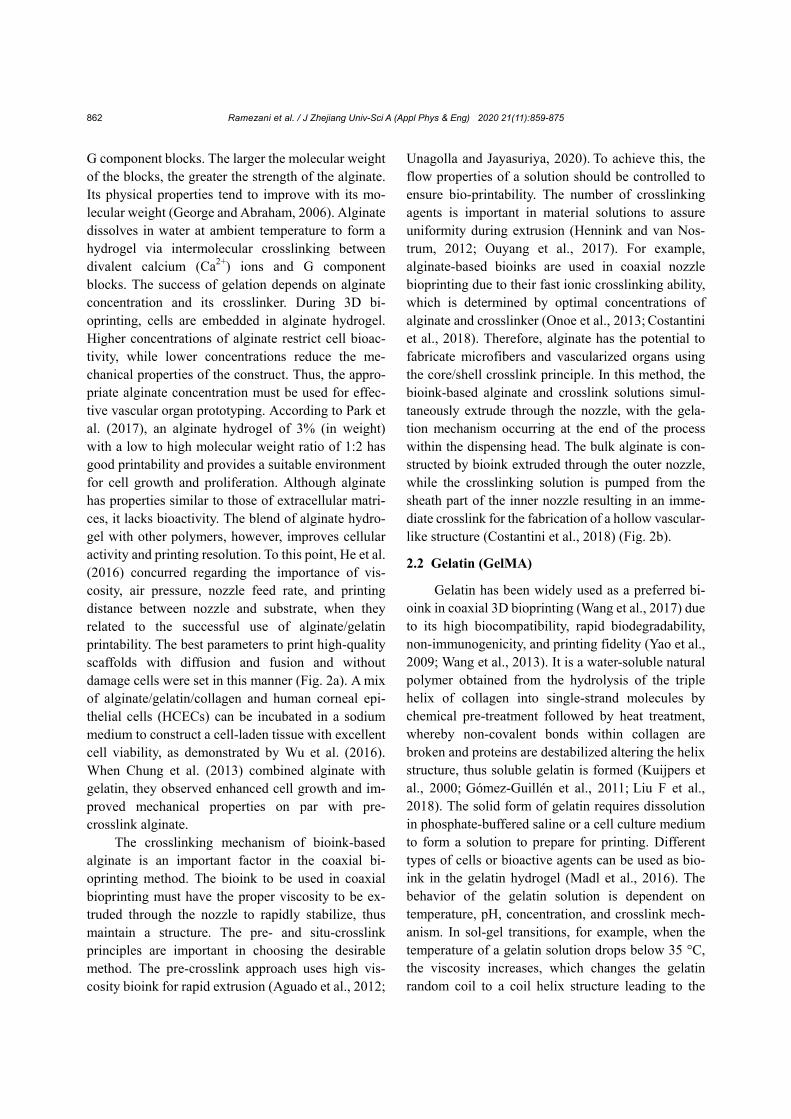
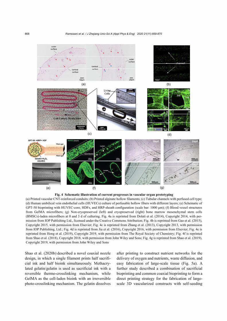
Shao et al. (2020b) described a novel coaxial nozzle design, in which a single filament prints half sacrifi-cial ink and half bioink simultaneously. Methacry-lated gelatin/gelatin is used as sacrificial ink with a reversible thermo-crosslinking mechanism, while GelMA as the cell-laden bioink with an irreversible photo-crosslinking mechanism. The gelatin dissolves
after printing to construct nutrient networks for the delivery of oxygen and nutrients, waste diffusion, and easy fabrication of large-scale tissue (Fig. 5a). A further study described a combination of sacrificial bioprinting and common coaxial bioprinting to form a direct printing strategy for the fabrication of large- scale 3D vascularized constructs with self-seeding
Fig. 4 Schematic illustration of current progresses in vascular organ prototyping (a) Printed vascular CNT-reinforced conduits; (b) Printed alginate hollow filaments; (c) Tubular channels with perfused cell type; (d) Human umbilical vein endothelial cells (HUVECs) culture of perfusable hollow fibers with different layers; (e) Schematic of GPT-50 bioprinting with HUVEC-core, HDFs, and HRP-sheath configuration (scale bar: 1000 μm); (f) Blood vessel structures from GelMA microfibers; (g) Non-cryopreserved (left) and cryopreserved (right) bone marrow mesenchymal stem cells (BMSCs)-laden microfibers at 0 and 3 d of culturing. Fig. 4a is reprinted from Dolati et al. (2014), Copyright 2014, with per-mission from IOP Publishing Ltd., licensed under the Creative Commons Attribution; Fig. 4b is reprinted from Gao et al. (2015), Copyright 2015, with permission from Elsevier; Fig. 4c is reprinted from Zhang et al. (2013), Copyright 2013, with permission from IOP Publishing, Ltd.; Fig. 4d is reprinted from Jia et al. (2016), Copyright 2016, with permission from Elsevier; Fig. 4e is reprinted from Hong et al. (2019), Copyright 2019, with permission from The Royal Society of Chemistry; Fig. 4f is reprinted from Shao et al. (2018), Copyright 2018, with permission from John Wiley and Sons; Fig. 4g is reprinted from Shao et al. (2019), Copyright 2019, with permission from John Wiley and Sons
(a) (b)
(c) (d)
(e) (f) (g)
Ramezani et al. / J Zhejiang Univ-Sci A (Appl Phys & Eng) 2020 21(11):859-875
869
ECs and without perfusion (Shao et al., 2020a). Complex bioprinted tissue constructs and vascular networks are fabricated simultaneously with this ap-proach. The two materials extruded from the same coaxial nozzle can rapidly print vascular constructs without changing the nozzle (Fig. 5b).
In this technique, the GelMA of tissue and ECs are extruded from an outside nozzle, whereas gelatin is present in the inside nozzle resulting in the fabri-cation of core-sheath fibers appropriate for printing large-scale vascularized tissue. ECs will automati-cally settle and adhere to the inner wall of the vascular networks forming the vascular structure. The size of relevant vascular tissue constructs therein was ≥1 cm, which were cultured for 20 d (Fig. 5c).
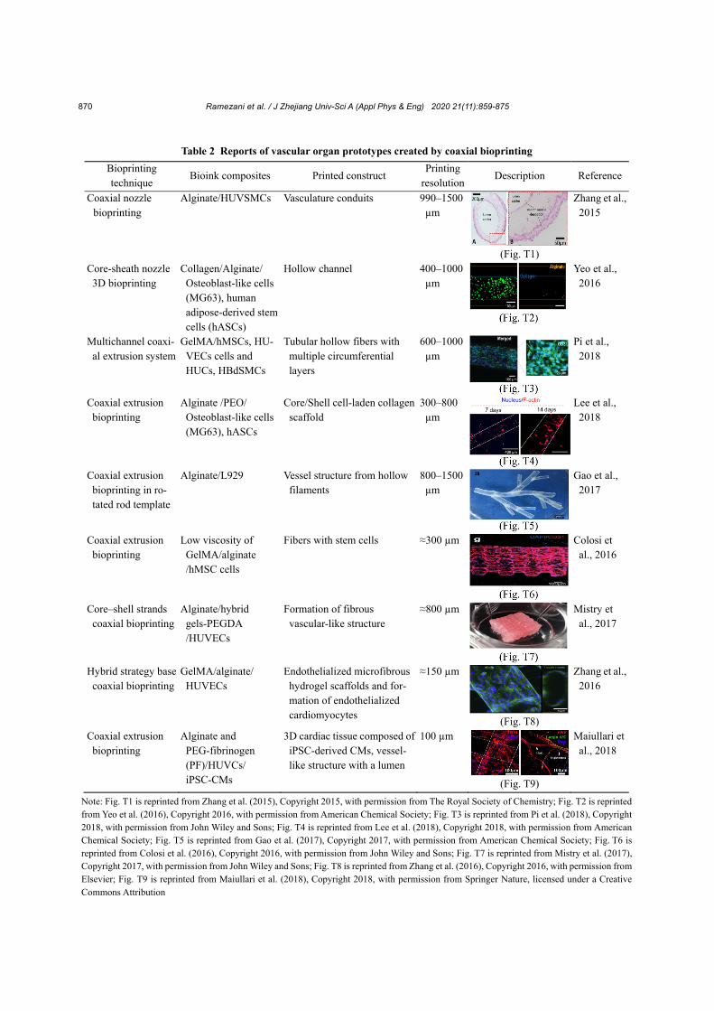
These advances indicate good potential for con-structing large-scale vascular tissues through tissue engineering applications and organ prototyping. Other attempts for prototyping vascular organs via coaxial bioprinting are detailed in Table 2. Coaxial 3D bioprinting has the potential for the construction
of vascular structures within organ prototypes. Novel coaxial bioprinting techniques have utilized bioinks both to support cell viability and to provide the ability of constructing a multilayered vessel-mimicking structure or hollow tubes by extrusion, thus allowing multiple kinds of cells and biomaterial to be cultured in a single-step process.
5 Conclusions and future directions Vascularization is a critical factor for the bio-
fabrication of volumetric tissue. In addition, vascular networks must be prefabricated to promote cell pro-liferation. This is a major impediment to successful tissue engineering and bioprinting. Coaxial 3D bi-oprinting, a novel technique in bioprinting, enables the formation of a directly deposited biomimetic vascular structure, potentially solving the problem of complex vascularized tissue construct fabrication. Classic coaxial bioprinting uses a core/shell nozzle
Fig. 5 3D bioprinting strategy for engineering large-scale tissue constructs with nutrient networks (a); 3D bioprintingmulti-compartmental construct and its vertical sections (b); confocal laser scanning microscopy (CLSM) images of thevascularized osteogenic tissue constructs (c) Fig. 5a is reprinted from Shao et al. (2020b), Copyright 2020, with permission from John Wiley and Sons; Figs. 5b and 5c arereprinted from Shao et al. (2020a), Copyright 2020, with permission from IOP Publishing, Ltd.
Ramezani et al. / J Zhejiang Univ-Sci A (Appl Phys & Eng) 2020 21(11):859-875
870
Table 2 Reports of vascular organ prototypes created by coaxial bioprinting
Bioprinting technique
Bioink composites Printed construct Printing
resolutionDescription Reference
Coaxial nozzle bioprinting
Alginate/HUVSMCs Vasculature conduits 990–1500 µm
(Fig. T1)
Zhang et al.,2015
Core-sheath nozzle 3D bioprinting
Collagen/Alginate/ Osteoblast-like cells (MG63), human adipose-derived stem cells (hASCs)
Hollow channel 400–1000 µm
Yeo et al., 2016
Multichannel coaxi-al extrusion system
GelMA/hMSCs, HU-VECs cells and HUCs, HBdSMCs
Tubular hollow fibers with multiple circumferential layers
600–1000 µm
Pi et al., 2018
Coaxial extrusion bioprinting
Alginate /PEO/ Osteoblast-like cells (MG63), hASCs
Core/Shell cell-laden collagen scaffold
300–800 µm
Lee et al., 2018
Coaxial extrusion bioprinting in ro-tated rod template
Alginate/L929 Vessel structure from hollow filaments
800–1500 µm
Gao et al., 2017
Coaxial extrusion bioprinting
Low viscosity of GelMA/alginate /hMSC cells
Fibers with stem cells ≈300 µm
Colosi et al., 2016
Core–shell strands coaxial bioprinting
Alginate/hybrid gels-PEGDA /HUVECs
Formation of fibrous vascular-like structure
≈800 µm
Mistry et al., 2017
Hybrid strategy base coaxial bioprinting
GelMA/alginate/ HUVECs
Endothelialized microfibrous hydrogel scaffolds and for-mation of endothelialized cardiomyocytes
≈150 µm
(Fig. T8)
Zhang et al.,2016
Coaxial extrusion bioprinting
Alginate and PEG-fibrinogen (PF)/HUVCs/ iPSC-CMs
3D cardiac tissue composed ofiPSC-derived CMs, vessel- like structure with a lumen
100 µm
(Fig. T9)
Maiullari et al., 2018
Note: Fig. T1 is reprinted from Zhang et al. (2015), Copyright 2015, with permission from The Royal Society of Chemistry; Fig. T2 is reprinted from Yeo et al. (2016), Copyright 2016, with permission from American Chemical Society; Fig. T3 is reprinted from Pi et al. (2018), Copyright 2018, with permission from John Wiley and Sons; Fig. T4 is reprinted from Lee et al. (2018), Copyright 2018, with permission from American Chemical Society; Fig. T5 is reprinted from Gao et al. (2017), Copyright 2017, with permission from American Chemical Society; Fig. T6 is reprinted from Colosi et al. (2016), Copyright 2016, with permission from John Wiley and Sons; Fig. T7 is reprinted from Mistry et al. (2017), Copyright 2017, with permission from John Wiley and Sons; Fig. T8 is reprinted from Zhang et al. (2016), Copyright 2016, with permission fromElsevier; Fig. T9 is reprinted from Maiullari et al. (2018), Copyright 2018, with permission from Springer Nature, licensed under a Creative Commons Attribution
Ramezani et al. / J Zhejiang Univ-Sci A (Appl Phys & Eng) 2020 21(11):859-875
871
(double-layered coaxial nozzle), whereby a cross-linked solution and cell-laden hydrogel are pumped from the inner/outer needles. A multi-layer coaxial nozzle can be used to attain a more biomimetic mul-tilayered vascular structure. Because of the popularity of the rapid ionic crosslink mechanism, alginate- based hydrogels are the most commonly used hy-drogel for encapsulating cells; however, the presence of alginate inevitably affects the behavior of encap-sulated cells. Thus, novel bioink combinations need to be developed to promote cell functionalization. Cur-rently, gelatin/GelMA is the optimal core/shell hy-drogel candidate for prototyping vascularized tissue constructs due to their superior biological performance and photo-/thermo-crosslinking mechanisms.
Looking forward, prototyping vascularized tis-sue constructs involves two crucial factors: (i) a fea-sible strategy for channel endothelialization and (ii) perfusable channels and extended perfusion cultures for angiogenesis. The new form of coaxial bioprinting that supports self-seeding ECs is promising, although perfusion culturing must progress further to promote angiogenesis. Subsequently, bioprinted tissues can slowly vascularize via angiogenesis for biomedical applications in vitro. Future advances may include the introduction of nerve cells for producing tissue con-structs with both vascular and neural functional capacity.
Nevertheless, limitations to the current approach of coaxial 3D bioprinting exist. Creating branched vascular structures in different ranges is challenging. The walls of blood vessels contain several layers of proteins and cells; they branch to form an intricate system throughout the body. The physiological func-tion of a blood vessel determines the number of layers and thickness of the vessel. Therefore, one goal of coaxial bioprinting is the successful fabrication of a branched vascular network capable of angiogenesis. A further limitation of coaxial 3D bioprinting is the inability to print high length vascular networks. Hence, it is important to foster coaxial bioprinting approaches that are most likely to produce large di-ameter constructs retaining shape fidelity without shrinkage during the printing process. Furthermore, it is difficult to bioprint submicron-sized capillaries with current coaxial bioprinting techniques. In order to keep pace with patient demand and ever-expanding clinical needs, we expect an increasing focus on co-
axial 3D bioprinting applications for the rapid pro-duction of vascularized tissue. Therefore, the goal of future efforts could be to print microvascular net-works concurrent with other large tissues.
Contributors
Hamed RAMEZANI and Yong HE designed the outline of this review. Hamed RAMEZANI wrote the first draft of the manuscript. Lu-yu ZHOU and Lei SHAO helped to organize the manuscript. Yong HE revised and edited the final version.
Conflict of interest
Hamed RAMEZANI, Lu-yu ZHOU, Lei SHAO, and Yong HE declare that they have no conflict of interest.
Reference Abraham LC, Zuena E, Perez-Ramirez, et al., 2008. Guide to
collagen characterization for biomaterial studies. Journal of Biomedical Materials Research-Part B Applied Biomaterials, 87B(1):264-285. https://doi.org/10.1002/jbm.b.31078
Aguado BA, Mulyasasmita W, Su J, et al., 2012. Improving viability of stem cells during syringe needle flow through the design of hydrogel cell carriers. Tissue Engineering Part A, 18(7-8):806-815. https://doi.org/10.1089/ten.tea.2011.0391
Ashammakhi N, Ahadian S, Xu C, et al., 2019. Bioinks and bioprinting technologies to make heterogeneous and biomimetic tissue constructs. Materials Today Bio, 1:100008. https://doi.org/10.1016/j.mtbio.2019.100008
Attalla R, Ling C, Selvaganapathy P, 2016. Fabrication and characterization of gels with integrated channels using 3D printing with microfluidic nozzle for tissue engineering applications. Biomedical Microdevices, 18(1):17. https://doi.org/10.1007/s10544-016-0042-6
Axpe E, Oyen ML, 2016. Applications of alginate-based bioinks in 3D bioprinting. International Journal of Molecular Sciences, 17(12):1976. https://doi.org/10.3390/ijms17121976
Bertassoni LE, Cecconi M, Manoharan V, et al., 2014. Hydrogel bioprinted microchannel networks for vascularization of tissue engineering constructs. Lab on a Chip, 14(13):2202-2211. https://doi.org/10.1039/c4lc00030g
Blaeser A, Duarte Campos DF, Fischer H, 2017. 3D bioprinting of cell-laden hydrogels for advanced tissue engineering. Current Opinion in Biomedical Engineering, 2:58-66. https://doi.org/10.1016/j.cobme.2017.04.003
Chung JHY, Naficy S, Yue ZL, et al., 2013. Bio-ink properties and printability for extrusion printing living cells. Biomaterials Science, 1(7):763-773. https://doi.org/10.1039/c3bm00012e
Ramezani et al. / J Zhejiang Univ-Sci A (Appl Phys & Eng) 2020 21(11):859-875
872
Colosi C, Shin SR, Manoharan V, et al., 2016. Microfluidic bioprinting of heterogeneous 3D tissue constructs using low-viscosity bioink. Advanced Materials, 28(4):677- 684. https://doi.org/10.1002/adma.201503310
Cornelissen DJ, Faulkner-Jones A, Shu WM, 2017. Current developments in 3D bioprinting for tissue engineering. Current Opinion in Biomedical Engineering, 2:76-82. https://doi.org/10.1016/j.cobme.2017.05.004
Costantini M, Colosi C, Świȩszkowski W, et al., 2018. Co-axial wet-spinning in 3D bioprinting: state of the art and future perspective of microfluidic integration. Biofabrication, 11(1):012001. https://doi.org/10.1088/1758-5090/aae605
Das S, Basu B, 2019. An overview of hydrogel-based bioinks for 3D bioprinting of soft tissues. Journal of the Indian Institute of Science, 99(3):405-428. https://doi.org/10.1007/s41745-019-00129-5
Datta P, Ayan B, Ozbolat IT, 2017. Bioprinting for vascular and vascularized tissue biofabrication. Acta Biomaterialia, 51:1-20. https://doi.org/10.1016/j.actbio.2017.01.035
Dolati F, Yu Y, Zhang YH, et al., 2014. In vitro evaluation of carbon-nanotube-reinforced bioprintable vascular con-duits. Nanotechnology, 25(14):145101. https://doi.org/10.1088/0957-4484/25/14/145101
Duarte Campos DF, Blaeser A, Buellesbach K, et al., 2016. Bioprinting organotypic hydrogels with improved mes-enchymal stem cell remodeling and mineralization prop-erties for bone tissue engineering. Advanced Healthcare Materials, 5(11):1336-1345. https://doi.org/10.1002/adhm.201501033
Gao Q, He Y, Fu JZ, et al., 2015. Coaxial nozzle-assisted 3D bioprinting with built-in microchannels for nutrients delivery. Biomaterials, 61:203-215. https://doi.org/10.1016/j.biomaterials.2015.05.031
Gao Q, Liu ZJ, Lin ZW, et al., 2017. 3D bioprinting of vessel-like structures with multilevel fluidic channels. ACS Biomaterials Science & Engineering, 3(3):399-408. https://doi.org/10.1021/acsbiomaterials.6b00643
George M, Abraham TE, 2006. Polyionic hydrocolloids for the intestinal delivery of protein drugs: alginate and chitosan —a review. Journal of Controlled Release, 114(1):1-14. https://doi.org/10.1016/j.jconrel.2006.04.017
Glowacki J, Mizuno S, 2008. Collagen scaffolds for tissue engineering. Biopolymers, 89(5):338-344. https://doi.org/10.1002/bip.20871
Gómez-Guillén MC, Giménez B, López-Caballero ME, et al., 2011. Functional and bioactive properties of collagen and gelatin from alternative sources: a review. Food Hydro-colloids, 25(8):1813-1827. https://doi.org/10.1016/j.foodhyd.2011.02.007
Griffith CK, Miller C, Sainson RCA, et al., 2005. Diffusion limits of an in vitro thick prevascularized tissue. Tissue Engineering, 11(1-2):257-266. https://doi.org/10.1089/ten.2005.11.257
Guvendiren M, Molde J, Soares RMD, et al., 2016. Designing biomaterials for 3D printing. ACS Biomaterials Science & Engineering, 2(10):1679-1693. https://doi.org/10.1021/acsbiomaterials.6b00121
Hann SY, Cui HT, Esworthy T, et al., 2019. Recent advances in 3D printing: vascular network for tissue and organ regeneration. Translational Research, 211:46-63. https://doi.org/10.1016/j.trsl.2019.04.002
Haycock JW, 2011. 3D cell culture: a review of current approaches and techniques. In: Haycock JW (Ed.), 3D Cell Culture. Humana Press, New York, USA, p.1-15. https://doi.org/10.1007/978-1-60761-984-0_1
He Y, Yang FF, Zhao HM, et al., 2016. Research on the printability of hydrogels in 3D bioprinting. Scientific Reports, 6:29977. https://doi.org/10.1038/srep29977
He Y, Xie M, Gao Q, et al., 2019. Why choose 3D bioprinting? Part I: a brief introduction of 3D bioprinting for the beginners. Bio-Design and Manufacturing, 2:221-224. https://doi.org/10.1007/s42242-019-00053-8
He Y, Gu Z, Xie M, et al., 2020. Why choose 3D bioprinting? Part II: methods and bioprinters. Bio-Design and Manu-facturing, 3:1-4. https://doi.org/10.1007/s42242-020-00064-w
Helary C, Bataille I, Abed A, et al., 2010. Concentrated collagen hydrogels as dermal substitutes. Biomaterials, 31(3):481-490. https://doi.org/10.1016/j.biomaterials.2009.09.073
Hennink WE, van Nostrum CF, 2012. Novel crosslinking methods to design hydrogels. Advanced Drug Delivery Reviews, 64(S1):223-236. https://doi.org/10.1016/j.addr.2012.09.009
Hong S, Kim JS, Jung B, et al., 2019. Coaxial bioprinting of cell-laden vascular constructs using a gelatin-tyramine bioink. Biomaterials Science, 7(11):4578-4587. https://doi.org/10.1039/c8bm00618k
Ji S, Almeida E, Guvendiren M, 2019. 3D bioprinting of complex channels within cell-laden hydrogels. Acta Biomaterialia, 95:214-224. https://doi.org/10.1016/j.actbio.2019.02.038
Jia WT, Gungor-Ozkerim PS, Zhang YS, et al., 2016. Direct 3D bioprinting of perfusable vascular constructs using a blend bioink. Biomaterials, 106:58-68. https://doi.org/10.1016/j.biomaterials.2016.07.038
Kolesky DB, Truby RL, Gladman AS, et al., 2014. 3D bioprinting of vascularized, heterogeneous cell-laden tissue constructs. Advanced Materials, 26(19):3124- 3130. https://doi.org/10.1002/adma.201305506
Kuijpers AJ, van Wachem PB, van Luyn MJA, et al., 2000. In vivo compatibility and degradation of crosslinked gelatin gels incorporated in knitted Dacron. Journal of Biomed-ical Materials Research, 51(1):136-145. https://doi.org/10.1002/(SICI)1097-4636(200007)51:1< 136::AID-JBM18>3.0.CO;2-W
Kyle S, Jessop ZM, Al-Sabah A, et al., 2017. ‘Printability’ of
Ramezani et al. / J Zhejiang Univ-Sci A (Appl Phys & Eng) 2020 21(11):859-875
873
candidate biomaterials for extrusion based 3D printing: state-of-the-art. Advanced Healthcare Materials, 6(16): 1700264. https://doi.org/10.1002/adhm.201700264
Lee A, Hudson AR, Shiwarski DJ, et al., 2019. 3D bioprinting of collagen to rebuild components of the human heart. Science, 365(6452):482-487. https://doi.org/10.1126/science.aav9051
Lee JM, Yeong WY, 2016. Design and printing strategies in 3D bioprinting of cell-hydrogels: a review. Advanced Healthcare Materials, 5(22):2856-2865. https://doi.org/10.1002/adhm.201600435
Lee JY, Koo YW, Kim GH, 2018. Innovative cryopreservation process using a modified core/shell cell-printing with a microfluidic system for cell-laden scaffolds. ACS Applied Materials & Interfaces, 10(11):9257-9268. https://doi.org/10.1021/acsami.7b18360
Lee VK, Kim DY, Ngo H, et al., 2014. Creating perfused functional vascular channels using 3D bio-printing technology. Biomaterials, 35(28):8092-8102. https://doi.org/10.1016/j.biomaterials.2014.05.083
Liu F, Chen QH, Liu C, et al., 2018. Natural polymers for organ 3D bioprinting. Polymers, 10(11):1278. https://doi.org/10.3390/polym10111278
Liu WJ, Heinrich MA, Zhou YX, et al., 2017. Extrusion bioprinting of shear-thinning gelatin methacryloyl bioinks. Advanced Healthcare Materials, 6(12):1601451. https://doi.org/10.1002/adhm.201601451
Liu WJ, Zhong Z, Hu N, et al., 2018. Coaxial extrusion bi-oprinting of 3D microfibrous constructs with cell- favorable gelatin methacryloyl microenvironments. Bio-fabrication, 10(2):024102. https://doi.org/10.1088/1758-5090/aa9d44
Madl CM, Katz LM, Heilshorn SC, 2016. Bio-orthogonally crosslinked, engineered protein hydrogels with tunable mechanics and biochemistry for cell encapsulation. Advanced Functional Materials, 26(21):3612-3620. https://doi.org/10.1002/adfm.201505329
Maiullari F, Costantini M, Milan M, et al., 2018. A multi- cellular 3D bioprinting approach for vascularized heart tissue engineering based on HUVECs and iPSC-derived cardiomyocytes. Scientific Reports, 8(1):13532. https://doi.org/10.1038/s41598-018-31848-x
Mandrycky C, Wang ZJ, Kim K, et al., 2016. 3D bioprinting for engineering complex tissues. Biotechnology Advances, 34(4):422-434. https://doi.org/10.1016/j.biotechadv.2015.12.011
McBeth C, Lauer J, Ottersbach M, et al., 2017. 3D bioprinting of GelMA scaffolds triggers mineral deposition by primary human osteoblasts. Biofabrication, 9(1):015009. https://doi.org/10.1088/1758-5090/aa53bd
Miller JS, Stevens KR, Yang MT, et al., 2012. Rapid casting of patterned vascular networks for perfusable engineered three-dimensional tissues. Nature Materials, 11(9):768- 774. https://doi.org/10.1038/nmat3357
Mironov V, Trusk T, Kasyanov V, et al., 2009. Biofabrication: a 21st Century manufacturing paradigm. Biofabrication, 1(2):022001. https://doi.org/10.1088/1758-5082/1/2/022001
Mistry P, Aied A, Alexander M, et al., 2017. Bioprinting using mechanically robust core–shell cell-laden hydrogel strands. Macromolecular Bioscience, 17(6):1600472. https://doi.org/10.1002/mabi.201600472
Murphy SV, Atala A, 2014. 3D bioprinting of tissues and organs. Nature Biotechnology, 32(8):773-785. https://doi.org/10.1038/nbt.2958
Nagel T, Kelly DJ, 2013. The composition of engineered cartilage at the time of implantation determines the likelihood of regenerating tissue with a normal collagen architecture. Tissue Engineering Part A, 19(7-8):824-833. https://doi.org/10.1089/ten.tea.2012.0363
Ng WL, Chua CK, Shen YF, 2019. Print me an organ! why we are not there yet. Progress in Polymer Science, 97: 101145. https://doi.org/10.1016/j.progpolymsci.2019.101145
Onoe H, Okitsu T, Itou A, et al., 2013. Metre-long cell-laden microfibres exhibit tissue morphologies and functions. Nature Materials, 12(6):584-590. https://doi.org/10.1038/nmat3606
Ouyang LL, Yao R, Zhao Y, et al., 2016. Effect of bioink properties on printability and cell viability for 3D bioplotting of embryonic stem cells. Biofabrication, 8(3): 035020. https://doi.org/10.1088/1758-5090/8/3/035020
Ouyang LL, Highley CB, Sun W, et al., 2017. A generalizable strategy for the 3D bioprinting of hydrogels from non-viscous photo-crosslinkable inks. Advanced Materials, 29(8):1604983. https://doi.org/10.1002/adma.201604983
Ozbolat IT, Chen H, Yu Y, 2014. Development of ‘Multi-arm bioprinter’ for hybrid biofabrication of tissue engineering constructs. Robotics and Computer-Integrated Manu-facturing, 30(3):295-304. https://doi.org/10.1016/j.rcim.2013.10.005
Parenteau-Bareil R, Gauvin R, Berthod F, 2010. Collagen- based biomaterials for tissue engineering applications. Materials, 3(3):1863-1887. https://doi.org/10.3390/ma3031863
Park J, Lee SJ, Chung S, et al., 2017. Cell-laden 3D bioprinting hydrogel matrix depending on different compositions for soft tissue engineering: characterization and evaluation. Materials Science and Engineering: C, 71:678-684. https://doi.org/10.1016/j.msec.2016.10.069
Paulsen SJ, Miller JS, 2015. Tissue vascularization through 3D printing: will technology bring us flow? Developmental Dynamics, 244(5):629-640. https://doi.org/10.1002/dvdy.24254
Pawar SN, Edgar KJ, 2012. Alginate derivatization: a review of chemistry, properties and applications. Biomaterials, 33(11):3279-3305. https://doi.org/10.1016/j.biomaterials.2012.01.007
Ramezani et al. / J Zhejiang Univ-Sci A (Appl Phys & Eng) 2020 21(11):859-875
874
Pereira RF, Bártolo PJ, 2015. 3D bioprinting of photocross-linkable hydrogel constructs. Journal of Applied Polymer Science, 132(48):42458. https://doi.org/10.1002/app.42458
Persikov AV, Ramshaw JAM, Kirkpatrick A, et al., 2005. Electrostatic interactions involving lysine make major contributions to collagen triple-helix stability. Biochem-istry, 44(5):1414-1422. https://doi.org/10.1021/bi048216r
Pi QM, Maharjan S, Yan X, et al., 2018. Digitally tunable microfluidic bioprinting of multilayered cannular tissues. Advanced Materials, 30(43):1706913. https://doi.org/10.1002/adma.201706913
Pinnock CB, Meier EM, Joshi NN, et al., 2016. Customizable engineered blood vessels using 3D printed inserts. Methods, 99:20-27. https://doi.org/10.1016/j.ymeth.2015.12.015
Radisic M, Yang LM, Boublik J, et al., 2004. Medium perfusion enables engineering of compact and contractile cardiac tissue. American Journal of Physiology-Heart and Circulatory Physiology, 286(2):H507-H516. https://doi.org/10.1152/ajpheart.00171.2003
Rouwkema J, Rivron NC, van Blitterswijk CA, 2008. Vascu-larization in tissue engineering. Trends in Biotechnology, 26(8):434-441. https://doi.org/10.1016/j.tibtech.2008.04.009
Rücker M, Laschke MW, Junker D, et al., 2006. Angiogenic and inflammatory response to biodegradable scaffolds in dorsal skinfold chambers of mice. Biomaterials, 27(29): 5027-5038. https://doi.org/10.1016/j.biomaterials.2006.05.033
Sasmal P, Datta P, Wu Y, et al., 2018. 3D bioprinting for modelling vasculature. Microphysiological Systems, 2:9. https://doi.org/10.21037/mps.2018.10.02
Sekine H, Shimizu T, Sakaguchi K, et al., 2013. In vitro fabrication of functional three-dimensional tissues with perfusable blood vessels. Nature Communications, 4:1399. https://doi.org/10.1038/ncomms2406
Shao L, Gao Q, Zhao HM, et al., 2018. Fiber-based mini tissue with morphology-controllable gelma microfibers. Small, 14(44):1802187. https://doi.org/10.1002/smll.201802187
Shao L, Gao Q, Xie CQ, et al., 2019. Bioprinting of cell-laden microfiber: can it become a standard product? Advanced Healthcare Materials, 8(9):1900014. https://doi.org/10.1002/adhm.201900014
Shao L, Gao Q, Xie CQ, et al., 2020a. Directly coaxial 3D bioprinting of large-scale vascularized tissue constructs. Biofabrication, 12(3):035014. https://doi.org/10.1088/1758-5090/ab7e76
Shao L, Gao Q, Xie CQ, et al., 2020b. Synchronous 3D bioprinting of large-scale cell-laden constructs with nutrient networks. Advanced Healthcare Materials, 9(15): 1901142. https://doi.org/10.1002/adhm.201901142
Shaw CJ, Ter Haar GR, Rivens IH, et al., 2014. Pathophysio-
logical mechanisms of high-intensity focused ultrasound- mediated vascular occlusion and relevance to non- invasive fetal surgery. Journal of the Royal Society Interface, 11(95):20140029. https://doi.org/10.1098/rsif.2014.0029
Spang MT, Christman KL, 2018. Extracellular matrix hydrogel therapies: in vivo applications and development. Acta Biomaterialia, 68:1-14. https://doi.org/10.1016/j.actbio.2017.12.019
Suntornnond R, An J, Yeong WY, et al., 2015. Biodegradable polymeric films and membranes processing and forming for tissue engineering. Macromolecular Materials and Engineering, 300(9):858-877. https://doi.org/10.1002/mame.201500028
Suntornnond R, An J, Chua CK, 2017. Bioprinting of thermoresponsive hydrogels for next generation tissue engineering: a review. Macromolecular Materials and Engineering, 302(1):1600266. https://doi.org/10.1002/mame.201600266
Townsend JM, Beck EC, Gehrke SH, et al., 2019. Flow be-havior prior to crosslinking: the need for precursor rhe-ology for placement of hydrogels in medical applications and for 3D bioprinting. Progress in Polymer Science, 91:126-140. https://doi.org/10.1016/j.progpolymsci.2019.01.003
Unagolla JM, Jayasuriya AC, 2020. Hydrogel-based 3D bi-oprinting: a comprehensive review on cell-laden hydro-gels, bioink formulations, and future perspectives. Ap-plied Materials Today, 18:100479. https://doi.org/10.1016/j.apmt.2019.100479
van den Bulcke AI, Bogdanov B, de Rooze N, et al., 2000. Structural and rheological properties of methacrylamide modified gelatin hydrogels. Biomacromolecules, 1(1): 31-38. https://doi.org/10.1021/bm990017d
Wang XH, He K, Zhang WM, 2013. Optimizing the fabrication processes for manufacturing a hybrid hierarchical poly-urethane-cell/hydrogel construct. Journal of Bioactive and Compatible Polymers, 28(4):303-319. https://doi.org/10.1177/0883911513491359
Wang XH, Ao Q, Tian XH, et al., 2017. Gelatin-based hydrogels for organ 3D bioprinting. Polymers, 9(9):401. https://doi.org/10.3390/polym9090401
Wu ZJ, Su X, Xu YY, et al., 2016. Bioprinting three- dimensional cell-laden tissue constructs with controllable degradation. Scientific Reports, 6:24474. https://doi.org/10.1038/srep24474
Xie M, Gao Q, Fu J, et al., 2020a. Bioprinting of novel 3D tumor array chip for drug screening. Bio-Design and Manufacturing, 3:175-188. https://doi.org/10.1007/s42242-020-00078-4
Xie M, Zheng Y, Gao Q, et al., 2020b. Facile 3D cell culture protocol based on photocurable hydrogels. Bio-Design and Manufacturing, in press.
Ramezani et al. / J Zhejiang Univ-Sci A (Appl Phys & Eng) 2020 21(11):859-875
875
https://doi.org/10.1007/s42242-020-00096-2 Xing Q, Yates K, Vogt C, et al., 2014. Increasing mechanical
strength of gelatin hydrogels by divalent metal ion removal. Scientific Reports, 4:4706. https://doi.org/10.1038/srep04706
Yao R, Zhang RJ, Yan YN, et al., 2009. In vitro angiogenesis of 3D tissue engineered adipose tissue. Journal of Bioactive and Compatible Polymers, 24(1):5-24. https://doi.org/10.1177/0883911508099367
Yeo MG, Lee JS, Chun W, et al., 2016. An innovative collagen-based cell-printing method for obtaining human adipose stem cell-laden structures consisting of core- sheath structures for tissue engineering. Biomacromole-cules, 17(4):1365-1375. https://doi.org/10.1021/acs.biomac.5b01764
Yu Y, Zhang YH, Martin JA, et al., 2013. Evaluation of cell viability and functionality in vessel-like bioprintable cell-laden tubular channels. Journal of Biomechanical Engineering, 135(9):091011. https://doi.org/10.1115/1.4024575
Zhang LW, Li GY, Shi M, et al., 2017. Establishment and characterization of an acute model of ocular hypertension by laser-induced occlusion of episcleral veins. Investiga-tive Ophthalmology & Visual Science, 58(10):3879-3886. https://doi.org/10.1167/iovs.16-20807
Zhang Y, Zhou DZ, Chen JW, et al., 2019. Biomaterials based on marine resources for 3D bioprinting applications. Marine Drugs, 17(10):555. https://doi.org/10.3390/md17100555
Zhang YH, Yu Y, Chen H, et al., 2013. Characterization of printable cellular micro-fluidic channels for tissue engineering. Biofabrication, 5(2):025004. https://doi.org/10.1088/1758-5082/5/2/025004
Zhang YH, Yu Y, Akkouch A, et al., 2015. In vitro study of directly bioprinted perfusable vasculature conduits. Biomaterials Science, 3(1):134-143. https://doi.org/10.1039/c4bm00234b
Zhang YS, Arneri A, Bersini S, et al., 2016. Bioprinting 3D microfibrous scaffolds for engineering endothelialized myocardium and heart-on-a-chip. Biomaterials, 110: 45-59. https://doi.org/10.1016/j.biomaterials.2016.09.003
中文概要
题 目:同轴生物 3D 打印器官原型——从营养输送到血
管化
概 要:组织/器官内的血供系统,为组织提供了必要的营
养及代谢交换。而在体外构造组织/器官原型时,
如何在大尺寸结构中构建营养网络,是长期以来
的技术难题。近年来,同轴生物 3D 打印技术为
该问题提供了一种极具潜力的解决方案。同轴生
物 3D 打印技术的基本原理是:使用同轴喷头将
外层的水凝胶材料和内层的牺牲材料共同挤出,
打印为所需的复杂结构,内层的牺牲材料去除后
形成的中空通路即成为后续培养中的营养网络。
该技术结合了传统生物打印方法和牺牲组分 3D
打印方法的优点,能够一步构造内置营养网络的
大尺寸仿生结构,在组织工程和器官重建等领域
具有突出的优势。
本文结合课题组近年围绕同轴生物 3D 打印技术
所做的一些工作,梳理和总结了该技术的最新研
究进展。主要关注以下几点:(1)在同轴 3D 打
印血管时必须考虑的因素;(2)首选生物材料清
单;(3)内皮化通道的制造原理及其潜在机制;
(4)同轴生物 3D 打印技术的最近进展;(5)未
来的挑战。
首先,本文概述了当前生物 3D 打印中常用的水
凝胶材料,包括海藻酸钠(Alginate)、明胶/甲基
丙烯酸酐化明胶( Gelatin/GelMA )和胶原
(Collagen)等,介绍了这些材料的生物相容性、
可打印性和打印原理等生物 3D 打印技术中重点
关注的因素。随后,论文详述了同轴生物 3D 打
印技术的基本原理、技术特点以及使用该技术构
造内含营养网络(特别是血管化)的大尺寸结构
的最新尝试。最后,论文展望了同轴生物 3D 技
术未来可能的发展方向。最新的研究进展表明,
该技术为快速制造血管化的组织/器官原型提供
了可能。
关键词:生物 3D 打印;同轴生物打印;血管化;生物
墨水