Embed Size (px)
Citation preview

Current Opinion in Colloid & Interface Science xxx (2013) xxx–xxx
COCIS-00866; No of Pages 8
Contents lists available at SciVerse ScienceDirect
Current Opinion in Colloid & Interface Science
j ourna l homepage: www.e lsev ie r .com/ locate /coc is
Small-scale structure in fluid cholesterol–lipid bilayers
Maikel C. Rheinstädter a,b, Ole G. Mouritsen c,⁎a Department of Physics and Astronomy, McMaster University, 1280 Main Street West, Hamilton, Ontario L8S 4M1,Canadab The Canadian Neutron Beam Centre, National Research Council of Canada, Chalk River Laboratories, Canadac MEMPHYS—Center for Biomembrane Physics, Department of Physics, Chemistry, and Pharmacy, University of Southern Denmark, 55 Campusvej, DK-5230 Odense M, Denmark
⁎ Corresponding author. Tel.: +45 6550 3528; fax: +E-mail addresses: [email protected] (M.C. R
[email protected] (O.G. Mouritsen).
1359-0294/$ – see front matter © 2013 Elsevier Ltd. Allhttp://dx.doi.org/10.1016/j.cocis.2013.07.001
Please cite this article as: Rheinstädter MC, M(2013), http://dx.doi.org/10.1016/j.cocis.201
a b s t r a c t
a r t i c l e i n f oArticle history:Received 19 June 2013Received in revised form 1 July 2013Accepted 2 July 2013Available online xxxx
Keywords:CholesterolLipid bilayerSmall-scale structureRaftCorrelation functionNeutron scatteringComputer simulation
Cholesterol is the single most abundant molecule in animal plasma membranes, in the range of 20–30 mol%,where it is known to modulate the lipid-bilayer component of the membrane and lead to increased mechan-ical stability, lower permeability, larger thickness, and a distinct lateral organization. The phase equilibria ofmembranes with cholesterol and the associated large- and small-scale structure have turned out to be aparticularly elusive problem. With the proposal that lipid domains and so-called ‘rafts’, characterized byhigh local levels of cholesterol in a liquid-ordered phase, are important for a wide range of cellular functions,an understanding and a quantitative assessment of the nature of these cholesterol-induced structures andtheir types of ordering have become urgent. Recent progress in neutron diffraction studies of lipid–cholesterolmodel membranes has now revealed details of the lateral ordering, and combined with earlier molecularmodel studies a picture emerges of the membrane as a locally structured liquid with small ordered ‘domains’of a highly dynamic nature.
© 2013 Elsevier Ltd. All rights reserved.
1. Introduction
Higher sterols are universally present in large amounts (20–30 mol%)in the plasmamembranes of all eukaryotic cells: cholesterol in animals,ergosterol in yeast and fungi, phytosterols in plants, and, e.g., fucosterolin algae [1]. The early work by Nobel Laureate Konrad Bloch demon-strated that the biochemical pathway to cholesterol required thepresence of molecular oxygen in order to remove a bottleneck betweenthe precursor lanosterol and the streamlining of higher sterols [2]. Bynoting the coincidence in time of the occurrence of eukaryotes andthe increase in oxygen pressure in the Earth's atmosphere, theevolutional path from prokaryotes to eukaryotes could be translatedinto the molecular ‘evolution of a small molecule,’ cholesterol [3].
The issue is thenwhat is so special about cholesterol (or other highersterols) [4]. It is known that some cellular functions have specificrequirements for cholesterol in small amounts [5,6], butwhy do all plas-ma membranes of eukaryotes universally have very large amounts ofcholesterol, in the order of 20–30 mol%, making cholesterol, by anycomparison, the singlemost abundantmolecule in plasmamembranes?Another question is, what is it cholesterol can do which its biochemicalprecursor lanosterol cannot? In his work, Konrad Bloch pointed out theeffectiveness of cholesterol to order fluid lipid bilayers, providing forlow passive permeability and increased mechanical strength [2,7].
45 6550 4048.heinstädter),
rights reserved.
ouritsen OG, Small-scale stru3.07.001
These effects have been demonstrated by a large body of literature fora wide range of model membranes [8].
In most cases biological function requires membranes to be in afluid (or liquid) state in the sense that it has to allow for rapid diffu-sion of the lipid molecules and the imbedded membrane proteins.Membranes in liquid phases are generally thinner and less mechani-cally stable than their solid counterparts, which on the other handdo not allow for rapid diffusion. The cholesterol molecule tends tostabilize the membrane by ordering lipid acyl chains in liquid mem-branes because of its rigid steroid structure and an α-face that ismolecularly smooth in contrast to lanosterol with three bumpymethyl groups decorating its α-face [9]. In solid membranes, choles-terol has the opposite effect due to packing constraints [10]. Soconsidering ordering, cholesterol molecules prefer solid phases,while when it comes to packing, cholesterol prefers liquid phases.This duality in cholesterol's affinity for liquid and solid lipid bilayerphases was cast into a thermodynamic phase diagram in 1987 bythe proposal that cholesterol in large amounts induces a newmesophase, the liquid-ordered (lo) phase, that dominates the phasediagram in the range of physiological relevant concentrations ofcholesterol, as shown in Fig. 1 [11]. By the introduction of this newmesophase, the traditional gel (solid) phase was renamed as thesolid-ordered phase and the traditional liquid-crystalline (fluid)phase was renamed as the liquid-disordered phase. It is noteworthythat the phase diagram has an upper critical point. In the neighbor-hood of this point, critical fluctuations will prevail and the correlationlength describing local dynamic domain formation can get very large[12,13]. The experimental observation of the liquid-ordered phase in
cture in fluid cholesterol–lipid bilayers, Curr Opin Colloid Interface Sci

Fig. 1. Generic phase diagram of phospholipid/cholesterol bilayers, such as DMPC/cholesterol and DPPC/cholesterol, displaying a solid-ordered phase (Pβ′), a liquid-disordered phase (Lα), a liquid-ordered phase (lo), as well as the regions of two-phase co-existence [100]. At high levels of cholesterol, cholesterol separates out as a pure crystallinephase [10].
2 M.C. Rheinstädter, O.G. Mouritsen / Current Opinion in Colloid & Interface Science xxx (2013) xxx–xxx
a cholesterol lipid binary mixture was for the first time reported in alandmark paper by Vist and Davis [14]. The quantitative determinationof binary lipid–cholesterol phase diagrams has turned out to be veryelusive, and only a few cases have been resolved and the same holdstrue for phase diagrams of binary mixtures of lipids with other highersterols, with lipid–ergosterol being a notorious exception [15]. In con-trast, a number of ternary phase diagrams have been determined forsystems involving cholesterol and two different lipid species, usually alipid with a high melting point (e.g., long-chain saturated phospho-lipids) and a lipid with a lowmelting point (e.g., sphingolipids or unsat-urated phospholipids) [16]. The liquid-ordered phase is a unique phasein membranology and no other molecules but the higher sterols havebeen shown to be able to stabilize this type of phase. In this sensecholesterol (and other higher sterols) is something special [4].
The liquid-ordered phase called the attention of the life sciencecommunity (for a recent review, see [17]) in 1997 when Simonsand Ikonen [18] proposed the existence of so-called rafts in biologicalmembranes based on some ideas from an earlier work on lipid sortingin epithelial cells [19]. The early literature in the field of rafts wasmarred by the use of a definition of rafts as a certain detergent-resistant fraction of the membranes, and as pointed out by Heerklotz[20], the detergent is likely to introduce artifacts. The rafts were sup-posed to be small, molecularly organized units providing for somelocal structure in fluid biological membranes and hence furnishing plat-forms for specific biological functions [18,21–29]. These rafts were sup-posed to be enriched in cholesterol making them more ordered andthicker and hence appropriate anchoring places for certain acylatedand/or hydrophobically-matched integral membrane proteins. Thehigh levels of cholesterol in these rafts led to the proposal that raftsare local manifestations of the liquid-ordered phase, although in mostcases the nature of the lipid ordering and the phase state were notestablished neither in cells nor in most model membrane studies.
Whereas the concept of dynamic local structures in disorderedliquid systems, even in one-phase regions, is well known to physicalscientists by terms such as structured fluids and microemulsions,the life-science community generally interpreted rafts as some kindof super-particles floating around in an otherwise structure-lessliquid membrane. However, early work in the physical chemistry oflipid bilayers pointed to the possibility of dynamic heterogeneity[30–33] in thermodynamic one-phase regions of, e.g., binary systems.The source of dynamic heterogeneity is cooperative molecularinteractions and thermal fluctuations that lead to density and/orcompositional fluctuations in space and time. These fluctuations arebest characterized by correlation functions or appropriate structure
Please cite this article as: Rheinstädter MC, Mouritsen OG, Small-scale stru(2013), http://dx.doi.org/10.1016/j.cocis.2013.07.001
factors. From these functions a length scale can be extracted whichis a measure of the range over which the liquid is correlated. This mo-tional coherence was observed experimentally in the fluid phase oflipid membranes [34] using quasi-elastic neutron scattering. Thelength scale associated with this coherence length is probably themost relevant and quantitative description of a domain or a ‘raft’ ina liquid membrane.
While in most cases it is relatively easy to design experiments todetermine large-scale domain structure and global phase separationin coexistence regions [29], it turned out to be much more involvedto perform an experiment to measure the dynamic local structureand the associated correlation length in liquids where transient,nanometer-sized local structures are predicted. This has been partic-ularly challenging for cholesterol-containing lipid membranes in theliquid-ordered phase, which is supposed to harbor the putativerafts. Lack of information on the nature of a possible small-scale struc-ture even in simple liquid membranes with large amounts of choles-terol has had a significant impact on the progress in the scientificunderpinning of the growing field of rafts in biological membranes[24,25,29,35–37]. In Section 2 we shall briefly review some of theexisting experimental data pointing to the existence of lipid domainsand small-scale structures in model membranes. Some insight intosmall-scale structure and dynamics of lipid–cholesterol membranesin the liquid-ordered phase has also been provided by model andcomputer simulation studies which we will review in Section 4.
Recently a break-through has been reported in the use of neutrondiffraction to quantitatively assess small-scale structure in binarylipid–cholesterol membranes in the liquid-ordered phase [38,39].The experiments revealed the existence of highly ordered lipiddomains in equilibrium with a disordered matrix. The lipids in thesedomains were found to be in a liquid-ordered state and thought tobe saturated with cholesterol molecules.
In the present short topical review we shall describe thisbreak-through in Section 3 and in Section 5 discuss the results togetherwith results frommodel studies described in Section 4 and thereby pro-vide a status of small-scale ordered domains in membranes with highlevels of cholesterol. We believe that the combined results from theseexperiments and the model simulations provide a much improved andsolid underpinning for future work on rafts in biological membranes.
2. Lateral structure in fluid model membranes with cholesterol
It is interesting to note that membranes and the question of their in-ternalmolecular organization resemble a ‘colloid-inside-a-colloid’ prob-lem. The lipid bilayer is an about 5 nanometer thin, self-assembledmolecular aggregate, bound together byweak physical (colloidal) forcesthat are strongly renormalized by temperature. The in-plane organiza-tion of this free-standing soft and liquid interface inwater is itself a ques-tion of physical forces that, influenced by the cooperative behavior andmany-bodyness of the assembly, can stabilize dynamic and fluctuatinglocal structures on varying length scales that also are subject to thermalrenormalization [1,40].
The generic cholesterol–lipid binary phase diagram in Fig. 1 containsa region with phase separation between the two liquid phases, theliquid-disordered and the liquid-ordered phase. It has proved very diffi-cult tomeasure the phase boundaries of this region and the nature of thecoexistence region is still in dispute. Whereas there is a large body ofliterature on phase separation and lipid domains in a wide range oflipid membrane systems with cholesterol in cases where solid (gel,solid-ordered) phases are involved [16,41–45], and there is some workon systems involving coexistence between liquid-disordered (fluid,liquid-crystalline) and liquid-ordered phases [15,46–49], only a fewstudies (to be reviewed in Sections 3 and 4) address the question oflocal structure in lipid mixtures with cholesterol being in a single-phase liquid-ordered phase.
cture in fluid cholesterol–lipid bilayers, Curr Opin Colloid Interface Sci

3M.C. Rheinstädter, O.G. Mouritsen / Current Opinion in Colloid & Interface Science xxx (2013) xxx–xxx
It should be pointed out that even if the liquid-ordered phase origi-nallywas proposed to exist in binary lipid–cholesterol systems, only lit-tle conclusive evidence is available of the presence of a macroscopicliquid-ordered phase and macroscopic phase coexistence betweenliquid-ordered–liquid-disordered phases in binary lipid–cholesterolbilayers. In fact, the available data suggests rather than macroscopiccoexistence the existence of smaller microscopic domains, possibly ofthe size of tens of nanometers [15,47]. It is difficult to reconcile these re-sults with a thermodynamic phase diagram unless non-equilibriumphenomena are taken into account.
Neutron and X-ray diffraction experiments in the liquid-orderedphase of membranes report broad correlation peaks related toshort-ranged order of the lipid acyl tails [50–56], indicative of a uni-form, fluid-like state. By using energy resolving neutron diffractionexperiments, it was shown that the origin of these correlations is dy-namic in nature [57], i.e., they are not only the result of short-rangedorder, but they are also short-lived, in the order of nanoseconds. Thefluid phase of membranes has, therefore, the properties of a real fluid,as there is no truly elastic scattering.
The lipid acyl chain correlation peak in single and multi-componentlipid membranes is the result of the close packing of lipid tails in thehydrophobic membrane core. This correlation peak occurs at a q||value of q|| ≈ 1.5 Å−1 (q|| being the in-plane component of the scatter-ing vector Q) in the solid-ordered (gel) phase and at q|| ≈ 1.4 Å−1 inliquid-disordered (fluid) membranes [39,50–55,57–59]. From thehigher order reflections of this peak it was found (see, e.g., [38]) thatthe tails form a hexagonally packed structure with a nearest neighbordistance of d|| = 4π/(√3q||), corresponding to distances of 4.8 Å and5.2 Å, respectively. From quasi-elastic neutron scattering experimentsit was reported that tail ordering is short ranged and dynamic, coupledover distances of about 30 Å (three to four lipid distances) and overtime scales of about 10 ns [34]. The corresponding equilibrated lipidarea is governed by a balance of forces resulting from the head groupand the hydrocarbon chains. The precise determination of lipid areasis an important issue in membrane research. As discussed in detail in[51], it is not straightforward to determine the area per lipid moleculedirectly from the inter-acyl chain correlation peak in fluid membranesbecause of the inherent, large fluctuations. Lipid areas are successfullydetermined with high accuracy by a combined approach using X-rayand neutron diffraction and computer simulations; see [60] for a recentreview.
The addition of cholesterol to fully hydrated lipid membranes inthe fluid phase usually leads to a significant decrease of the area perlipid molecule and at the same time a thickening of the membrane,i.e., an increase in the head group–head groupdistance across the bilayer.This is the result of cholesterol's condensation effect (i.e., suppression offluctuations andordering of a lipid's hydrocarbon chains) [44,45]. The ab-sence of pronounced features in X-ray and neutron in-plane diffractionpatterns of membranes containing cholesterol was interpreted as indica-tion that the cholesterol is uniformly distributed in the lipid matrix, withthe lipid tails arranging themselves in a hexagonally packed structure.
Several models describing the interaction between lipids and cho-lesterol are currently being discussed. Of these, the most prevalentare the umbrella model [61,62], the complex model [63], and thesuper-lattice model [45,64]. In the umbrella model [61,62], eachlipid head group can ‘host’ two cholesterol molecules, thus shieldingthe mostly hydrophobic cholesterol molecule from the aqueousenvironment. It was speculated that cholesterol might form orderedstructures, so-called super-lattices in membranes at certain ‘magical’concentrations [65,66], depending on temperature and lipid composi-tion. Since this suggestion is based on studies using fluorescencespectroscopy they are unable to assess the actual real-space natureof this putative local structure.
Experimental observations of membrane heterogeneities in realmembranes and live cells have proven challenging, as the heteroge-neity is thought to be local and short-lived [23,67–69]. Therefore, in
Please cite this article as: Rheinstädter MC, Mouritsen OG, Small-scale stru(2013), http://dx.doi.org/10.1016/j.cocis.2013.07.001
order for experimental techniques to unambiguously observe suchstructures, they must be capable of simultaneously accessing small(nanometer to micrometer) length scales and fast (nanosecond to mi-crosecond) time regimes. To date, the detection of functional domainsin living cells has mostly relied on indirect methods, such as deter-gent extraction and cholesterol depletion, as they are now believedto be too small to be observed with the presently available microsco-py techniques [70]. One of the most recent and convincing studies of‘rafts’ in membranes of live cells uses stimulated emission depletionmicroscopy [26], which is an elegant way of going around the diffrac-tion limit of visible light. The study shows the presence of nanoscopicdomains of sizes around 20 nm where plasma membrane proteinsdwell in periods of 10–20 ms. The study also demonstrates thatthese special domains owe in part their existence to sphingolipidsin the cholesterol-enriched rafts [26].
The existence of nanometer-sizeddomains in binary lipid–cholesterolbilayers was recently reported experimentally by Armstrong et al. [38].Neutron diffraction experiments manipulating the coherence length ofthe neutron beam, performed on DPPC/cholesterol membranes gave ev-idence for the existence of highly ordered lipid domains in equilibriumwith a disordered matrix. The lipids in these domains were reported tobe in a gel-like state, and were thought to be saturated with cholesterolmolecules (66 mol%, in agreement with the umbrella model). Thisstudy reports on two important results. Firstly, that raft formation wasobserved in a binary system,while it was previously thought that choles-terol in the presence of several different types of lipids was needed toform raft-like domains. Secondly, small, nanometer-sized domains onthe order of ~100 Å were observed, in equilibrium with a disorderedmatrix.
In Sections 3 and 4 we shall now review results from experimentaland theoretical studies of binary lipid–cholesterol systems where thefocus is on the small-scale structure in the liquid-ordered phase.Major new insight has been obtained specifically from neutron scat-tering studies and a variety of computer-simulation calculations onsimple membrane models.
3. Neutron scattering studies of fluid bilayers with cholesterol
Scattering of X-rays and neutrons has significantly contributed toour understanding of the nature of the liquid state of matter [71]and is the proper way of probing the local structure of the disorderedliquid state in terms of correlation functions and structure factors.Neutron and X-ray scattering are similar in that both techniques arecapable of providing dynamical and structural information. However,whereas X-rays are scattered primarily by electrons, neutrons arefundamental particles scattered primarily by their interaction withatomic nuclei. Although the scattering ‘ability’ of X-rays increases ina simple way with atomic number, in the case of neutron scatteringthis depends in a complex manner on the nucleus' mass, spin, and en-ergy levels. Since neutrons interact uniquely with different nuclei, in-cluding with the various isotopes of elements, this fashion ofneutrons interacting with atoms allows for the powerful and com-monly used contrast variation method. In the case of biological sam-ples inherently rich in hydrogen (1H), the classic example is thesubstitution of 1H for its isotope deuterium (2H). This method for se-lectively tuning the sample's ‘contrast’ is used to accentuate or nullifythe scattering from particular regions of a macromolecular complex.
Vesicles, or liposomes, which are bilayers closed in roughly spher-ical shapes and enclosing a fixed volume of water or solution, areoften used in diffraction studies. They enable for instance the controlof the composition not only of the lipid bilayers, but also of both theouter and inner media. The environment can be tailored (pH, ionicstrength, etc.) to match those found under physiological conditions.Aligned, solid-supported single membranes and multilamellar mem-brane stacks further allow the determination of the membrane'slateral structure and profile perpendicular to the bilayer. The use of
cture in fluid cholesterol–lipid bilayers, Curr Opin Colloid Interface Sci

4 M.C. Rheinstädter, O.G. Mouritsen / Current Opinion in Colloid & Interface Science xxx (2013) xxx–xxx
aligned lipid/water systems in particular provided insights into thestructure of a variety of lipid phases.
The cooperative character of the liquid-disordered state offluidmem-branes manifests itself in their dynamic properties. The cooperativityimpacts for instance on the lateral diffusion of lipid molecules, whichshows distinct flow-like properties [72–75]. While continuous diffusioncan be pictured as the Brownian motion of individual lipid molecules, aflow-like motion involves the coherent movement of several lipid mole-cules. Evidence for these flow patterns, which are the result of local andtransient density fluctuations, were observed in computer simulationsby Falck et al. [74] and in quasi-elastic neutron scattering experimentson nanoscopic length scales by Busch et al. [75] and Armstrong et al.[73], and on sub-nanometer length scales by Armstrong et al. [72].These findings point out the importance of spontaneous collective fluctu-ations for modeling dynamics in lipid membranes. Phonon-like dynam-ics, related to in-plane and trans-membrane transport were observed inpure lipid bilayers [54,76] using inelastic X-ray and neutron scattering;however, and maybe even more importantly, cooperativity was alsofound in membranes containing ethanol [59] and cholesterol [56,77,78]indicating that this type of dynamicsmight be important tomodel mem-brane properties such as permeability and elastic properties.
The main reason that small, nanometer-sized domains in theliquid-ordered phase have not been previously reported by scatteringtechniques may be related to the fact that the X-ray and neutronprobes coherently average over a given area or volume. As scatteringexperiments use the wave properties of X-rays and neutrons, their(longitudinal) coherence length, ξ, is defined by ξ = λ2/Δλ [79].The higher the wavelength resolution, which results in a highmomentum and energy resolution, the larger the coherence lengthof the X-ray or neutron beam. Small structures may not be visiblebecause only coherent spatial averages are observed. Increasing thespatial resolution of the experiment by using a configuration with acoarse energy and momentum resolution will result in a drastic de-crease of the coherence length of the neutron beam, enabling smallermembrane patches to be studied. Typical values for the coherencelength in X-ray experiments are several thousand Angstroms. Thelower monochromaticity of neutron beams results in typical coher-ence lengths for cold neutrons in the order of about 500 Å. Thesmallest coherence length obtained in the neutron scattering experi-ments in [38] was about 30 Å. These coherence length dependent dif-fraction experiments have recently been conducted in aligned lipidbilayers to study the co-existence of gel and fluid domains in a singlecomponent lipid membrane [39] and in particular to determine a pos-sible lateral domain structure in membranes containing cholesterol.
In a powder diffraction experiment, the size of the crystallites inthe powder is in the order of typically a few micrometers (when nostructure is visible with the naked eye). Ideally, the crystallites inthe powder all have the same chemical composition. The longitudinalX-ray coherence length in a high-resolution instrument can reachabout 0.3 μm. This leads to narrow Bragg peaks and a high spatial res-olution in the diffraction experiment. The coherently scattered signalsfrom different crystallites add incoherently. It is well known thatsmall structures, such as nanoparticles, lead to a significant broaden-ing of the Bragg peaks up to the point, where no Bragg peaks can beobserved. The relevant length scale, which defines ‘too small’, ismost likely related to the coherence length of the X-ray or neutronbeam. In this language, the size of cholesterol domains, which coexistwith a disordered membrane, would indeed be so small that thedomains cannot be observed in a typical diffraction experiment. Byreducing the coherence length of the beam a situation similar to theabove-mentioned powder experiment is re-established enabling thestructure determination of the small domains.
The results for a DPPC membrane containing 32.5 mol% cholesterolare shown in Fig. 2 [38]. The lipid tails were selectively deuterated(by using partially, tail-deuterated DPPC-d62) to make the experimentsensitive to the structure of the hydrophobic membrane core. A
Please cite this article as: Rheinstädter MC, Mouritsen OG, Small-scale stru(2013), http://dx.doi.org/10.1016/j.cocis.2013.07.001
standard, high-resolution setup (Fig. 2a), which integrated over largeareas of the membranes, gave rise to a diffraction pattern typical offluid-like, disordered systems. This data suggest a uniform distributionof cholesterolmoleculeswithin the lipidmatrix. The high spatial resolu-tion setup in Fig. 2b on the other hand resulted in distinct correlationpeaks due to a local ordering of the lipid acyl chains. The area per lipidmolecule in these domains could be determined from the diffractedintensities and it was found that the domains are saturatedwith choles-terol molecules, i.e., two cholesterol molecules per lipid molecule, assuggested by the umbrella model [61].
Dynamical properties of lipid molecules on nanometer lengthscales in fluid, gel and liquid disordered phases were studied byinelastic neutron scattering [56] and complete dispersion relationsfor lipids in the three phases were obtained. From this nanoscaledynamics, the high-cholesterol liquid-ordered phase appeared to be‘softer’ than fluid bilayers, however better ordered than bilayers inthe gel phase. The corresponding excitations were observed simulta-neously, indicating a co-existence of the different phases. The elasticneutron scattering experiments in [38] together with these inelasticneutron scattering experiments now present strong experimentalevidence for small-scale domains of ordered lipid phases, which arein dynamic equilibrium with the less ordered parts of the membrane.
4. Model simulation studies of fluid bilayers with cholesterol
Whereas thermodynamic phase diagrams and static equilibriumproperties can readily be derived from theory on various levels, seee.g., [11,80,81], detailed information on fluctuations as well aspossible small-scale structures and dynamic heterogeneity insidethermodynamic phases most often requires computer simulation ofstatistical mechanical particle models including some level of molec-ular detail and involving different strategies for coarse graining [82].
The most detailed models involve atomistic degrees of freedomand use Molecular Dynamics simulations to determine model proper-ties. There is now a substantial body of literature based on thisapproach specifically directed towards a study of raft formation internary bilayers, typically based on a canonical raft mixture consistingof cholesterol and two different kinds of lipids, one being a partlyunsaturated lipid species and the other being as saturated lipid or asphingolipid [23,83–85]. Much less work has been done on theseemingly simpler cholesterol–lipid binary mixture, possibly becausethis mixture has turned out to be more difficult to study and possiblyalso because the canonical raft mixture and more complex multi-component systems are considered to be more realistic models ofbiological membranes.
In the present context there are basically two types of models thatapproach the problem of local structure in binary lipid–cholesterol mix-tures in the liquid-ordered phase fromdifferent levels of coarse graining.
The simplest one is a minimal off-lattice model whose virtue isthat it only incorporates the simplest possible molecular variablesthat allow for two distinct and coupled order-disorder processes, cor-responding to the translational and internal (conformational) degreesof lipid molecules [86]. Only one monolayer leaflet of the bilayer isconsidered and an interfacial pressure assures the integrity of the sys-tem. The molecules have a hard core and are interacting via a soft po-tential like that of Lennard–Jones. The lipid molecules have internaldegrees of freedom in contrast to the sterol molecules, which do not.The properties of the model are derived by Monte Carlo simulations.The model reproduces the generic phase diagram for cholesterol–lipidmixtures. A detailed analysis of the structure factor in the liquid-ordered phase, cf. Fig. 3b, reveals highly non-ideal mixing of the twocomponents and a diffuse peak around 0.4 × 2π/d, where d is thehard-core diameter of the molecules. This corresponds to a severallipid molecules. Inspection and analysis of real-space images showdynamic domains that are enriched in cholesterol, around 50 mol% ormore, depending on the temperature. Although it is difficult to put an
cture in fluid cholesterol–lipid bilayers, Curr Opin Colloid Interface Sci

Fig. 2. In-plane diffraction of DPPC-d62 bilayers with 32.5 mol% cholesterol using a) a conventional high-energy resolution setup with a large coherence length ξ, and b) a low-energyresolution setup with a coherence length of ξ ≈ 30 Å. A disordered structure was observed in a) with a hexagonal packing of the lipid tails. The sharp features in b) are indicative ofthepresenceof highly ordered lipid domainswith the lipid tails forming amonoclinic unit cell. Top views of the correspondingmolecular structures are depicted in the insets: Blue squaresrepresent lipid head groups, yellow circles correspond to lipid tails, and red circle are cholesterol molecules. The structure in b) is saturatedwith cholesterol, with a cholesterol content of66 mol%. c) Schematic of a lipid bilayer containing a lipid domain as studied by using low (top) and high (bottom) spatial resolution setups.Figure adapted from [38].
5M.C. Rheinstädter, O.G. Mouritsen / Current Opinion in Colloid & Interface Science xxx (2013) xxx–xxx
absolute scale on the extension of these domains, an approximate valueis around 20–60 Å. Furthermore, this length scale is very dependent onthe proximity to the critical point denoting the upper terminus ofliquid-disordered–liquid-ordered coexistence region. Fig. 3a shows asnapshot of a transient local structure that is characteristic of these do-mains in the liquid-ordered phase. This structure displays a stringy,thread-like structure with parallel strands of cholesterol and lipids intheir conformationally ordered state. The mechanism behind this localorder is that the individual cholesterol molecule, as a stiff moleculethat only has a weak affinity for other cholesterol molecules, tends tominimize its contact with other cholesterol molecules and at the sametime optimize contact with lipid acyl chains in the conformationallyordered state (characteristic of the liquid-ordered phase) whoseinteraction with the cholesterol molecules is stronger than withtheir conformationally disordered counterparts (characteristic of theliquid-disordered phase). Related local collective ordering of cholesterolhas been found in atomisticMolecular Dynamics simulations on amodelof lipid–cholesterol mixtures [87].
Fig. 3 also includes data for the same type of off-lattice minimalmodel simulations but with lanosterol instead of cholesterol. It is seenthat the signature of a local structure is now much less pronounced.Hence lipid–lanosterol mixtures only display week dynamic heteroge-neity. This correlates with the absence of a liquid-ordered phase in thelanosterol–lipid phase diagram [88]. It has been suggested that thelack of ability of lanosterol to induce a liquid-ordered phase may be areason why cholesterol imparted cells with evolutionary advantagesthat led to the evolution of the eukaryotes [89].
Please cite this article as: Rheinstädter MC, Mouritsen OG, Small-scale stru(2013), http://dx.doi.org/10.1016/j.cocis.2013.07.001
On the next level of modeling of lipid–cholesterol mixtures wefind a variety of coarse-grained models that retain different levels ofconformational details of the lipid acyl chains [84,85,90]. Recently,Meinhardt et al. [91] reported a large-scale Molecular Dynamics sim-ulation study of a dipalmitoylphosphatidylcholine (DPPC)/cholesterolsystem. Using a coarse-grained model that included 20,000 lipidmolecules, a microemulsion-type state was observed containingnanometer-size (about 100–120 Å) liquid-ordered domains in aliquid-disordered environment. A snapshot of a configuration ofthese calculations is shown in Fig. 4. As noted from this figure, and as ra-tionalized by Meinhardt et al. via an elastic continuum theory, the con-figurations are associated with local monolayer curvature, induced bythe propensity of liquid-ordered domains to curve inwards. The reasonfor this curvature is the effective conical shape of the cholesterol mole-cule. This effect tends to stabilize the domains in a fashion similar to thatcontrolling the structure of a microemulsion. This observation is also inline with a theoretical mechanism suggested by Schick [92] by whichmembrane curvature is coupled to a difference in composition betweenthe two monolayer leaflets of the membrane. A related mechanisminvolving domain line tension and coupling to fluctuations in thethird dimension was proposed by Semrau and Schmidt [93]. It shouldbe noted, however, that there is a conceptual difference betweena microemulsion and dynamic fluctuating domains in a standardone-phase region of amulticomponent system, in that amicroemulsionis a true thermodynamic phase.
Another Molecular Dynamics simulation of a coarse-grained modelwith united-atom representation of a lipid bilayer incorporated with
cture in fluid cholesterol–lipid bilayers, Curr Opin Colloid Interface Sci
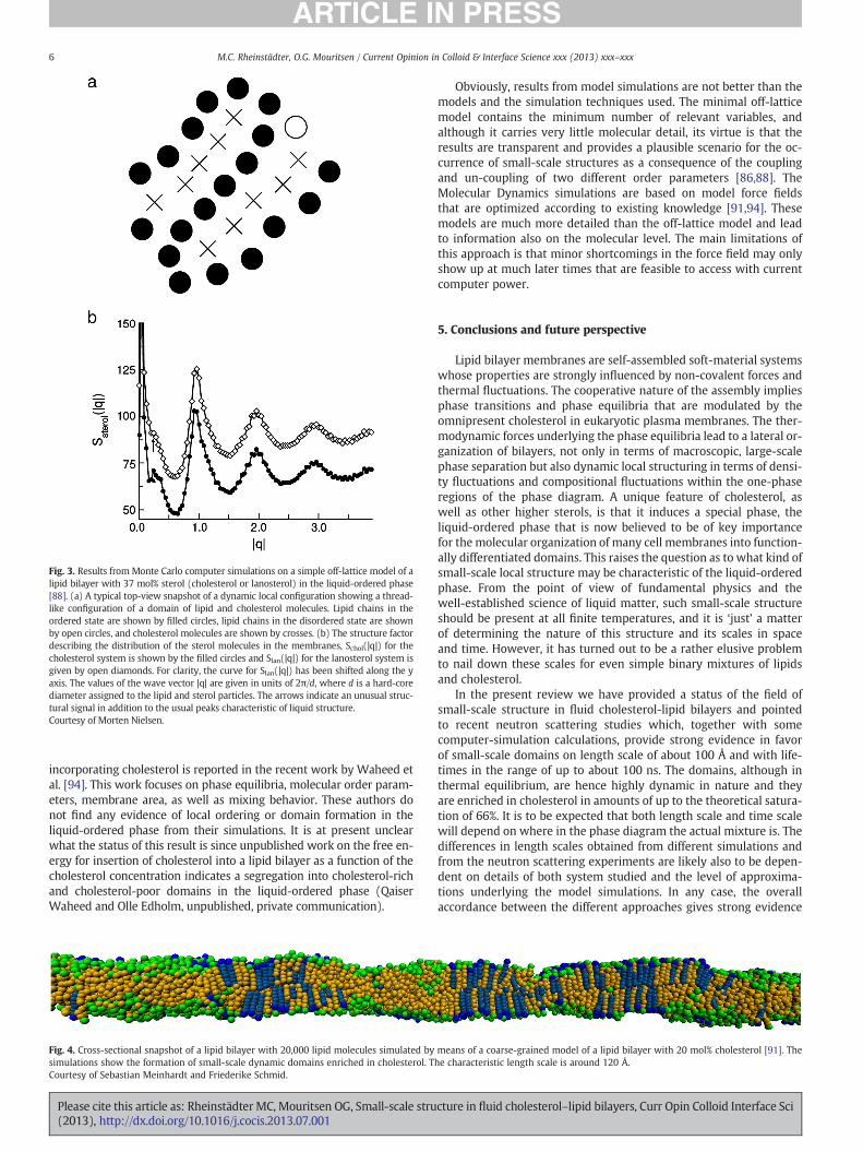
Fig. 3. Results from Monte Carlo computer simulations on a simple off-lattice model of alipid bilayer with 37 mol% sterol (cholesterol or lanosterol) in the liquid-ordered phase[88]. (a) A typical top-view snapshot of a dynamic local configuration showing a thread-like configuration of a domain of lipid and cholesterol molecules. Lipid chains in theordered state are shown by filled circles, lipid chains in the disordered state are shownby open circles, and cholesterol molecules are shown by crosses. (b) The structure factordescribing the distribution of the sterol molecules in the membranes, Schol(|q|) for thecholesterol system is shown by the filled circles and Slan(|q|) for the lanosterol system isgiven by open diamonds. For clarity, the curve for Slan(|q|) has been shifted along the yaxis. The values of the wave vector |q| are given in units of 2π/d, where d is a hard-corediameter assigned to the lipid and sterol particles. The arrows indicate an unusual struc-tural signal in addition to the usual peaks characteristic of liquid structure.Courtesy of Morten Nielsen.
6 M.C. Rheinstädter, O.G. Mouritsen / Current Opinion in Colloid & Interface Science xxx (2013) xxx–xxx
incorporating cholesterol is reported in the recent work by Waheed etal. [94]. This work focuses on phase equilibria, molecular order param-eters, membrane area, as well as mixing behavior. These authors donot find any evidence of local ordering or domain formation in theliquid-ordered phase from their simulations. It is at present unclearwhat the status of this result is since unpublished work on the free en-ergy for insertion of cholesterol into a lipid bilayer as a function of thecholesterol concentration indicates a segregation into cholesterol-richand cholesterol-poor domains in the liquid-ordered phase (QaiserWaheed and Olle Edholm, unpublished, private communication).
Fig. 4. Cross-sectional snapshot of a lipid bilayer with 20,000 lipid molecules simulated bysimulations show the formation of small-scale dynamic domains enriched in cholesterol. TCourtesy of Sebastian Meinhardt and Friederike Schmid.
Please cite this article as: Rheinstädter MC, Mouritsen OG, Small-scale stru(2013), http://dx.doi.org/10.1016/j.cocis.2013.07.001
Obviously, results from model simulations are not better than themodels and the simulation techniques used. The minimal off-latticemodel contains the minimum number of relevant variables, andalthough it carries very little molecular detail, its virtue is that theresults are transparent and provides a plausible scenario for the oc-currence of small-scale structures as a consequence of the couplingand un-coupling of two different order parameters [86,88]. TheMolecular Dynamics simulations are based on model force fieldsthat are optimized according to existing knowledge [91,94]. Thesemodels are much more detailed than the off-lattice model and leadto information also on the molecular level. The main limitations ofthis approach is that minor shortcomings in the force field may onlyshow up at much later times that are feasible to access with currentcomputer power.
5. Conclusions and future perspective
Lipid bilayer membranes are self-assembled soft-material systemswhose properties are strongly influenced by non-covalent forces andthermal fluctuations. The cooperative nature of the assembly impliesphase transitions and phase equilibria that are modulated by theomnipresent cholesterol in eukaryotic plasma membranes. The ther-modynamic forces underlying the phase equilibria lead to a lateral or-ganization of bilayers, not only in terms of macroscopic, large-scalephase separation but also dynamic local structuring in terms of densi-ty fluctuations and compositional fluctuations within the one-phaseregions of the phase diagram. A unique feature of cholesterol, aswell as other higher sterols, is that it induces a special phase, theliquid-ordered phase that is now believed to be of key importancefor the molecular organization of many cell membranes into function-ally differentiated domains. This raises the question as to what kind ofsmall-scale local structure may be characteristic of the liquid-orderedphase. From the point of view of fundamental physics and thewell-established science of liquid matter, such small-scale structureshould be present at all finite temperatures, and it is ‘just’ a matterof determining the nature of this structure and its scales in spaceand time. However, it has turned out to be a rather elusive problemto nail down these scales for even simple binary mixtures of lipidsand cholesterol.
In the present review we have provided a status of the field ofsmall-scale structure in fluid cholesterol-lipid bilayers and pointedto recent neutron scattering studies which, together with somecomputer-simulation calculations, provide strong evidence in favorof small-scale domains on length scale of about 100 Å and with life-times in the range of up to about 100 ns. The domains, although inthermal equilibrium, are hence highly dynamic in nature and theyare enriched in cholesterol in amounts of up to the theoretical satura-tion of 66%. It is to be expected that both length scale and time scalewill depend on where in the phase diagram the actual mixture is. Thedifferences in length scales obtained from different simulations andfrom the neutron scattering experiments are likely also to be depen-dent on details of both system studied and the level of approxima-tions underlying the model simulations. In any case, the overallaccordance between the different approaches gives strong evidence
means of a coarse-grained model of a lipid bilayer with 20 mol% cholesterol [91]. Thehe characteristic length scale is around 120 Å.
cture in fluid cholesterol–lipid bilayers, Curr Opin Colloid Interface Sci

7M.C. Rheinstädter, O.G. Mouritsen / Current Opinion in Colloid & Interface Science xxx (2013) xxx–xxx
in favor of the presence of nano-scale structures in the liquid-orderedphase induced by cholesterol.
It is our contention that these small-scale domains are the nucleiand/or the virtual nucleation sites that may lead to ‘rafts’ in biologicalmembranes. Obviously, real biological membranes are very differentfrom simple model membranes in thermodynamic equilibrium. Firstof all, biological membranes are associated with integral and periph-erally bound proteins that modulate the lateral structure. It has beenproposed that integral membrane proteins can ‘harvest’ the lipid do-mains and pick up the correlations in the lipid matrix [95,96]. This canon the one side lead to lipid-mediated protein–protein interactionsand protein lateral organization and on the other side to stabilizationand increase in lifetime as well as enlargement of the small-scale lipiddomains. Secondly, biological membranes are not in thermal equilib-rium and fluxes of material and energy will influence the membraneproperties and small-scale structures [97,98]. It has indeed beenpointed out, that rafts in biological membranes may be dynamicstructures maintained by active processes [99].
Acknowledgments
The idea behind this paper nucleated during the conferenceNeutrons and Life Sciences organized by the European SpallationSource in Lund in May 2013. Useful subsequent correspondencewith Profs. Friederike Schmid, Ilpo Vattulainen, and Olle Edholm isgratefully acknowledged. The work of OGM is supported by a grant(11-107269) from the Danish Council for Independent ResearchNatural Sciences. MCR was funded by the Natural Sciences and Engi-neering Research Council of Canada (NSERC), the National ResearchCouncil Canada (NRC), the Canada Foundation for Innovation (CFI)and the Ontario Ministry of Economic Development and Innovation.MCR is the recipient of an Early Researcher Award of the Provinceof Ontario.
References
[1] Mouritsen OG. Life-as a matter of fat. The emerging science of lipidomics.Heidelberg: Springer; 2005.
[2] Bloch KE. Sterol, structure and membrane function. CRC Crit Rev Biochem MolBiol 1983;14:47–92.
[3] BloomM,Mouritsen OG. The evolution ofmembranes. In: Lipowsky R, Sackmann E,editors. Amsterdam: Elsevier; 1995. p. 65–95.
[4] Mouritsen OG, Zuckermann MJ. What's so special about cholesterol? Lipids2004;39:1101–13.
[5] Yeagle PL. Modulation of membrane function by cholesterol. Biochimie 1991;73:1303–10.
[6] Kurzchalia TV, Ward S. Why do worms need cholesterol? Nat Cell Biol 2003;5:684–8.
[7] Yeagle PL. Lanosterol and cholesterol have different effects on phospholipid acylchain ordering. Biochim Biophys Acta 1985;815:33–6.
[8] Vance DE, van den Bosch H, editors. Cholesterol in the year 2000, 1529. BiochimBiophys Acta. Special issue (dedicated to the memory of Konrad Bloch); 2000.p. 1–375.
[9] Pitman MC, Suits F, MacKerell AD, Feller SE. Molecular-level organization ofsaturated and polyunsaturated fatty acids in a phosphatidylcholine bilayercontaining cholesterol. Biochemistry 2004;43:15318–28.
[10] Barrett MA, Zheng S, Toppozini LA, Alsop RJ, Dies H, Wang A, Jago N, Moore M,Rheinstädter MC. Solubility of cholesterol in lipid membranes and the formationof immiscible cholesterol plaques at high cholesterol concentrations. Soft Matter2013 (accepted for publication).
[11] Ipsen JH, Karlstrom G, Mouritsen OG, Wennerstrom H, Zuckermann MJ. Phaseequilibria in the phosphatidylcholine–cholesterol system. Biochim BiophysActa 1987;905:162–72.
[12] Risbo J, Sperotto MM, Mouritsen OG. Theory of phase equilibria and criticalmixing points in binary lipid bilayers. J Chem Phys 1995;103:3643–56.
[13] Honerkamp-Smith AR, Veatch SL, Keller SL. An introduction to critical points forbiophysicists: observations of compositional heterogeneity in lipid membranes.Biochim Biophys 2009;1788:53–63.
[14] Vist MR, Davis JH. Phase equilibria of cholesterol–dipalmitoyl phosphatidylcholinemixtures: 2H nuclear magnetic resonance and differential scanning calorimetry.Biochemistry 1990;29:451–64.
[15] Hsueh YW, Gilbert K, TrandumC, ZuckermannM, Thewalt J. The effect of ergosterolon dipalmitoylphosphatidylcholine bilayers: a deuterium NMR and calorimetricstudy. Biophys J 2005;88:1799–808.
Please cite this article as: Rheinstädter MC, Mouritsen OG, Small-scale stru(2013), http://dx.doi.org/10.1016/j.cocis.2013.07.001
[16] Marsh D. Liquid-ordered phases induced by cholesterol: a compendium of binaryphase diagrams. Biochim Biophys Acta 2010;1798:688–99.
[17] Mouritsen OG. The liquid-ordered state comes of age. Biochim Biophys Acta2010;1798:1286–8.
[18] Simons K, Ikonen E. Functional rafts in cell membranes. Nature 1997;387:569–72.
[19] Simons K, van Meer G. Lipid sorting in epithelial cells. Biochemistry 1988;27:6197–202.
[20] Heerklotz H. Triton promotes domain formation in lipid raft mixtures. Biophys J2002;83:2693–701.
[21] Simons K, Ikonen E. How cells handle cholesterol. Science 2000;290:1721–6.[22] Engelman DM. Membranes are more mosaic than fluid. Nature 2005;438:
578–80.[23] Niemelä PS, Ollila S, Hyvonen MT, Karttunen M, Vattulainen I. Assessing the
nature of lipid raft membranes. PLoS Comput Biol 2007;3:e34.[24] Pike LJ. The challenge of lipid rafts. J Lipid Res 2009;50:S323–8.[25] Lingwood D, Simons K. Lipid rafts as a membrane-organizing principle. Science
2009;327:46–50.[26] Eggeling C, Ringemann C, Medda R, Schwarzmann G, Sandhoff K, Polyakova S,
Belov VN, Hein B, von Middendorf C, Schonle A, Hell SW. Direct observation ofthe nanoscale dynamics of membrane lipids in a living cell. Nature 2009;457:1159–62.
[27] van der Goot FG, Harder T. Raft membrane domains: from a liquid-orderedmembrane phase to a site of pathogen attack. Semin Immunol 2001;13:89–97.
[28] Lenne P-F, Nicolas A. Physics puzzles on membrane domains posed by cellbiology. Soft Matter 2009;5:2841–8.
[29] Simons K, Gerl MJ. Revitalizing membrane rafts: new tools and insights. Nat RevMol Cell Biol 2010;11:688–99.
[30] Dibble AR, Hinderliter AK, Sando JJ, Biltonen RL. Lipid lateral heterogeneity inphosphatidylcholine/phosphatidylserine/diacylglycerol vesicles and its influ-ence on protein kinase C activation. Biophys J 1996;71:1877–90.
[31] Mouritsen OG, Biltonen RL. Protein–lipid interactions and membrane heteroge-neity. New Compr Biochem 1993;25:1–39.
[32] Mouritsen OG, Jørgensen K. Dynamical order and disorder in lipid bilayers. ChemPhys Lipids 1994;73:3–25.
[33] Mouritsen OG, Jørgensen K. Small-scale lipid membrane structure: simulationvs. experiment. Curr Opin Struct Biol 1997;7:518–27.
[34] Rheinstädter MC, Das J, Flenner EJ, Brüning B, Seydel T, Kosztin I. Motionalcoherence in fluid phospholipid membranes. Phys Rev Lett 2008;101:248106.
[35] Jacobson K, Mouritsen OG, Anderson RGW. Lipid rafts: at a crossroad betweencell biology and physics. Nat Cell Biol 2007;9:7–14.
[36] Van Meer G, Voelker DR, Feigenson GW. Membrane lipids: where they are andhow they behave. Nat Rev Mol Cell Biol 2008;9:112–24.
[37] Bagatolli LA, Ipsen JH, Simonsen AC, Mouritsen OG. An outlook on organizationof lipids in membranes: searching for a realistic connection with the organiza-tion of biological membranes. Prog Lipid Res 2010;49:378–89.
[38] Armstrong CL, Marquardt D, Dies H, Kucerka N, Yamani Z, Harroun TA, Katsaras J,Shi A-C, Rheinstädter MC. The observation of highly ordered domains in mem-branes with cholesterol. PLoS One 2013;8:e66162.
[39] Armstrong CL, Barrett MA, Toppozini L, Kucerka N, Yamani Z, Katsaras J, FragnetoG, Rheinstädter MC. Co-existence of gel and fluid domains in single-componentphospholipid membranes. Soft Matter 2012;8:4687–94.
[40] Ninham BW, Lo Nostro P. Molecular forces and self assembly in colloid, nanosciences and biology. Cambridge: Cambridge Molecular Science; 2010 .
[41] Vist R, Davis JH. Phase equilibria of cholesterol/dipalmitoylphosphatidylcholinemixtures: deuterium nuclear magnetic resonance and differential scanningcalorimetry. Biochemistry 1990;29:451–64.
[42] Almeida PFF, Vaz WLC, Thompson TE. Lateral diffusion in the liquid phases ofdimyristoylphosphatidylcholine/cholesterol lipid bilayers: a free volume analy-sis. Biochemistry 1992;31:6739–47.
[43] Thewalt JL, Bloom M. Phosphatidylcholine/cholesterol phase diagrams. Biophys J1992;63:1176–81.
[44] deMeyer F, Smit B. Effect of cholesterol on the structure of a phospholipid bilayer.Proc Natl Acad Sci U S A 2009;106:3654–8.
[45] de Meyer FJ, Benjamini A, Rodgers JM, Misteli Y, Smit B. Molecular simulation ofthe DMPC–cholesterol phase diagram. J Phys Chem B 2010;114:10451–61.
[46] Veatch SL, Polozov IV, Gawrisch K, Keller SL. Liquid domains in vesicles investi-gated by NMR and fluorescence microscopy. Biophys J 2004;86:2910–22.
[47] Davis JH, Clair JJ, Juhasz J. Phase equilibria in DOPC/DPPC-d62/cholesterolmixtures. Biophys J 2009;96:521–39.
[48] Silvius JR, del Guidice D, Lafleur M. Cholesterol at different bilayer concentra-tions can promote or antagonize lateral segregation of phospholipids of differingacyl chain length. Biochemistry 1996;35:15198–208.
[49] Xu X, London E. The effect of sterol structure on membrane lipid domains revealshow cholesterol can induce lipid domain formation. Biochemistry 2000;39:843–9.
[50] Pan J, Mills TT, Tristram-Nagle S, Nagle JF. Cholesterol perturbs lipid bilayersnonuniversally. Phys Rev Lett 2008;100:198103.
[51] Mills TT, Toombes GE, Tristram-Nagle S, Smilgies DM, Feigenson GW, Nagle JF.Order parameters and areas in fluid-phase oriented lipid membranes usingwide angle X-ray scattering. Biophys J 2008;95:669–81.
[52] Mills TT, Tristram-Nagle S, Heberle FA, Morales NF, Zhao J, Wu J, Toombes GE,Nagle JF, Feigenson GW. Liquid–liquid domains in bilayers detected by wideangle X-ray scattering. Biophys J 2008;95:682–90.
[53] Mills TT, Huang J, Feigenson GW, Nagle JF. Effects of cholesterol and unsaturatedDOPC lipid on chain packing of saturated gel-phase DPPC bilayers. Gen PhysiolBiophys 2009;28:126–39.
cture in fluid cholesterol–lipid bilayers, Curr Opin Colloid Interface Sci

8 M.C. Rheinstädter, O.G. Mouritsen / Current Opinion in Colloid & Interface Science xxx (2013) xxx–xxx
[54] Rheinstädter MC, Ollinger C, Fragneto G, Demmel F, Saldit T. Collective dynamicsof lipid membranes studied by inelastic neutron scattering. Phys Rev Lett2004;93:108107.
[55] Brüning B, Rheinstädter MC, Hiess A, Weinhausen B, Reusch T, Aeffner S, SaldittT. Influence of cholesterol on the collective dynamics of the phospholipid acylchains in model membranes. Eur Phys J E 2010;31:419–28.
[56] Armstrong CL, Barrett MA, Hiess A, Salditt T, Katsaras J, Shi A-C, Rheinstädter MC.Effect of cholesterol on the lateral nanoscale dynamics of fluid membranes. EurBiophys J 2012;41:901–13.
[57] Rheinstädter MC, Seydel T, Salditt T. Nanosecond molecular relaxations in lipidbilayers studied by high energy resolution neutron scattering and in-situ diffrac-tion. Phys Rev E 2007;75:011907.
[58] Rheinstädter MC, Ollinger C, Fragneto G, Salditt T. Collective dynamics in phos-pholipid bilayers investigated by inelastic neutron scattering: exploring thedynamics of biological membranes with neutrons. Physica B 2004;350:136–9.
[59] Kaye MD, Schmalzl K, Nibali VC, Tarek M, Rheinstädter MC. Ethanol enhancescollective dynamics of lipid membranes. Phys Rev E 2011;83:050907.
[60] Pabst G, Kucerka N, Nieh MP, Rheinstädter MC, Katsaras J. Applications ofneutron and X-ray scattering to the study of biologically relevant modelmembranes. Chem Phys Lipids 2010;163:460–79.
[61] Huang J, Feigenson GW. A microscopic interaction model of maximum solubilityof cholesterol in lipid bilayers. Biophys J 1999;76:2142–57.
[62] Dai J, Alwarawrah M, Huang J. Instability of cholesterol clusters in lipid bilayersand the cholesterol's umbrella effect. J Phys Chem B 2010;114:840–8.
[63] Radhakrishnan A, McConnell HM. Condensed complexes of cholesterol andphospholipids. Biophys J 1999;77:1507–17.
[64] Andoh Y, Oono K, Okazaki S, Hatta I. Subcellular colocalization of the cellular andscrapie prion proteins in caveolae-like membranous domains. J Chem Phys2012;136:155104.
[65] Chong PL. Evidence for regular distribution of sterols in liquid crystallinephosphatidylcholine bilayers. Proc Natl Acad Sci U S A 1994;91:10069–73.
[66] Liu F, Chong PL. Evidence for a regulatory role of cholesterol superlattices in thehydrolytic activity of secretory phospholipase A2 in lipid membranes. Biochem-istry 1999;38:3867–73.
[67] Ehrig J, Petrov EP, Schwille P. Phase separation and near-critical fluctuations intwo-component lipid membranes: Monte Carlo simulations on experimentallyrelevant scales. New J Phys 2011;13:045019.
[68] Murtola T, Rog T, Falck E, Karttunen M, Vattulainen I. Transient ordered domainsin single-component phospholipid bilayers. Phys Rev Lett 2006;97:238102.
[69] Brüning B, Wald E, Schrader W, Behrends R, Kaatze U. Slowing down in lipidbilayers: domain structure fluctuations and axial diffusion. Soft Matter 2009;5:3340–6.
[70] London E. How principles of domain formation in model membranes mayexplain ambiguities concerning lipid raft formation in cells. Biochim BiophysActa 2005;1746:203–20.
[71] Egelstaff PA. An introduction to the liquid state. London: Academic Press; 1967 .[72] Armstrong CL, Trapp M, Peters J, Seydel T, Rheinstädter MC. Short range ballistic
motion in fluid lipid bilayers studied by quasi-elastic neutron scattering. SoftMatter 2011;7:8358–62.
[73] Armstrong CL, Toppozini L, Dies H, Faraone A, Nagao M, Rheinstädter MC.Incoherent neutron spin-echo spectroscopy as an option to study long-rangelipid diffusion. ISRN Biophys 2013;2013:439758.
[74] Falck E, Rog T, Karttunen M, Vattulainen I. Lateral diffusion in lipid membranesthrough collective flows. J Am Chem Soc 2008;130:44–5.
[75] Busch S, Smuda C, Pardo LC, Unruh T. Molecular mechanism of long-range diffu-sion in phospholipid membranes studied by quasielastic neutron scattering.J Am Chem Soc 2010;132:3232–3.
Please cite this article as: Rheinstädter MC, Mouritsen OG, Small-scale stru(2013), http://dx.doi.org/10.1016/j.cocis.2013.07.001
[76] Chen SH, Liao CY, Huang HW, Weiss TM, Bellisent-Funel MC, Sette F. Collectivedynamics in fully hydrated phospholipid bilayers studied by inelastic X-rayscattering. Phys Rev Lett 2001;86:740–3.
[77] Poe-Jou Chen YL, Weiss TM, Huang HW, Sinn H, Alp EE, Alatas A, Said A, ChenS-H. Studies of short-wavelength collective molecular motions in lipid bilayersusing high resolution inelastic X-ray scattering. Biophys Chem 2003;105:721–41.
[78] Weiss TM, Chen PJ, Sinn H, Alp EE, Chen SH, Huang HW. Collective chain dynam-ics in lipid bilayers by inelastic X-ray scattering. Biophys J 2003;84:3767–76.
[79] Rauch H. Reality in neutron interference experiments. Found Phys 1993;23:7–36.
[80] Pandit SA, Khelashvili G, Jakobsson E, Grama A, Scott HL. Lateral organization inlipid–cholesterol mixed bilayers. Biophys J 2007;92:440–7.
[81] Almeida PF. A simple thermodynamic model of the liquid-ordered state and theinteractions between phospholipids and cholesterol. Biophys J 2011;100:420–9.
[82] Merz KM, Roux B. Biological membranes: a molecular perspective from compu-tation and experiment. Birkhäuser Boston; 1996 .
[83] Risselada HJ, Marrink SJ. The molecular face of lipid rafts in model membranes.Proc Natl Acad Sci U S A 2008;105:17367–72.
[84] Berkowitz ML. Detailed Molecular Dynamics simulations of model biologicalmembranes containing cholesterol. Biochim Biophys Acta 2009;1788:86–96.
[85] Bennett WF, Tieleman DP. Computer simulations of lipid membrane domains.Biochim Biophys Acta 2013;1828:1765–76.
[86] Nielsen M, Miao L, Ipsen JH, Zuckermann MJ, Mouritsen OG. Off-lattice model forthe phase behavior of lipid–cholesterol bilayers. Phys Rev E 1999;59:5790–803.
[87] Martinez-Seara H, Rog T, Karttunen M, Vattulainen I, Reigada R. Cholesterolinduces specific spatial and orientational order in cholesterol/phospholipidmembranes. PLoS One 2010;5:e11162.
[88] Miao L, Nielsen M, Thewalt J, Ipsen JH, Bloom M, Zuckermann MJ, Mouritsen OG.From lanosterol to cholesterol: structural evolution and differential effects onlipid bilayers. Biophys J 2002;82:1429–44.
[89] Nielsen M, Thewalt J, Miao L, Ipsen JH, Bloom M, Zuckermann MJ, Mouritsen OG.Sterol evolution and the physics of membranes. Europhys Lett 2000;52:368–74.
[90] Vattulainen I, Rog T. Lipid simulations: a perspective on lipids in action. ColdSpring Harb Perspect Biol 2011;3:91–103.
[91] Meinhardt S, Vink RLC, Schmid F. Monolayer curvature stabilizes nanoscale raftdomains in mixed lipid bilayers. Proc Natl Acad Sci U S A 2013;110:4476–81.
[92] Schick M. Membrane heterogeneity: manifestation of a curvature-inducedmicroemulsion. Phys Rev E 2012;85:031902.
[93] Semrau S, Schmidt T. Membrane heterogeneity—from lipid domains to curvatureeffects. Soft Matter 2009;5:3174–86.
[94] Waheed Q, Tjornhammar R, Edholm O. Phase transitions in coarse-grained lipidbilayers containing cholesterol by Molecular Dynamics simulations. Biophys J2012;103:2125–33.
[95] Gil T, Sabra MC, Ipsen JH, Mouritsen OG. Wetting and capillary condensation asmeans of protein organization in membranes. Biophys J 1997;73:1728–41.
[96] Mouritsen OG. Self-assembly and organization of lipid–protein membranes. CurrOpin Colloid Interface Sci 1998;3:78–87.
[97] Sabra MC, Mouritsen OG. Steady-state compartmentalization of lipid mem-branes by active proteins. Biophys J 1998;74:745–52.
[98] Bouvrais H, Cornelius F, Ipsen JH, Mouritsen OG. Intrinsic reaction-cycle timescale of Na+, K+-ATPase manifests itself in the lipid–protein interactions ofnon-equilibrium membranes. Proc Natl Acad Sci U S A 2012;109:18442–6.
[99] Mayor S, Rao M. Rafts: scale-dependent, active lipid organization at the cellsurface. Traffic 2004;5:231–40.
[100] Ipsen JH, Mouritsen OG, Zuckermann MJ. Theory of thermal anomalies in thespecific heat of lipid bilayers containing cholesterol. Biophys J 1989;56:661–7.
cture in fluid cholesterol–lipid bilayers, Curr Opin Colloid Interface Sci