Embed Size (px)
Citation preview
Urinary cholesterol: its association with a macromolecular protein-lipid complex
Dieter Jiingst, Herbert Weiser, Elmar Siess,* and Hans J. Karl Department of Medicine 2, Klinikum Grosshadern, University of Munich, and Institute of Diabetes Research,* Miinchen FRG
Abstract The cholesterol-containing complexes in the urine of normal subjects and patients with diseases accompanied by hyperexcretion of urinary cholesterol were characterized. In normal subjects, the major portion of the recovered urinary cholesterol was eluted in the void volume fractions after gel chromatography on Bio-Gel A-5m; this suggested an association with a macromolecular complex above 5 X 1 O6 daltons. A com- parable elution pattern was seen in most of the urines of the patients with benign or malignant diseases of the kidneys or the urogenital tract. However, in single patients with hyperexcretion of urinary cholesterol, considerable amounts of cholesterol were detected in the included volume of the column. This was caused by additional excretion of high density lipoproteins or both high and low density lipoproteins in the urine which could be iden- tified in these fractions by agarose electrophoresis and immu- nodiffusion. These results indicate that the macromolecular complex represents the majority of the recovered urinary cho- lesterol in normal subjects and in disease states with known hyperexcretion. Macroscopically, the isolated cholesterol-con- taining complex in the void volume fractions was turbid, and electron microscopy showed lipoprotein-like particles with di- ameters ranging from 300 to 700 A. The chemical analysis revealed median values of protein (46.0%), triglycerides (1 6.3%), cholesterol (8.2%), and phospholipids (29.5%) in normal subjects and comparable results in the patients with benign or malignant diseases of the kidney and the urogenital tract. Ethanolamine glycerophospholipids, phosphatidylcholine, sphingomyelin, and phosphatidylserine were the main phospholipid components. After ultracentrifugation in a CsCl gradient, the cholesterol- containing complex was found between densities 1.1 and 1.3 g/ml. By SDS polyacrylamide electrophoresis, up to 17 protein subunits in the molecular weight range of 14,000 to 87,500 were separated. Immunodiffusion studies showed in about 40% precipitin lines against anti-human albumin, but no reactions against anti-human apoHDL and anti-human apoLDL. However, immunodiffusion of the macromolecular complex against anti- liver-specific and anti-kidney-specific lipoproteins revealed single precipitin 1ines.M In conclusion, the isolated cholesterol-con- taining urinary complex showed many characteristics of mem- brane-associated protein-lipid particles of the human kidney and even the liver. These proteolipids are the major source of urinary cholesterol in normal and disease states. They could be derived partially from the kidney, but in addition possibly from other parts of the urogenital system, especially the bladder.-Jiingt, D., H. Weisn, E. Siear, and H. J. Karl. Urinary cholesterol: its association with a macromolecular protein-lipid complex. J. Lipid Res. 1984. 25: 655-664.
Supplementary key words gel chromatography ultrafiltration SDS- PAGE electrophoresis immunodiffusion lipoproteins
Small amounts of cholesterol, mainly nonesterified, are present in normal urine (1-7), whereas elevated levels have been reported in patients with benign and malignant diseases of the kidney and the urogenital tract (8-27). On the basis of ultrafiltration studies, excretion as a pro- tein-bound complex has been suggested (28). In normal urine most of the cholesterol seems to be associated with a light particulate fraction, similar to plasma membranes (29). However, in patients suffering from the nephrotic syndrome, a urinary loss of plasma high density (HDL) and in some cases also of low density (LDL) lipoproteins has been reported (30, 31).
It was the aim of this study to characterize the cho- lesterol-containing complexes in the urine of normal sub- jects and in disease states with known hyperexcretion. We demonstrate that cholesterol in the urine is associated mainly to a macromolecular protein-lipid complex which might be of cellular origin. In addition, in some cases HDL and LDL could be identified, especially in patients with the nephrotic syndrome.
MATERIALS AND METHODS
Normal subjects and collection of urine The group of normal subjects included four men and
five women, 18 to 46 years old, who had no history or clinical or biochemical signs of a renal or urogenital dis- ease. Microscopic examination of the urine samples was normal, as well as the results of the N-Multistix reagent strips for protein, glucose, bilirubin, blood cells, ketone
Abbreviations: HDL, high density lipoprotein; LDL, low density lipoprotein; VLDL, very low density lipoprotein; GLC, gas-liquid chromatography; SDS-PAGE, sodium dodecyl sulfate polyacrylamide gel electrophoresis; LSP, liver-specific lipoprotein; KSP, kidney-specific lipoprotein; TLC, thin-layer chromatography.
Journal of Lipid Research Volume 25, 1984 655
by guest, on June 23, 2018w
ww
.jlr.orgD
ownloaded from

bodies, nitrate, and urobilinogen. The concentrations of creatinine and total protein in serum were normal in all cases and the excretion of urinary protein was below 100 mg/24 hr (Table 1). During the collection period of 24 hr the urine was kept refrigerated (4°C) and sodium azide (0.02%) was added to prevent bacterial growth. Frac- tionation of urinary cholesterol started within 1 hour after the end of the collection period.
Patients The group of patients consisted of 12 with benign and
9 with malignant diseases of the kidney or the urogenital tract. Diagnoses were established by routine clinical pro- cedures including urinary protein quantitation, intrave- nous pyelography, cystoscopy, renal ultrasound, and his- tological examination of biopsies or resected material. Further clinical data of these patients including protein and creatinine in serum and 24-hr excretion of protein in the urine are given in Tables 2 and 3.
Fractionation of urinary cholesterol For fractionation of urinary cholesterol usually the
whole 24-hr urine volumes were used, and were passed through a Selecta filter no. 1 1 17 1/2 (Schleicher Schull). This filtration removes larger particles (5-10 pm in di- ameter) such as intact cells and larger cellular debris. After filtration NaCl was added to the urinary samples (final concentration 0.56 M) to precipitate the Tamm- Horsefall mucoprotein (32). This was removed by cen- trifugation (8000 rpm for 10 min) and the supernatant, considered as the upper 90% of the volume, was collected. Recovery studies of urinary cholesterol in single specimens revealed a loss of - 10% during these procedures. Ul- trafiltration was done in a stirred cell (Amicon model 402 and Amicon model 52) using PM 10 membranes at 4°C to a final volume of 3-5 ml. This concentration step was the most critical in regard to the loss of urinary cho- lesterol since some of the lipid perhaps adhered to the PM 10 membranes or might even pass the membrane. Aliquots of 2-3 ml of the concentrated urine were used for gel chromatography on a 1.2 m X 2.0 cm Bio-Gel A-5 m column (BioRad), mesh 200-400, according to Sata, Havel, and Jones (33). Elution was performed with 0.15 M NaCl in water containing 0.01 M EDTA and 0.02% NaNs at pH 7.0. Usually 48 to 50 fractions (8.5 ml) were collected. Absorbance at 280 nm was determined in a spectrophotometer (Beckman, model 24).
gradient ultracentrifugation, immunodiffusion, agarose electrophoresis, and SDS-polyacrylamide gel electropho- resis. Cholesterol-containing fractions in the included volume of the column were studied separately by agarose electrophoresis and immunodiffusion. A considerable amount of urinary cholesterol was lost as a consequence of the different fractionation procedures, especially the two ultrafiltration steps. Recovery studies in single urinary specimens for cholesterol showed a total loss of urinary cholesterol between 30 and 50% at the end of the second ultrafiltration.
Chemical assays
Total cholesterol was analyzed in 1.0-ml aliquots of the 24-hr urine collection and the 8.5-ml fractions after gel chromatography. After extraction with 8 ml of chlo- roform-methanol 3:l (v/v) and centrifugation, 5 ml of chloroform was removed and dried under a stream of nitrogen. The residue was hydrolyzed with 0.5 ml of ethanolic KOH at 60°C for 60 min and reextracted with 6 ml of n-hexane and evaporated. The residue from 5 ml of the extract was dissolved in 0.1 ml of the internal standard solution (1 0 mg of androstendione/dl of isooc- tane). Gas-liquid chromatographic determination of cho- lesterol was performed using a 1.8 X 2 mm, 1% XE 60 column, column temperature 220"C, with flame ioniza- tion detectors.
Triglycerides were measured enzymatically with com- mercial test kits (Boehringer Mannheim) (34).
Phospholipids in the lipid extracts (chloroform-meth- anol 3: 1, v/v) were determined by phosphorus analysis (35). The phospholipids were fractionated by TLC on 20 X 20 cm glass plates coated with silica gel (Merck). After two-dimensional TLC in the solvent systems chlo- roform-methanol-25% aq. ammonia 60:20:5 and chlo- roform-acetone-methanol-acetic acid-water 3:4: 1 : 1 :0.5, the separated phospholipids were made visible with iodine vapor; they were individually recovered and assayed for phosphorus content (36).
Protein was measured by the method of Kashyap, Hynd, and Robinson (37).
Electron microscopy
Electron microscopy was performed with a Siemens Typ 101 instrument and a negative staining tech- nique (38).
Total cholesterol was measured by gas-liquid chro- matography. The cholesterol-containing fractions were
Density gradient centrifugation - . , -
eluted usually at the void volume. These fractions were pooled and concentrated with PM 10 membranes (Ami- con) to a final volume of 1-2 ml. Further characterization was done by chemical assay, electron microscopy, density
Ultracentrifugation was performed on a CsC1-gradient (density 1.05-1.35 g/ml) at 50,000 rpm in a Beckman SW-60 rotor for 68 hr. Eleven or 12 fractions were ob- tained by pipetting from the top. Absorbance at 280 nm
656 Journal of Lipid Research Volume 25, 1984
by guest, on June 23, 2018w
ww
.jlr.orgD
ownloaded from

and total cholesterol content were measured in each frac- tion (39).
Electrophoresis and immunodiffusion
Electrophoretic separation was done on 1 % agarose gels according to Noble (40). For SDS-polyacrylamide gel electrophoresis, aliquots of the fractions were lyophilized and then mixed with Tris-HC1 (37.5 mM, pH 8.8) con- taining 2% SDS, 4% 2-mercaptoethanol, and 10% sucrose. After incubation for 2 hr at 50°C, protein subunits were separated in 1.5-mm vertical slab gels using 4-22.5% acrylamide concentrations (41). After fixing the gel for 30 min in 20% (w/v) trichloracetic acid, the proteins were stained with Coomassie brilliant blue (G 250). Mo- lecular weights were estimated comparing with the low molecular weight calibration kit (Pharmacia Fine Chem- icals) (42).
Ouchterlony immunodiffusion was performed in 1.5% agar (Difco) at 2OoC on glass plates (5 X 2 cm) for 48 hr. Antisera against human serum, human albumin, a- and &lipoproteins were obtained from Behring (Mar- burg). Special antisera against cellular lipoproteins of liver (anti-LSP) and kidney (anti-KSP) were kindly provided by U. Behrens, Immunopathology Laboratory, VAM Center, Bronx, NY. The antibodies against a- and /3-lipoproteins were identical with antibodies to apoHDL and apoLDL, respectively. The manufacturer used LDL and HDL isolated by preparative ultracentrifugation from normal human plasma for immunization. Density intervals of 1.020-1.050and 1.063-1.21 g/ml were used for LDL and HDL antigen isolation, respectively. The anti-HDL and anti-LDL antibodies did not react with lipoprotein- free plasma obtained by preparative ultracentrifugation of plasma at d > 1.25 g/ml. There was no cross-reactivity between anti-HDL with LDL and anti-LDL with HDL.
For the preparation of antibodies against LSP and KSP, the antigens were isolated by Behrens from normal human liver and kidney that showed no significant histologic changes nor evidence of autolysis (43). Essentially the same method was used as described by McFarlane et al. (44). Tissue slices were washed free of blood in 0.25 M sucrose, adjusted to pH 8.0 with 1 M Tris, and then homogenized. The homogenate was centrifuged at 100,000 g in a SW-27 rotor and the supernatant was placed on a column of Sephadex G-100 (gel bed 90 X 2.5 cm), previously equilibrated with 0.1 M Tris-HCL-0.2 M NaCl-1 mM EDTA, pH 8.0 (Tris/EDTA buffer). The first peak fractions were concentrated and then chro- matographed on a column of Sepharose 6 B using the Tris/EDTA buffer for equilibration and elution. The first peak material contained LSP and KSP and was fur- ther subjected to ultracentrifugal flotation in a CsCl gra- dient.
LSP and KSP were recovered between the densities 1.107 and 1.202 g/ml. Analysis by SDS-PAGE revealed that the relative mobilities of LSP and KSP components were similar and the molecular weights of the monomers ranged from 40,000 to 96,000. At least seven out of eight recognizable LSP and KSP bands had the same R f value, with one of them corresponding to that of human al- bumin. Antisera against the purified antigens were pro- duced in rabbits and absorbed with lyophilized human plasma. Anti-LSP sera were tested by indirect immuno- fluorescence and immunoenzyme light microscopy on cryostat sections. They reacted with cell membranes not only of liver cells but also of smooth muscle fibers, thyroid follicles, intestine, spleen, kidney tubules, and glomeruli (43). Identical staining patterns were obtained with anti- KSP sera by Behrens and Paronetto (43). These findings of Behrens and Paronetto demonstrate the non-organ specificity of LSP and KSP. By immunodiffusion anti- LSP serum formed precipitin lines with LSP and KSP, as well as with SDS- or papain-solubilized isolated liver cell membranes (43). No reaction of anti-LSP and anti- KSP sera was seen by immunodiffusion against human albumin or total human plasma. These observations were made with the anti-LSP and anti-KSP sera used in this study.
RESULTS
Normal subjects After gel filtration on the Bio-Gel A-5 m column, urines
of normal subjects showed similar elution patterns. The characteristic profile illustrated in Fig. 1 shows that the major portion of the cholesterol recovered from urine was eluted in the void volume. The total amount of uri- nary cholesterol in the 24-hr samples and the percentage
081 0 2 1
0 6 1 9015.
IO 20 30 LO 50 FRACTIONS
Fig. 1. Gel chromatographic elution pattern of a concentrated U~~MI-Y
sample from a normal subject on Bio-Gel A-5m. Column size 120 X 2.0 cm. Individual points represent values of cholesterol (0 - 0, mg/ 10-ml fraction) and the absorbance at 280 nm (0 - 0).
Jiingst et al. Urinary cholesterol 657
by guest, on June 23, 2018w
ww
.jlr.orgD
ownloaded from

TABLE 1 . Clinical data and relative percentage of cholesterol in the void volume fractions after gel chromatography on the Bic-Gel A-5m column in normal subjects
Serum Serum Urinary Urinary % Cholesterol Subject Protein Creatinine Protein Cholesterol in Void Volume
gldl
7.8 8.3 7.4 7.9 7.5 8.2 7.2 7.3 7.8
mgldl
0.8 0.7 1 .o 0.6 0.8 0.9 1 .O 0.6 1 .o
mgi24 hr
65 50 42 83 92 34 76 52 67
mg124 hi
0.6 0.9 0.4 1.8 2.1 0.8 0.7 0.8 1.3
84.0 96.0 93.4 95.3 86.2 91.7 94.6 87.5 98.8
of the recovered cholesterol in the void volume fractions is given in Table 1. Between 84 and 99% of the cholesterol appeared in the void volume, indicating that it was as- sociated with a macromolecular complex of more than 5 X lo6 daltons. Only a minor portion of the cholesterol was found in the included volume of the column. Al- though it is possible that small amounts of HDL were present in these fractions, immunodiffusion against anti- a- and &lipoproteins and agarose-electrophoresis failed to demonstrate these lipoproteins.
Patients In 8 out of 12 urines of patients with benign diseases
of the kidneys and the urogenital tract a comparable elu- tion pattern as in normal subjects (Fig. 1) was seen after gel filtration on the Bio-Gel A-5 m column. In these cases 84 to 98% of the cholesterol was found in the void volume fractions after gel chromatography (Table 2).
However, in one patient with a renal cyst and in three
patients with the nephrotic syndrome, only 44 to 75% of the fractionated urinary cholesterol was eluted in the void volume (Table 2). Measurements of cholesterol in the single fractions revealed two (Fig. 2) or three (Fig. 3) cholesterol peaks. In the second peak immunodiffusion showed precipitin lines against anti-apoLDL and in the third peak precipitin lines against anti-apoHDL were seen (Fig. 4 and Fig. 5).
In patients with malignant disease of the kidneys or the urogenital tract, 44 to 100% of the urinary cholesterol was found in the void volume fractions (Table 3). In single cases, HDL and LDL were detected in the lower molecular weight fractions as judged by agarose electro- phoresis and immunodiffusion against anti-apoLDL and anti-apoHDL. These results demonstrate that urinary cholesterol is mainly excreted as a macromolecular com- plex in normal subjects, as well as in patients with benign or malignant diseases of the kidneys or the urogenital tract.
TABLE 2. Clinical data and relative percentage of urinary cholesterol in the void volume fractions after gel chromatography on the Bio-Gel A-5m column in patients with benign
diseases of the kidneys and the urogenital tract
Serum Serum Urinary Urinary Cholesterol % Diagnosis Subject Protein Creatinine Protein Cholesterol in Void Volume
Urolithiasis
Renal cyst
Nephrotic syndrome Glomerular nephritis Lupus erythematosus Diabetic glomerular
Glomerular nephritis Glomerular nephritis Prostatic adenoma
sclerosis
J K L
M N
0 P
Q R S T U
gld l
7.5 8.1 7.7
8.3 7.2
5.5 5.2
5.8 5.9 5.5 8.5 7.9
mgldl g124 hr
0.9 0.18 1 . 1 0.14 0.8 0.32
2.2 0.41 1 . 1 0.35
1.4 8.2 2.5 12.1
2.0 4.3 1.2 5.5 1 . 1 3.7 0.9 0.22 0.8 0.34
mgi24 hr
3.5 0.7 5.4
1.8 7.3
59.0 81.0
18.1 5.9
14.7 3.4
11.5
85.2 98.4 87.3
88.6 68.1
58.3 44.2
75.2 98.6 92.3 84.3 95.8
658 Journal of Lipid Research Volume 25, 1984
by guest, on June 23, 2018w
ww
.jlr.orgD
ownloaded from
"1 201 A * /
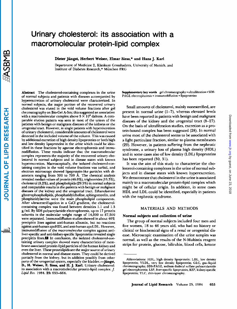
Fig. 2. Gel chromatographic elution pattern of a concentrated urinary sample from a patient with a nephrotic syndrome on Bio-Gel A-5m. Column size 120 X 2.0 cm. Individual points represent values of cho- lesterol (0 - 0, mg/lO-ml fraction) and the absorbance at 280 nm (0 - 0).
Characterization of the macromolecular urinary complex
Macroscopically, the isolated cholesterol-containing complex in the void volume fractions was turbid, appeared to be labile, and showed a tendency to aggregate after storage at 4OC for several days. In regard to the exclusion limit of the Bio-Gel A-5 m column, the estimated mo- lecular weight of this complex was above 5 X l O6 daltons.
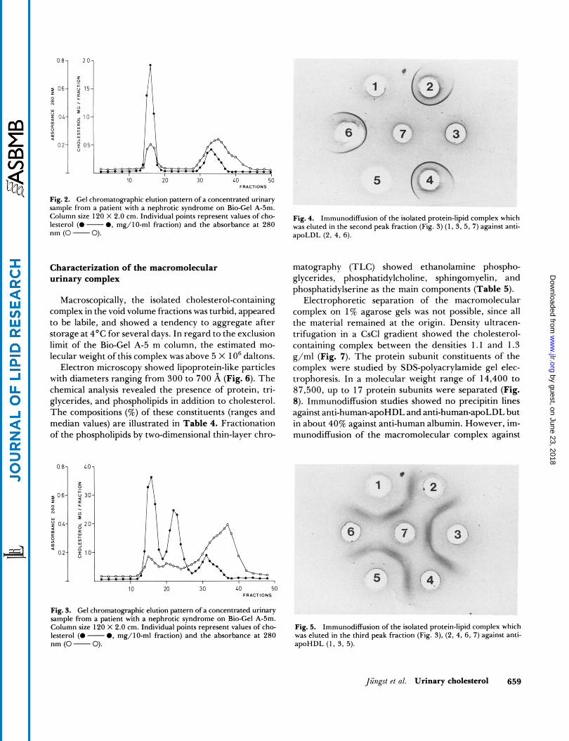
Electron microscopy showed lipoprotein-like particles with diameters ranging from 300 to 700 A (Fig. 6). The chemical analysis revealed the presence of protein, tri- glycerides, and phospholipids in addition to cholesterol. The compositions (%) of these constituents (ranges and median values) are illustrated in Table 4. Fractionation of the phospholipids by two-dimensional thin-layer chro-
IO 20 30 LO 50 FRACTIONS
Fig. 3. Gel chromatographic elution pattern of a concentrated urinary sample from a patient with a nephrotic syndrome on Bio-Gel A-5m. Column size 120 X 2.0 cm. Individual points represent values of cho- lesterol (0 - 0, mg/lO-ml fraction) and the absorbance at 280 nm (0 - 0).
Fig. 4. Immunodiffusion of the isolated protein-lipid complex which was eluted in the second peak fraction (Fig. 3) (1, 3. 5 , 7) against anti- apoLDL (2, 4, 6).
matography (TLC) showed ethanolamine phospho- glycerides, phosphatidylcholine, sphingomyelin, and phosphatidylserine as the main components (Table 5).
Electrophoretic separation of the macromolecular complex on 1% agarose gels was not possible, since all the material remained at the origin. Density ultracen- trifugation in a CsCl gradient showed the cholesterol- containing complex between the densities 1.1 and 1.3 g/ml (Fig. 7). The protein subunit constituents of the complex were studied by SDS-polyacrylamide gel elec- trophoresis. In a molecular weight range of 14,400 to 87,500, up to 17 protein subunits were separated (Fig. 8). Immunodiffusion studies showed no precipitin lines against anti-human-apoHDL and anti-human-apoLDL but in about 40% against anti-human albumin. However, im- munodiffusion of the macromolecular complex against
Fig. 5. Immunodiffusion of the isolated protein-lipid complex which was eluted in the third peak fraction (Fig. 3), (2, 4, 6, 7) against anti- apoHDL (1, 3. 5).
liinmf t f al. Urinary cholesterol 659
by guest, on June 23, 2018w
ww
.jlr.orgD
ownloaded from
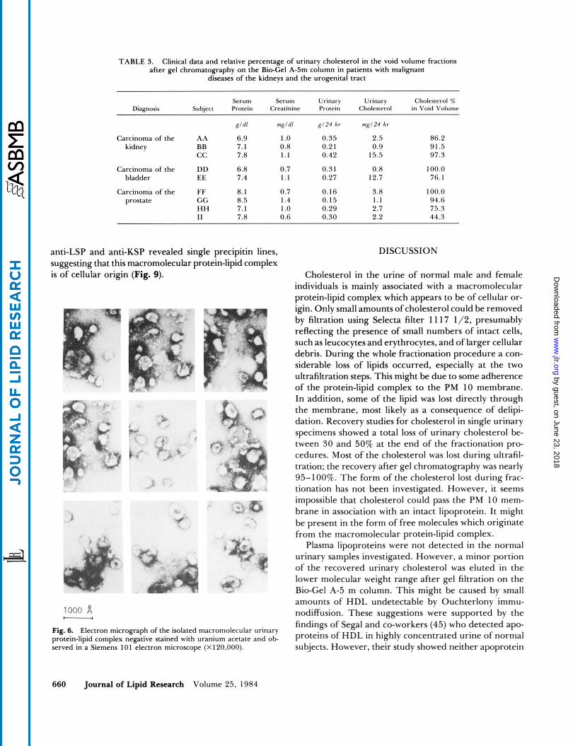
TABLE 3. Clinical data and relative percentage of urinary cholesterol in the void volume fractions after gel chromatography on the Bio-Gel A-5m column in patients with malignant
diseases of the kidneys and the urogenital tract
Serum Serum Urinary Urinary Cholesterol o/c Diagnosis Subject Protein Creatinine Protein Cholesterol in Void Volume
gld l mgldl g124 hr 1ng124 hr
Carcinoma of the AA 6.9 1 .O 0.35 2.5 86.2 kidney BB 7.1 0.8 0.2 1 0.9 91.5
cc 7.8 1.1 0.42 15.5 97.3
Carcinoma of the DD 6.8 0.7 0.3 1 0.8 100.0 bladder EE 7.4 1.1 0.27 12.7 76.1
Carcinoma of the FF 8.1 0.7 0.16 3.8 100.0 prostate GG 8.5 1.4 0.15 1.1 94.6
HH 7.1 1 .o 0.29 2.7 75.3 I1 7.8 0.6 0.30 2.2 44.3
anti-LSP and anti-KSP revealed single precipitin lines, suggesting that this macromolecular protein-lipid complex is of cellular origin (Fig. 9).
1000 A - Fig. 6. Electron micrograph of the isolated macromolecular urinary protein-lipid complex negative stained with uranium acetate and ob- served in a Siemens 10 1 electron microscope (X 120,000).
DISCUSSION
Cholesterol in the urine of normal male and female individuals is mainly associated with a macromolecular protein-lipid complex which appears to be of cellular or- igin. Only small amounts of cholesterol could be removed by filtration using Selecta filter 1 1 17 1/2, presumably reflecting the presence of small numbers of intact cells, such as leucocytes and erythrocytes, and of larger cellular debris. During the whole fractionation procedure a con- siderable loss of lipids occurred, especially at the two ultrafiltration steps. This might be due to some adherence of the protein-lipid complex to the PM 10 membrane. In addition, some of the lipid was lost directly through the membrane, most likely as a consequence of delipi- dation. Recovery studies for cholesterol in single urinary specimens showed a total loss of urinary cholesterol be- tween 30 and 50% at the end of the fractionation pro- cedures. Most of the cholesterol was lost during ultrafil- tration; the recovery after gel chromatography was nearly 95-100%. The form of the cholesterol lost during frac- tionation has not been investigated. However, it seems impossible that cholesterol could pass the PM 10 mem- brane in association with an intact lipoprotein. I t might be present in the form of free molecules which originate from the macromolecular protein-lipid complex.
Plasma lipoproteins were not detected in the normal urinary samples investigated. However, a minor portion of the recovered urinary cholesterol was eluted in the lower molecular weight range after gel filtration on the Bio-Gel A-5 m column. This might be caused by small amounts of HDL undetectable by Ouchterlony immu- nodiffusion. These suggestions were supported by the findings of Segal and co-workers (45) who detected apo- proteins of HDL in highly concentrated urine of normal subjects. However, their study showed neither apoprotein
660 Journal of Lipid Research Volume 25, 1984
by guest, on June 23, 2018w
ww
.jlr.orgD
ownloaded from
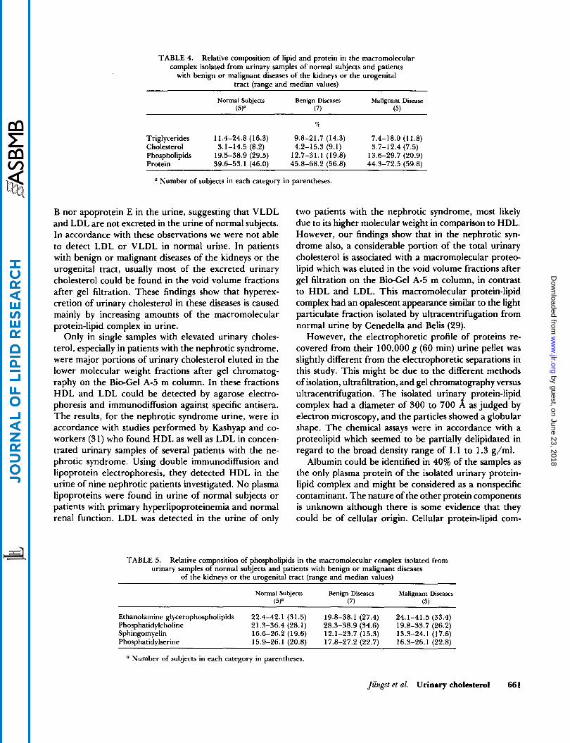
TABLE 4. Relative composition of lipid and protein in the macromolecular complex isolated from urinary samples of normal subjects and patients
with benign or malignant diseases of the kidneys or the urogenital tract (range and median values)
Normal Subjects Benign Diseases Malignant Disease (5Y (7) 15)
a Triglycerides 11.4-24.8 (16.3) 9.8-21.7 (14.3) 7.4-18.0 (11.8)
Phospholipids 19.5-38.9 (29.5) 12.7-31.1 (19.8) 13.6-29.7 (20.9) Cholesterol 3.1-14.5 (8.2) 4.2-15.3 (9.1) 3.7-12.4 (7.5)
Protein 39.6-53.1 (46.0) 45.8-68.2 (56.8) 44.3-72.5 (59.8)
a Number of subjects in each category in parentheses.
B nor apoprotein E in the urine, suggesting that VLDL and LDL are not excreted in the urine of normal subjects. In accordance with these observations we were not able to detect LDL or VLDL in normal urine. In patients with benign or malignant diseases of the kidneys or the urogenital tract, usually most of the excreted urinary cholesterol could be found in the void volume fractions after gel filtration. These findings show that hyperex- cretion of urinary cholesterol in these diseases is caused mainly by increasing amounts of the macromolecular protein-lipid complex in urine.
Only in single samples with elevated urinary choles- terol, especially in patients with the nephrotic syndrome, were major portions of urinary cholesterol eluted in the lower molecular weight fractions after gel chromatog- raphy on the Bio-Gel A-5 m column. In these fractions HDL and LDL could be detected by agarose electro- phoresis and immunodiffusion against specific antisera. The results, for the nephrotic syndrome urine, were in accordance with studies performed by Kashyap and co- workers (31) who found HDL as well as LDL in concen- trated urinary samples of several patients with the ne- phrotic syndrome. Using double immunodiffusion and lipoprotein electrophoresis, they detected HDL in the urine of nine nephrotic patients investigated. No plasma lipoproteins were found in urine of normal subjects or patients with primary hyperlipoproteinemia and normal renal function. LDL was detected in the urine of only
two patients with the nephrotic syndrome, most likely due to its higher molecular weight in comparison to HDL. However, our findings show that in the nephrotic syn- drome also, a considerable portion of the total urinary cholesterol is associated with a macromolecular proteo- lipid which was eluted in the void volume fractions after gel filtration on the Bio-Gel A-5 m column, in contrast to HDL and LDL. This macromolecular protein-lipid complex had an opalescent appearance similar to the light particulate fraction isolated by ultracentrifugation from normal urine by Cenedella and Belis (29).
However, the electrophoretic profile of proteins re- covered from their 100,000 g (60 min) urine pellet was slightly different from the electrophoretic separations in this study. This might be due to the different methods of isolation, ultrafiltration, and gel chromatography versus ultracentrifugation. The isolated urinar protein-lipid
electron microscopy, and the particles showed a globular shape. The chemical assays were in accordance with a proteolipid which seemed to be partially delipidated in regard to the broad density range of 1.1 to 1.3 g/ml.
Albumin could be identified in 40% of the samples as the only plasma protein of the isolated urinary protein- lipid complex and might be considered as a nonspecific contaminant. The nature of the other protein components is unknown although there is some evidence that they could be of cellular origin. Cellular protein-lipid com-
complex had a diameter of 300 to 700 x as judged by
TABLE 5. Relative composition of phospholipids in the macromolecular complex isolated from urinary samples of normal subjects and patients with benign or malignant diseases
of the kidneys or the urogenital tract (range and median values)
Normal Subjects Benign Diseases Malignant Diseases (5)n (7) ( 5 )
Ethanolamine glycerophospholipids 22.4-42.1 (31.5) 19.8-38.1 (27.4) 24.1-41.5 (33.4) Phosphatidylcholine 21.3-36.4 (28.1) 28.3-38.9 (34.6) 19.8-33.7 (26.2) Sphingomyelin 16.6-26.2 (19.6) 12.1-23.7 (15.3) 13.3-24.1 (17.6) Phosphatidylserine 15.9-26.1 (20.8) 17.8-27.2 (22.7) 16.3-26.1 (22.8)
a Number of subjects in each category in parentheses.
Jiingst et al. Urinary cholesterol 661
by guest, on June 23, 2018w
ww
.jlr.orgD
ownloaded from

C 8
z 0.6
00 (Y
W 0
m
0 K
v)
5 O L
$ 0 2
0 2 1
," 015 . u s
IO 1.05 1.1 115 1 2 125 1.3 1.35
DENSITY G I ML
Fig. 7. Density ultracentrifugation in a CsCl gradient of the isolated macromolecular urinary protein-lipid complex in a Beckman SW-60 rotor (68 hr). Individual points represent values of cholesterol (0 - 0, mg/fraction) and the absorbance at 280 nm (0 - 0).
plexes have been isolated from the liver and recently from the kidney (43,46). They were found in the 150,000 g supernatant of liver or kidney homogenates, but im- munofluorescent studies indicate that they were derived from cell membranes (43, 44). After gel filtration on
1 2 3 4
Fig. 8. SDS-PACE of the isolated macromolecular urinary protein- lipid complex in 1.5-mm vertical slab gels using 4-22.5% acrylamide concentrations (lanes 1, 2, 3). Standard proteins phosphorylase b (94.000), albumin (67.000), ovalbumin (43,000), carbonic anhydrase (30,000). trypsin inhibitor (20,100). and a-lactalbumin (14.000) were used (lane 4).
662 Journal of Lipid Research Volume 25, 1984
Fig. 9. Immunodiffusion of the isolated macromolecular urinary pro- tein-lipid complex (1, 3. 5, 7) in 1.58 agar (Difco) at 2OoC for 48 hr against anti-KSP (2, 4) and anti-LSP (6).
Sepharose 6B columns, these protein-lipid complexes were eluted in the void volume fractions, suggesting a high molecular weight above 5 X lo6 daltons (43, 44). SDS- PAGE of the associated proteins separated up to 13 sub- units in a molecular weight range between 30,000 and 90,000 daltons. Since there were similarities between these cellular proteolipids and the isolated urinary protein- lipid complex, further identification by immunodiffusion studies was undertaken. Anti-LSP and anti-KSP sera were provided by Behrens and Paronetto (43). These antisera did not react with plasma lipoproteins or other plasma proteins but showed precipitin lines against cytosolic pro- teins in liver or kidney cells which are not associated with lipids (43). However, they reacted with cell membranes not only of liver or kidney cells but also of smooth muscle fibers, thyroid follicles, intestine, and spleen. No studies were performed against tissue of the bladder or other parts of the urinary tract. In our study immunodiffusion of these antisera against the isolated macromolecular uri- nary proteolipid revealed single precipitin lines which support the view that cells were the source of this complex. In kidney disease we suppose that most of this urinary complex is of renal origin. In diseases of the bladder it is much more likely that this organ is the major source of urinary cholesterol. In regard to the immunofluores- cent studies, the urinary protein-lipid complex might be a fragment of a plasma membrane. This is supported by the findings of Cenedella and Belis (29). These authors described a high activity of 5'-nucleotidase, a marker en- zyme for plasma membranes, in their light particulate cholesterol-containing fraction.
In conclusion, the isolated macromolecular cholesterol- containing urinary complex showed many characteristics of membrane-associated protein-lipid particles of the hu-
by guest, on June 23, 2018w
ww
.jlr.orgD
ownloaded from

man kidney and even the liver and is most likely a frag- ment of the plasma membrane.l The authors are grateful to Mrs. Benedikte Hauserer for her technical assistance. We thank Dr. U. Behrens of the Immu- nopathology Laboratory, VA Medical Center, Bronx, NY, for
VI. Its excretion in women with inoperable inflammatory carcinoma of the breast. Cancer. 3 4 1727-1736.
19. Acevedo, H. F., E. A. Campbell, J. C. Frich Jr., L. P. Mer- kow, D. Hayeslip, and J. Gilmore. 1975. Urinary cholesterol. VII. The significance of the excretion of nonesterified cho- lesterol in patients with uterine carcinomas. Cancer. 36:
providing antisera against liver and kidney lipoproteins. Manuscript received 20 July 1983.
1459-1 469: 20. Acevedo, H. F., E. A. Campbell, J. C. Frich Jr., D. W.
Hayeslip, and J. Gilmore. 1976. Urinary cholesterol. VIII.
1.
2.
3.
4.
5.
6.
7.
8.
9.
10.
1 1 .
12.
13.
14.
15.
16.
17.
18.
REFERENCES
Bing, J., and U. Starup. 1935. Investigations of hyperlipemia and cholesterinuria. Acta Med. Scand. 8 6 12,2 1 . Butenandt, A,, and H. Dannenbaum. 1937. Uber die Aus- scheidung von Cholesterin im Harn. 2. Physiol. Chem. 248
Gardner, J. A., and H. Gainsborough. 1925. Cholesterol secretion in the urine. Part I. Biochem. J. 1 9 667-671. Gerard, E. 191 1 . Sur la presence de traces de cholesterine dans les urines normales. J. Pharm. Chim. 7: 998-1000. Grunke, W. 1922. Uber die Ausscheidungdes Cholesterins im Harn. Biochem. 2. 132: 543-555. Vela, B. A,, and H. F. Acevedo. 1969. Urinary cholesterol. I. Isolation and characterization of “free” cholesterol in normal and pregnant subjects. J . Clin. Endocrinol. Metab.
Vela, B. A., and H. F. Acevedo. 1969. Determination of urinary cholesterol by gas-liquid chromatography. Steroids.
Bloch, E., and H. Sobotka. 1938. Urinary cholesterol in cancer. J . Biol. Chem. 124 567-572. Sobotka, H., and E. Bloch. 1939. Urinary extractives in cancer. Am. J. Cancer. 35: 50-54. Sobotka, H., Bloch, E., and A. B. Rosenbloom. 1940. Uri- nary cholesterol in cancer. 11. Am. J. Cancer. 3 8 253-256. Trappe, W. 1942. Uber den Cholesteringehalt des Hams von Geschwulstkranken. Z. Krebsforsch. 53: 47-56. Bruger, M., and S. B. Ehrlich. 1943. Cholesterol content of the urine in patients with cancer. Arch. Intern. Med. 72:
Burchell, M. M., J. H. 0. Earle, and N. F. Maclagan. 1949. Urinary cholesterol in cancer: urinary cholesterol excretion in cancer patients and control subjects. Br. J. Cancer. 3:
Frick, J., and G. Spiteller. 1968. Cholesterin und Harn- trakterkrankungen. Z. Urol. 61: 833-838. Spiteller-Friedmann, M., G. Spiteller, H. Spiteller, and J. Frick. 197 1 . Zur Frage der Ausscheidung von Cholesterin im Harn bei verschiedenen Erkrankungen (vornehmlich bei Tumorerkrankungen des Urogenitaltrakts). Oesterr. Z. Erforsch. Bekaempf: Krebskr. 2 6 25-35. Klahr, S., K. Tripathy, and 0. Bolanos. 1967. Qualitative and quantitative analysis of urinary lipids in the nephrotic syndrome. J . Clin. Invest. 16: 1475-1481. Acevedo, H. F., E. A. Campbell, E. L. Saier, J. C. Frich Jr., L. P. Merkow, D. W. Hayeslip, S. P. Bartok, R. C. Grauer, and J. L. Hamilton. 1973. Urinary cholesterol. V. Its excretion in men with testicular and prostatic neoplasms. Cancer. 32: 196-205. Acevedo, H. F., E. A. Campbell, J. C. Frich Jr., P. J. Dugan, E. L. Saier, and L. P. Merkow. 1974. Urinary cholesterol.
151-154.
2 9 1251-1258.
1 4 499-5 17.
108-114.
42-51.
Its excretion in women with ovarian neoplasms. Cancer. 37:
21. Chu, T. M., S. K. Shukla, A. Mittelman, and G. P. Murphy. 1975. Comparative evaluation of serum acid phosphatase, urinary cholesterol and androgens in diagnosis of prostatic cancer. Urology. 6 291-294.
22. Belis, J. A., and R. J. Cenedella. 1979. Urinary nonesterified cholesterol excretion in adenocarcinoma of the prostate. Cancer. 43: 1840-1 846.
23. Jungst, D., A. Pickel, E. Elgsser, F. J. Marx, and H. J. Karl. 1979. Urinary cholesterol excretion in men with benign prostatic hyperplasia and carcinoma of the prostate. Cancer. 43: 353-359.
24. Jungst, D., A. Pickel, A. Stadler, F. J. Marx, E. Ellsser, and H. J. Karl. 1979. Comparative evaluation of nones- terified and total urinary cholesterol in papilloma and car- cinoma of the bladder. Cancer. 43: 2486-249 1 .
25. Jungst, D., J. Wallner, and H. J. Karl. 1980. Correlation of total cholesterol and protein in urine in patients with the nephrotic syndrome. Klin. Wochenschr. 58: 12 15-1 2 16.
26. Jungst, D., J. Wallner, A. Pickel, A. Stadler, W. Eiermann, F. J. Marx, and H. J. Karl. 1981. Studies on the clinical significance of nonesterified and total cholesterol in urine. Klin. Wochenschr. 5 9 545-552.
27. Jungst, D., M. Osterholzer, R. Tauber, and H. J. Karl. 1982. Urinary cholesterol in cancer screening. Urology. 2 0
28. Acevedo, H. F., and E. A. Campbell. 1970. Urinary cho- lesterol. 111. Its excretion as a protein-bound complex. Ste- roids. 16: 569-577.
29. Cenedella, R. J., and J. A. Belis. 1981. Studies on the source of urinary cholesterol in the normal human male. J. Lipid Res. 22: 122-130.
30. de Mendoza S. G., M. L. Kashyap, C. Y. Chen, and R. F. Lutmer. 1976. High density lipoproteinuria in the nephrotic syndrome. Metabolism. 25: 1143-1 149.
31. Kashyap, M. L., B. S. Ooi, B. A. Hynd, C. J. Glueck, V. E. Pollak, and K. Robinson. 1979. Sequestration and excretion of high density and low density lipoproteins by the kidney in human nephrotic syndrome. Artery. 6 108- 121.
32. McQueen, E. G., and G. B. Engel. 1966. Factors determining the aggregation of urinary mucoprotein.]. Clin. Pathol. 1 9
33. Sata, T., R. J. Havel, and A. L. Jones. 1972. Characterization of subfractions of triglyceride-rich lipoproteins separated by gel Chromatography from blood plasma of normolipemic and hyperlipemic humans. J. Lipid Res. 13: 757-768.
34. Eggstein, M. 1966. Eine neue Bestimmung der Neutralfette im Blutserum und Gewebe. 11. Mitteilung. Zuverlassigkeit der Methode, andere Neutralfettbestimmungen, Normal- werte Fur Triglyzeride und Glycerin im menschlichen Blut. Klin. Wochenschr. 44: 267-273.
2847-2857.
495-498.
392-396.
Jiingst et al. Urinary cholesterol 663
by guest, on June 23, 2018w
ww
.jlr.orgD
ownloaded from
35. Fiske, C. H., and Y. Subbarow. 1925. The colorimetric determination of phosphorus. J. Biol. Chem. 6 6 375-400.
36. Holzl, J. 1976. Pharmacokinetic studies on phosphatidyl- choline and phosphatidylinositol. In Phosphatidylcholine- Biochemical and Clinical Aspects of Essential Phospholipids. H. Peters, editor. Springer-Verlag; Berlin, Heidelberg, New York. 66-79.
37. Kashyap, M. L., B. A. Hynd, and K. Robinson. 1980. A rapid and simple method for measurement of total protein in very low density lipoproteins by the Lowry assay. J. Lipid Res. 21: 491-495.
38. Jones, 0. C., and J. M. Price. 1968. Some methods of elec- tron microscopic visualization of lipoproteins in plasma and chyle. J. Histochem. Cytochem. 1 6 366.
39. Griffith, 0. M., 1976. Techniques of preparative zonal and continuous flow ultracentrifugation. Applications Research Department, Spinco Division, Beckman Instruments, Inc., Palo Alto, CA. 5.
40. Noble, R. P., 1968. Electrophoretic separation of plasma lipoproteins in agarose gel. J. Lifid Res. 9: 693-700.
4 1 . Maizel, J. V. Jr. 197 1 . Polyacrylamide gel electrophoresis of viral proteins. Methods Virol. 5: 179-246.
42. Weber, K., and M. Osborn. 1969. The reliability of mo- lecular weight determinations by dodecyl sulfate-polyacryl- amide gel electrophoresis. J . Biol. Chem. 244: 4406-4412.
43. Behrens, U. J., and F. Paronetto. 1979. Studies on “liver- specific” antigens. I. Evaluation of the liver specificity of “LSP” and “LP-2”. Gastroenterology. 77: 1045-1052.
44. McFarlane, G., B. M. Wojcicka, G. M. Zucker, A. L. W. F. Eddlestone, and R. Williams. 1977. Purification and characterization of human liver-specific membrane li- poprotein (LSP). Clin. Exp. Immunol. 27: 381-390.
45. Segal, P., L. I. Gidez, G. L. Vega, D. Edelstein, H. A. Eder, and P. S. Roheim. 1979. Apoproteins of high density li- poproteins in the urine of normal subjects. J . Lipid Res. 2 0 784-788.
46. Meyer zum Buschenfelde, K. H., and P. A. Miescher. 1972. Liver-specific antigens. Purification and characterization. Clin. Exp. Immunol. 10: 89-102.
664 Journal of Lipid Research Volume 25, 1984
by guest, on June 23, 2018w
ww
.jlr.orgD
ownloaded from