Embed Size (px)
Citation preview
W&M ScholarWorks W&M ScholarWorks
VIMS Articles Virginia Institute of Marine Science
11-2017
Collection techniques for the analyses of pathogens in Collection techniques for the analyses of pathogens in
crustaceans crustaceans
Jeffrey D. Shields Virginia Institute of Marine Science
Follow this and additional works at: https://scholarworks.wm.edu/vimsarticles
Part of the Marine Biology Commons
Recommended Citation Recommended Citation Shields, Jeffrey D., "Collection techniques for the analyses of pathogens in crustaceans" (2017). VIMS Articles. 1663. https://scholarworks.wm.edu/vimsarticles/1663
This Article is brought to you for free and open access by the Virginia Institute of Marine Science at W&M ScholarWorks. It has been accepted for inclusion in VIMS Articles by an authorized administrator of W&M ScholarWorks. For more information, please contact [email protected].

© The Author 2017. Published by Oxford University Press on behalf of The Crustacean Society. All rights reserved. For permissions, please e-mail: [email protected]
Journal of Crustacean Biology Advance Access published 18 September 2017
Journal of
Crustacean BiologyJournal of Crustacean Biology 37(6), 753–763, 2017. doi:10.1093/jcbiol/rux077
The Crustacean Society
Collection techniques for the analyses of pathogens in crustaceans
Jeffrey D. ShieldsThe Virginia Institute of Marine Science, College of William & Mary, Gloucester Point, VA 23062, USA
Correspondence: J.D. Shields; email: [email protected]
(Received 1 May 2017; accepted 14 July 2017)
ABSTRACT
Outbreaks of diseases have been reported from a number of ecologically or commercially important crustaceans in tropical, temperature, and boreal waters. The etiology of a disease is often unknown prior to these outbreaks and the effect of the pathogen on the host popula-tion is poorly understood. Various techniques can be used to collect, identify, and monitor host populations for pathogens. These include classical methods, such as visual or histological assessment, to more refined techniques, such as simple and quantitative polymerase chain reaction assays. The strengths and weaknesses of the different methods are presented as well as some general guidelines for managing data associated with disease surveys in conjunction with field collections.
Key words: crabs, diagnostics, diseases, electron microscopy, field collections, histolopathol-ogy, lobsters, pathogens, shrimps
INTRODUCTION
Most of our knowledge of pathogens and diseases in crustaceans has come from the accidental discovery of discolored or missha-pen hosts in museum collections, cryptic infections from unrelated studies, or from natural outbreaks that have killed commercially important hosts. Identification of the pathogens can be difficult because of poor fixation and improper handling of appropriate host specimens collected for other reasons. In the case of museum collections, the parasitic or pathogenic agents are often poorly fixed for preservation because they were not identified as such until the hosts have been examined in the laboratory. In addition, the methods used to preserve museum specimens or to prepare hosts for other studies are not always appropriate for proper fix-ation and handling of their pathogens. In the case of fisheries col-lections, the etiological agents are typically unknown prior to an outbreak, and that can lead to poor preservation and identification of the proper agent (Shields, 2012). During outbreaks in fisheries, managers typically collect dead hosts, which are very difficult to necropsy properly. Live diseased or moribund hosts are typically culled at sea, leaving the pathologist with few specimens for diag-nostic studies. Moreover, the pathogens of crustaceans represent a diverse biota, and disparate pathogens require different methods for proper assessment and identification. Although some of the pathogens of crustaceans have counterparts in well-studied verte-brates, such as Platyhelminthes, most are specialists on Crustacea,
and thus may be more difficult for the non-specialist to identify, such as parasitic dinoflagellates and rhizocephalans.
Given the myriad ways in which crustaceans are used and stud-ied, one can imagine that there are several methods for analyzing their tissues for pathogens and associated pathologies. The sim-plest method for diagnosis is to use macroscopic, or visual, assess-ment for disease, but this relies on the pathogen having specific, or pathognomonic, signs of gross infection. Macroscopic assess-ment can work quite well for some infectious disease agents, espe-cially when combined with more refined diagnostic techniques to confirm the findings. Additional methods include cytological, histological, and molecular techniques; and these typically require development, testing, and comparison with visual assessments. Here I briefly cover methods used in collecting and diagnosing pathogens from crustaceans with an emphasis on field surveys and proper handling for further processing and diagnosis in the laboratory setting.
VISUAL ASSESSMENT METHODS
Macroscopic diagnostics can be as simple as reporting the number of discolored hosts in a sample (e.g., Pestal et al., 2003), or counting hosts with obviously misshapen features, such as bopyrid isopods that inflate the gill chambers of their hosts (e.g., Chaplin-Ebanks & Curran, 2007), or rhizocephalan barnacles that protrude from
Dow
nloaded from https://academ
ic.oup.com/jcb/article-abstract/37/6/753/4161633 by Serials D
ept -- College of W
illiam and M
ary user on 08 August 2019

J. D. SHIELDS
754
the marsupium (Figs. 1, 2) (e.g., Sloan et al., 1984; Hawkes et al., 1985). Field-based surveys and a few long-standing monitoring programs that implemented visual assessments have provided significant insights into the biology of several pathogens in bio-mass surveys from trap or trawl hauls (Meyers et al., 1987; Field et al.; 1992; Castro & Angell, 2000; Stentiford et al., 2001). With the exception of epizootic shell disease, these studies have relied
on previous identification of the agents. In most cases, the agents must have pathognomonic signs of infection, which means that the macroscopic sign of infection is specific to the pathogen caus-ing the disease. It is imperative that the epidemiological sensitivity and specificity be determined for the diagnostic method, particu-larly when using macroscopic or visual assessment, as this will have bearing on the estimation of prevalence in the host popula-tion (Pestal et al., 2003; Shields et al., 2015a). Visual assessment is particularly effective when combined with other methods to verify the specificity of the condition as well as to account for the preva-lence of subpatent infections. By way of example, Hematodinium-like infections discolor the carapace of the snow crab, Chionoecetes opilio (O. Fabricius, 1788), but the discoloration occurs only in hosts with advanced infections (Fig. 1). Using just the discolored carapace for assessment under-reports the actual prevalence by as much as 50%, but the discoloration is highly specific to the patho-gen in this system making it useful for diagnostic purposes (Pestal et al., 2003).
A significant advantage to macroscopic, or visual, diagnosis is that the method is not lethal and prevalence data can be read-ily incorporated into shipboard or field sampling protocols. Where possible, macroscopic signs of disease should be incorporated into field surveys, particularly those involving commercial fisheries. One example will demonstrate the powerful insights that can be gained by incorporating disease data into field surveys. Epizootic shell disease (ESD) emerged in lobsters, Homarus americanus H. Milne Edwards, 1837, from Long Island Sound in the late 1990s (Fig. 2F) (Castro & Angell, 2000). The etiology of the disease and its effect on the host population were unknown at that time (see Shields, 2013 for review). It was originally thought that the dis-ease would have minor effects on the lobster population because the affected animals could molt out of it; however, over time the landings in Long Island Sound declined precipitously and in nega-tive correlation with increasing prevalence of ESD (Wahle et al., 2009). Dominion Resource Services, Inc., undertook biweekly mark-recapture studies of lobsters starting in 1982 as part of their mandated environmental monitoring services. They incor-porated the presence of ESD into their routine data collection for lobster surveys in the late 1990s (Landers, 2005). Their data-set now encompasses over 35 years of mark-recapture data and allows for enhanced statistical analyses to estimate relative survival of lobsters with ESD in relation to healthy lobsters. The analyses show that ESD imposes a significant increase in mortality rates on affected lobsters and likely resulted in the precipitous decline in landings in Long Island Sound (Hoenig et al., 2017). Improved estimates of natural mortality were possible because of the field data on ESD. The natural resource agencies of states bordering Long Island Sound also incorporated data on ESD into their sur-veys, which helped to establish important environmental relation-ships with ESD (Howell et al., 2005; Glenn & Pugh, 2006)
Microscopic assessment of pleopods, or pleopodal staging, is another non-lethal technique used to evaluate disease in crusta-ceans. For this technique a lightly sclerotized, translucent pleopod is removed from the host and examined immediately with a com-pound microscope. This method can be used to detect systemic protozoal infections before they cause obvious discoloration to the carapace of the host (Field & Appleton, 1995). Hematodinium-like infections in the Norway lobster Nephrops norvegicus (Linnaeus, 1758) have been assessed by pleopodal staging with a stereo or compound microscope (Field et al., 1992, 1998; Stentiford et al., 2001). Infections were even categorized by their relative intensity in the host. The swimming leg, or fifth pereopod, of the blue crab, Callinectes sapidus Rathbun, 1896 has been evaluated for detecting Hematodinium infections in live juvenile crabs (Messick, 1994), but the method is not routinely used for this species and still requires a microscope for evaluation. It is easier and more reliable in practical terms to bleed the animal and evaluate the hemolymph directly, either shipboard or in the laboratory (pers. obs.).
Figure 1. A, Snow crabs, Chionoecetes opilio, captured by crab pots and awaiting post-capture processing by personnel from the Department of Fisheries and Oceans, Canada. Crabs were caught in Conception Bay, Newfoundland. In cases where too many crabs were caught, five baskets would be randomly selected and all of the crabs therein would be processed (Pestal et al., 2003). Photo by D. Taylor. B, Dorsal view of crabs with bit-ter crab disease (left) caused by infection with Hematodinium sp. Crabs with heavy infections can be visually diagnosed by their cooked appearance. C, Ventral view of the same crabs for comparative purposes. Photo P. O’Keefe from Pestal et al. (2003).
Dow
nloaded from https://academ
ic.oup.com/jcb/article-abstract/37/6/753/4161633 by Serials D
ept -- College of W
illiam and M
ary user on 08 August 2019

COLLECTION FOR PATHOLOGICAL ANALYSIS
755
Visual and microscopic assessment can be used to find egg predators such as nemertean worms or nicothoid copepods that live on the eggs of crabs and lobsters (Wickham & Kuris, 1988; Kuris et al., 1991). Embryos on setae or whole pleopods from affected clutches can be removed with scissors and fixed in 10% neutral buffered formalin for further microscopic evaluation (Fig. 2C). Formalin is the fixative of choice for embryos because the pigments within the developing larvae are retained, whereas they are washed out with ethanol preservation. Some specimens should, however, be fixed and preserved in 95% ethanol for later molecular identification and diagnostics. Depending on the study objectives, whole pleopods can be removed and examined microscopically as the distribution of predators within individual pleopods can give clues to the life history of the parasites and other symbionts (Shields et al., 1990a, b) or provide information regarding the host-symbiont association (Kuris et al., 1991).
HEMOLYMPH COLLECTION AND SMEARS
There are several methods for collecting hemolymph from crus-taceans, and these can be adapted easily for the study of differ-ent pathogens. Hemolymph samples from larger crustaceans (> 15 mm carapace width or carapace length) can be obtained with a 27 ga syringe from the arthrodial membranes in the leg joints or from between the juncture of the carapace and the abdo-men. The sample area should be swabbed with 70–95% ethanol if possible to avoid potential contaminants such as ciliates or diatoms that are often found on the external surfaces. Probably the sim-plest method to analyze hemolymph is to prepare wet smears and view them directly for altered cells or pathogens (Fig. 3A, B). One can alternately use a vital stain such as 0.3% neutral red or Janus green B in buffer or in invertebrate saline to help differentiate
Figure 2. Examples of external signs of infection in shrimps, crabs, and lobsters. A, Penaeus aztecus Ives, 1891 from Wachapreague Creek, VA. The shrimp on the bottom has a white body indicative of a microsporidian infection (image brightness increased slightly to enhance effect). See also Fig. 3A. Photo: H.J. Small. B, Thalamita sp. from off Lizard Island, Great Barrier Reef, Australia, with a patent externa of a rhizocephalan barnacle (arrow). C, Embryos in the clutch of Paralithodes camtschatica (Tilesius, 1815) showing empty eggs eaten by Carcinonemertes regicides Shields, Wickham, & Kuris, 1989 (arrows). Insets show the fixed pleopod (top) and the plethora of worms shaken free from a formalin-fixed pleopod (bottom). As many as 600,000 worms have been estimated from one crab clutch (Kuris et al. 1991). D, Homarus americanus from Narragansett Bay, RI, showing a white lesion in the eye indicative of blindness (arrow). Such obvious external signs of this disease are rare. E, Petrolisthes lamarki (Leach, 1820) from Heron Island, Great Barrier Reef, Australia, with a deformed carapace due to infection by the bopyrid isopod Aporobopyrina sp. Inset shows the isopod dissected away from the carapace. F, Homarus americanus from Narragansett Bay, RI, exhibiting a heavy level of epizootic shell disease. This individual was part of the “100 Lobsters” project (Shields et al., 2012b). This figure is available in color at Journal of Crustacean Biology online.
Dow
nloaded from https://academ
ic.oup.com/jcb/article-abstract/37/6/753/4161633 by Serials D
ept -- College of W
illiam and M
ary user on 08 August 2019
J. D. SHIELDS
756
between hemocytes and parasites (Fig. 4) (e.g., Chatton & Poisson, 1931; Stentiford & Shields, 2005). Neutral red is taken up differ-entially by phagosomes within cells, and many protozoan parasites take up the dye, whereas host hemocytes (mainly granulocytes) show only a little uptake, and stray connective tissue cells have modest uptake. These dye preparations tend to form small crystals in buffer preparations and may need coarse filtration to remove them. Direct observation of wet smears is one of the easiest meth-ods for observing pathogens in the hemolymph, but it does require advanced training to diagnose some agents, particularly parasitic dinoflagellates, amebae, and microsporidians. Epifluorescence microscopy can be used to observe bacteria in moderate infections and special fluorochrome dyes can be used to enhance diagnoses of other pathogens as well (see below). Direct observation requires good photographic documentation at different magnifications for
later reference; and this can be difficult aboard a ship or in rustic field conditions.
If fresh smears are unsuitable, prepared slides are relatively easy to make and provide a permanent record. An easy but less pre-ferred method is to make air-dried hemolymph smears (Fig. 4A). Thin smears of hemolymph are placed on ethanol-cleaned, poly-l-lysine or gelatin-subbed slides, air dried, fixed in 100% methanol, then stained with Giemsa stain or other Wright-stain derivatives (e.g., Humason 1979). Air-dried smears of crustacean hemolymph should, however, be avoided if possible, because unlike mamma-lian blood smears, dried hemolymph introduces too many artifacts that can make interpretation difficult, particularly when looking for cytoplasmic inclusions or nuclear alterations. Wet-fixed smears give much better results (Fig. 4B). For these, thin smears are made on poly-l-lysine-coated slides. The smears are placed horizontally
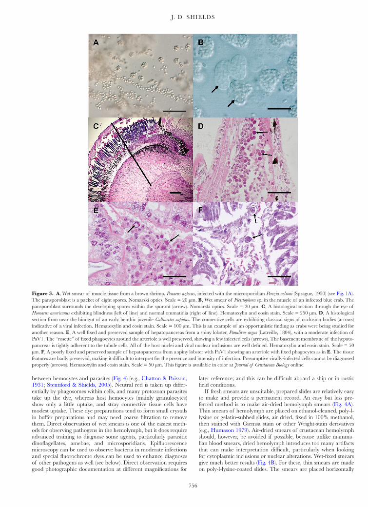
Figure 3. A, Wet smear of muscle tissue from a brown shrimp, Penaeus aztecus, infected with the microsporidian Perezia nelsoni (Sprague, 1950) (see Fig. 1A). The pansporoblast is a packet of eight spores. Nomarski optics. Scale = 20 µm. B, Wet smear of Pleistophora sp. in the muscle of an infected blue crab. The pansporoblast surrounds the developing spores within the sporont (arrow). Nomarski optics. Scale = 20 µm. C, A histological section through the eye of Homarus americanus exhibiting blindness (left of line) and normal ommatidia (right of line). Hematoxylin and eosin stain. Scale = 250 µm. D, A histological section from near the hindgut of an early benthic juvenile Callinectes sapidus. The connective cells are exhibiting classical signs of occlusion bodies (arrows) indicative of a viral infection. Hematoxylin and eosin stain. Scale = 100 µm. This is an example of an opportunistic finding as crabs were being studied for another reason. E, A well fixed and preserved sample of hepatopancreas from a spiny lobster, Panulirus argus (Latreille, 1804), with a moderate infection of PaV1. The “rosette” of fixed phagocytes around the arteriole is well preserved, showing a few infected cells (arrows). The basement membrane of the hepato-pancreas is tightly adherent to the tubule cells. All of the host nuclei and viral nuclear inclusions are well defined. Hematoxylin and eosin stain. Scale = 50 µm. F, A poorly fixed and preserved sample of hepatopancreas from a spiny lobster with PaV1 showing an arteriole with fixed phagocytes as in E. The tissue features are badly preserved, making it difficult to interpret for the presence and intensity of infection. Presumptive virally-infected cells cannot be diagnosed properly (arrows). Hematoxylin and eosin stain. Scale = 50 µm. This figure is available in color at Journal of Crustacean Biology online.
Dow
nloaded from https://academ
ic.oup.com/jcb/article-abstract/37/6/753/4161633 by Serials D
ept -- College of W
illiam and M
ary user on 08 August 2019
COLLECTION FOR PATHOLOGICAL ANALYSIS
757
in a humid chamber for 2–3 minutes to allow adherence of the cells, and then the preparation is fixed in Bouin’s solution or 10% neutral buffered formalin in a Coplin jar. The slides are then returned to the laboratory and processed through a routine hema-toxylin and eosin or other staining procedures (e.g., Messick & Shields, 2000; Pestal et al., 2003). Many staining procedures can be adapted from standard histology texts (e.g., Luna 1968; Humason 1979) depending on specific pathogens and their staining attrib-utes. These methods can be used in the field and aboard ships, and the only significant problem is taking suitable safety precau-tions when transporting fixatives or using them in confined, poorly ventilated spaces.
Quantitative assessments of pathogens can be made from hemolymph samples preserved with a fixative. Using a 27 ga syringe, 100 µl aliquots of hemolymph can be fixed in a 1:10 ratio with ice-cold 10% neutral-buffered formalin (900 µl) or other fixa-tives (e.g., 2% glutaraldehyde in 0.2M sodium cacodylate buffer). Quantitative cell counts of hemocytes or pathogens can then be made using a hemacytometer or flow cytometer. Total hemocytes counts are relatively easy to quantify with a hemacytometer, but differential cell counts require training. For pathogens that can be differentiated from host hemocytes (e.g., take up vital stains), cell counts using a hemacytometer can provide key data on inten-sity of infections, the presence of different life history stages and the relative abundance of pathogens in relation to host cells (e.g., Shields & Squyars, 2000).
The collection of preserved hemolymph samples can be adapted to field collections; however, there are some significant pitfalls to consider. Cells can adhere to the container used for fixation, they can clump due to handling, or the hemolymph constituents can form clots or precipitates with the fixatives, thus skewing the results or making the preparations difficult to assess. Hemolymph with high protein or lipid content, such as that from a female host
undergoing oogenesis, can form flocculants in fixatives making cell densities difficult to estimate in fixed samples (JDS, unpubl. data). In practical experience, polypropylene containers reduce adher-ence of cells and higher ratios of fixative can help reduce clotting, but the type of plastic container and the type of fixative should be tested prior to use (JDS, unpubl. data). Storage periods should also be considered as some fixatives require refrigeration or additional post processing and cells can degrade or clump if held too long (i.e., longer than 1–2 weeks).
Epifluorescence microscopy may also be useful for rapid diag-nosis of pathogens. Hemolymph samples can be fixed directly in cold 10% neutral-buffered formalin or 5% paraformaldehyde. The cells are stained in the laboratory with various fluorescent stains such as fluoroscein isothiocyanate (FITC) or 4’,6-diamid-ino-2-phenylindole (DAPI), gently centrifuged, washed in buffer, and viewed as wet smears with epifluorescence microscopy. DAPI works particularly well for examining nuclear details for Hematodinium infections, because the nucleus of the parasite is often in a metaphase-like state, and the dye shows this character quite well. Fluorochrome dyes also work well for ciliate infections, because their macro- and micronuclei can be visualized as well as dye uptake within the basal bodies of the kineties. Janus green and fast green may provide some contrast to the nuclei of for-malin-fixed cells with suitable results for some protozoans (JDS, pers. obs.).
Agar isolation
Bacterial infections are ubiquitous in crustaceans. In fact, many decapods do not have sterile hemolymph (i.e., Shields et al., 2015b for Brachyura). Bacterial infections can become pathogenic when the host is stressed by handling or environmental stressors, and infected hosts can die quickly to bacterial infections. The bacterial
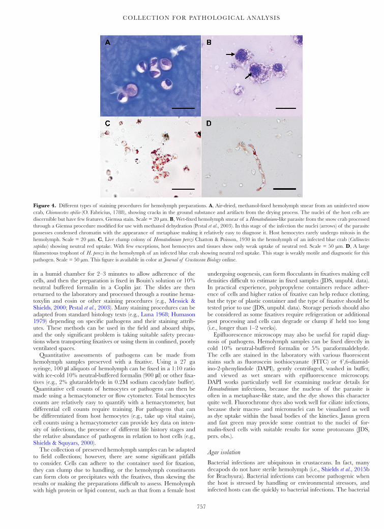
Figure 4. Different types of staining procedures for hemolymph preparations. A, Air-dried, methanol-fixed hemolymph smear from an uninfected snow crab, Chionoecetes opilio (O. Fabricius, 1788), showing cracks in the ground substance and artifacts from the drying process. The nuclei of the host cells are discernible but have few features. Giemsa stain. Scale = 20 µm. B, Wet-fixed hemolymph smear of a Hematodinium-like parasite from the snow crab processed through a Giemsa procedure modified for use with methanol dehydration (Pestal et al., 2003). In this stage of the infection the nuclei (arrows) of the parasite possesses condensed chromatin with the appearance of metaphase making it relatively easy to diagnose it. Host hemocytes rarely undergo mitosis in the hemolymph. Scale = 20 µm. C, Live clump colony of Hematodinium perezi Chatton & Poisson, 1930 in the hemolymph of an infected blue crab (Callinectes sapidus) showing neutral red uptake. With few exceptions, host hemocytes and tissues show only weak uptake of neutral red. Scale = 50 µm. D, A large filamentous trophont of H. perezi in the hemolymph of an infected blue crab showing neutral red uptake. This stage is weakly motile and diagnostic for this pathogen. Scale = 50 µm. This figure is available in color at Journal of Crustacean Biology online.
Dow
nloaded from https://academ
ic.oup.com/jcb/article-abstract/37/6/753/4161633 by Serials D
ept -- College of W
illiam and M
ary user on 08 August 2019

J. D. SHIELDS
758
flora also overgrows the hemolymph of dead crustaceans very quickly rendering it difficult to determine the underlying cause of death. The classical method for examining bacterial infections is to streak sterilely-collected hemolymph onto agar plates or broth. Some species such as Aerococcus viridans Williams, Hirch & Cowan, 1953, specifically A. viridans var. homari, the causative agent of gaffkemia in lobsters, and Vibrio spp. are isolated and grown on specific agars (Lavallée et al., 2001; Shields et al., 2012a) that are commercially available. Others are isolated on a general medium such as marine agar (Difco® 2216, Thermo Fisher, Waltham, MA, USA) and then purified or assessed on other media. For larger crabs and lobsters, hemolymph is taken from the juncture of the basis and ischium of the fifth pereopod with a 27 ga nee-dle on a 1 ml tuberculin syringe. The sample site is swabbed with 95% ethanol prior to the hemolymph draw and a few drops of hemolymph are expressed directly on agar plates and incubated at room temperature. Plates are assessed for colony growth after 48–72 hrs. For smaller crustaceans, glass pipettes or capillary tubes can be drawn out into very small syringe-like needles over an open flame and then used to obtain microliter quantities of hemolymph from an arthrodial membrane or heart puncture. These mini-syringes require some skill to make and the procedure is best done in the laboratory setting following good laboratory practices and safety protocols. Bacterial isolates can be stored frozen in sterile 10% glycerol for later reconstitution and identification.
HISTOLOGICAL METHODS
Histology remains a standard assessment tool for disease diagno-sis in histopathology and disease assessment. It provides informa-tion on the state of the host tissues, the etiology of disease, the level of infection, and pathological alterations of affected tissues. Histological identification and assessment of diseases requires proper fixation and handling of the tissues of interest. There are several concerns for tissues collected from field samples. Appropriate dissection and handling of the tissues must be con-ducted to ensure proper penetration of fixative into the tissues of interest and several tissues or organs have specific requirements with respect to fixation and embedding. For example, an important feature of nearly all crustaceans is that their cuticle provides an impervious barrier to fixation; hence dissection is usually required to provide good penetration of the fixatives into the tissues of interest. Cuticle, foregut, or gill preparations may also require a short decalcification period for proper sectioning, whereas gonads often require different fixatives as well as longer embedding times in paraffin. This necessitates the use of multiple cassettes, specific cataloging systems for identification numbers and specimen track-ing, assorted fixatives for different tissues, storage containers, and management of hazardous wastes when collecting specimens for analysis. There are excellent texts on histological techniques (e.g., Luna, 1968; Humason, 1979) and ideally more than one should be used for reference.
Fixatives and fixation
The correct fixation of the tissues is one of the most important steps for proper diagnosis, because poor fixation can render tissues useless for histological assessment. This often happens in field situations when there is an incomplete understand-ing of the role of proper fixation, or when resources are lim-ited. Three issues are absolutely critical to proper fixation: appropriate dissection, correct choice of fixative, and a suit-able fixation method that includes proper ratios of tissue to fixative. Appropriate dissection means that specimens are dis-sected and processed such that the fixatives penetrate the tissues of interest. The fixative must penetrate rapidly into the tissues
to produce good results. For small crustaceans such as copep-ods or small amphipods, no dissection may be necessary or small pin pricks may provide suitable entry points for fixation. For larger specimens, a break in the cuticle is required for the proper penetration of the fixatives; even larger specimens, such as market-sized crabs or lobsters, must be dissected and their tissues placed directly into fixative. Sometimes field sampling requires fixing whole specimens. In such cases, large amphipods, small crabs (< 30 mm carapace width) and juvenile lobsters (< 30 mm carapace length) should be cut in half prior to fixation. For larger specimens, placing tissue samples directly into histol-ogy cassettes will save time and effort as they can be processed through from fixation to paraffin embedding with little addi-tional handling. Many cassettes are designed to fit directly into the chuck on the microtome, further reducing handling time.
The choice of fixative is important because tissues have dif-ferent requirements for appropriate fixation and preservation. Bouin’s solution and Davidson’s fixative are good general fixa-tives because they have good penetration into the tissues, prepare the tissue for histological stains, and give superior staining results with hematoxylin and eosin stains and other staining techniques. Bouin’s solution, however, has two significant limitations: picric acid can be difficult to work with in certain situations, because when it dries out it can form unstable crystals that are unstable and potentially explosive, and it hydrolyzes DNA more rapidly than other fixatives, making it less desirable for molecular-based in situ hybridization techniques (e.g., visualization of a patho-genic virus). The picric acid in Bouin’s solution nonetheless serves as a counter stain and provides outstanding color to tis-sues prepared for hematoxylin and eosin stains. Tissues should be fixed in a few different fixatives, such as Bouin’s solution, 10% neutral buffered formalin, Z-fix® (Anatech, Battle Creek, MI, USA) or SafeFix® II (Thermo Fisher, Waltham, MA, USA) to provide good assessment of the fixation protocol. SafeFix® II is easier to employ in field settings because it has less volatility. Neutral-buffered formalin, Z-fix®, and SafeFix® II are fixatives of choice for molecular applications, such as in situ hybridiza-tions (e.g., Bruce et al., 1993; Carnegie et al., 2003; Li et al., 2006; Small et al., 2007); however, commercial fixatives can be sig-nificantly more expensive than 10% neutral-buffered formalin. Davidson’s fixative can be modified with seawater to provide a buffered fixative. It is an excellent fixative for histology as well as for whole mounts of helminthes.
Proper fixation is the most important step in histological assess-ment (Fig. 3C–F). The ratio of tissue to fixative is critical, and it should never be less than 1:10. The size of the tissue sample and the container used in fixation are also important because the fixa-tive has to penetrate throughout the tissue for proper fixation. For paraffin histology, pieces of tissue no larger than 10–20 mm in size are ideal, but larger sizes can be processed depending on the nature of the tissue. If possible, the tissues should be moved out of the fixative into a preservative (70% ethanol) no later than 48–72 hours after fixation, but fixatives such as 10% neutral buffered for-malin can be used to hold specimens for longer periods (weeks) if necessary.
Some tissues require special care and handling for good his-tological assessment. For example, the hepatopancreas degrades very rapidly and is highly sensitive to weak fixation, poor buffer-ing, and poor penetration of fixatives (Fig. 3F). The hepatopan-creas of crustaceans that have been dead for longer than 10–15 minutes rapidly degrade due to autolysis, so post-mortem changes can render this organ difficult to assess even after short periods. Muscle and spongy connective tissues, however, can often provide useful information in post-mortem situations. Lipid-rich tissues such as ovaries may require fixatives with good penetration, such as alcohol-formalin-acetic acid (AFA), longer paraffin infiltration times, or longer times in vacuum-assisted paraffin infiltration.
Dow
nloaded from https://academ
ic.oup.com/jcb/article-abstract/37/6/753/4161633 by Serials D
ept -- College of W
illiam and M
ary user on 08 August 2019

COLLECTION FOR PATHOLOGICAL ANALYSIS
759
Steedman’s ester wax method is a useful alternative embedding technique for tissues rich in lipids (Humason, 1979).
Cuticular structures and decalcification
The crustacean cuticle presents a significant barrier to the histolo-gist. For lightly sclerotized cuticles, such as gills and pleopods, short decalcification periods (3–6 hours) in decalcification fluid (e.g., the formic acid - sodium citrate method of Luna, 1968) may be all that is required. For heavily sclerotized pieces, such as eyestalks (Fig. 3C), sections of carapace, claw, and whole bodies, longer decalcification is necessary (overnight or > 12 h). After decalcifi-cation, specimens may require additional trimming or processing. For example, after decalcification, lobster eyes can be safely halved with a single-edged razor blade (not a scalpel) with much less damage to the delicate support network holding the ommatidia in place (Maniscalco & Shields, 2007; Magel et al., 2009; Shields et al., 2012a). Bisecting the eye without decalcification ruins these delicate structures. In another example, an ongoing epidemic of shell disease in the American lobster (see Castro & Somers, 2012; Shields, 2013) required examining the cuticle of affected animals in some detail. Using bone shears or heavy scissors, cuticle samples were taken from the dorsal and ventral surfaces and other loca-tions from symptomatic and asymptomatic hosts. Cuticle pieces included a portion of the underlying epidermis, which revealed clues to the presence of a pseudomembrane that partially defined the disease syndrome (Smolowitz et al., 2005a, b; Shields et al., 2012a). One problem with decalcification is that the reagents, par-ticularly formic acid, can damage the integrity of nucleic acids, and thus interfere with in situ hybridization techniques, so decal-cification times should be reduced if the technique is to be used. Some histological procedures cause tissues such as the cuticle to become brittle, but limiting preservation time and reducing dehy-dration times may resolve this development.
ELECTRON MICROSCOPY
For certain diseases, transmission electron microscopy (TEM) is a critical diagnostic tool in support of field collections (e.g., Stentiford & Feist, 2005); and seminal papers on viral etiologies in crustaceans include TEM as a primary tool for identification. Although the method has lost its prominence in many diagnostic laboratories, it still offers a powerful means of identifying micro-bial pathogens such as viral and rickettsial agents. Many of the methods used in TEM can be difficult to employ, and tissues require tedious embedding in plastics as well as significant techni-cal training in ultramicrotomy. Some methods, however, can be quite simple, such as in the negative staining of hemolymph sam-ples for spiroplasms in Eriocheir sinensis H. Milne Edwards, 1853 as in Wang & Gu (2002), or negative staining of viral particles from some types of viral purifications as in Adams & Bonami (1991).
Histological assessment, with additional sampling for TEM, should be included in field studies or field-based collections where known or suspect viral pathogens are present. There are signifi-cant constraints to using TEM for processing large numbers of samples. The fixatives employed in TEM are hazardous, par-ticularly glutaraldehyde and osmium tetra-oxide. A single good fixative for both paraffin histology and TEM is problematic, but 1G:4F (1 part glutaraldehyde to 4 parts formalin; Luna, 1968) has been used with mixed results in surveys of oyster diseases (e.g., Carver et al., 2010). Tissues often can be left in the primary fixa-tive in the refrigerator for short periods, up to a few weeks, so it is possible to use glutaraldehyde in short-term field situations, but it can be difficult to transport as it presents a significant chemical hazard. It is best to ship whole live animals showing signs of infec-tion to the diagnostic laboratory where tissues can be processed for TEM as well as other assessments. It may not be possible to
ship infected hosts across international boundaries, however, even if the specimens have been biologically fixed. One solution is to contact a research hospital in the area of collection because they may offer services for TEM fixation and plastic embedding (e.g., Xu et al., 2007). Once embedded in paraffin or plastic, specimens no longer represent a chemical hazard and can be shipped safely and easily.
MOLECULAR METHODS
Molecular methods are now routine for disease diagnosis. Molecular assays, such as the polymerase chain reaction (PCR), in situ hybridization (ISH) and quantitative PCR, are extremely valu-able diagnostic tools. These methods give information on a specific pathogen but provide little to no information on other organisms that may be present in a sample. It is therefore beneficial to include other methods, such as direct observation and histology, in the repertoire of routine diagnostic techniques for assessment of path-ogens. High-throughput sequencing has been used in some situa-tions to help identify a pathogen but it is not practical for use in most field or fishery applications (Hewson et al., 2014).
As with histological fixation, nucleic acid analysis requires cor-rect preservation of tissues and pathogens. There are two standard methods for preserving tissues for later molecular analysis: freez-ing specimens at –20º C or lower or preserving samples in 95% ethanol. For work with microbial pathogens, hemolymph or other tissue samples can be adequately preserved by these methods, and the preservation can last many years. For most DNA-based diag-nostics, hemolymph samples can be stored frozen in anticoagu-lants such as citrate-EDTA buffers (see Söderhäll & Smith, 1983; Durliat & Vranckx, 1989), or stored neat in frozen 1 ml aliquots, or by preserving 1:10 in 95% ethanol and storing at room temper-ature. A minimum ratio of 1 part tissue to 10 parts preservative is best for proper preservation. Other fixatives, such as rum or iso-propyl alcohol, may work in short-term or emergency situations. For example, we have used rum to successfully preserve samples as a short-term alternative to avoid purchasing and shipping reagent-grade ethanol to and from overseas locations (Moss et al., 2013). Note that rum or 70% ethanol is not an adequate long-term pre-servative for DNA diagnostics (Dean et al., 2001; Vink et al., 2005), and lysis buffers used in many DNA extraction kits are not pre-servatives but rather buffers used in extraction protocols. Samples to be preserved or archived must be stored either in the freezer or in 95% ethanol with an eye to long-term curation. For expres-sion or transcriptomic work, tissues should be stored routinely in fixatives such as RNAlater® (Thermo Fisher, Waltham, MA, USA) that allow gene expression studies (e.g., Hasson et al., 1997). Freshly collected tissue samples in RNAlater® can be stored for several months at –80º C for later gene expression studies (Beale et al., 2008).
Validation of molecular tools
Molecular tools are valuable for establishing the presence of a pathogen, but they do require interpretation. Pathogens can for instance be present within or on a prospective host organism, but they may not necessarily be infecting that organism; hence, it is critical to confirm that the pathogen is indeed within the tissues of the host (Burreson, 2008). This is crucial because many labo-ratories rely heavily on polymerase chain reaction (PCR) meth-ods, which simply give presence or absence of a pathogen, and sometimes the primers are not designed or tested adequately to diagnose a pathogen correctly (Claydon et al., 2004). Moreover, PCR-based assays often have a very high specificity to strains of an organism from a specific region. If an assay is to be used on samples from outside the parameters used for its development, then samples and parameters should be re-validated to avoid the
Dow
nloaded from https://academ
ic.oup.com/jcb/article-abstract/37/6/753/4161633 by Serials D
ept -- College of W
illiam and M
ary user on 08 August 2019

J. D. SHIELDS
760
possibility of false negatives (Carnegie et al., 2016). Validation includes sequencing samples to prove that the DNA from the pathogen of interest is being amplified appropriately (Claydon et al., 2004; Burreson, 2008).
Where possible, one should evaluate hosts using a variety of techniques, including in situ hybridization (ISH) methods to con-firm infections (e.g., Carnegie et al., 2003; Li et al., 2006; Small et al., 2007; Burreson, 2008). ISH can be used to validate the pres-ence of a pathogen in the tissues or whether it is adhering to the carapace of exposed animals or is passing through the digestive tract without actually infecting host tissues. This is important for microbial agents that may be present in the water column, per-haps adhering to potential hosts but not infecting them. ISH is also useful for attempting to piece together complex life cycles, tis-sue affinities, or to find rare life history stages that may use several host species (e.g., Audemard et al. 2002).
Molecular and immunological methods allow for the rapid screening of large numbers of samples. It can nevertheless be costly to perform PCR assays on hundreds of individual sam-ples, particularly if prevalence of a pathogen is very low. With the caveat that one must define presence versus infection as indicated above, pooling samples is an excellent way to conserve resources as well as screen large numbers of samples. Several pooling algo-rithms have been developed for estimating disease prevalence in host populations (Worlund & Taylor, 1983; Litvak et al., 1994, Hedt & Pagano, 2008). Simple pooling is efficient when preva-lence is low (< 5%), and the lowest confidence intervals can be obtained from equal pool sizes (Litvak et al., 1994; Williams & Moffitt, 2001). There can be issues, however, with sample size, quantity of host versus pathogen DNA, and equal versus une-qual sample sizes in pooled samples (Worlund & Taylor, 1983; Thorburn, 1996; Williams & Moffitt, 2001). Unequal sample sizes can be used to improve the estimates of confidence intervals when prevalence approaches 10% or more (Williams & Moffitt, 2001). The size of the sample pool and the efficiency (sensitivity) of the PCR method as well as the quantity of host versus pathogen DNA has to be optimized for the method to work well.
Even with the advent of molecular diagnostics, suitably pre-served reference specimens may still be difficult to obtain. Such was the case during the outbreak of Neoparamoeba pemaquidensis (Page, 1980) in the American lobster in 1999, when there was a lack of properly preserved samples of the closely related Paramoeba perniciosa Sprague, Beckett and Sawyer, 1969 for much needed comparisons (Mullen et al., 2004). With the emergence of new pathogens, retrospective studies of properly preserved specimens of related species can provide important insights into diagnostics. Paramoeba perniciosa is a rare parasite of blue crabs, and it is difficult to diagnose using cytological methods (Messick, 2002). Samples of P. perniciosa were initially not available in suitable fixatives for molecular comparisons. My existing samples of P. perniciosa were unfortunately lost when an ultracold freezer malfunctioned; and this, further demonstrates that collections used in field samples must be properly preserved, stored, and curated to retain their value (e.g., Shields et al., 2012b).
FIELD COLLECTIONS
Populations of many commercially important crustaceans are monitored through intensive pre- or post-harvest biomass sur-veys. Nonetheless, when disease outbreaks arise in commercially exploited crustaceans, it can be difficult to obtain well-fixed and preserved tissues that are suitable for diagnosis of the patho-gens involved. Further issues often arise in terms of sample size, observer bias, and the management of data, including important metadata, or the resulting data stream can overwhelm existing storage resources. Some thought must therefore be given to host and sample collection, the number of samples to be collected and
processed, and data management and collective sharing of the information.
For estimates of prevalence, it is important to have reasonable sample sizes and to minimize sample or collection biases (e.g., Gregory & Blackburn, 1991, Jovani & Tella, 2006, Shields et al. 2017). Prevalence can be biased by low sample sizes; hence, req-uisite sample sizes must be considered before beginning any field assessments of disease. It also can be difficult to process large sam-ple sizes of hosts in a timely manner, both in the field and in the laboratory. Pilot studies or trial runs can help determine bottle-necks in processing or provide insights into methods to avoid. One relatively straightforward means to sample large field collections is through randomization of host individuals. Randomization can be as simple as sampling every other host or every xth host in a trap or trawl, with the caveat that the first host in the sample stream must be randomly selected. Trap or trawl hauls also can be simi-larly randomized for sampling and all individuals in the selected trap or trawl then sampled. More focused or biased collection of diseased animals is also important as it provides material for identification and documentation of the pathogen(s) and allow for the optimization of additional diagnostics (e.g., Pestal et al., 2003; Shields et al., 2005, 2007).
Managing the data stream
Data management can be one of the more difficult aspects of coordinating large field-based programs. A collection of large numbers of animals from disparate workers can rapidly generate an overwhelming amount of data. I prefer to use a project abbrevi-ation with consecutive accession numbers for specimens, much like that used in museum collections. Other methods also work well. For example, one large field-based study on PaV1 in the Caribbean spiny lobster used the initials of the boat captains followed by con-secutive numbers for each host animal that was sampled (JDS, unpubl. data). The boat crews were recorded separately by date making it relatively easy to cross reference and access the data.
For large studies, it is often necessary to assign several identifica-tion numbers to samples because of the different data generated by collaborators. That is, field and host data may reside in one data set whereas histological or PCR data may reside in another, and the identification numbers are cross-referenced for ease in ref-erence and quality assurance. Although this may seem trivial, it can be difficult to implement and track data sets through time, particularly after the completion of the data collection component of a study. A good identification system is thus required. Although computer databases can be specifically designed for such track-ing, their implementation requires additional labor, training and documentation.
Field collections often have a veritable “boat load” of environ-mental or collection data that adds to the complexity and wealth of information needed to assess the ecology of the hosts and their diseases. The associated metadata must be curated properly so it is not lost or poorly managed between collaborators. Data sets and databases must be designed to incorporate and archive metadata collected for field samples (e.g., environmental conditions, station data) as well as for that generated in the laboratory (e.g., differ-ent methods for diagnosis, different observers). Such databases require significant design and layout to incorporate the varieties of data to be stored and analyzed (e.g. Fig. 5). We routinely scan all data sheets and retain them in a “cloud”-based storage service with photographic documentation of histological samples and field specimens. Processed data files are stored similarly when they are completed, thus maintaining a coherent storage area for all of the data.
By way of example, in response to the severe decline of the American lobster, H. americanus, in the waters of Long Island Sound, NY, USA, concomitant with increased prevalence of epizootic shell disease, a $2.3 million research initiative was
Dow
nloaded from https://academ
ic.oup.com/jcb/article-abstract/37/6/753/4161633 by Serials D
ept -- College of W
illiam and M
ary user on 08 August 2019

COLLECTION FOR PATHOLOGICAL ANALYSIS
761
funded to monitor and study lobster health. Part of the initia-tive included the “100 Lobsters” project in which one laboratory served as a central point for dissecting, distributing, and archiving tissues as well as for data storage (Shields et al., 2012a, b). Project goals were to sample 100 lobsters for joint analyses among several participating laboratories. Carapace, hemolymph, and various tissues were dissected, preserved accordingly and sent to several
collaborators for further assessment (histology, gene expression, metal contamination, contaminants exposure). A data sheet was developed prior to the work that served as a checklist to ensure that tissues and data were obtained for each animal entering the study (Fig. 5). It also facilitated the later distribution of tis-sue samples. A component of the project involved coordinating data access for the 100 lobsters to researchers, fisheries managers,
Figure 5. Sample data sheet from the “100 Lobsters” project used by laboratory personnel to facilitate tissue sampling, storage, shipment and receipt of samples (Shields et al., 2012b). The data sheet was vetted among research groups to include tissues of interest to all collaborators.
Dow
nloaded from https://academ
ic.oup.com/jcb/article-abstract/37/6/753/4161633 by Serials D
ept -- College of W
illiam and M
ary user on 08 August 2019
J. D. SHIELDS
762
fishermen, and laypeople in the form of a website (www.uglylob-ster.org). The project is a work in progress but it serves as a useful tool for understanding the complexity of this disease phenom-enon in Long Island Sound.
ACKNOWLEDGEMENTS
I thank K. Wheeler and Drs. C. Li, J. Moss and H. Small for reviewing an early version of the manuscript. This work was par-tially supported by NSF grants OCE-0929086 and OCE BE-UF-0723662. This is contribution no. 3647 from the Virginia Institute of Marine Science.
REFERENCESAdams, J.R. & Bonami, J. R. 1991. Atlas of invertebrate viruses, CRC Press,
Boca Raton, FL.Audemard, C., Le Roux,F., Barnaud, A., Collins, C., Sautour, B., Sauriau,
P.G., De, Montaudouin, X., Coustau, C., Combes, C. & Berthe, F. 2002. Needle in a haystack: involvement of the copepod a Paracartia grani in the life-cycle of the oyster pathogen marteilia refringens. Parasitology, 124: 315–323.
Beale, K.M., Towle, D.W., Jayasundara, N., Smith, C.M., Shields, J.D., Small, H.J. & Greenwood, S.J. 2008. Anti-lipopolysaccharide factors in the American lobster Homarus americanus: Molecular characteriza-tion and transcriptional response to Vibrio fluvialis challenge. Comparative Biochemistry and physiology. Part D, Genomics & Proteomics, 3: 263–269.
Bruce, L.D., Redman, R.M., Lightner, D.V. & Bonami, J.R. 1993. Application of gene probes to detect a penaeid shrimp baculovirus in fixed tissue using in situ hybridization. Diseases of Aquatic Organisms, 17: 215–221.
Burreson, E.M. 2008. Misuse of PCR assay for diagnosis of mollusc pro-tistan infections. Diseases of Aquatic Organisms, 80: 81–83.
Carnegie, R.B., Arzul, I. & Bushek, D. 2016. Managing marine mollusc diseases in the context of regional and international commerce: pol-icy issues and emerging concerns. Philosophical Transactions of the Royal Society, London, B, 371(1689): 20150215.
Carnegie, R.B., Barber, B.J. & Distel, D.L. 2003. Detection of the oyster parasite Bonamia ostreae by fluorescent in situ hybridization. Diseases of Aquatic Organisms, 55: 247–252.
Carver, C.E., Theriault, I. & Mallet, A.L. 2010. Infection of cultured east-ern oysters Crassostrea virginica by the boring sponge Cliona celata, with emphasis on sponge life history and mitigation strategies. Journal of Shellfish Research, 29: 905–915.
Castro, K. M. & Angell, T.E. 2000. Prevalence and progression of shell dis-ease in American lobster, Homarus americanus, from Rhode Island waters and the offshore canyons. Journal of Shellfish Research, 19: 691–700.
Castro, K.M. & Somers, B.A. 2012. Observations of epizootic shell disease in American lobsters, Homarus americanus, in southern New England. Journal of Shellfish Research, 31: 423–430.
Chaplin-Ebanks, S.A. & Curran, M.C. 2007. Prevalence of the bopyrid isopod Probopyrus pandalicola in the grass shrimp, Palaemonetes pugio, in four tidal creeks on the South Carolina-Georgia coast. Journal of Parasitology, 93: 73–77.
Chatton, E. and Poisson, R. 1931. Sur l’existance, dans le sang des crabes, de péridiniens parasites Hematodinium perezi n.g., n.sp. (Syndinidae). Comptes Rendus des Séances de l’Societé biologique de Paris, 105: 553–557.
Claydon, K., Cullen, B. & Owens, L. 2004. OIE white spot syndrome virus PCR gives false-positive results in Cherax quadricarinatus. Diseases of Aquatic Organisms, 62: 265–268.
Dean, M.D. & Ballard, J.W. 2001. Factors affecting mitochondrial DNA quality from museum preserved Drosophila simulans. Entomologia Experimentalis et Applicata, 98: 279–283.
Durliat, M. & Vranckx, R. 1981. Action of various anticoagulants on hemolymph of lobsters and spiny lobsters. Biological Bulletin, 160: 55–68.
Fabricius, O. 1788. Beskrivelse over den store Gronlandske krabbe. Kongelige Danske Videnskabernes Selskab Skrivter, Nye Samling, 3: 181–190.
Field, R.H. & Appleton, P.L. 1995. A Hematodinium-like infection of the Norway lobster Nephrops norvegicus: observations on pathology and pro-gression of infection. Diseases of Aquatic Organisms, 22: 115–128.
Field, R.H., Chapman, C.J., Taylor, A.C., Neil, D.M. & Vickerman, K. 1992. Infection of the Norway lobster Nephrops norvegicus by a Hematodinium-like species of dinoflagellate on the west coast of Scotland. Diseases of Aquatic Organisms, 13:1–15.
Field, R.H., Hills, J.M., Atkinson, R.J.A., Magill, S. & Shanks, A.M. 1998. Distribution and seasonal prevalence of Hematodinium sp. infection of the Norway lobster (Nephrops norvegicus) around the west coast of Scotland. ICES Journal of Marine Science, 55: 846–858.
Glenn, R.P. & Pugh, T.L. 2006. Epizootic shell disease in American lob-ster (Homarus americanus) in Massachusetts coastal waters: interactions of temperature, maturity, and intermolt duration. Journal of Crustacean Biology, 26: 639–645.
Gregory, R.D. & Blackburn, T.M. 1991. Parasite prevalence and host sam-ple size. Parasitology Today, 7: 316–318.
Hasson, K.W., Hasson, J., Aubert, H., Redman, R.M. & Lightner, D.V. 1997. A new RNA-friendly fixative for the preservation of penaeid shrimp samples for virological detection using cDNA genomic probes. Journal of Virological Methods, 66: 227–236.
Hawkes, C.R., Meyers, T.R. & Shirley, T.C. 1985. The prevalence of the rhizocephalan Briarosaccus callosus Boschma, a parasite in blue king crabs, Paralithodes platypus (Brandt), of southeastern Alaska. In: Proceedings of the International King Crab Symposium, Anchorage, Alaska, January 22–24, 1985, pp. 353–363. University of Alaska Sea Grant, Report 85-12. Anchorage, AK.
Hedt, B.L. & Pagano, M. 2008. A matrix pooling algorithm for disease detection. Harvard University Biostatistics Working Paper Series, paper 57 [www.bepress.com/harvardbiostat/paper57].
Hewson, I., Button, J.B., Gudenkauf, B.M., Miner, B., Newton, A.L., Gaydos, J.K., Wynne, J., Groves, C.L., Hendler, G., Murray, M. & Fradkin, S. 2014. Densovirus associated with sea-star wasting disease and mass mortality. Proceedings of the National Academy of Sciences of the United States, 111: 17278–17283.
Hoenig, J.M., Groner, M.L., Smith, M.W., Vogelbein, W.K., Taylor, D.M., Landers, D.F. Jr., Gauthier, D.T., Sadler, P., Matsche, M., Haines, A., Small, H.J., & Shields, J.D. 2017. Impact of disease on survival of three commercially fished species. Ecological Applications [doi: 10.1002/eap.1595].
Howell, P., Giannini, C. & Benway, J. 2005. Status of shell disease in Long Island Sound. In: State of lobster science: lobster shell disease workshop (Tlusty, M.F., Halvorson, H.O., Smolowitz, R. & Sharma, U., eds.), pp. 106–114, Aquatic Forum Series 05-1. New England Aquarium, Boston, MA.
Humason, G. 1979. Animal tissue techniques, Edn. 4. W.H. Freeman, San Francisco.
Ives, J.E. 1891. Crustacea from the northern coast of Yucatan, the har-bor of Vera Cruz, the west coast of Florida and the Bermuda Islands. Proceedings of the Academy of Natural Sciences of Philadelphia, 43: 176–207.
Jovani, R. & Tella, J.L. 2006. Parasite prevalence and sample size: miscon-ceptions and solutions. Trends in Parasitology, 22: 214–218.
Kuris, A.M., Blau, S.F., Paul, A.J., Shields, J.D. & Wickham, D.E. 1991. Infestation by brood symbionts and their impact on egg mortality in the red king crab, Paralithodes camtschatica, in Alaska: Geographic and temporal variation. Canadian Journal of Fisheries and Aquatic Sciences, 48: 559–568.
Landers, D. F., Jr. 2005. Prevalence and severity of shell disease in American lobster Homarus americanus from eastern Long Island Sound, Connecticut. In: State of lobster science: lobster shell disease workshop (Tlusty, M.F., Halvorson, H.O., Smolowitz, R. & Sharma, U., eds.), pp. 94–97, Aquatic Forum Series 05-1. New England Aquarium, Boston, MA.
Lavallée, J., Hammell, K.L., Spangler, E.S. & Cawthorn, R.J. 2001. Estimated prevalence of Aerococcus viridans and Anophryoides haemophila in American lobsters Homarus americanus freshly captured in the waters of Prince Edward Island, Canada. Diseases of Aquatic Organisms, 46: 231–236.
Latreille, P.A. 1804. Des Langoustes du Muséum national d’Histoire naturelle. Annales du Muséum National d’Histoire Naturelle (Paris), 3: 388–395.
Leach, W.E. 1820. Galatéadées. Dictionnaire des Sciences Naturelles, vol. 18, pp. 49–56. F. G. Levreault, Paris.
Li, C., Shields, J.D., Small, H.J., Reece, K.S., Hartwig, C.L., Cooper, R. & Ratzlaff, R.E. 2006. Diagnosis of Panulirus argus virus 1 (PaV1) in the Caribbean spiny lobster using fluorescence in situ hybridization. Diseases of Aquatic Organisms, 72: 185–192.
Dow
nloaded from https://academ
ic.oup.com/jcb/article-abstract/37/6/753/4161633 by Serials D
ept -- College of W
illiam and M
ary user on 08 August 2019

COLLECTION FOR PATHOLOGICAL ANALYSIS
763
Linnaeus, C. 1758. Systema Naturae per Regna Tria Naturae, Secundum Classes, Ordines, Genera, Species, cum Characteribus, Differentiis, Synonymis, Locis. Vol. 1, Edn. 10. Reformata. Laurentii Salvii, Holmiae [= Stockholm].
Litvak, E., Tu, X.M. & Pagano, M. 1994. Screening for the presence of a disease by pooling sera samples. Journal of the American Statistical Association, 89: 424–434.
Luna, L.G. 1968. Manual of histologic staining methods of the Armed Forces Institute of Pathology, Edn.3. McGraw-Hill, New York.
Magel, DR., Shields, J.D. & Brill, R 2009. Idiopathic lesions and visual deficits in the American lobster (Homarus americanus) from Long Island Sound, NY. Biological Bulletin, 217: 95–101.
Maniscalco, A.M. & Shields, J.D. 2006. Histopathology of idiopathic lesions in the eyes of Homarus americanus from Long Island Sound. Journal of Invertebrate Pathology, 91: 88–97.
Messick, G.A. 1994. Hematodinium perezi infections in adult and juven-ile blue crabs Callinectes sapidus from coastal bays of Maryland and Virginia, USA. Diseases of Aquatic Organisms, 19: 77–82.
Messick, G.A. 2002. A survey for prevalence of Paramoeba spp. in blue crabs along the Atlantic and Gulf coasts. Proceedings of the Annual Conference of the Southeastern Association of Fish and Wildlife Agencies, 56: 105–113.
Messick, G.A. & Shields, J.D. 2000. Epizootiology of the parasitic dinofla-gellate hematodinium sp. In the american blue crab callinectes sapi-dus. Diseases of Aquatic Organisms, 43: 139–152.
Meyers, T.R., Koeneman, T.M., Botelho, C. & Short, S. 1987. The epi-zootiology of a parasitic dinoflagellate, Hematodinium sp., in American blue crabs, Callinectes sapidus. Diseases of Aquatic Organisms, 3: 195–216.
Milne Edwards, H. 1837. Histoire naturelle des Crustacés, comprenant l’anatomie, la physiologie et la classification de ces animaux, Vol. 2. Librairie Encyclopédique de Roret, Paris.
Milne Edwards, H. 1853. Mémoire sur la famille des Ocypodides. Suite (1). Deuxiéme Tribu Principale. Annales des Sciences Naturelles, séries 3, 20: 163–228, pls. 6–11.
Moss, J., Behringer, D., Shields, J.D., Baeza, A., Aguilar-Perera, A., Bush, P.G., Dromer, C., Herrera-Moreno, A., Gittens, L., Matthews, T.R. & McCord, M.R. 2013. Distribution, prevalence, and genetic analysis of Panulirus argus virus 1 (PaV1) from the Caribbean Sea. Diseases of Aquatic Organisms, 104: 129–140.
Mullen, T.E., Russell, S., Tucker, M.T., Maratea, J.L., Koerting, C., Hinckley, L., De Guise, S., Frasca, S. Jr, French, R.A., Burrage, T.G. & Perkins, C., 2004. Paramoebiasis associated with mass mortality of American lobster Homarus americanus in Long Island Sound, USA. Journal of Aquatic Animal Health, 16: 29–38.
Pestal, G.P., Taylor, D.M., Hoenig, J.M., Shields, J.D. & Pickavance, R. 2003. Monitoring the presence of the lethal parasite Hematodinium sp. in snow crabs from Newfoundland. Diseases of Aquatic Organisms, 53: 67–75.
Rathbun, M.J. 1896. The genus Callinectes. Proceedings of the United States National Museum, 18: 349–375, pls. 12–28.
Shields, J.D. 2012. The impact of pathogens on exploited populations of decapod crustaceans. Journal of Invertebrate Pathology, 110: 211–224.
Shields, J.D. 2013. Complex etiologies of emerging diseases in lobsters (Homarus americanus) from Long Island Sound. Canadian Journal of Fisheries and Aquatic Sciences, 70: 1576–1587.
Shields, J.D., Huchin-Mian, J.P., O’Leary, P.A. & Small, H.J. 2017. New insight into the transmission dynamics of the crustacean pathogen Hematodinium perezi (Dinoflagellata) using a novel sentinel methodology. Marine Ecology Progress Series, 573: 73–84.
Shields, J.D. & Squyars, C.M. 2000. Mortality and hematology of blue crabs, Callinectes sapidus, experimentally infected with the parasitic dino-flagellate Hematodinium perezi. Fishery Bulletin, 98: 139–152.
Shields, J.D., Okazaki, R.K. & Kuris, A.M. 1990a. Brood mortality and egg predation by Carcinonemertes epialti on the yellow crab, Cancer antho-nyi. Canadian Journal of Fisheries and Aquatic Sciences, 47: 1275–1281.
Shields, J.D., Sullivan, S.E. & Small, H.J. 2015a. Overwintering of the parasitic dinoflagellate Hematodinium perezi in dredged blue crabs (Callinectes sapidus) from Wachapreague Creek, Virginia. Journal of Invertebrate Pathology, 130: 124–132.
Shields, J.D., Taylor, D.M., O’Keefe, P.G., Colbourne, E. & Hynick, E. 2007. Epidemiological determinants in outbreaks of bitter crab disease (Hematodinium sp.) in snow crabs Chionoecetes opilio from Newfoundland Canada. Diseases of Aquatic Organisms 77: 61–72.
Shields, J.D., Taylor, D.M., Sutton, S.G., O’Keefe, P.G., Ings, D. & Party, A. 2005. Epizootiology of bitter crab disease (Hematodinium sp.) in snow crabs, Chionoecetes opilio from Newfoundland, Canada. Diseases of Aquatic Organisms, 64: 253–264.
Shields, J.S., Wheeler, K.N., & Moss, J. 2012a. Histological assessment of lobsters in the “100 Lobsters” Project. Journal of Shellfish Research, 31: 439–447.
Shields, J.D., Wheeler, K.N., Moss, J., Somers, B. & Castro, K. 2012b. The “100 Lobsters” Project: a cooperative demonstration project for health assessments of lobsters from Rhode Island. Journal of Shellfish Research, 31: 431–438.
Shields, J.D., Wickham, D.E., Blau, S.F. & Kuris, A.M. 1990b. Some impli-cations of egg mortality caused by symbiotic nemerteans for data acqui-sition and management strategies of the red king crab. Proceedings of the International Symposium on King and Tanner Crabs, November 28–30, 1989. Anchorage, Alaska, pp. 397–402. Alaska Sea Grant College Program Report No. 90-04. Anchorage, AK.
Shields, J.D., Williams, J.D. & Boyko, C.B. 2015b. Parasites and patho-gens of Brachyura. In: Decapoda: Brachyura, Treatsie on zoology – anatomy, taxonomy, biology (P. Castro, P. Davie, D. Guinot, F.R. Schram & J.C. von Vaupel Klein, eds.), Vol. 9C-II, pp. 639–774. Brill, Leiden, The Netherlands.
Sloan, N.A. 1984. Incidence and effects of parasitism by the rhizocephalan barnacle, Briarosaccus callosus Boschma, in the golden king crab, Lithodes aequispina Benedict, from deep fjords in northern British Columbia, Canada. Journal of Experimental Marine Biology and Ecology, 84:111–131.
Small, H.J., Shields, J.D., Hudson, K.L. and Reece, K.S. 2007. Molecular detection of the Hematodinium sp. infecting the blue crab, Callinectes sapi-dus. Journal of Shellfish Research, 26: 131–136.
Smolowitz, R., Chistoserdov, A.Y. & Hsu, A. 2005a. A description of the pathology of epizootic shell disease in the American lobster, Homarus amer-icanus H. Milne Edwards 1837. Journal of Shellfish Research, 24: 749–756.
Smolowitz, R., Chistoserdov, A.Y. & Hsu, A. 2005b. Epizootic shell disease in the American lobster, Homarus americanus. In: State of lobster science: lobster shell disease workshop (Tlusty, M.F., Halvorson, H.O., Smolowitz, R. & Sharma, U., eds.), pp. 2–11, Aquatic Forum Series 05-1. New England Aquarium, Boston, MA.
Söderhäll, K. & Smith, V.J. 1983. Separation of the haemocyte popu-lations of Carcinus maenas and other marine decapods, and proph-enoloxidase distribution. Developmental and Comparative Immunology, 7: 229–239.
Stentiford, G.D. & Feist, S.W. 2005. A histopathological survey of shore crab (Carcinus maenas) and brown shrimp (Crangon crangon) from six estuar-ies in the United Kingdom. Journal of Invertebrate Pathology, 88: 136–146.
Stentiford, G.D. & Shields, J.D. 2005. A review of the parasitic dinoflag-ellates Hematodinium species and Hematodinium-like infections in marine crustaceans. Diseases of Aquatic Organisms, 66: 47–70.
Stentiford, G.D., Neil, D.M. & Atkinson, R.J.A. 2001. The relationship of Hematodinium infection prevalence in a Scottish Nephrops norvegicus popu-lation to season, moulting and sex. ICES Journal of Marine Science, 58: 814–823.
Thorburn, M.A. 1996. Apparent prevalence of fish pathogens in asymp-tomatic salmonid populations and its effect on misclassifying popula-tion infection status. Journal of Aquatic Animal Health, 8: 271–277.
Tilesius, W.C., 1815. De Cancris Camtschaticis, Oniscis, Entomostracis et Cancellis marinis microscopicis noctilucentibus, Cum tabulis IV: Aneaeis et appendice adnexo de Acaris et Ricinis Camtschaticis. Auctore Tilesio. Conventui exhibuit die 3 Februarii 1813. Mémoires de l’Académie Impériale des Sciences de Saint-Pétersbourg, 5: 331–405.
Vink, C.J., Thomas, S.M., Paquin, P., Hayashi, C.Y. & Hedin, M. 2005. The effects of preservatives and temperatures on arachnid DNA. Inverterbrate Systematics, 19: 99–104.
Wang, W. & Gu, Z. 2002. Rickettsia-like organism associated with tremor disease and mortality of the Chinese mitten crab Eriocheir sinensis. Diseases of Aquatic Organisms, 48: 149–153.
Wahle, R.A., Gibson, M. & Fogarty, M.J. 2009. Distinguishing disease impacts from larval supply effects in a lobster fishery collapse. Marine Ecology Progress Series, 376: 185–192.
Wickham, D.E. & Kuris, A.M. 1988. Diversity among nemertean egg predators of decapod crustaceans. Hydrobiologia, 156: 23–30.
Williams, C.J. & Moffitt, C.M. 2001. A critique of methods of sampling and reporting pathogens in populations of fish. Journal of Aquatic Animal Health, 12: 300–209.
Worlund, D.D. & Taylor, G. 1983. Estimation of disease incidence in fish populations. Canadian Journal of Fisheries and Aquatic Sciences, 40: 2194–2197.
Xu, W.J., Shi, H., Xu, H.X. & Small, H.J. 2007. Preliminary study on the Hematodinium infection in cultured Portunus trituberculatus. Acta Hydrobiologia Sinica, 31: 637–642 [in Chinese].
Dow
nloaded from https://academ
ic.oup.com/jcb/article-abstract/37/6/753/4161633 by Serials D
ept -- College of W
illiam and M
ary user on 08 August 2019