Embed Size (px)
Citation preview
© 2012 The Korean Academy of Medical Sciences.This is an Open Access article distributed under the terms of the Creative Commons Attribution Non-Commercial License (http://creativecommons.org/licenses/by-nc/3.0) which permits unrestricted non-commercial use, distribution, and reproduction in any medium, provided the original work is properly cited.
pISSN 1011-8934eISSN 1598-6357
Collision of Three Histologically Distinct Endometrial Cancers of the Uterus
A collision tumor is defined by the presence of two separate masses in one organ, which are pathologically distinct. We described a 70-yr-old patient who complained of abnormal vaginal bleeding with a collision tumor of the uterine corpus. The patient received total hysterectomy, bilateral salphingo-oophorectomy, bilateral pelvic-paraaortic lymphadenectomy, omentectomy, and intraperitoneal chemotherapy. The uterine corpus revealed three separate masses, which were located at the fundus, anterior and posterior wall. Each tumor revealed three pathologically different components, which were malignant mixed müllerian tumor, papillary serous carcinoma, and endometrioid adenocarcinoma. Among these components, only the papillary serous carcinoma component invaded the underlying myometrium and metastasized to the regional lymph node. Adjuvant chemotherapy and radiation therapy were performed. The patient is still alive and has been healthy for the last 8 yr. We have reviewed previously reported cases of collision tumors which have occurred in the uterine corpus.
Key Words: Uterus; Collision Tumor; Endometrial Neoplasms
Ki-Seok Jang1, Won-Moo Lee2, Young-Jae Kim2, and Sam-Hyun Cho2
Departments of 1Pathology and 2Obstetrics and Gynecology, College of Medicine, Hanyang University, Seoul, Korea
Received: 2 June 2011Accepted: 1 November 2011
Address for Correspondence:Sam-Hyun Cho, MDDepartment of Obstetrics and Gynecology, College of Medicine, Hanyang University, 345 Wangsimni-ro, Seongdong-gu, Seoul 133-791, KoreaTel: +82.2-2296-8412, Fax: +82.2-2290-8417E-mail: [email protected]
http://dx.doi.org/10.3346/jkms.2012.27.1.89 • J Korean Med Sci 2012; 27: 89-92
CASE REPORTOncology & Hematology
INTRODUCTION
A collision tumor is defined by the coexistence of two adjacent, but histologically distinct tumor components. This tumor is con-sidered a multiple synchronous tumor in a single organ, because these components are separated from each other by stroma with-out histologic admixture (1). Collision tumors have been report-ed in various organs, such as esophagus, stomach, colon, lung, skin, thyroid gland, breast, ovary, and uterus (2). Previously re-ported uterine collision tumors are mainly composed of two different histological components; serous or endometrioid ad-enocarcinoma, and sarcoma or neuroendocrine tumor (2-8). In this report, we describe the clinicopathologic features of an un-usual uterine collision tumor. The tumor is composed of three distinct histologic components, endometrioid adenocarcino-ma, papillary serous carcinoma, and malignant mixed mülleri-an tumor.
CASE DESCRIPTION
On November 14, 2003, a 70-yr-old female visited our hospital due to abnormal uterine bleeding and abdominal pain. Her gy-necologic history included seven full-term pregnancies, six nor-mal deliveries, one still birth, and two abortions. Menarche had occurred at the age of 17 yr and menstruation was on a regular basis. Menopause occurred at the age of 50 yr, and she has never
received hormone replacement therapy. Past medical history includes hypertension, controlled by medication for the past 15 yr. She has no family history of cancer. Pelvic examination dis-closed a normal vagina, unremarkable cervix, and an enlarged uterus. Transvaginal ultrasonography revealed a 9.2 × 5.9 cm-sized mass with a mixed echogenic shadow in the uterus, sug-gesting an endometrial malignancy. Pelvic computed tomogra-phy displayed a large mass, which occupied the entire endome-trial cavity with suspicious infiltration to the myometrium and parametrial soft tissue. The adjacent organs, such as urinary blad-der, rectum, and sigmoid colon, were free of tumor extension. However, the nodular infiltration of omentum was noted, sug-gesting the possible omental seeding. Magnetic resonance im-aging revealed poorly enhancing inhomogeneous mass within the uterine cavity with tumor infiltration around the uterus with no definitive evidence of lymphadenopathy or extension to the adjacent organs. The laboratory test was within normal limit for routine blood count, electrolytes, and chemistry. The tumor marker, CA125, was elevated to 1,132 U/mL (reference range 0.1-35 U/mL). On November 17, 2003, exploratory laparotomy was performed. There was no evidence of distant metastasis on the surface of liver, spleen, diaphragm and peritoneum. However, suspicious metastatic nodules were found on omentum and the surface of sigmoid colon. The patient underwent total abdomi-nal hysterectomy with bilateral salphingo-oophorectomy, total omentectomy, removal of the mass on the surface of sigmoid
Jang K-S, et al. • Collision Tumor of the Uterus
90 http://jkms.org http://dx.doi.org/10.3346/jkms.2012.27.1.89
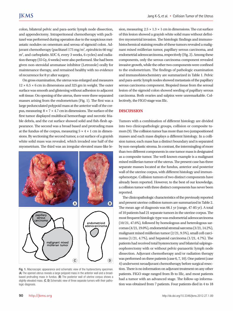
colon, bilateral pelvic and para-aortic lymph node dissection, and appendectomy. Intraperitoneal chemotherapy with pacli-taxel was performed during operation due to the suspicious met-astatic nodules on omentum and serosa of sigmoid colon. Ad-juvant chemotherapy (paclitaxel 175 mg/m2, epirubicin 60 mg/m2, and carboplatin AUC 6, every 3 weeks, 6 cycles) and radia-tion therapy (55 Gy, 6 weeks) were also performed. She had been given non-steroidal aromatase inhibitor (Letrozole) orally for maintenance therapy, and remained healthy with no evidence of recurrence for 8 yr after surgery. On gross examination, the uterus was enlarged and measures 12 × 6.5 × 6 cm in dimensions and 325 gm in weight. The outer surface was smooth and glistening without adhesion to adjacent soft tissue. On opening of the uterus, there were three separated masses arising from the endometrium (Fig. 1). The first was a large pedunculated polypoid mass at the anterior wall of the cor-pus, measuring 8 × 7 × 4.7 cm in dimensions. The surface of the first tumor displayed multifocal hemorrhage and necrotic fria-ble debris, and the cut surface showed solid and fish-flesh ap-pearance. The second was a broad based and protruding mass at the fundus of the corpus, measuring 5 × 4 × 1 cm in dimen-sions. By sectioning the second tumor, a cut surface of a grayish white solid mass was revealed, which invaded one half of the myometrium. The third was an irregular elevated mass-like le-
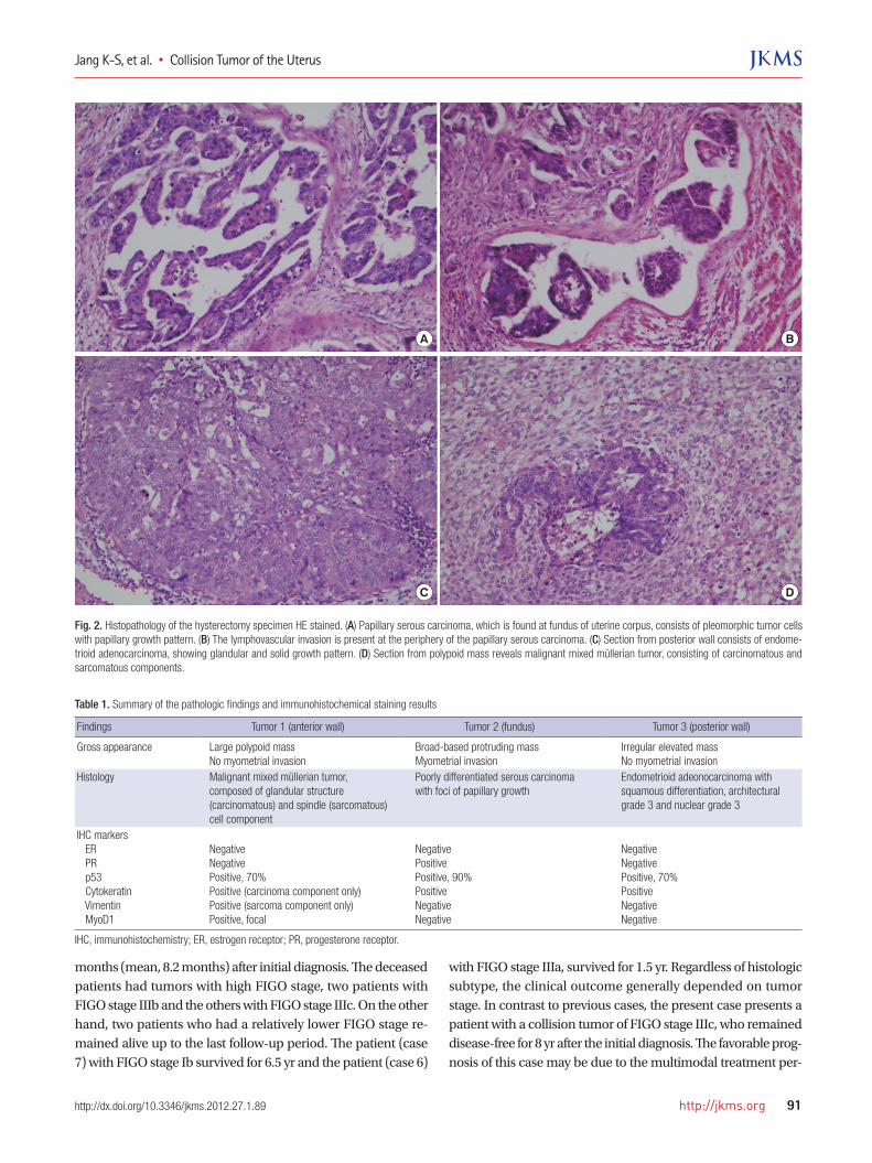
sion, measuring 2.5 × 1.3 × 1 cm in dimensions. The cut surface of this lesion showed a grayish white solid mass without defini-tive myometrial invasion. The histologic findings and immuno-histochemical staining results of these tumors revealed a malig-nant mixed müllerian tumor, papillary serous carcinoma, and endometrial adenocarcinoma, respectively (Fig. 2). Among these components, only the serous carcinoma component revealed invasive growth, while the other two components were confined to the endometrium. The findings of pathologic examination and immunohistochemistry are summarized in Table 1. Pelvic and para-aortic lymph nodes showed metastasis of the papillary serous carcinoma component. Biopsied tissue from the serosal lesion of the sigmoid colon showed seeding of papillary serous carcinoma. Both ovaries and salpinx were unremarkable. Col-lectively, the FIGO stage was IIIc.
DISCUSSION
Tumors with a combination of different histology are divided into two clinicopathologic groups, collision or composite tu-mors (9). The collision tumor has more than two juxtapositioned masses and each mass displays a different histology. In a colli-sion tumor, each mass has a distinct boundary and is separated by non-neoplastic stroma. In contrast, the intermingling of more than two different components in one tumor mass is designated as a composite tumor. The well-known example is a malignant mixed müllerian tumor of the uterus. The present case has three separate masses located at the fundus, anterior and posterior wall of the uterine corpus, with different histology and immun-ophenotype. Collision tumors of two distinct components have already been reported. However, to the best of our knowledge, a collision tumor with three distinct components has never been reported. The clinicopathologic characteristics of the previously reported and present uterine collision tumors are summarized in Table 2. The mean age of diagnosis was 66.1 yr (range, 47-85 yr). A total of 10 patients had 21 separate tumors in the uterine corpus. The most frequent histologic type was endometrial adenocarcinoma (10/21, 47.6%), followed by homologous and heterologous sar-comas (4/21, 19.0%), endometrial stromal sarcoma (3/21, 14.2%), malignant mixed müllerian tumor (2/21, 9.5%), small cell carci-noma (1/21, 4.7%), and hepatoid carcinoma (1/21, 4.7%). The patients had received total hysterectomy and bilaterial salpingo-oophorectomy with or without pelvic-paraaortic lymph node dissection. Adjuvant chemotherapy and/or radiation therapy was performed on three patients (case 6, 7, 10). One patient (case 4) underwent neoadjuvant chemotherapy before surgical resec-tion. There is no information on adjuvant treatment on any other patients. FIGO stage ranged from Ib to IIIc, and most patients had a tumor with an advanced stage. The follow-up informa-tion was obtained from 7 patients. Four patients died in 4 to 18
Fig. 1. Macroscopic appearance and schematic view of the hysterectomy specimen. (A) The opened uterus reveals a large polypoid mass in the anterior wall and a broad-based protruding mass in fundus. (B) The posterior wall of uterine corpus shows a slightly elevated mass. (C, D) Schematic view of three separate tumors with their patho-logic diagnosis.
A
C
B
D
Jang K-S, et al. • Collision Tumor of the Uterus
http://jkms.org 91http://dx.doi.org/10.3346/jkms.2012.27.1.89
Table 1. Summary of the pathologic findings and immunohistochemical staining results
Findings Tumor 1 (anterior wall) Tumor 2 (fundus) Tumor 3 (posterior wall)
Gross appearance Large polypoid massNo myometrial invasion
Broad-based protruding massMyometrial invasion
Irregular elevated massNo myometrial invasion
Histology Malignant mixed müllerian tumor, composed of glandular structure (carcinomatous) and spindle (sarcomatous) cell component
Poorly differentiated serous carcinoma with foci of papillary growth
Endometrioid adeonocarcinoma with squamous differentiation, architectural grade 3 and nuclear grade 3
IHC markers ER PR p53 Cytokeratin Vimentin MyoD1
NegativeNegativePositive, 70%Positive (carcinoma component only)Positive (sarcoma component only)Positive, focal
NegativePositivePositive, 90%PositiveNegativeNegative
NegativeNegativePositive, 70%PositiveNegativeNegative
IHC, immunohistochemistry; ER, estrogen receptor; PR, progesterone receptor.
A B
C D
Fig. 2. Histopathology of the hysterectomy specimen HE stained. (A) Papillary serous carcinoma, which is found at fundus of uterine corpus, consists of pleomorphic tumor cells with papillary growth pattern. (B) The lymphovascular invasion is present at the periphery of the papillary serous carcinoma. (C) Section from posterior wall consists of endome-trioid adenocarcinoma, showing glandular and solid growth pattern. (D) Section from polypoid mass reveals malignant mixed müllerian tumor, consisting of carcinomatous and sarcomatous components.
months (mean, 8.2 months) after initial diagnosis. The deceased patients had tumors with high FIGO stage, two patients with FIGO stage IIIb and the others with FIGO stage IIIc. On the other hand, two patients who had a relatively lower FIGO stage re-mained alive up to the last follow-up period. The patient (case 7) with FIGO stage Ib survived for 6.5 yr and the patient (case 6)
with FIGO stage IIIa, survived for 1.5 yr. Regardless of histologic subtype, the clinical outcome generally depended on tumor stage. In contrast to previous cases, the present case presents a patient with a collision tumor of FIGO stage IIIc, who remained disease-free for 8 yr after the initial diagnosis. The favorable prog-nosis of this case may be due to the multimodal treatment per-
Jang K-S, et al. • Collision Tumor of the Uterus
92 http://jkms.org http://dx.doi.org/10.3346/jkms.2012.27.1.89
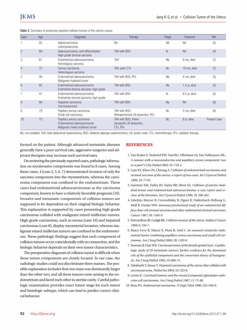
Table 2. Summary of previously reported collision tumors of the uterine corpus
Case Age Diagnosis Therapy Stage Outcome Ref.
1 55 AdenocarcinomaLeiomyosarcoma
NA NA NA (5)
2 69 Adenocarcinoma, well differentiatedHigh-grade stromal sarcoma
TAH with BSO Ib NA (4)
3 67 Endometrioid adenocarcinomaHomologous sarcoma
TAH IIIb 6 mo, died (7)
4 72 Serous carcinomaHeterologous sarcoma
TAH, prior CTx IIIc 18 mo, died (7)
5 49 Endometrioid adenocarcinomaMalignant rhabdoid tumor
TAH with BSO, RTx IIIb 4 mo, died (3)
6 85 Endometrioid adenocarcinomaEndmetrial stromal sarcoma, high-grade
TAH with BSO IIIa 1.5 yr, alive (2)
7 47 Endometrioid adenocarcinomaEndmetrial stromal sarcoma, high-grade
TAH with BSO Ib 6.5 yr, alive (2)
8 68 Hepatoid carcinomaCarcinosarcoma
TAH with BSO IIIa NA (8)
9 79 Papillary serous carcinomaSmall cell carcinoma
TAH with BSO, Retroperitoneal LN dissection, RTx
IIIc 5 mo, died (6)
10 70 Papillary serous carcinomaEndometrioid adenocarcinomaMalignant mixed müllerian tumor
TAH with BSO, Pelvic-paraaortic LN dissection,CTx, RTx
IIIc 8 yr, alive Present case
NA, not available; TAH, total abdominal hysterectomy; BSO, bilateral salpingo-oophorectomy; LN, lymph node; CTx, chemotherapy; RTx, radiation therapy.
formed on the patient. Although advanced metastatic diseases generally have a poor survival rate, aggressive surgeries and ad-juvant therapies may increase such survival rates. On reviewing the previously reported cases, pathologic informa-tion on myoinvasive components was found in 8 cases. Among these cases, 4 (case 2, 5, 6, 7) demonstrated invasion of only the sarcoma component into the myometrium, whereas the carci-noma component was confined to the endometrium. These cases had endometrioid adenocarcinomas as the carcinoma component, known to have a relatively favorable prognosis (10). Invasive and metastatic components of collision tumors are supposed to be dependent on their original biologic behavior. This explanation is supported by cases presenting high-grade carcinomas collided with malignant mixed müllerian tumors. High-grade carcinomas, such as serous (case 10) and hepatoid carcinomas (case 8), display myomterial invasion, whereas ma-lignant mixed müllerian tumors are confined to the endometri-um. These pathologic findings suggest that each component of collision tumors occur coincidentally with no connection, and the biologic behavior depends on their own tumor characteristics. The preoperative diagnosis of collision tumor is difficult when these tumor components are closely located. In our case, the radiologic studies could not discriminate three masses. The pos-sible explanation includes that one mass was dominantly larger than the other two, and all these tumors were arising in the en-dometrium and faced each other in uterine cavity. Careful patho-logic examination provides exact tumor stage for each tumor and histologic subtype, which can lead to predict correct clini-cal behavior.
REFERENCES
1. Van Eeden S, Nederlof PM, Taal BG, Offerhaus GJ, Van Velthuysen ML.
A tumour with a neuroendocrine and papillary serous component: two
or a pair? J Clin Pathol 2002; 55: 710-4.
2. Lam KY, Khoo US, Cheung A. Collision of endometrioid carcinoma and
stromal sarcoma of the uterus: a report of two cases. Int J Gynecol Pathol
1999; 18: 77-81.
3. Gaertner EM, Farley JH, Taylor RR, Silver SA. Collision of uterine rhab-
doid tumor and endometrioid adenocarcinoma: a case report and re-
view of the literature. Int J Gynecol Pathol 1999; 18: 396-401.
4. Lifschitz-Mercer B, Czernobilsky B, Dgani R, Dallenbach-Hellweg G,
Moll R, Franke WW. Immunocytochemical study of an endometrial dif-
fuse clear cell stromal sarcoma and other endometrial stromal sarcomas.
Cancer 1987; 59: 1494-9.
5. Patwardhan JR, Gadgil RK. Collision tumour of the uterus. Indian J Cancer
1969; 6: 194-7.
6. Shaco-Levy R, Manor E, Piura B, Ariel I. An unusual composite endo-
metrial tumor combining papillary serous carcinoma and small cell car-
cinoma. Am J Surg Pathol 2004; 28: 1103-6.
7. Sreenan JJ, Hart WR. Carcinosarcomas of the female genital tract. A patho-
logic study of 29 metastatic tumors: further evidence for the dominant
role of the epithelial component and the conversion theory of histogene-
sis. Am J Surg Pathol 1995; 19: 666-74.
8. Takahashi Y, Inoue T. Hepatoid carcinoma of the uterus that collided with
carcinosarcoma. Pathol Int 2003; 53: 323-6.
9. Lewin K. Carcinoid tumors and the mixed (composite) glandular-endo-
crine cell carcinomas. Am J Surg Pathol 1987; 11: 71-86.
10. Rose PG. Endometrial carcinoma. N Engl J Med 1996; 335: 640-9.