Embed Size (px)
Citation preview
Combinatorial Expression Patterns of LIM-Homeodomain andOther Regulatory Genes Parcellate Developing Thalamus
Yasushi Nakagawa and Dennis D. M. O’Leary
Molecular Neurobiology Laboratory, The Salk Institute, La Jolla, California 92037
The anatomical and functional organization of dorsal thalamus(dTh) and ventral thalamus (vTh), two major regions of thediencephalon, is characterized by their parcellation into distinctcell groups, or nuclei, that can be histologically defined inpostnatal animals. However, because of the complexity of dThand vTh and difficulties in histologically defining nuclei at earlydevelopmental stages, our understanding of the mechanismsthat control the parcellation of dTh and vTh and the differenti-ation of nuclei is limited. We have defined a set of regulatorygenes, which include five LIM-homeodomain transcription fac-tors (Isl1, Lhx1, Lhx2, Lhx5, and Lhx9) and three other genes(Gbx2, Ngn2, and Pax6), that are differentially expressed in dThand vTh of early postnatal mice in distinct but overlappingpatterns that mark nuclei or subsets of nuclei. These genesexhibit differential expression patterns in dTh and vTh as earlyas embryonic day 10.5, when neurogenesis begins; the expres-
sion of most of them is detected as progenitor cells exit the cellcycle. Soon thereafter, their expression patterns are very similarto those that we observe postnatally, indicating that uniquecombinations of these genes mark specific cell groups from thetime they are generated to their later differentiation into nuclei.Our findings suggest that these genes act in a combinatorialmanner to control the specification of nuclei-specific propertiesof thalamic cells and the differentiation of nuclei within dTh andvTh. These genes may also influence the pathfinding and tar-geting of thalamocortical axons through both cell-autonomousand non-autonomous mechanisms.
Key words: dorsal thalamus; neuronal specification; thalamicnuclei; thalamocortical projection; transcription factors; LIM-homeodomain; Isl1; Lhx1; Lhx2; Lhx5; Lhx9; Pax6; Gbx2;Ngn2; RPTPd; ventral thalamus
The brain and spinal cord are comprised of hundreds of distinctcell groups that constitute layers of laminated structures, such asthe cerebral cortex and cerebellum, and nuclei of nonlaminatedstructures, such as the diencephalon and hindbrain. These orga-nizations are the culmination of progressive developmental pro-cesses controlled by regulatory genes. Investigations of the rolesof regulatory genes in these processes have focused on earlyparcellation of the brain into major regions and the later specifi-cation of cell types. This study describes regulatory genes thatmay control the specification and differentiation of the nuclei ofdorsal thalamus (dTh) and ventral thalamus (vTh), two regions ofthe diencephalon.
The dTh is parcellated into over one dozen nuclei. The prin-cipal sensory nuclei, dorsal lateral geniculate (dLG), ventropos-terior (VP), and ventral medial geniculate (MGv), relay sensoryinformation from the periphery to primary sensory areas of theneocortex, visual, somatosensory, and auditory, respectively, viathalamocortical axons (TCAs). Other nuclei, such as posterior(Po) and lateral posterior (LP), project broadly to cortex (Jones,1985, 1998). The vTh has three major nuclei, reticular (RT), zonaincerta (ZI), and ventral lateral geniculate (vLG) (Lin et al., 1990;
Kolmac and Mitrofanis, 1998). Different domains of embryonicvTh are required for TCA pathfinding (Tuttle et al., 1999).
The vTh and dTh have been defined as adjacent domains of theembryonic diencephalic alar plate based on expression of thehomeodomain transcription factors Dlx2 and Gbx2, respectively(Bulfone et al., 1993; Puelles and Rubenstein, 1993; Puelles, 1995;Rubenstein et al., 1998), and restrictions in cell movement (Fig-dor and Stern, 1993). However, little is known about the organi-zation of embryonic dTh and vTh into discrete cell groups thatpresage their differentiation into nuclei, because the morphologyand connections that define nuclei (Jones, 1985) emerge late indevelopment. We have identified regulatory genes expressed insubsets of nuclei postnatally and then used them as markers toanalyze the early patterning and progressive parcellation of dThand vTh.
The LIM-homeodomain (LIM-HD) family of transcriptionfactors, as well as Gbx2, Pax6, and Neurogenin2 (Ngn2), arecandidates to be differentially expressed within dTh and vTh andcontrol their parcellation. The LIM-HD genes Lhx1 and Lhx5are expressed in early embryonic diencephalon (Fujii et al., 1994;Sheng et al., 1997), Lhx2 and Lhx9 in embryonic dTh (Retaux etal., 1999), and Isl1 in adult RT (Thor et al., 1991). LIM-HD genesare intriguing because their unique combinations mark subsets ofspinal neurons and specify their phenotypes, including axonalprojections (Hobert and Westphal, 2000; Jurata et al., 2000). Gbx2is expressed broadly early in dTh (Bulfone et al., 1993) and laterin a subset of nuclei that require it for their differentiation, as wellas for the development of the TCA projection (Miyashita-Lin etal., 1999). Pax6, a paired-box transcription factor, is expressedbroadly early in vTh (Walther and Gruss, 1991), later morediscretely (Stoykova and Gruss, 1994; Stoykova et al., 1996;
Received Oct. 9, 2000; revised Jan. 10, 2001; accepted Jan. 11, 2001.This work was supported by National Institutes of Health Grant R01 NS31558.
Y.N. has been supported by the Human Frontier Science Program, the UeharaMemorial Foundation, and the Sam Hersch Cerebral Palsy Foundation. We thankD. Anderson, S. Bertuzzi, G. Chapman, L. Jurata, Q. Ma, S. Pfaff, and L. Sommerfor cDNAs, M. Goulding, T. Jessell, and S. Pfaff for antibodies, K. Lee, K. Sharma,and J. Thalor for advice on immunostaining, and S. Bertuzzi, G. Lemke, S. Pfaff, R.Tuttle, and D. van Myel for comments on this manuscript.
Correspondence should be addressed to Dennis D. M. O’Leary, MolecularNeurobiology Laboratory, The Salk Institute, 10010 North Torrey Pines Road, LaJolla, CA 92037. E-mail: [email protected] © 2001 Society for Neuroscience 0270-6474/01/212711-15$15.00/0
The Journal of Neuroscience, April 15, 2001, 21(8):2711–2725
Kawano et al., 1999), and is required for development of RT, ZI,and vLG (Stoykova et al., 1996; Grindley et al., 1997; Warren andPrice, 1997) and TCA pathfinding (Kawano et al., 1999). Ngn2, abasic helix–loop–helix transcription factor expressed in a subsetof progenitor cells in dTh (Gradwohl et al., 1996; Sommer et al.,1996), is required for sensory neuron differentiation and dorso-ventral patterning of the telencephalon (Fode et al., 1998; Ma etal., 1999; Fode et al., 2000). Here we show that these regulatorygenes are expressed in distinct yet often overlapping patterns,suggesting that they cooperate to control the specification anddifferentiation of thalamic nuclei and cell types.
MATERIALS AND METHODSAnimals. Embryos and postnatal pups were obtained from timed preg-nant ICR mice (Harlan Sprague Dawley, Indianapolis, IN). The day ofinsemination and birth are designated embryonic day 0.5 (E0.5) andpostnatal day 0 (P0), respectively. Embryos younger than E14 were alsostaged according to external features (Kaufman, 1995).
Anatomical and axial nomenclature. Identification of dTh and vThnuclei at P2 is based on atlases (Paxinos et al., 1994; Franklin andPaxinos, 1997) and patterns of cytochrome oxidase (CO) histochemistry(Figs. 1, 2) (Nicolelis et al., 1995). Retrograde labeling of dTh neuronsfrom different neocortical areas was also used to help identify dTh nuclei(our unpublished data). Histological boundaries between nuclei are notclear at early embryonic stages. Therefore, we have tentatively identifiedearly embryonic cell groups as prospective nuclei by comparing geneexpression patterns with those defined at P2 and late embryonic stages.
To facilitate the comparison with other studies, for E12.5 and olderbrains, we used an axial nomenclature conventionally used for laterdevelopmental stages. Sections cut perpendicular to the base of forebrainare referred to as “coronal.” On the other hand, at E10.5, coronalsections were cut perpendicular to the true longitudinal axis, with dThlocated caudal to vTh, not dorsal to it (Puelles, 1995).
In situ hybridization. In situ hybridization and counterstaining on 20mm cryosections were performed as described by Tuttle et al. (1999). Thefollowing digoxigenin-labeled RNA probes were used: Lhx2 (mousefull-length clone; from L. Jurata, Salk Institute, La Jolla, CA); Lhx9(mouse full-length clone; from S. Bertuzzi, Salk Institute); Gbx2 (mousefull-length clone; from G. Chapman, University of Adelaide, Adelaide,Australia); Ngn2 (rat full-length clone; from Q. Ma, California Instituteof Technology, Pasadena, CA); Lhx1 (mouse full-length clone; from S.Pfaff, Salk Institute); Lhx5 (mouse full-length clone; from S. Bertuzzi);Isl1 and Isl2 (rat full-length clones; from S. Pfaff); RPTPd (rat 39 UTR;from D. Anderson, California Institute of Technology); and Pax6 (ratpartial clone; obtained by reverse transcription-PCR). Expression pat-terns of two different genes in adjacent sections were compared byoverlaying panels using Photoshop 5.02 (Adobe Systems, San Jose, CA).
Immunostaining. Immunostaining was performed on 20 mm cryosec-tions according to Liem et al. (1997). Primary antibodies used includedanti-Lhx2/9 (rabbit polyclonal, diluted at 1:4000; from T. Jessell, Colum-bia University, New York, NY) (Liem et al., 1997), anti-Lhx1/5 (mousemonoclonal, 1:10; clone 4F2 from Developmental Study HybridomaBank, University of Iowa, Iowa City, IA; and a rabbit polyclonal, 1:1000;from S. Pfaff) (Tsuchida et al., 1994), anti-Isl1/2 (rabbit polyclonal,1:4000; from S. Pfaff) (Tsuchida et al., 1994), anti-class III b-tubulin(TiJ1; mouse monoclonal, 1:500; Babco, Richmond, CA), and anti-bromodeoxyuridine (BrdU) (rat monoclonal, 1:200; Harlan SpragueDawley). Because Isl2 is not expressed in either dTh or vTh (data notshown), immunoreactivity with anti-Isl1/2 antibody in dTh or vTh indi-cates the presence of Isl1 protein. BrdU was injected at 100 mg/gm bodyweight 1.5 hr before removing embryos. Fluorescence material wasanalyzed using a confocal microscope (LSM510; Zeiss, Oberkochen,Germany) and Photoshop 5.02.
Cytochrome oxidase histochemistry. CO histochemistry was performedon fixed, 20 mm cryosections as described by Wong-Riley (1979). Sectionswere incubated overnight at 37°C in 0.1 M phosphate buffer containing5% sucrose, 0.03% cytochrome c, 0.02% catalase, and 0.05% DAB andthen dehydrated, cleared, and mounted in DPX.
RESULTSMost cells in the dTh and vTh of mice become postmitoticbetween E10.5 and E14.5 (Angevine, 1970). E10.5 is the onset of
neurogenesis for cells that form the caudal dTh nuclei, includingdLG, VP, LP, PF, and medial geniculate nucleus (MG) (Ange-vine, 1970; Altman and Bayer, 1989b,c). The generation of neu-rons that will form the more rostral and medial dTh nuclei begins;1–2 d later (Angevine, 1970). Cells of the three vTh nuclei aregenerated between E10.5 and E13.5 (Angevine, 1970). Our anal-ysis of gene expression was first done at P2, an age when thalamicnuclei can be readily defined histologically, to determine therelationship of the expression patterns to the nuclei, and then atE16.5, E14.5, E12.5, and E10.5 to cover the period of neurogen-esis and the formation of the nuclei. Abbreviations of the ana-tomical structures used in this study are summarized in Table 1.
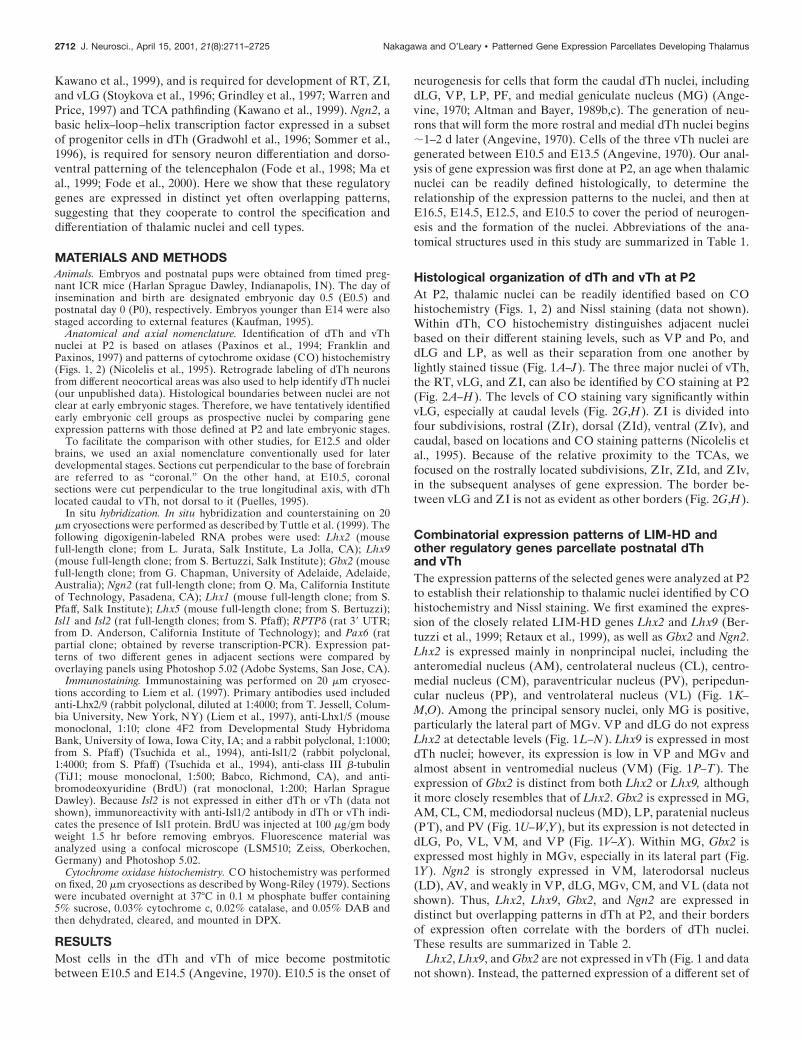
Histological organization of dTh and vTh at P2At P2, thalamic nuclei can be readily identified based on COhistochemistry (Figs. 1, 2) and Nissl staining (data not shown).Within dTh, CO histochemistry distinguishes adjacent nucleibased on their different staining levels, such as VP and Po, anddLG and LP, as well as their separation from one another bylightly stained tissue (Fig. 1A–J). The three major nuclei of vTh,the RT, vLG, and ZI, can also be identified by CO staining at P2(Fig. 2A–H). The levels of CO staining vary significantly withinvLG, especially at caudal levels (Fig. 2G,H). ZI is divided intofour subdivisions, rostral (ZIr), dorsal (ZId), ventral (ZIv), andcaudal, based on locations and CO staining patterns (Nicolelis etal., 1995). Because of the relative proximity to the TCAs, wefocused on the rostrally located subdivisions, ZIr, ZId, and ZIv,in the subsequent analyses of gene expression. The border be-tween vLG and ZI is not as evident as other borders (Fig. 2G,H).
Combinatorial expression patterns of LIM-HD andother regulatory genes parcellate postnatal dThand vThThe expression patterns of the selected genes were analyzed at P2to establish their relationship to thalamic nuclei identified by COhistochemistry and Nissl staining. We first examined the expres-sion of the closely related LIM-HD genes Lhx2 and Lhx9 (Ber-tuzzi et al., 1999; Retaux et al., 1999), as well as Gbx2 and Ngn2.Lhx2 is expressed mainly in nonprincipal nuclei, including theanteromedial nucleus (AM), centrolateral nucleus (CL), centro-medial nucleus (CM), paraventricular nucleus (PV), peripedun-cular nucleus (PP), and ventrolateral nucleus (VL) (Fig. 1K–M,O). Among the principal sensory nuclei, only MG is positive,particularly the lateral part of MGv. VP and dLG do not expressLhx2 at detectable levels (Fig. 1L–N). Lhx9 is expressed in mostdTh nuclei; however, its expression is low in VP and MGv andalmost absent in ventromedial nucleus (VM) (Fig. 1P–T). Theexpression of Gbx2 is distinct from both Lhx2 or Lhx9, althoughit more closely resembles that of Lhx2. Gbx2 is expressed in MG,AM, CL, CM, mediodorsal nucleus (MD), LP, paratenial nucleus(PT), and PV (Fig. 1U–W,Y), but its expression is not detected indLG, Po, VL, VM, and VP (Fig. 1V–X). Within MG, Gbx2 isexpressed most highly in MGv, especially in its lateral part (Fig.1Y). Ngn2 is strongly expressed in VM, laterodorsal nucleus(LD), AV, and weakly in VP, dLG, MGv, CM, and VL (data notshown). Thus, Lhx2, Lhx9, Gbx2, and Ngn2 are expressed indistinct but overlapping patterns in dTh at P2, and their bordersof expression often correlate with the borders of dTh nuclei.These results are summarized in Table 2.
Lhx2, Lhx9, and Gbx2 are not expressed in vTh (Fig. 1 and datanot shown). Instead, the patterned expression of a different set of
2712 J. Neurosci., April 15, 2001, 21(8):2711–2725 Nakagawa and O’Leary • Patterned Gene Expression Parcellates Developing Thalamus
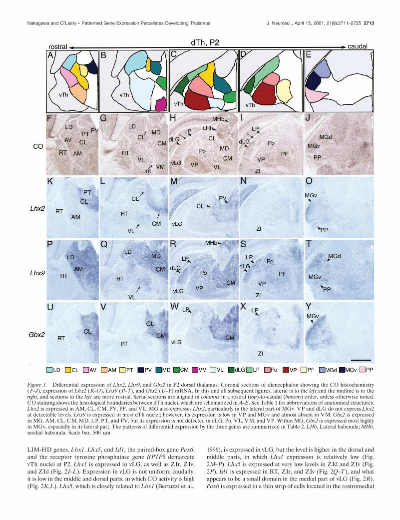
LIM-HD genes, Lhx1, Lhx5, and Isl1, the paired-box gene Pax6,and the receptor tyrosine phosphatase gene RPTPd demarcatevTh nuclei at P2. Lhx1 is expressed in vLG, as well as ZIr, ZIv,and ZId (Fig. 2I–L). Expression in vLG is not uniform; caudally,it is low in the middle and dorsal parts, in which CO activity is high(Fig. 2K,L). Lhx5, which is closely related to Lhx1 (Bertuzzi et al.,
1996), is expressed in vLG, but the level is higher in the dorsal andmiddle parts, in which Lhx1 expression is relatively low (Fig.2M–P). Lhx5 is expressed at very low levels in ZId and ZIv (Fig.2P). Isl1 is expressed in RT, ZIr, and ZIv (Fig. 2Q–T), and whatappears to be a small domain in the medial part of vLG (Fig. 2R).Pax6 is expressed in a thin strip of cells located in the rostromedial
Figure 1. Differential expression of Lhx2, Lhx9, and Gbx2 in P2 dorsal thalamus. Coronal sections of diencephalon showing the CO histochemistry(F–J), expression of Lhx2 (K–O), Lhx9 (P–T), and Gbx2 (U–Y) mRNA. In this and all subsequent figures, lateral is to the lef t and the midline is to theright, and sections to the lef t are more rostral. Serial sections are aligned in columns in a rostral (top)-to-caudal (bottom) order, unless otherwise noted.CO staining shows the histological boundaries between dTh nuclei, which are schematized in A–E. See Table 1 for abbreviations of anatomical structures.Lhx2 is expressed in AM, CL, CM, PV, PP, and VL. MG also expresses Lhx2, particularly in the lateral part of MGv. VP and dLG do not express Lhx2at detectable levels. Lhx9 is expressed in most dTh nuclei; however, its expression is low in VP and MGv and almost absent in VM. Gbx2 is expressedin MG, AM, CL, CM, MD, LP, PT, and PV, but its expression is not detected in dLG, Po, VL, VM, and VP. Within MG, Gbx2 is expressed most highlyin MGv, especially in its lateral part. The patterns of differential expression by the three genes are summarized in Table 2. LHb, Lateral habenula; MHb,medial habenula. Scale bar, 500 mm.
Nakagawa and O’Leary • Patterned Gene Expression Parcellates Developing Thalamus J. Neurosci., April 15, 2001, 21(8):2711–2725 2713
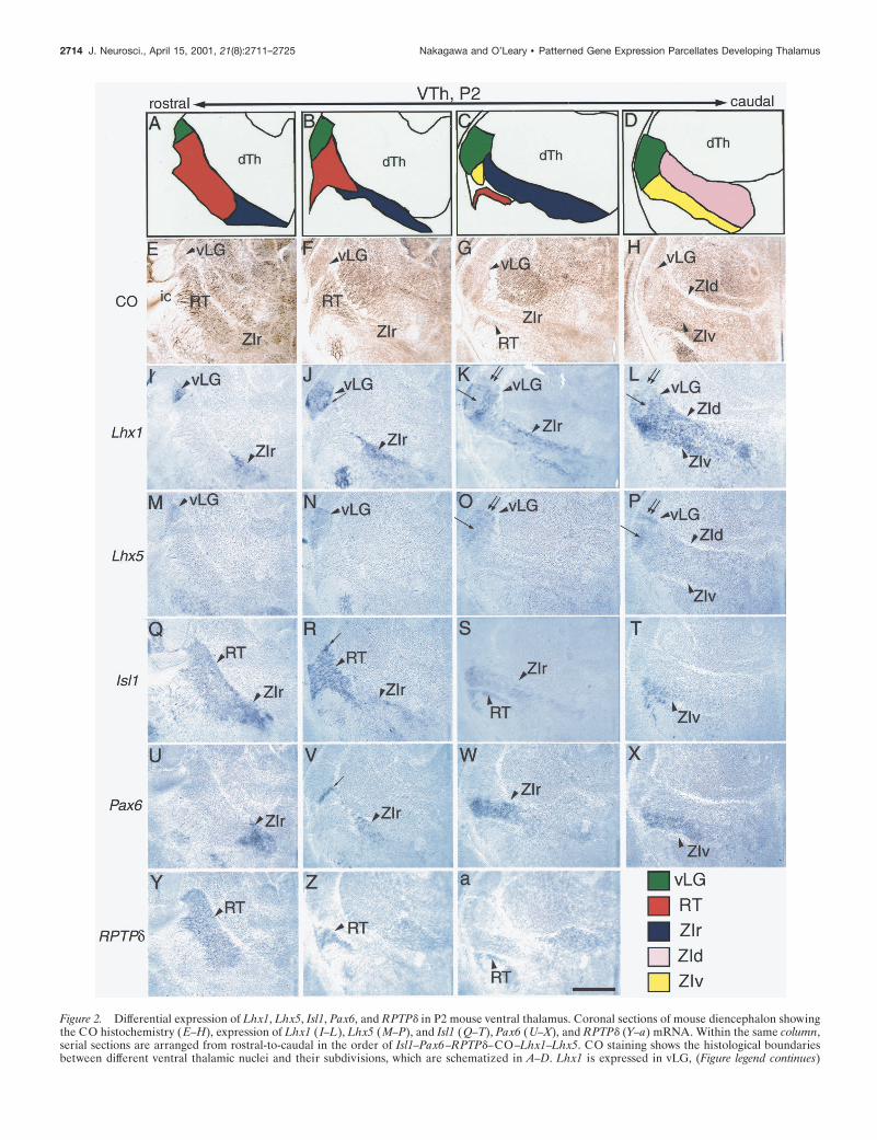
Figure 2. Differential expression of Lhx1, Lhx5, Isl1, Pax6, and RPTPd in P2 mouse ventral thalamus. Coronal sections of mouse diencephalon showingthe CO histochemistry (E–H), expression of Lhx1 ( I–L), Lhx5 (M–P), and Isl1 (Q–T), Pax6 (U–X), and RPTPd (Y–a) mRNA. Within the same column,serial sections are arranged from rostral-to-caudal in the order of Isl1–Pax6–RPTPd–CO–Lhx1–Lhx5. CO staining shows the histological boundariesbetween different ventral thalamic nuclei and their subdivisions, which are schematized in A–D. Lhx1 is expressed in vLG, (Figure legend continues)
2714 J. Neurosci., April 15, 2001, 21(8):2711–2725 Nakagawa and O’Leary • Patterned Gene Expression Parcellates Developing Thalamus
portion of vLG (Fig. 2V ), as well as in ZIr and ZIv but not inRT (Fig. 2U–X ). The boundary between RT and ZIr is alsoclearly defined by the expression of RPTPd (Mizuno et al.,1993; Sommer et al., 1997; Tuttle et al., 1999), which is ex-pressed in RT but not in ZIr (Fig. 2Y,Z). These results aresummarized in Table 3.
In summary, the regulatory genes analyzed here have distinctbut overlapping expression patterns in subsets of dTh and vThnuclei; the expression patterns often correlate with the histolog-ically defined borders of nuclei. In addition, potential subdomainswithin the same nuclei (e.g., MGv and vLG) are suggested bysome of the more discrete expression patterns. Thus, the combi-natorial expression of these transcription factors may regulate the
postnatal development of dTh and vTh nuclei. Because thesegenes may also serve as markers to define the organization of theembryonic dTh and vTh into nascent nuclei, or cell groups thatwill later form these nuclei, we examined their expression atembryonic stages.
Patterned expression of regulatory genes inembryonic dThAlthough dTh has undergone considerable architectonic differen-tiation by E16.5, dTh nuclei cannot be as easily distinguished inCO- or Nissl-stained sections as at P2 (data not shown); forexample, the dLG borders with LP and MG do not appear as acell-free band, and the levels of staining are not clearly different
Table 1. Names and their abbreviations of the anatomical structures used in this study are based onPaxinos et al. (1994) and are listed below
Nuclei of dTh (dorsal thalamus)
AM anteromedial nucleusAV anteroventral nucleusCL centrolateral nucleusCM centromedial nucleusdLG dorsal lateral geniculate nucleusLD laterodorsal nucleusMD mediodorsal nucleusMG medial geniculate nucleusMGv ventral subdivision of medial geniculate nucleusMGd dorsal subdivision of medial geniculate nucleusLP lateral posterior nucleusPF parafascicular nucleusPo posterior complexPP peripeduncular nucleusPT paratenial nucleusPV paraventricular nucleusVL ventrolateral nucleusVM ventromedial nucleusVP ventroposterior nucleus
Nuclei of vTh (ventral thalamus)
RT reticular nucleusvLG ventral lateral geniculate nucleusZI zona incertaZIr rostral subdivision of zona incertaZIv ventral subdivision of zona incertaZId dorsal subdivision of zona increta
Other abbreviations
TCAS thalamocortical axonsic internal capsuleZLI zona limitans intrathalamicaCO cytochrome oxidaseLHb lateral habenulaMHb medial habenula
4
ZIr, ZId, and ZIv. Expression in vLG is not uniform and relatively low in middle and dorsal parts (K, L; arrows and double arrows, respectively). Lhx5is expressed in vLG and more weakly in ZId and ZIv. Lhx5 expression in vLG is higher in the dorsal and middle parts (O, P; arrows and arrowheads,respectively) and is complementary to that of Lhx1. Isl1 is expressed in RT, ZIr, and ZIv. It is also possibly expressed in a very small, ventromedial partof vLG (R; arrow). Pax6 is expressed in a thin band in vLG (V; arrow), ZIr, and ZIv. RPTPd is expressed in RT. The patterns of differential expressionexhibited by these genes are summarized in Table 3. Scale bar, 500 mm.
Nakagawa and O’Leary • Patterned Gene Expression Parcellates Developing Thalamus J. Neurosci., April 15, 2001, 21(8):2711–2725 2715

between these nuclei. Nonetheless, it is evident that Lhx2,Lhx9, and Gbx2 are differentially expressed in patterns similarto those at P2 (Fig. 3). Robust Lhx2 expression is present inthe rostromedial nuclei, i.e., the putative CL, CM, and PV(Fig. 3 A, B), as well as in MG and PP (Fig. 3C), whereas itsexpression in dLG and LP is very low and is undetectable inVP (Fig. 3 B, C). As at P2, Lhx9 is expressed at high levels inmost nuclei except in VM, VP, and MGv (Fig. 3D–F ). Gbx2expression is high in MGv and the dorsal subdivision of medialgeniculate nucleus (MGd) and is not detected in VP and dLG(Fig. 3G–I ). Although the putative border between LP anddLG is not apparent in CO- or Nissl-stained sections at E16.5,the expression pattern of Gbx2 appears to mark it, with mod-erate expression in LP and undetectable expression in dLG.The expression pattern of Ngn2 in dTh at E16.5 is similar tothat at P2 (Fig. 3J–L) and is partially complementary to Lhx2and Gbx2 expression. This suggests that, in dTh, Gbx2/Lhx2and Ngn2 negatively regulate each others expression, or cellsexpressing Gbx2/Lhx2 and cells expressing Ngn2 do not mixwith each other and thereby remain as distinct cell groups. Insummary, the expression patterns of Lhx2, Lhx9, Gbx2, andNgn2 observed in dTh at P2 are already evident at E16.5,
suggesting that these genes are useful markers to follow theparcellation of dTh into molecularly distinct cell groups thatwill form specific dTh nuclei.
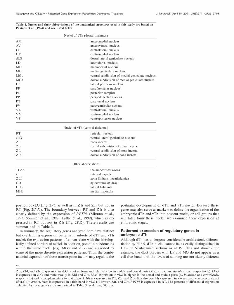
It is not possible to histologically distinguish dTh nuclei at E12.5or E14.5. However, Lhx2, Lhx9, Gbx2, and Ngn2 already showdistinct patterns of expression at these ages, and the overall pat-terns evident at E14.5 are similar to those at E16.5 and P2. Forexample, caudally, the putative MG already expresses Lhx2, Lhx9,Gbx2, and Ngn2 in a pattern reminiscent of that observed at laterages (Fig. 4C,F,I,L). Lhx2 and Gbx2 are expressed at high levels inthe lateral part of the mantle zone, and Lhx9 and Ngn2 areexpressed at low levels in the putative MGv. More rostrally, theputative dLG expresses Lhx9 and Ngn2 but not Lhx2 or Gbx2 (Fig.4B,E,H,K).
Figure 3. Gene expression patterns in E16.5 mouse dorsal thalamus aresimilar to those at P2. Coronal sections of diencephalon showing theexpression of Lhx2 (A–C), Lhx9 (D–F), Gbx2 (G–I), and Ngn2 ( J–L)mRNA. At the rostral level, Lhx9 is strongly expressed in the putative LD(D), in which Lhx2 is only weakly expressed and Gbx2 is undetectable (A,G). Ngn2 is expressed in LD ( J). At the middle level, the square-shapedregion (asterisks) expresses Lhx9 and Ngn2 but not Lhx2 or Gbx2, compat-ible with dLG (B, E, H, K; asterisks). The putative LP, located immediatelydorsal to dLG, expresses high levels of Lhx9 and Gbx2 (E, H; arrows) butonly very low levels of Lhx2 and Ngn2 (B, K; arrows). The putative VP isnegative for Lhx2 and Gbx2 and weakly positive for Lhx9 and Ngn2. Theputative VM is positive for Ngn2 and negative for the others (B, E, H, K;arrowheads). At the caudal level, MGv is strongly positive for Lhx2 andGbx2 (C, I; asterisks) and weaker for Lhx9 and Ngn2 (F, L; asterisks). Theputative PP is positive for Lhx2 and Lhx9 (C, F; arrowheads) and negativefor Gbx2 and Ngn2 (F, L; arrowheads). MGd expresses Lhx2, Lhx9, andGbx2 (E, C, F; arrows) but not Ngn2 (L; arrow). Scale bar, 500 mm.
Table 2. Differential expression of LIM-HD and other transcriptionfactors in P2 mouse dorsal thalamus
Lhx2 Lhx9 Gbx2 Ngn2
Principal sensory nucleiVP 2 12 2 12
dLG 2 1 2 12
MGv 1 12 1 12
Other nucleiAM 12 1 12 1
AV 2 1 2 1
CL 1 1 1 2
CM 1 1 1 12
LD 2 1 2 1
MD 2 1 1 2
MGd 12 1 1 2
LP 2 1 1 2
Po 2 1 2 2
PP 1 1 2 2
PT 2 1 1 2
PV 1 1 1 2
VL 1 1 2 12
VM 2 2 2 1
1, Expressed; 12, weakly expressed; 2, not detectable.
Table 3. Differential expression of LIM-HD and other transcriptionfactors in P2 mouse ventral thalamus
Lhx1 Lhx5 Isl1 Pax6
vLG 1*a 1*a 1*b 1*b
ZIZIr 1 12 1 1
ZId 1 12 2 2
ZIv 1 12 1 1
RT 2 2 1 2
1, expressed; 12, weakly expressed; 2, not detectable.*a Expression of Lhx1 and Lhx5 in vLG is nearly complementary.*b Expression of Isl1 and Pax6 is only in small, ventral parts of vLG.
2716 J. Neurosci., April 15, 2001, 21(8):2711–2725 Nakagawa and O’Leary • Patterned Gene Expression Parcellates Developing Thalamus
Although the similarities in gene expression patterns at earlyand late stages are striking, some differences are also apparent.At E16.5, the expression domains of Lhx2 overlap with those ofLhx9 except for MGv, in which Lhx2 is highly expressed butLhx9 expression is low. However, at E14.5, a rostromedial partof dTh exhibits a high level of Lhx2 expression but a very lowlevel of Lhx9 expression (Fig. 4 A, D), which is not found atE16.5. This band of cells is located just outside of the ventric-ular zone and expresses Gbx2 at a high level and Ngn2 at a verylow level (Fig. 4G,J ). An approximately similar pattern isalready apparent at E12.5 (Fig. 4 M–T ). Another example of adifference in the patterns of gene expression is found betweenE12.5 and E14.5 in the putative dLG; the overall patterncharacteristic of dLG after E14.5 is not evident at the putative
location of this nucleus in the caudolateral portion of dTh (Fig.4 N–T ).
The band of Gbx2-expressing cells located just outside of theventricular zone (Fig. 4G,Q, asterisk s) has been described to bein the thalamic subventricular zone (Bulfone et al., 1993;Miyashita-Lin et al., 1999). Lhx2 is expressed in a similar band.However, pulse labeling with BrdU ;1 hr before fixation showsthat this expression domain of Gbx2 and Lhx2 is BrdU-negative at these ages (data not shown; also see below and Fig. 8),suggesting that this domain is not a proliferative zone and thatthese cells are postmitotic. A similar study in rat using tritiatedthymidine reported the labeling of a few scattered cells in thisdomain just lateral to the ventricular zone, leading the authors toterm this zone the subependymal layer, but noted that a large
Figure 4. Differential gene expression patterns already exist in the dorsal thalamus at E12.5. Coronal sections of diencephalon showing the expressionof Lhx2 (A–C, M, N ), Lhx9 (D–F, O, P), and Gbx2 (G–I, Q, R), and Ngn2 (J–L, S, T ) mRNA. A–L are for E14.5, and M–T are for E12.5. At E14.5, Lhx9and Ngn2 are expressed in the lateral–ventral part of dTh at the rostral level, corresponding to LD, but Lhx2 and Gbx2 are not (A, D, G, J; arrows). Aband of strong Lhx2 and Gbx2 expression (A, G, asterisks) is located more medially to a band of strong Lhx9 expression (D; cross). Ngn2 is weak in theband of Lhx2 and Gbx2 expression (J; asterisk) but strong in the putative ventricular zone (J, K; double-headed arrow), in which Lhx2, Lhx9, and Gbx2are negative. More caudally, a region expresses Lhx9 and Ngn2 but not Lhx2 or Gbx2, which is likely to be dLG (B, E, H, K; asterisks). The putative LP,located dorsally to dLG (B, E, H, K; arrows), expresses high levels of Lhx9 and Gbx2 but only low levels of Lhx2 and Ngn2. The putative VP is negativefor Lhx2 and Gbx2 and weakly positive for Lhx9 and Ngn2 (B, E, H, K ). The putative PP is positive for Lhx2 and Lhx9 and negative for Gbx2 and Ngn2(C, F, I, L; arrowheads), whereas MGv strongly expresses Lhx2 and Gbx2 and weakly expresses Lhx9 and Ngn2 (C, F, I, L; asterisks). These patterns ofdifferential expression are not still apparent at E12.5, but the band with high levels of Lhx2/Gbx2 expression is already located medial to the band withthe high Lhx9 expression (L, N–R; asterisks), similar to E14.5. Ngn2 is expressed in the ventricular zone, as well as the mantle zone, but is weak in theband with strong Lhx2/Gbx2 expression (S, T; asterisk). Scale bars, 200 mm.
Nakagawa and O’Leary • Patterned Gene Expression Parcellates Developing Thalamus J. Neurosci., April 15, 2001, 21(8):2711–2725 2717
proportion of the cells in this layer must be postmitotic (Altmanand Bayer, 1989a).
In summary, the differential expression patterns of genes thatmark dTh nuclei at later ages are already evident in dTh as earlyas E12.5. These patterns undergo some changes until E16.5, butthen the patterns appear to be stable to P2. It is unclear whetherchanges in these expression patterns between E12.5 and E16.5 areattributable to changes in expression per se or whether the ex-
pressing population is the same but the patterns change as a resultof cell movements (see Discussion).
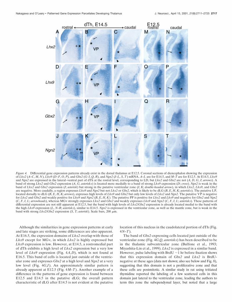
Patterned expression of regulatory genes inembryonic vThAt E16.5, CO histochemistry suggests that the organization of vThis similar to that at P2 (Fig. 5A–D). RT is identified as a sheet ofcells interposed between dTh, vLG, ZIr, and the internal capsule
Figure 5. Gene expression patterns in E16.5 mouse ventral thalamus are similar to those at P2. Coronal sections of diencephalon showing the COhistochemistry (A–D) and the expression of Lhx1 (E–H), Lhx5 ( I–L), Isl1 (M–P), Pax6 (Q–T), and RPTPd (U–X) mRNA. Within each column, serialsections are aligned in a rostral-to-caudal order from Lhx1 to RPTPd and then CO. CO staining shows similar patterns to P2 and delineates the nucleiof vTh (A–D). Lhx1 is expressed in vLG, ZIr, ZId, and ZIv. Expression in vLG is high in the ventromedial (E–G; arrows) and ventrolateral (F, G; doublearrows) parts. In addition, Lhx1 is expressed in ZLI (E, F ). Lhx5 is expressed in vLG, most highly in the dorsal part ( I–L), and in the most rostral partof ZLI ( I ). It is also weakly expressed in ZId and ZIv ( L). Part of hypothalamus is positive for Lhx5 (I, J; asterisk). Isl1 is expressed in RT, ZIr, ZIv(M–P), and possibly in a small, rostroventral part of vLG (N; arrow). Pax6 is expressed in ZIr, ZIv (S, T ), and a rostroventral part of vLG (R; arrow).RPTPd is expressed in RT and ZIv. Scale bar, 500 mm.
2718 J. Neurosci., April 15, 2001, 21(8):2711–2725 Nakagawa and O’Leary • Patterned Gene Expression Parcellates Developing Thalamus
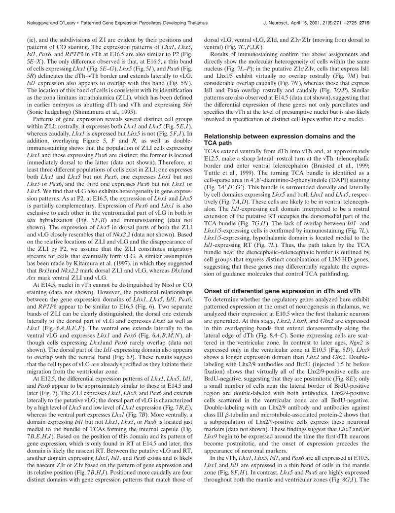
(ic), and the subdivisions of ZI are evident by their positions andpatterns of CO staining. The expression patterns of Lhx1, Lhx5,Isl1, Pax6, and RPTPd in vTh at E16.5 are also similar to P2 (Fig.5E–X). The only difference observed is that, at E16.5, a thin bandof cells expressing Lhx1 (Fig. 5E–G), Lhx5 (Fig. 5I), and Pax6 (Fig.5R) delineates the dTh–vTh border and extends laterally to vLG.Isl1 expression also appears to overlap with this band (Fig. 5N).The location of this band of cells is consistent with its identificationas the zona limitans intrathalamica (ZLI), which has been definedin earlier embryos as abutting dTh and vTh and expressing Shh(Sonic hedgehog) (Shimamura et al., 1995).
Patterns of gene expression reveals several distinct cell groupswithin ZLI; rostrally, it expresses both Lhx1 and Lhx5 (Fig. 5E,I),whereas caudally, Lhx1 is expressed but Lhx5 is not (Fig. 5F,J). Inaddition, overlaying Figure 5, F and R, as well as double-immunostaining shows that the population of ZLI cells expressingLhx1 and those expressing Pax6 are distinct; the former is locatedimmediately dorsal to the latter (data not shown). Therefore, atleast three different populations of cells exist in ZLI; one expressesboth Lhx1 and Lhx5 but not Pax6, one expresses Lhx1 but notLhx5 or Pax6, and the third one expresses Pax6 but not Lhx1 orLhx5. We find that vLG also exhibits heterogeneity in gene expres-sion patterns. As at P2, at E16.5, the expression of Lhx1 and Lhx5is partially complementary. Expression of Pax6 and Lhx1 is alsoexclusive to each other in the ventromedial part of vLG in both insitu hybridization (Fig. 5F,R) and immunostaining (data notshown). The expression of Lhx5 in dorsal parts of both the ZLIand vLG closely resembles that of Nkx2.2 (data not shown). Basedon the relative locations of ZLI and vLG and the disappearance ofthe ZLI by P2, we assume that the ZLI constitutes migratorystreams for cells that eventually form vLG. A similar assumptionhas been made by Kitamura et al. (1997), in which they suggestedthat Brx1and Nkx2.2 mark dorsal ZLI and vLG, whereas Dlx1andArx mark ventral ZLI and vLG.
At E14.5, nuclei in vTh cannot be distinguished by Nissl or COstaining (data not shown). However, the positional relationshipsbetween the gene expression domains of Lhx1, Lhx5, Isl1, Pax6,and RPTPd appear to be similar to E16.5 (Fig. 6). Two separatebands of ZLI can be clearly distinguished; the dorsal one extendslaterally to the dorsal part of vLG and expresses Lhx5 as well asLhx1 (Fig. 6A,B,E,F). The ventral one extends laterally to theventral vLG and expresses Lhx1 and Pax6 (Fig. 6A,B,M,N), al-though cells expressing Lhx1and Pax6 rarely overlap (data notshown). The dorsal part of the Isl1-expressing domain also appearsto overlap with the ventral band (Fig. 6J). These results suggestthat the cell types of vLG are already specified as they initiate theirmigration from the ventricular zone.
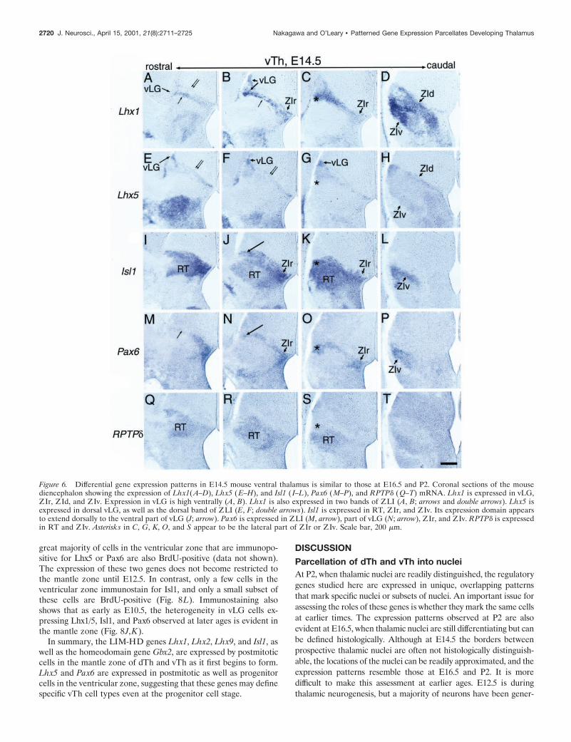
At E12.5, the differential expression patterns of Lhx1, Lhx5, Isl1,and Pax6 appear to be approximately similar to those at E14.5 andlater (Fig. 7). The ZLI expresses Lhx1, Lhx5, and Pax6 and extendslaterally to the putative vLG; the dorsal part of vLG is characterizedby a high level of Lhx5 and low level of Lhx1 expression (Fig. 7B,E),whereas the ventral part expresses Lhx1 (Fig. 7B). More ventrally, adomain expressing Isl1 but not Lhx1, Lhx5, or Pax6 is located justmedial to the bundle of TCAs forming the internal capsule (Fig.7B,E,H,J). Based on the position of this domain and its pattern ofgene expression, which is only found in RT at E14.5 and later, thisdomain is likely the nascent RT. Between the putative vLG and RT,another domain expressing Lhx1, Isl1, and Pax6 exists and is likelythe nascent ZIr or ZIv based on the pattern of gene expression andits relative position (Fig. 7B,H,J). Positioned more caudally are fourdistinct domains with gene expression patterns that match those of
dorsal vLG, ventral vLG, ZId, and ZIv/ZIr (moving from dorsal toventral) (Fig. 7C,F,I,K).
Results of immunostaining confirm the above assignments anddirectly show the molecular heterogeneity of cells within the samenucleus (Fig. 7L–P); in the putative ZIr/ZIv, cells that express Isl1and Lhx1/5 exhibit virtually no overlap rostrally (Fig. 7M) butconsiderable overlap caudally (Fig. 7N), whereas those that expressIsl1 and Pax6 overlap rostrally and caudally (Fig. 7O,P). Similarpatterns are also observed at E14.5 (data not shown), suggesting thatthe differential expression of these genes not only parcellates andspecifies the vTh at the level of presumptive nuclei but is also likelyinvolved in specification of distinct cell types within these nuclei.
Relationship between expression domains and theTCA pathTCAs extend ventrally from dTh into vTh and, at approximatelyE12.5, make a sharp lateral–rostral turn at the vTh–telencephalicborder and enter ventral telencephalon (Braisted et al., 1999;Tuttle et al., 1999). The turning TCA bundle is identified as acell-sparse area in 49,69-diaminino-2-phenylindole (DAPI) staining(Fig. 7A9,D9,G9). This bundle is surrounded dorsally and laterallyby cell domains expressing Lhx5 and both Lhx1 and Lhx5, respec-tively (Fig. 7A,D). These cells are likely to be in ventral telenceph-alon. The Isl1-expressing cell domain interpreted to be a rostralextension of the putative RT occupies the dorsomedial part of theTCA bundle (Fig. 7G,H). The lack of overlap between Isl1- andLhx1/5-expressing cells is confirmed by immunostaining (Fig. 7L).Lhx1/5-expressing, hypothalamic domain is located medial to theIsl1-expressing RT (Fig. 7L). Thus, the path taken by the TCAbundle near the diencephalic–telencephalic border is outlined bycell groups that express distinct combinations of LIM-HD genes,suggesting that these genes may differentially regulate the expres-sion of guidance molecules that control TCA pathfinding.
Onset of differential gene expression in dTh and vThTo determine whether the regulatory genes analyzed here exhibitpatterned expression at the onset of neurogenesis in thalamus, weanalyzed their expression at E10.5 when the first thalamic neuronsare generated. At this stage, Lhx2, Lhx9, and Gbx2 are expressedin thin overlapping bands that extend dorsoventrally along thelateral edge of dTh (Fig. 8A–C). Some expressing cells are scat-tered in the ventricular zone. In contrast to later ages, Ngn2 isexpressed only in the ventricular zone at E10.5 (Fig. 8D). Lhx9shows a longer expression domain than Lhx2 and Gbx2. Double-labeling with Lhx2/9 antibodies and BrdU (injected 1.5 hr beforefixation) shows that virtually all of the Lhx2/9-positive cells areBrdU-negative, suggesting that they are postmitotic (Fig. 8E); onlya small number of cells near the lateral border of BrdU-positiveregion are double-labeled with both antibodies. Lhx2/9-positivecells scattered in the ventricular zone are all BrdU-negative.Double-labeling with an Lhx2/9 antibody and antibodies againstclass III b-tubulin and microtubule-associated protein-2 shows thata subpopulation of Lhx2/9-positive cells express these neuronalmarkers (data not shown). These findings suggest that Lhx2 and/orLhx9 begin to be expressed around the time the first dTh neuronsbecome postmitotic, and the onset of expression precedes theappearance of neuronal markers.
In the vTh, Lhx1, Lhx5, Isl1, and Pax6 are all expressed at E10.5.Lhx1 and Isl1 are expressed in a thin band of cells in the mantlezone (Fig. 8F,H). In contrast, Lhx5 and Pax6 are highly expressedthroughout both the mantle and ventricular zones (Fig. 8G,I). The
Nakagawa and O’Leary • Patterned Gene Expression Parcellates Developing Thalamus J. Neurosci., April 15, 2001, 21(8):2711–2725 2719
great majority of cells in the ventricular zone that are immunopo-sitive for Lhx5 or Pax6 are also BrdU-positive (data not shown).The expression of these two genes does not become restricted tothe mantle zone until E12.5. In contrast, only a few cells in theventricular zone immunostain for Isl1, and only a small subset ofthese cells are BrdU-positive (Fig. 8L). Immunostaining alsoshows that as early as E10.5, the heterogeneity in vLG cells ex-pressing Lhx1/5, Isl1, and Pax6 observed at later ages is evident inthe mantle zone (Fig. 8J,K).
In summary, the LIM-HD genes Lhx1, Lhx2, Lhx9, and Isl1, aswell as the homeodomain gene Gbx2, are expressed by postmitoticcells in the mantle zone of dTh and vTh as it first begins to form.Lhx5 and Pax6 are expressed in postmitotic as well as progenitorcells in the ventricular zone, suggesting that these genes may definespecific vTh cell types even at the progenitor cell stage.
DISCUSSION
Parcellation of dTh and vTh into nucleiAt P2, when thalamic nuclei are readily distinguished, the regulatorygenes studied here are expressed in unique, overlapping patternsthat mark specific nuclei or subsets of nuclei. An important issue forassessing the roles of these genes is whether they mark the same cellsat earlier times. The expression patterns observed at P2 are alsoevident at E16.5, when thalamic nuclei are still differentiating but canbe defined histologically. Although at E14.5 the borders betweenprospective thalamic nuclei are often not histologically distinguish-able, the locations of the nuclei can be readily approximated, and theexpression patterns resemble those at E16.5 and P2. It is moredifficult to make this assessment at earlier ages. E12.5 is duringthalamic neurogenesis, but a majority of neurons have been gener-
Figure 6. Differential gene expression patterns in E14.5 mouse ventral thalamus is similar to those at E16.5 and P2. Coronal sections of the mousediencephalon showing the expression of Lhx1(A–D), Lhx5 (E–H), and Isl1 ( I–L), Pax6 (M–P), and RPTPd (Q–T) mRNA. Lhx1 is expressed in vLG,ZIr, ZId, and ZIv. Expression in vLG is high ventrally (A, B). Lhx1 is also expressed in two bands of ZLI (A, B; arrows and double arrows). Lhx5 isexpressed in dorsal vLG, as well as the dorsal band of ZLI (E, F; double arrows). Isl1 is expressed in RT, ZIr, and ZIv. Its expression domain appearsto extend dorsally to the ventral part of vLG (J; arrow). Pax6 is expressed in ZLI (M, arrow), part of vLG (N; arrow), ZIr, and ZIv. RPTPd is expressedin RT and ZIv. Asterisks in C, G, K, O, and S appear to be the lateral part of ZIr or ZIv. Scale bar, 200 mm.
2720 J. Neurosci., April 15, 2001, 21(8):2711–2725 Nakagawa and O’Leary • Patterned Gene Expression Parcellates Developing Thalamus
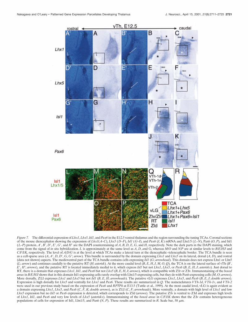
Figure 7. The differential expression of Lhx1, Lhx5, Isl1, and Pax6 in the E12.5 ventral thalamus and the region surrounding the tuning TCAs. Coronal sectionsof the mouse diencephalon showing the expression of Lhx1(A–C), Lhx5 (D–F), Isl1 (G–I), and Pax6 (J, K) mRNA and Lhx1/5 (L–N), Pax6 (O, P), and Isl1(L–P) protein. A9, B9, D9, E9, G9, and H9 are the DAPI counterstaining of A, B, D, E, G, and H, respectively. Note the dark parts in the DAPI staining, whichcome from the signal of in situ hybridization. L is approximately at the same level as A, D, and G, whereas M/O and N/P are at similar levels to B/E/H/J andC/F/I/K, respectively. The level of A/D/G is at the level at which TCAs make a lateral turn at the diencephalic–telencephalic border. The TCA bundle is seenas a cell-sparse area (A, A9, D, D9, G, G9; arrow). This bundle is surrounded by the domain expressing Lhx1 and Lhx5 on its lateral, dorsal (A, D), and rostral(data not shown) aspects. The medioventral part of the TCA bundle contains cells expressing Isl1 (G; arrowhead). This domain does not express Lhx1 or Lhx5(L; arrow) and continues caudally to the putative RT (H; asterisk). At the more caudal level (B, E, H, J, M, O, Q), the TCA is on the lateral surface of vTh (B9,E9, H9; arrows), and the putative RT is located immediately medial to it, which express Isl1 but not Lhx1, Lhx5, or Pax6 (B, E, H, J; asterisks). Just dorsal toRT, there is a domain that expresses Lhx1, Isl1, and Pax6 but not Lhx5 (B, E, H, J; arrows), which is compatible with ZIr or ZIv. Immunostaining of the boxedareas in B/E/H/J shows that in this domain Isl1-expressing cells rarely overlap with Lhx1/5-expressing cells, but they do with Pax6-expressing cells (M, O; arrows).More dorsally, ZLI expresses Lhx1 and Lhx5 but not Isl1 (B, E, H; arrowheads). The putative vLG expresses Lhx1, Lhx5, and Pax6 (B, E, J; double arrows).Expression is high dorsally for Lhx5 and ventrally for Lhx1 and Pax6. These results are summarized in Q. The nomenclatures VTh-1d, VTh-1v, and VTh-2were used in our previous study based on the expression of Pax6 and RPTPd at E13.5 (Tuttle et al., 1999). At the most caudal level, vLG is again evident asa domain expressing Lhx1, Lhx5, and Pax6 (C, F, K; double arrows), as is ZLI (C, F; arrowheads). More ventrally, a domain with high level of Lhx1 and lowLhx5 expression but no Isl1 or Pax6 expression is detected, which corresponds to ZId (arrows). The putative ZIv is ventral to ZId and expresses high levelsof Lhx1, Isl1, and Pax6 and very low levels of Lhx5 (asterisks). Immunostaining of the boxed areas in C/F/I/K shows that the ZIv contains heterogeneouspopulations of cells for expression of Isl1, Lhx1/5, and Pax6 (N, P). These results are summarized in R. Scale bar, 50 mm.
Nakagawa and O’Leary • Patterned Gene Expression Parcellates Developing Thalamus J. Neurosci., April 15, 2001, 21(8):2711–2725 2721
ated and many have completed their migration and established amantle zone within which nuclei will differentiate. Even at this earlydevelopmental stage, the expression patterns of the eight genesrelative to each other are approximately similar to those seen later.
Thus, thalamic neurons appear to express the same subset of theseregulatory genes between E12.5 and P2. Each of these genes isexpressed in a unique pattern as early as E10.5, when the firstthalamic neurons are generated; thus, they may mark distinct subsetsof thalamic neurons beginning around the time they are generatedthrough the time they form nuclei. Based on the expression patternsand known functions of these genes, they are good candidates to actin a combinatorial manner to control the specification of nuclei-specific properties of thalamic cells and the differentiation of nuclei.This suggestion is supported by analyses of Gbx2-deficient mice(Miyashita-Lin et al., 1999). Although the mutant exhibits a severedisorganization of dTh, some parts that correspond to VP, dLG, Po,and MG persist. These results are consistent with our finding thatGbx2 is not expressed in VP, dLG, and Po, and only in part of MG.Furthermore, the “sparing” of these nuclei in the mutant suggeststhat the cell populations that express Gbx2 do not significantlychange over embryonic development.
The expression patterns observed in dTh do not appear tochange between E16.5 and P2. However, at E14.5 and E12.5, aband of cells in rostromedial dTh exhibits a Lhx2/Gbx2-high, Lhx9/Ngn2-low expression not seen later. One possible explanation forthe difference is that this population is still present after E14.5 but
is mixed with other populations. This possibility is supported by thefact that neurons of rostromedial dTh nuclei are born later thancaudolateral dTh nuclei (Altman and Bayer, 1979; Altman andBayer, 1988) and may mingle with each other during migration.Another possibility is that these cells express a different combina-tion of genes at E16.5 than at E14.5, similar to the finding that somespinal motor neurons initially express Lhx3 and Lhx4 but later donot (Sharma et al., 1998).
Heterogeneity in gene expression within a nucleusBecause thalamic nuclei contain multiple types of neurons, regu-latory genes might exhibit not only nuclei-specific expression butalso differential expression within a nucleus. For example, eachsubdivision of ZI contains heterogeneous populations of cells thatexpress different neurotransmitters and calcium binding proteins(Kolmac and Mitrofanis, 1998). In the putative ZI at E12.5, ourimmunostaining for Lhx1/5, Isl1, and Pax6 reveals at least four (andpossibly 18) different subsets of cells; in rostral ZI, cells onlyexpress Isl1 or Lhx1/5, whereas in caudal ZI, many cells expressIsl1 and Lhx1/5. Therefore, the differential expression of Lhx1/5,Isl1, and Pax6 may have a role in regulating the differentiation ofspecific cell types and subdivisions within this nucleus.
Lhx1, Lhx5, and Pax6 may have a similar role within vLG,because it is subdivided by their differential expression. This het-erogeneity of gene expression is also evident in the ZLI, which atthis stage of development appears to be a migratory stream con-
Figure 8. Expression of Lhx2, Lhx9, Gbx2, and Ngn2 in dTh and Lhx1, Lhx5, Isl1, and Pax6 in vTh is apparent at E10.5. Coronal sections of the mousedTh ( A–E) and vTh (F–L). Lhx2, Lhx9, and Gbx2 are expressed in the lateral part of dTh (A–C; arrows). Patterns of Lhx2 and Gbx2 areindistinguishable, and expression of Lhx9 extends further dorsally compared with Lhx2 and Gbx2. Ngn2 is expressed only in the ventricular zone andnot in the lateral part in which Lhx2, Lhx9, and Gbx2 are expressed (D; arrow). Lhx2/9 is mostly localized in BrdU-negative cells at the mantle zone,and only a small number of Lhx2/9-positive cells are BrdU-positive (E; arrowheads). Lhx1, and Isl1 are expressed in the mantle zone of vTh, whereasLhx5 and Pax6 are expressed both in the mantle and ventricular zones (F–I; arrow). Isl1 is expressed only in BrdU-negative cells ( J). The mantle zoneis already composed of heterogeneous cell populations for the expression of Lhx1, Isl1, and Pax6 (K, L). Scale bars: A–D, F–I, 100 mm; E, J–L, 50 mm.
2722 J. Neurosci., April 15, 2001, 21(8):2711–2725 Nakagawa and O’Leary • Patterned Gene Expression Parcellates Developing Thalamus
taining vLG neurons. Based on the expression of the regulatorygenes Dlx1, Arx, Brx1, and Nkx2.2, Kitamura et al. (1997) havesuggested that ZLI is composed of two cell groups that give rise todifferent parts of vLG. Together our findings show that vLG issubdivided into multiple domains based on the differential expres-sion of regulatory genes and that this molecular heterogeneity isalready evident while vLG neurons are migrating within the ZLI.
Potential roles in control of TCA axon pathfindingCombinations of LIM-HD genes expressed by subsets of motorneurons in vertebrates and Drosophila constitute a “LIM-HD com-binatorial code” that dictates their axonal pathfinding (Tsuchida etal., 1994; Sharma et al., 1998; Thor et al., 1999). In addition, theDrosophila LIM-HD gene apterous, the ortholog of Lhx2 and Lhx9,is required for the axonal pathfinding of a subset of interneurons(Lungdren et al., 1995). Similarly, the differential expression ofLIM-HD genes in dTh nuclei might regulate TCA pathfinding in acell-autonomous manner.
Candidate genes regulated by the potential “combinatorial tran-scription factor code” include Eph receptor tyrosine kinases (Gaoet al., 1998; Mackarehtschian et al., 1999; Vanderhaeghen et al.,2000) and cadherins such as Cad6, Cad8, and Cad11, which havematching expression between dTh nuclei and their target corticalareas (Suzuki et al., 1997; Inoue et al., 1998). Interestingly, Lhx2expression also shows a correlation between dTh and neocortex;the auditory area expresses the highest level of Lhx2 in the corticalplate (Nakagawa et al., 1999), the target of MGv axons, the onlyprincipal sensory nucleus that highly expresses Lhx2 (presentstudy). The visual and somatosensory areas express lower levels ofLhx2 (Nakagawa et al., 1999), and the principal sensory nuclei thatproject to them, VP and dLG, do not express Lhx2 (present study).Because Lhx2 expression in cortex is established independent ofTCAs (Nakagawa et al., 1999), the matching of Lhx2 expressionbetween dTh and cortex may independently regulate the expres-sion of molecules involved in TCA targeting from MGv to theauditory area.
Our results also suggest a role for these genes in controlling TCApathfinding through a non-cell-autonomous mechanism. At E12.5,Lhx1, Lhx5, Isl1, and Pax6 are expressed in distinct patterns thatmark domains along the TCA pathway through vTh to ventraltelencephalon and may influence the expression of guidance mol-ecules. For example, RT expresses RPTPd (Tuttle et al., 1999;present study), which could function as both a ligand and a receptorfor axon guidance (Wang and Bixby, 1999), and Slit1 (J. E.Braisted, T. Ringstedt, and D. D. M. O’Leary, unpublished obser-vations), a repulsive axon guidance molecule and a ligand for roboreceptors (Brose et al., 1999; Kidd et al., 1999; Li et al., 1999),which are expressed in dTh (Braisted, Ringstedt, and O’Leary,unpublished observations). The perireticular nucleus, which is ap-posed to the TCA path near the border of vTh and ventral telen-cephalon (Clemence and Mitrofanis, 1992; Earle and Mitrofanis,1996), has been proposed to be a guidepost for TCAs (Mitrofanisand Guillery, 1993) (but see Coleman and Mitrofanis, 1999). Al-though molecular markers defining this nucleus have not beenreported for early stages, it appears to coincide with the rostral partof the Isl1-positive, putative RT (present study), and/or ventraltelencephalic cells expressing Nkx2.1 (Tuttle et al., 1999).
Regionalization of diencephalon into dTh and vThThe sets of genes that we show to be expressed in dTh and vTh aredistinct from one another and similar to those expressed in dorsal
and ventral spinal cord, respectively. This similarity suggests thatthe expression patterns in thalamus might be established by mech-anisms similar to those in spinal cord. In spinal cord, inductivesignals from the roof plate and floor plate control neuronal fatealong the dorsoventral axis (Tanabe and Jessell, 1996; Lee andJessell, 1999). Signals from the roof plate, such as TGFb familymembers, are required in dorsal spinal cord for the induction ofLhx2 and Lhx9, which define D1A and D1B interneurons, respec-tively (Liem et al., 1997; Lee and Jessell, 1999; Lee et al., 2000). Inventral spinal cord, distinct classes of motor neurons and ventralinterneurons are generated by a graded signaling activity of Shh(Briscoe et al., 1999, 2000). Shh controls these neural fates byestablishing different progenitor cell populations defined by theirexpression of Pax6 and Nkx2.2. Pax6 establishes distinct popula-tions of ventral progenitor cells and controls the identity of motorneurons and V1 and V2 interneurons (Ericson et al., 1997),whereas Nkx2.2 specifies the identity of V3 interneurons at a moreventral location (Briscoe et al., 1999). These genes appear to beessential intermediaries for Shh to regulate the differential expres-sion of LIM-HD proteins, including Lhx1, Lhx3, Lhx4, Lhx5, Isl1,and Isl2.
In diencephalon, Shh is transiently expressed as early as E9.5 inthe ZLI, which at this stage is a narrow cell domain interposedbetween prospective dTh and vTh (Shimamura et al., 1995; Kita-mura et al., 1997). Similar to ventral spinal cord, Nkx2.2 and Pax6are also expressed in progenitor cells in vTh. Shh induces in vitrothe expression of Isl1 in chick forebrain explants and neuroepithe-lial cells from rat forebrain (Ericson et al., 1995; Nakagawa et al.,1996). Therefore, ZLI-derived Shh may specify progenitor celltypes in vTh to produce different neuronal subtypes, which aredetermined by the subset of LIM-HD and other transcriptionfactors expressed by these neurons. Interestingly, dTh, which isadjacent to the ZLI, does not express any of the LIM-HD genesinduced by Shh and expressed in vTh. Ngn2, which is expressed byprogenitor cells of dTh but not vTh, could act to limit the respon-siveness of dTh to an Shh-mediated induction of vTh-typeLIM-HD genes, which may be a crucial step in regionalization ofthe diencephalon.
REFERENCESAltman J, Bayer SA (1979) Development of the diencephalon in the rat.
IV. Quantitative study of the time of origin of neurons and the inter-nuclear chronological gradients in the thalamus. J Comp Neurol188:455–471.
Altman J, Bayer SA (1988) Development of the rat thalamus. I. Mosaicorganization of the thalamic neuroepithelium. J Comp Neurol275:346–377.
Altman J, Bayer SA (1989a) Development of the rat thalamus. IV. Theintermediate lobule of the thalamic neuroepithelium and the time andsite of origin and settling pattern of neurons of the ventral nuclearcomplex. J Comp Neurol 284:534–566.
Altman J, Bayer SA (1989b) Development of the rat thalamus. V. Theposterior lobule of the thalamic neuroepithelium and the time and site oforigin and settling pattern of neurons of the medial geniculate body.J Comp Neurol 284:567–580.
Altman J, Bayer SA (1989c) Development of the rat thalamus. VI. Theposterior lobule of the thalamic neuroepithelium and the time and site oforigin and settling pattern of neurons of the lateral geniculate and lateralposterior nuclei. J Comp Neurol 284:581–601.
Angevine Jr JB (1970) Time of neuron origin in the diencephalon of themouse. An autoradiographic study. J Comp Neurol 139:129–187.
Bertuzzi S, Sheng HZ, Copeland NG, Gilbert DJ, Jenkins NA, Taira M,Dawid IB, Westphal H (1996) Molecular cloning, structure, and chro-mosomal localization of the mouse LIM/homeobox gene Lhx5. Genom-ics 36:234–239.
Bertuzzi S, Porter FD, Pitts A, Kumar M, Agulnick A, Wassif C, WestphalH (1999) Characterization of Lhx9, a novel LIM/homeobox gene ex-pressed by the pioneer neurons in the mouse cerebral cortex. Mech Dev81:193–198.
Nakagawa and O’Leary • Patterned Gene Expression Parcellates Developing Thalamus J. Neurosci., April 15, 2001, 21(8):2711–2725 2723
Braisted JE, Tuttle R, O’Leary DDM (1999) Thalamocortical axons areinfluenced by chemorepellent and chemoattractant activities localized todecision points along their path. Dev Biol 208:430–440.
Briscoe J, Sussel L, Serup P, Hartigan-O’Connor D, Jessell TM, Ruben-stein JLR, Ericson J (1999) Homeobox gene Nkx2.2 and specification ofneuronal identity by graded Sonic hedgehog signalling. Nature398:622–627.
Briscoe J, Pierani A, Jessell TM, Ericson J (2000) A homeodomain pro-tein code specifies progenitor cell identity and neuronal fate in theventral neural tube. Cell 101:435–445.
Brose K, Bland KS, Wang KH, Arnott D, Henzel W, Goodman CS,Tessier-Lavigne M, Kidd T (1999) Slit proteins bind Robo receptorsand have an evolutionarily conserved role in repulsive axon guidance.Cell 96:795–806.
Bulfone A, Puelles L, Porteus MH, Frohman MA, Martin GR, RubensteinJLR (1993) Spatially restricted expression of Dlx-1, Dlx-2 (Tes-1),Gbx-2, and Wnt-3 in the embryonic day 12.5 mouse forebrain definespotential transverse and longitudinal segmental boundaries. J Neurosci13:3155–3172.
Clemence AE, Mitrofanis J (1992) Cytoarchitectonic heterogeneities inthe thalamic reticular nucleus of cats and ferrets. J Comp Neurol322:167–180.
Coleman KA, Mitrofanis J (1999) Does the perireticular thalamic nucleusproject to the neocortex? Anat Embryol (Berl) 200:521–531.
Earle KL, Mitrofanis J (1996) Genesis and fate of the perireticular tha-lamic nucleus during early development. J Comp Neurol 367:246–263.
Ericson J, Muhr J, Placzek M, Lints T, Jessell TM, Edlund T (1995) Sonichedgehog induces the differentiation of ventral forebrain neurons: acommon signal for ventral patterning within the neural tube. Cell81:747–756.
Ericson J, Rashbass P, Schedl A, Brenner-Morton S, Kawakami A, vanHeyningen V, Jessell TM, Briscoe J (1997) Pax6 controls progenitorcell identity and neuronal fate in response to graded Shh signaling. Cell90:169–180.
Figdor MC, Stern CD (1993) Segmental organization of embryonic dien-cephalon. Nature 363:630–634.
Fode C, Gradwohl G, Morin X, Dierich A, LeMeur M, Goridis C, Guil-lemot F (1998) The bHLH protein NEUROGENIN 2 is a determina-tion factor for epibranchial placode-derived sensory neurons. Neuron20:483–494.
Fode C, Ma Q, Casarosa S, Ang SL, Anderson DJ, Guillemot F (2000) Arole for neural determination genes in specifying the dorsoventral iden-tity of telencephalic neurons. Genes Dev 14:67–80.
Franklin KBJ, Paxinos G (1997) The mouse brain in stereotaxic coordi-nates. San Diego: Academic.
Fujii T, Pichel JG, Taira M, Toyama R, Dawid IB, Westphal H (1994)Expression patterns of the murine LIM class homeobox gene lim1 in thedeveloping brain and excretory system. Dev Dyn 199:73–83.
Gao PP, Yue Y, Zhang JH, Cerretti DP, Levitt P, Zhou R (1998) Reg-ulation of thalamic neurite outgrowth by the Eph ligand ephrin-A5:implications in the development of thalamocortical projections. Proc NatlAcad Sci USA 95:5329–5334.
Gradwohl G, Fode C, Guillemot F (1996) Restricted expression of a novelmurine atonal-related bHLH protein in undifferentiated neural precur-sors. Dev Biol 180:227–241.
Grindley JC, Hargett LK, Hill RE, Ross A, Hogan BL (1997) Disruptionof PAX6 function in mice homozygous for the Pax6Sey-1Neu mutationproduces abnormalities in the early development and regionalization ofdiencephalon. Mech Dev 64:111–126.
Hobert I, Westphal I (2000) Functions of LIM-homeobox genes. TrendsGenet 16:75–83.
Inoue T, Tanaka T, Suzuki SC, Takeichi M (1998) Cadherin-6 in thedeveloping mouse brain: expression along restricted connection systemsand synaptic localization suggest a potential role in neuronal circuitry.Dev Dyn 211:338–351.
Jones EG (1985) The thalamus. New York: Plenum.Jones EG (1998) A new view of specific and nonspecific thalamocortical
connections. Adv Neurol 77:49–71.Jurata LW, Thomas JB, Pfaff SL (2000) Transcriptional mechanisms in
the development of motor control. Curr Opin Neurobiol 10:72–79.Kaufman MH (1995) The atlas of mouse development. San Diego:
Academic.Kawano H, Fukuda T, Kubo K, Horie M, Uyemura K, Takeuchi K, Osumi
N, Eto K, Kawamura K (1999) Pax-6 is required for thalamocorticalpathway formation in fetal rats. J Comp Neurol 408:147–160.
Kidd T, Bland KS, Goodman CS (1999) Slit is the midline repellent forthe robo receptor in Drosophila. Cell 96:785–794.
Kitamura K, Miura H, Yanazawa M, Miyashita T, Kato K (1997) Expres-sion patterns of Brx1 (Rieg gene), Sonic hedgehog, Nkx2.2, Dlx1 and Arxduring zona limitans intrathalamica and embryonic ventral lateral genic-ulate nuclear formation. Mech Dev 67:83–96.
Kolmac C, Mitrofanis J (1998) Distribution of various neurochemicalswithin the zona incerta: an immunocytochemical and histochemicalstudy. Anat Embryol (Berl) 199:265–280.
Lee KJ, Jessell TM (1999) The specification of dorsal cell fates in thevertebrate central nervous system. Annu Rev Neurosci 22:261–294.
Lee KJ, Dietrich P, Jessell TM (2000) Genetic ablation reveals that theroof plate is essential for dorsal interneuron specification. Nature403:734–740.
Li HS, Chen JH, Wu W, Fagaly, T, Zhou L, Yuan W, Dupuis S, Jiang ZH,Nash W, Gick C, Ornitz DM, Wu JY, Rao Y (1999) Vertebrate slit, asecreted ligand for the transmembrane protein roundabout, is a repellentfor olfactory bulb axons. Cell 96:807–818.
Liem Jr KF, Tremml G, Jessell TM (1997) A role for the roof plate and itsresident TG Fbeta-related proteins in neuronal patterning in the dorsalspinal cord. Cell 91:127–138.
Lin CS, Nicolelis MA, Schneider JS, Chapin JK (1990) A major directGABAergic pathway from zona incerta to neocortex. Science248:1553–1556.
Lundgren SE, Callahan CA, Thar S, Thomas JB (1995) Control ofneuronal pathway selection by the Drosophila L/M homeodomain geneapterous. Development 121:1769–1773.
Ma Q, Fode C, Guillemot F, Anderson DJ (1999) Neurogenin1 and neu-rogenin2 control two distinct waves of neurogenesis in developing dorsalroot ganglia. Genes Dev 13:1717–1728.
Mackarehtschian K, Lau CK, Caras I, McConnell SK (1999) Regionaldifferences in the developing cerebral cortex revealed by ephrin-A5expression. Cereb Cortex 9:601–610.
Mitrofanis J, Guillery RW (1993) New views of the thalamic reticularnucleus in the adult and the developing brain. Trends Neurosci16:240–245.
Miyashita-Lin EM, Hevner R, Wassarman KM, Martinez S, RubensteinJLR (1999) Early neocortical regionalization in the absence of thalamicinnervation. Science 285:906–909.
Mizuno K, Hasegawa K, Katagiri T, Ogimoto M, Ichikawa T, Yakura H(1993) MPTP delta, a putative murine homolog of HPTP delta, isexpressed in specialized regions of the brain and in the B-cell lineage.Mol Cell Biol 13:5513–5523.
Nakagawa Y, Kaneko T, Ogura T, Suzuki T, Torii M, Kaibuchi K, Arai K,Nakamura S, Nakafuku M (1996) Roles of cell-autonomous mecha-nisms for differential expression of region-specific transcription factors inneuroepithelial cells. Development 122:2449–2464.
Nakagawa Y, Johnson JE, O’Leary DDM (1999) Graded and arealexpression patterns of regulatory genes and cadherins in embryonicneocortex independent of thalamocortical input. J Neurosci19:10877–10885.
Nicolelis MA, Chapin JK, Lin RC (1995) Development of directGABAergic projections from the zona incerta to the somatosensorycortex of the rat. Neuroscience 65:609–631.
Paxinos G, Ashwell KWS, Tork I (1994) Atlas of the developing ratnervous system. San Diego: Academic.
Puelles L (1995) A segmental morphological paradigm for understandingvertebrate forebrains. Brain Behav Evol 46:319–337.
Puelles L, Rubenstein JLR (1993) Expression patterns of homeobox andother putative regulatory genes in the embryonic mouse forebrain sug-gest a neuromeric organization. Trends Neurosci 16:472–479.
Retaux S, Rogard M, Bach I, Failli V, Besson MJ (1999) Lhx9: a novelLIM-homeodomain gene expressed in the developing forebrain. J Neu-rosci 19:783–793.
Rubenstein JLR, Shimamura K, Martinez S, Puelles L (1998) Regional-ization of the prosencephalic neural plate. Annu Rev Neurosci21:445–477.
Sharma K, Sheng HZ, Lettieri K, Li H, Karavanov A, Potter S, WestphalH, Pfaff SL (1998) LIM homeodomain factors Lhx3 and Lhx4 assignsubtype identities for motor neurons. Cell 95:817–828.
Sheng HZ, Bertuzzi S, Chiang C, Shawlot W, Taira M, Dawid I, WestphalH (1997) Expression of murine Lhx5 suggests a role in specifying theforebrain. Dev Dyn 208:266–277.
Shimamura K, Hartigan DJ, Martinez S, Puelles L, Rubenstein JLR(1995) Longitudinal organization of the anterior neural plate and neuraltube. Development 121:3923–3933.
Sommer L, Ma Q, Anderson DJ (1996) neurogenins, a novel family ofatonal-related bHLH transcription factors, are putative mammalian neu-ronal determination genes that reveal progenitor cell heterogeneity in thedeveloping CNS and PNS. Mol Cell Neurosci 8:221–241.
Sommer L, Rao M, Anderson DJ (1997) RPTP delta and the novelprotein tyrosine phosphatase RPTP psi are expressed in restricted re-gions of the developing central nervous system. Dev Dyn 208:48–61.
Stoykova A, Gruss P (1994) Roles of Pax-genes in developing and adultbrain as suggested by expression patterns. J Neurosci 14:1395–1412.
Stoykova A, Fritsch R, Walther C, Gruss P (1996) Forebrain patterningdefects in Small eye mutant mice. Development 122:3453–3465.
Suzuki SC, Inoue T, Kimura Y, Tanaka T, Takeichi M (1997) Neuronalcircuits are subdivided by differential expression of type-II classic cad-herins in postnatal mouse brains. Mol Cell Neurosci 9:433–447.
Tanabe Y, Jessell TM (1996) Diversity and pattern in the developingspinal cord. Science 274:1115–1123.
Thor S, Ericson J, Brannstrom T, Edlund T (1991) The homeodomain
2724 J. Neurosci., April 15, 2001, 21(8):2711–2725 Nakagawa and O’Leary • Patterned Gene Expression Parcellates Developing Thalamus

LIM protein Isl-1 is expressed in subsets of neurons and endocrine cellsin the adult rat. Neuron 7:881–889.
Thor S, Andersson SG, Tomlinson A, Thomas JB (1999) A LIM-homeodomain combinatorial code for motor-neuron pathway selection.Nature 397:76–80.
Tsuchida T, Ensini M, Morton SB, Baldassare M, Edlund T, Jessell TM,Pfaff SL (1994) Topographic organization of embryonic motor neuronsdefined by expression of LIM homeobox genes. Cell 79:957–970.
Tuttle R, Nakagawa Y, Johnson JE, O’Leary DDM (1999) Defects inthalamocortical axon pathfinding correlate with altered cell domains inMash-1-deficient mice. Development 126:1903–1916.
Vanderhaeghen P, Lu Q, Prakash N, Frisen J, Walsh CA, Frostig RD,
Flanagan JG (2000) A mapping label required for normal scale of bodyrepresentation in the cortex. Nat Neurosci 3:358–365.
Walther C, Gruss P (1991) Pax-6, a murine paired box gene, is expressedin the developing CNS. Development 113:1435–1449.
Wang J, Bixby JL (1999) Receptor tyrosine phosphatase-delta is a ho-mophilic, neurite-promoting cell adhesion molecule for CNS neurons.Mol Cell Neurosci 14:370–384.
Warren N, Price DJ (1997) Roles of Pax-6 in murine diencephalic devel-opment. Development 124:1573–1582.
Wong-Riley M (1979) Changes in the visual system of monocularly su-tured or enucleated cats demonstrable with cytochrome oxidase histo-chemistry. Brain Res 171:11–28.
Nakagawa and O’Leary • Patterned Gene Expression Parcellates Developing Thalamus J. Neurosci., April 15, 2001, 21(8):2711–2725 2725