Embed Size (px)
Citation preview
Common Oral Lesions
Gwen Cohen Brown DDS, FAAOMP
Professor
New York City College of Technology
Oral Health
As Dentists we want other health care
providers, Physicians, nurses, PAs, to
know when and how to treat common oral
lesions associated with HIV/AIDS and
when to refer to specific oral care
providers.

Oral Health
We want you to look in the mouth.
Recognize and observe normal v.
abnormal findings.
Periodontal disease (Gum disease)
Visible decay (Caries)
Consider the oral cavity as part of the
whole body
Oral Health
There are known links between oral health
and systemic health.
Diabetes
Pulmonary
Other links are suggestive, not as clean a
correlation.
Cardiovascular
Pregnancy
Oral Health
We want the primary care provider to feel
comfortable with the basics of oral disease
and to know when they can treat and
when they need to refer.
Work with the dental clinics on site and
learn which facility has the specialists you
may need.
Oral Health
Although many diseases have been
associated with HIV/AIDS in the past
including Kaposi’s Sarcoma, NHL and
Hairy Leukoplakia, the diseases which are
currently the cause of major concern
include:
Oral Candidiasis
HPV
Xerostomia

Candidiasis
Acute
Acute Atrophic Candidiasis
Pseudomembranous Candidiasis
Chronic
Median Rhomboid Glossitis
Denture Stomatitis
Angular Cheilitis
Chronic Hyperplastic Candidiasis
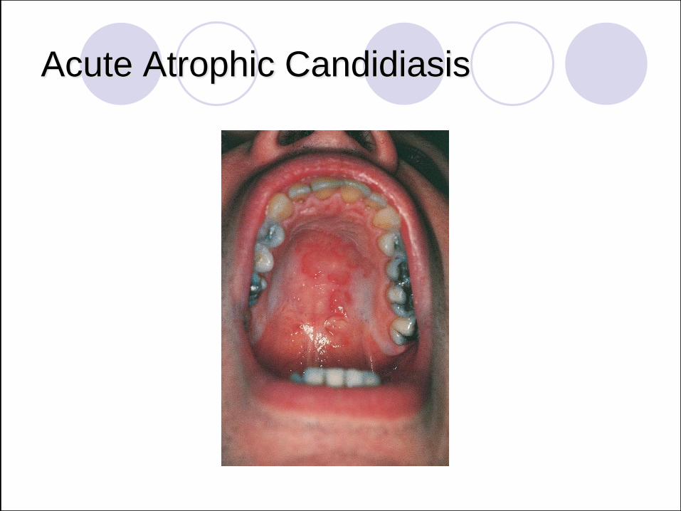
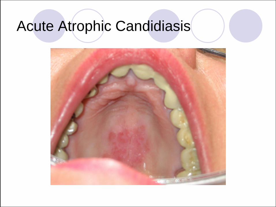
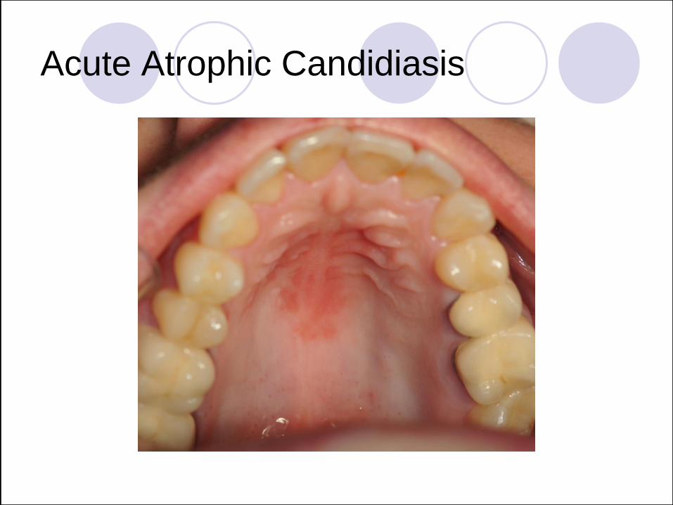
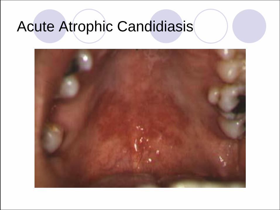
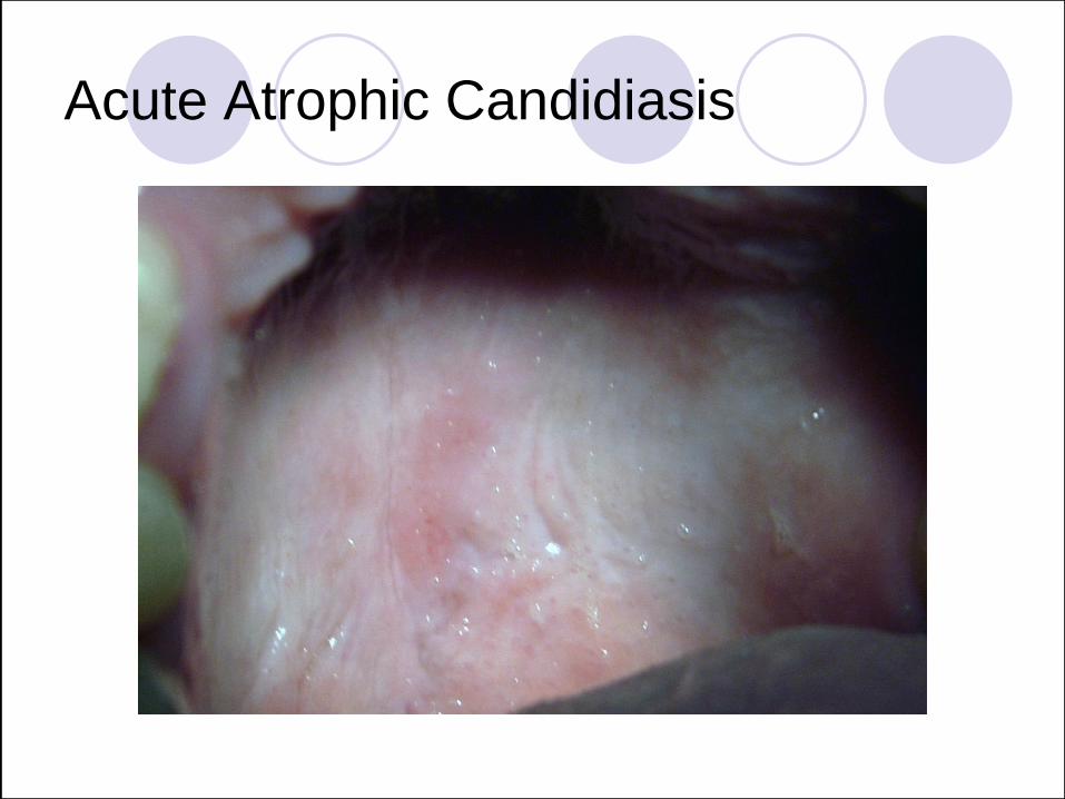
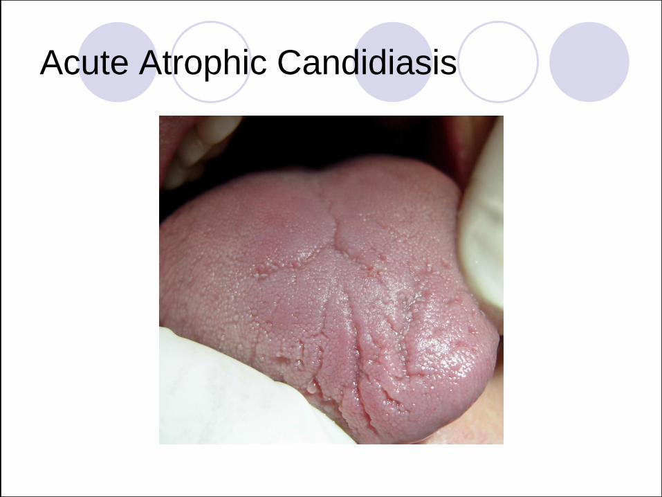
Acute Atrophic Candidiasis
Clinical: Erythematous mucosal macules
and/or patchy-depapillation of the dorsal
tongue.
Definitive diagnosis:
Identifying fungal hyphae within the lesion by
cytology or biopsy.
Response to antifungal treatment.
Acute Atrophic Candidiasis
Acute Atrophic Candidiasis
Acute Atrophic Candidiasis
Acute Atrophic Candidiasis
Acute Atrophic Candidiasis
Acute Atrophic Candidiasis
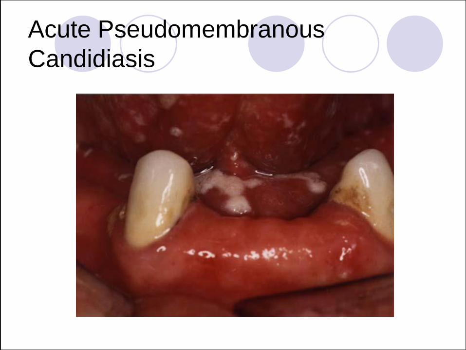
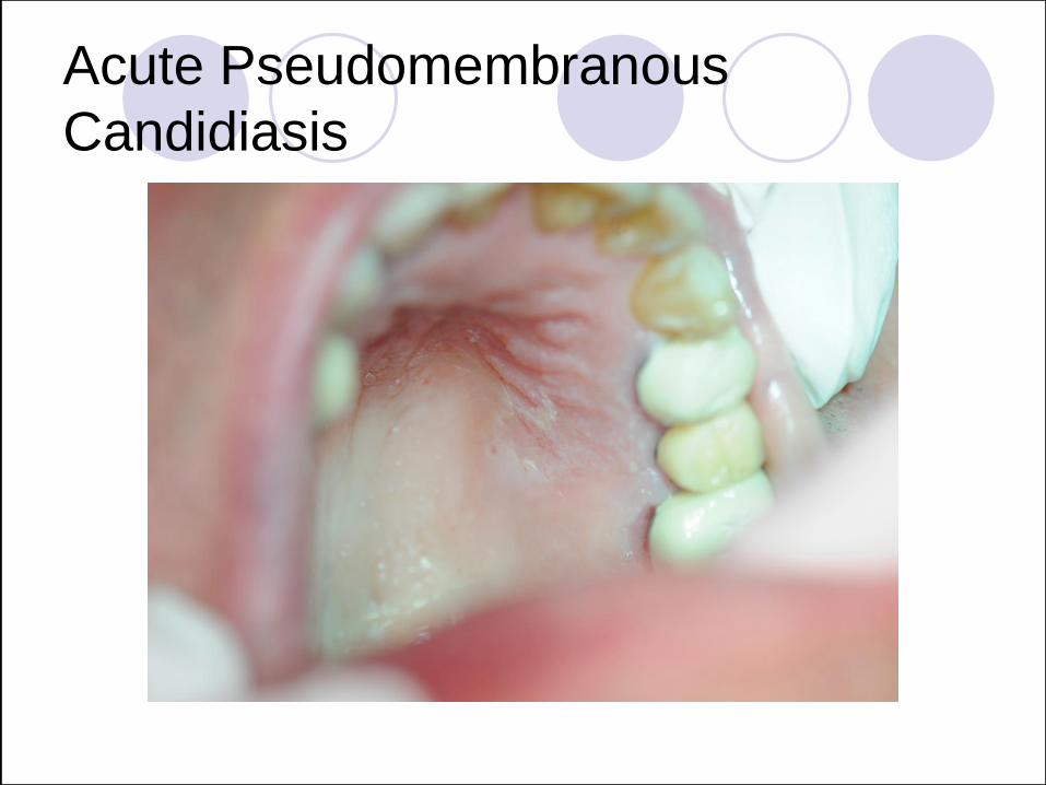
Acute Pseudomembranous Candidiasis
Appearance: White “curd-like” material that wipes off revealing an
underlying erythematous mucosa.
Clinical Diagnosis: Generally made
on the basis of appearance.

Acute Pseudomembranous
Candidiasis
Acute Pseudomembranous
Candidiasis
Acute Pseudomembranous
Candidiasis

Acute Pseudomembranous Candidiasis

Acute Pseudomembranous
Candidiasis

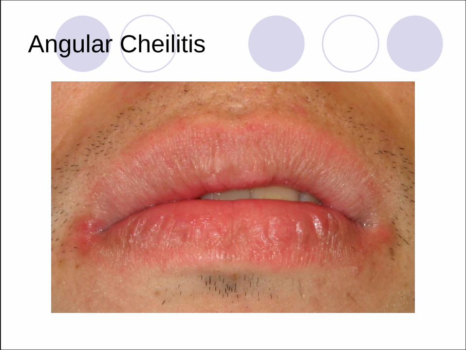
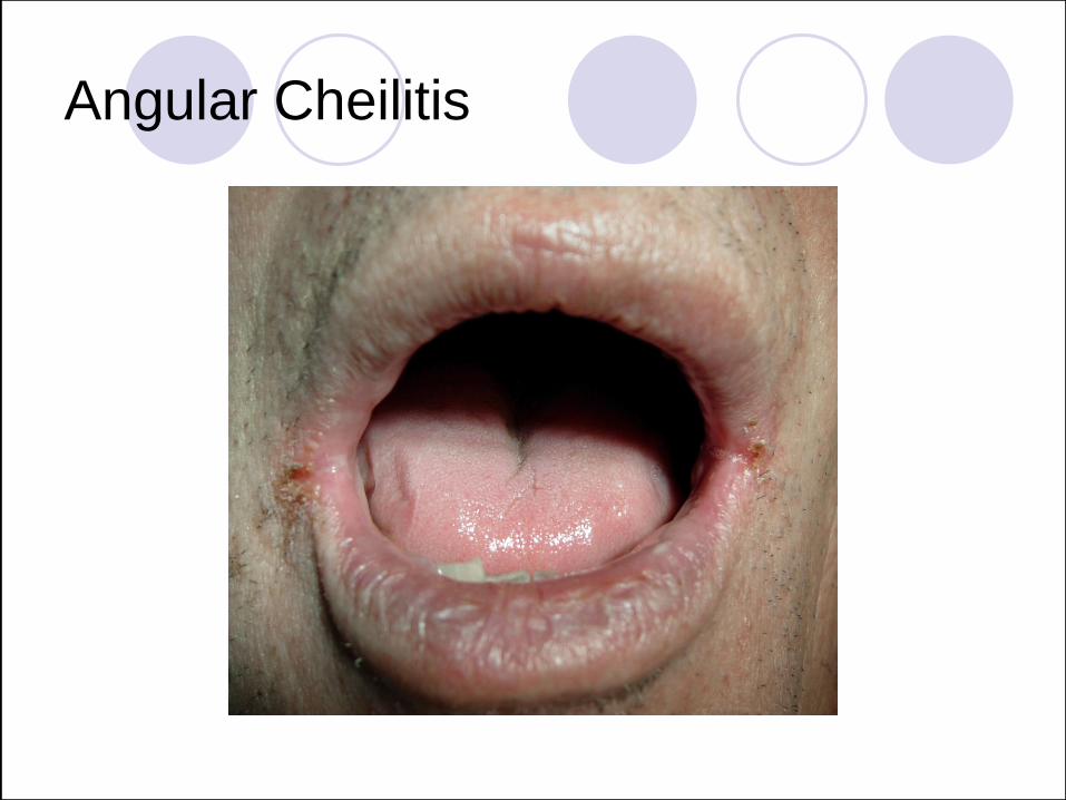
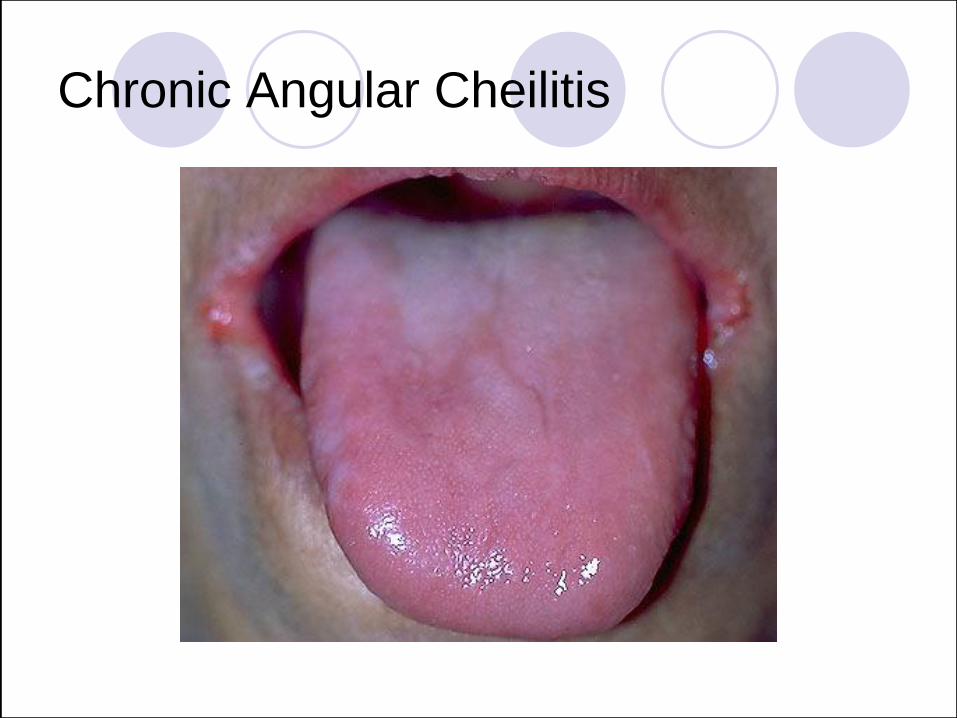
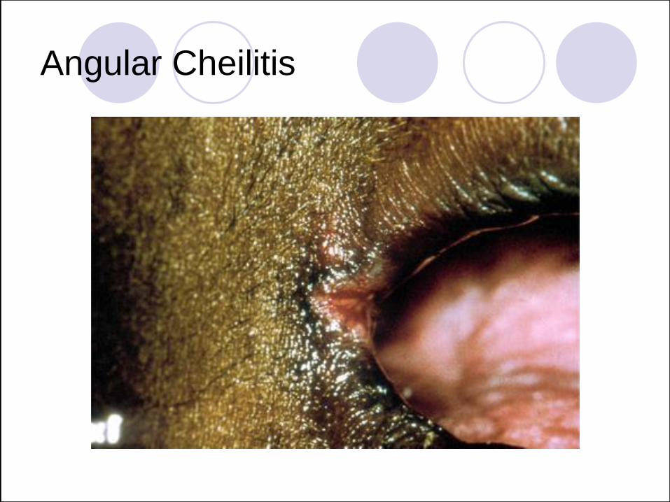
Chronic Angular Cheilitis
Clinical: Angular Cheilitis appears as cracked, eroded and encrusted surfaces at the corner of the mouth.
Frequently accompanies intraoral
candidiasis and/or a reduction in vertical
dimension
Etiology: Saliva accumulating at the
commisures of the mouth.
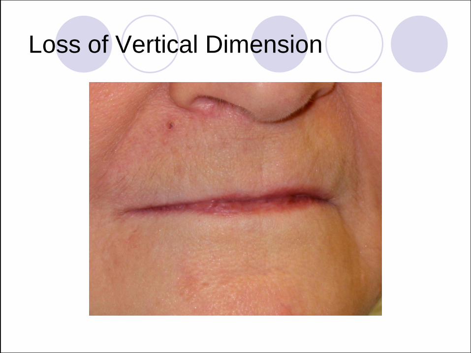
Loss of Vertical Dimension

Chronic Angular Cheilitis
Angular Cheilitis
Angular Cheilitis
Chronic Angular Cheilitis
Angular Cheilitis
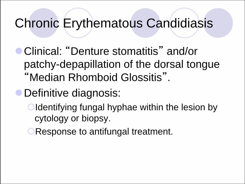
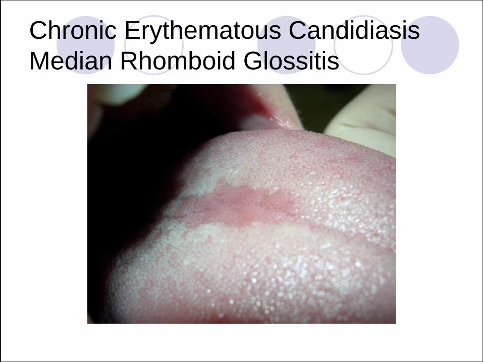
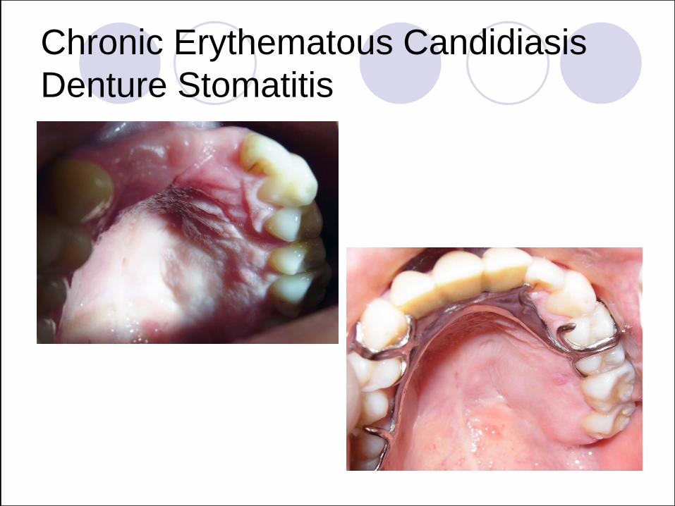
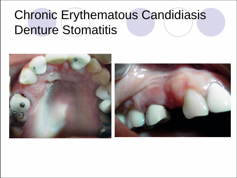
Chronic Erythematous Candidiasis
Clinical: “Denture stomatitis” and/or
patchy-depapillation of the dorsal tongue
“Median Rhomboid Glossitis”.
Definitive diagnosis:
Identifying fungal hyphae within the lesion by
cytology or biopsy.
Response to antifungal treatment.
Chronic Erythematous Candidiasis
Median Rhomboid Glossitis
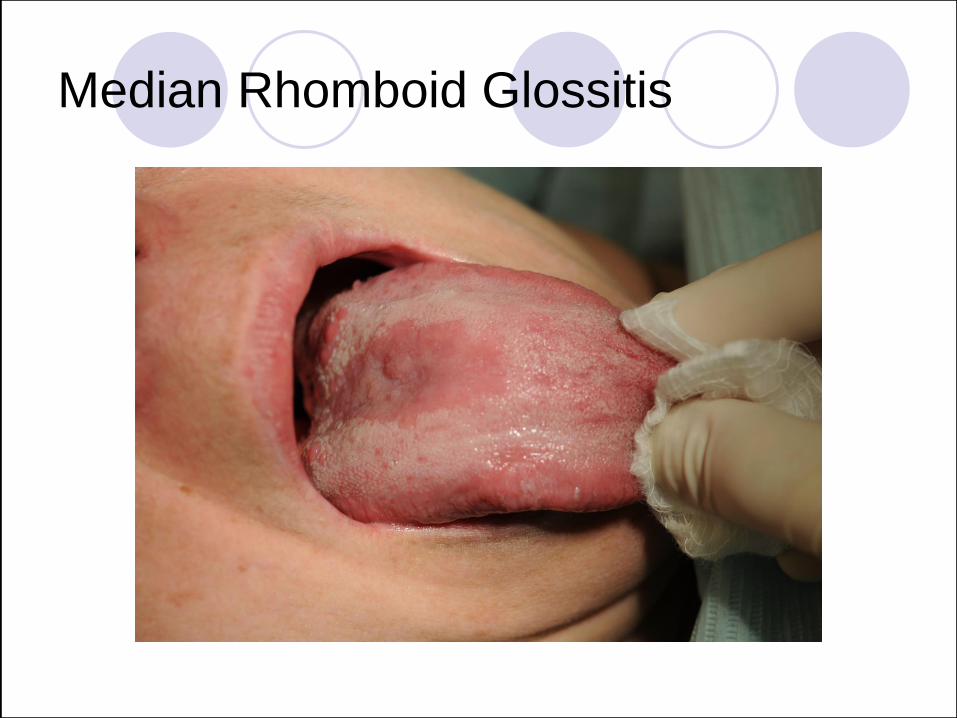
Median Rhomboid Glossitis

Median Rhomboid Glossitis
Chronic Erythematous Candidiasis
Denture Stomatitis
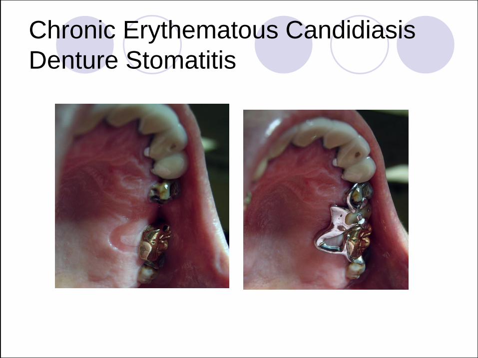
Chronic Erythematous Candidiasis
Denture Stomatitis
Chronic Erythematous Candidiasis
Denture Stomatitis

Chronic Erythematous Candidiasis
Denture Stomatitis
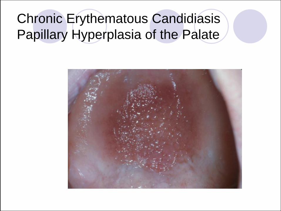
Chronic Erythematous Candidiasis
Papillary Hyperplasia of the Palate
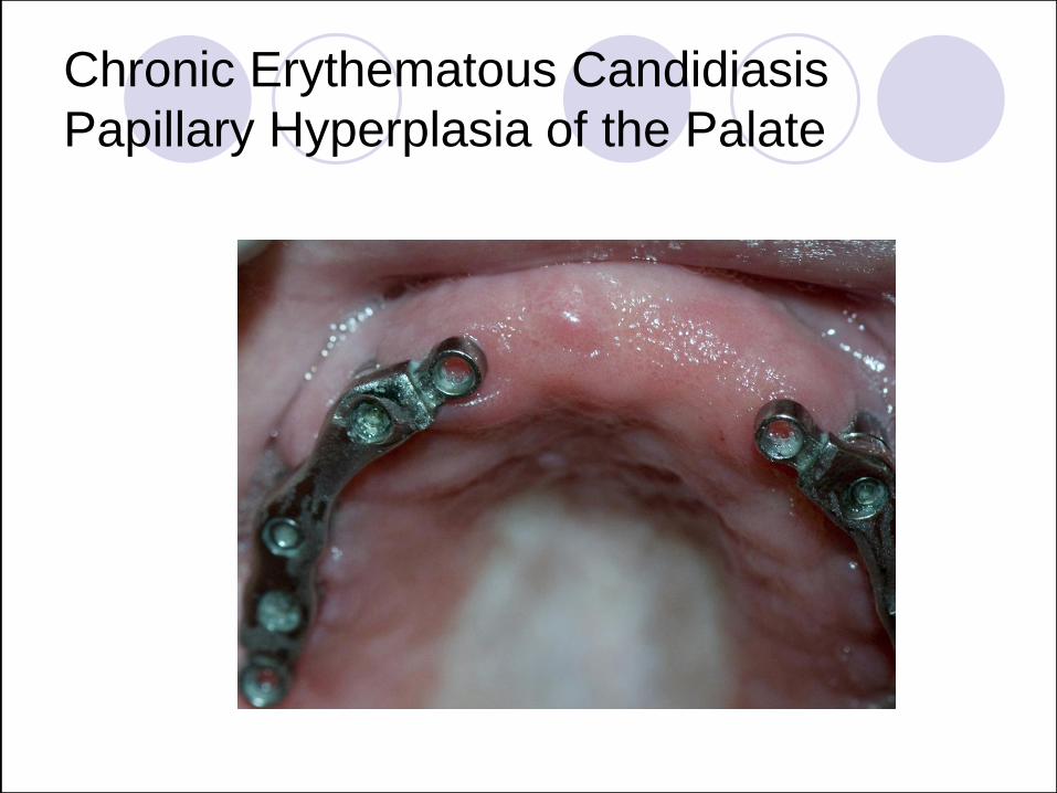
Chronic Erythematous Candidiasis
Papillary Hyperplasia of the Palate
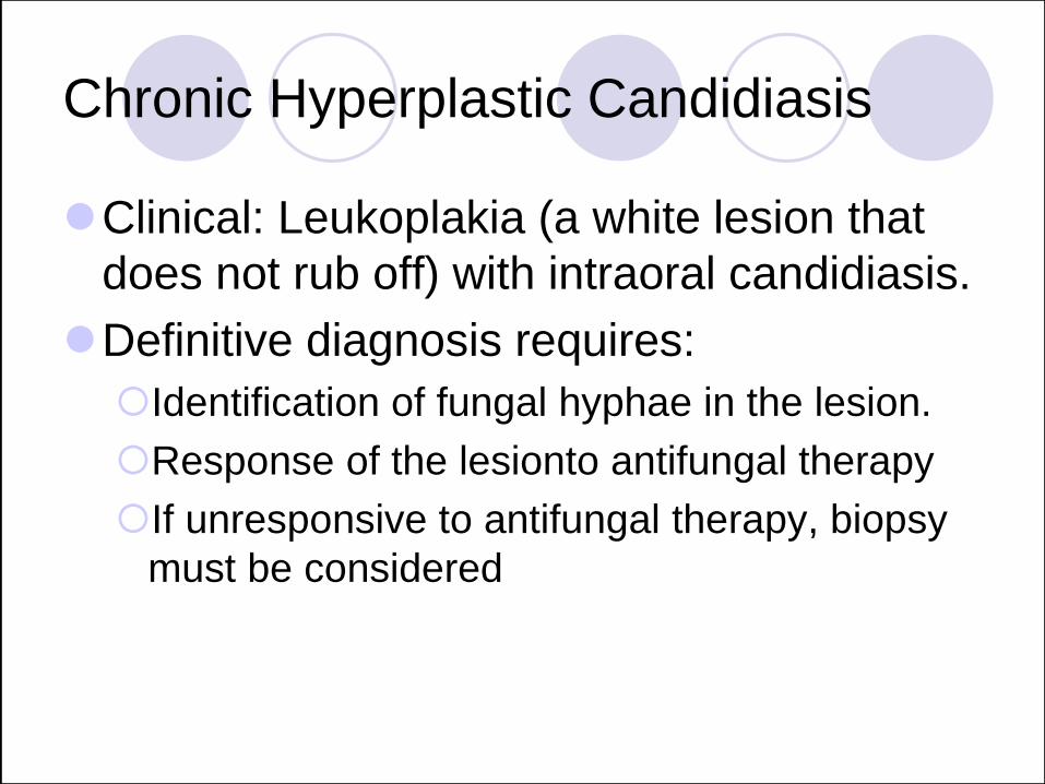
Chronic Hyperplastic Candidiasis
Clinical: Leukoplakia (a white lesion that
does not rub off) with intraoral candidiasis.
Definitive diagnosis requires:
Identification of fungal hyphae in the lesion.
Response of the lesionto antifungal therapy
If unresponsive to antifungal therapy, biopsy
must be considered

Chronic Hyperplastic Candidiasis

Chronic Hyperplastic Candidiasis

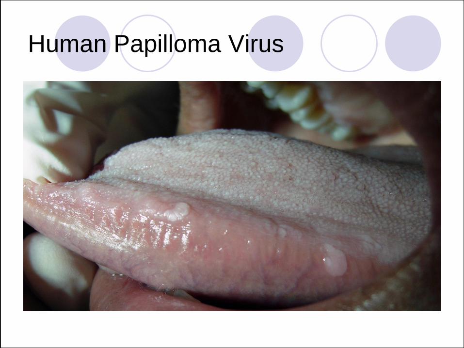
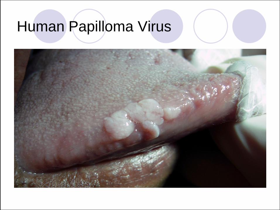
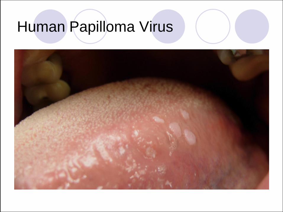
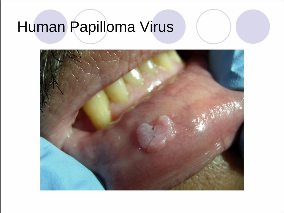
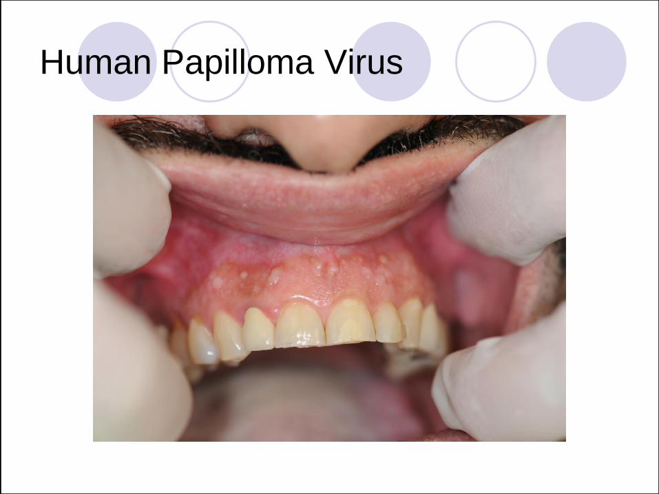
Human Papilloma Virus
Condyloma Acuminatum
Etiology: Several strains of HPV have
been reported to cause oral lesions.
Clinical: may appear cauliflower-like,
spiked or raised with a flat surface
anywhere within the oral cavity and lips.
Often present with multiple warts, difficult
to treat due to a high risk of recurrence.
Human Papilloma Virus
Human Papilloma Virus
Human Papilloma Virus
Human Papilloma Virus

Human Papilloma Virus

Human Papilloma Virus
Human Papilloma Virus

Xerostomia
Inadequate saliva production.
Objective vs. Subjective findings.
Dental visit are necessary to prevent
and treat root/coronal caries.
Frequent recalls help avoid tooth loss.
Xerostomia
Alcohol free fluoride rinses.
Use salivary substitutes containing methylcellulose or a mucin base, to provide lubrication.
Sugarless chewing gum/lozenges help to stimulate salivary flow.

Xerostomia

Xerostomia

Xerostomia

Xerostomia

Xerostomia
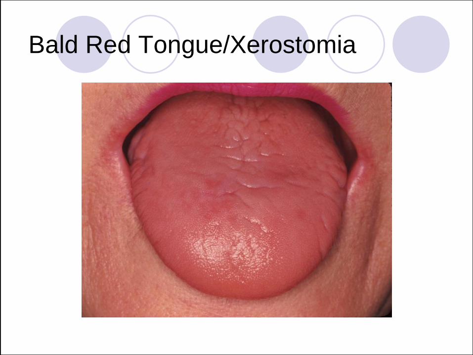
Bald Red Tongue/Xerostomia
Weblinks
Weblinks