Embed Size (px)
Citation preview
PIGMENTED LESIONS OF ORAL
CAVITY

Pigmented lesions are commonly found in the mouth. Such
lesions represent a variety of clinical entities, ranging from
physiologic changes to manifestations of systemic illnesses and
malignant neoplasm
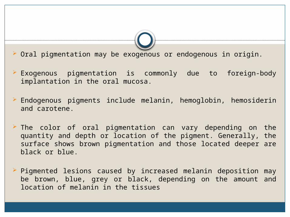
Oral pigmentation may be exogenous or endogenous in origin.
Exogenous pigmentation is commonly due to foreign-body implantation in the oral mucosa.
Endogenous pigments include melanin, hemoglobin, hemosiderin and carotene.
The color of oral pigmentation can vary depending on the quantity and depth or location of the pigment. Generally, the surface shows brown pigmentation and those located deeper are black or blue.
Pigmented lesions caused by increased melanin deposition may be brown, blue, grey or black, depending on the amount and location of melanin in the tissues
Differential diagnosis of pigmented lesions of oral cavity
The history should include the onset and duration of the lesion The presence of associated skin hyperpigmentation The presence of systemic signs and symptoms Use of prescription and nonprescription medications, Smoking habitsThe number, distribution, size, shape and colour of intraoral pigmented lesions should be assessed. Clinical tests -diascopy and radiography and laboratory investigations
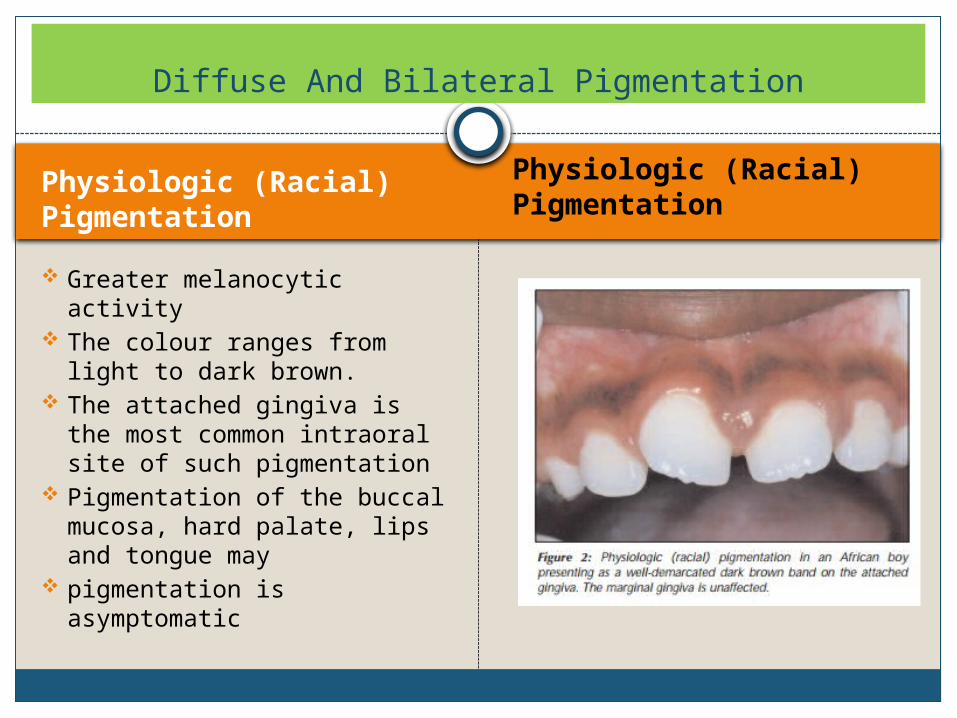
Physiologic (Racial) Pigmentation
Physiologic (Racial) Pigmentation
Greater melanocytic activity
The colour ranges from light to dark brown.
The attached gingiva is the most common intraoral site of such pigmentation
Pigmentation of the buccal mucosa, hard palate, lips and tongue may
pigmentation is asymptomatic
Diffuse And Bilateral Pigmentation
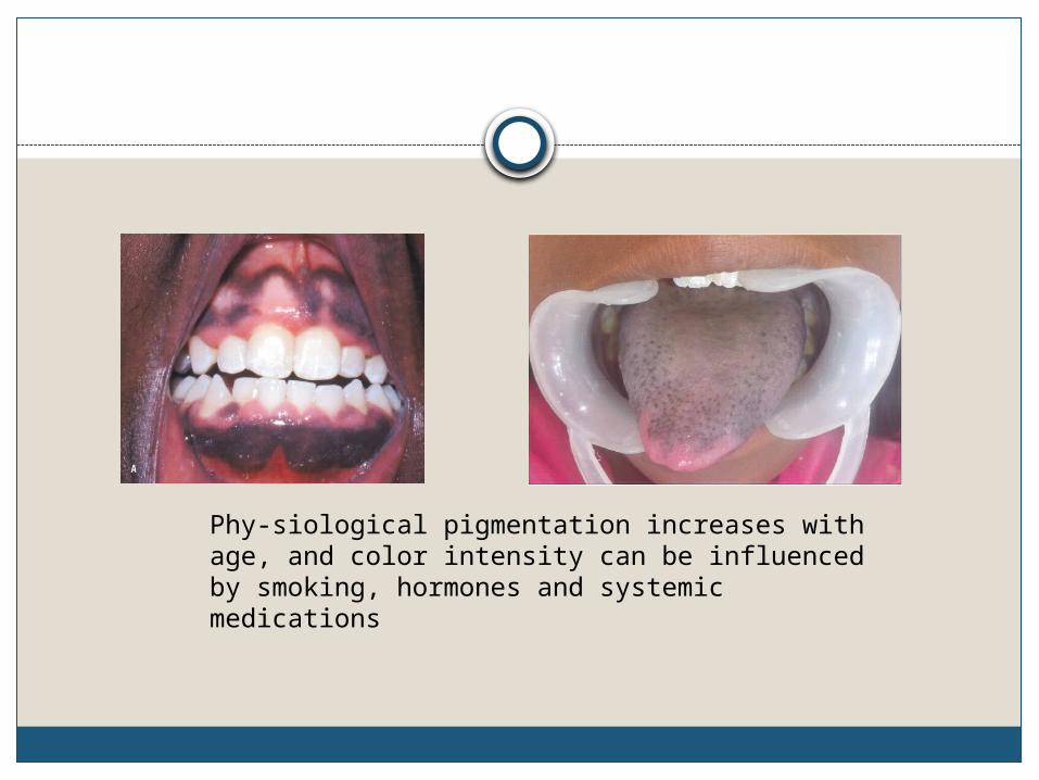
Phy-siological pigmentation increases with age, and color intensity can be influenced by smoking, hormones and systemic medications
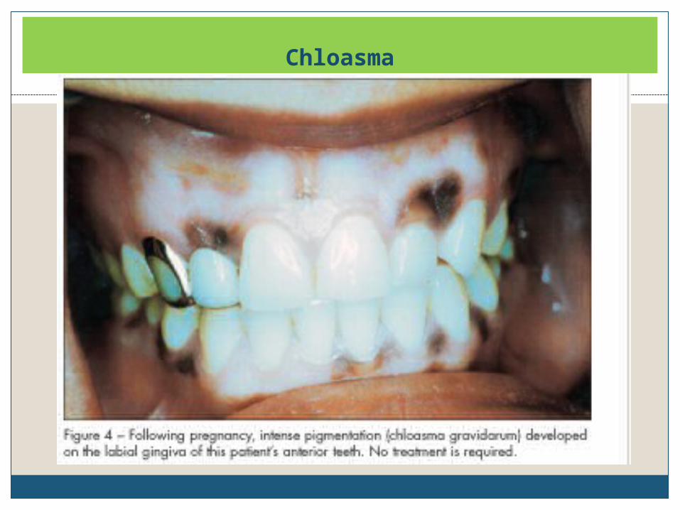
Chloasma
Peutz-Jeghers Syndrome
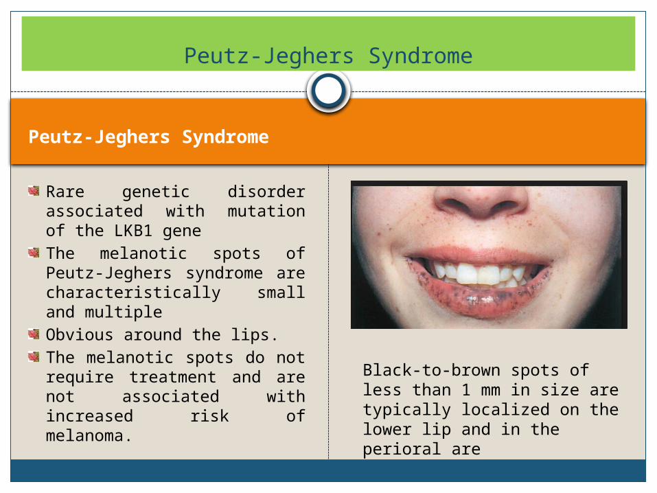
Rare genetic disorder associated with mutation of the LKB1 geneThe melanotic spots of Peutz-Jeghers syndrome are characteristically small and multipleObvious around the lips.The melanotic spots do not require treatment and are not associated with increased risk of melanoma.
Peutz-Jeghers Syndrome
Black-to-brown spots of less than 1 mm in size are typically localized on the lower lip and in the perioral are
Peutz-Jeghers Syndrome
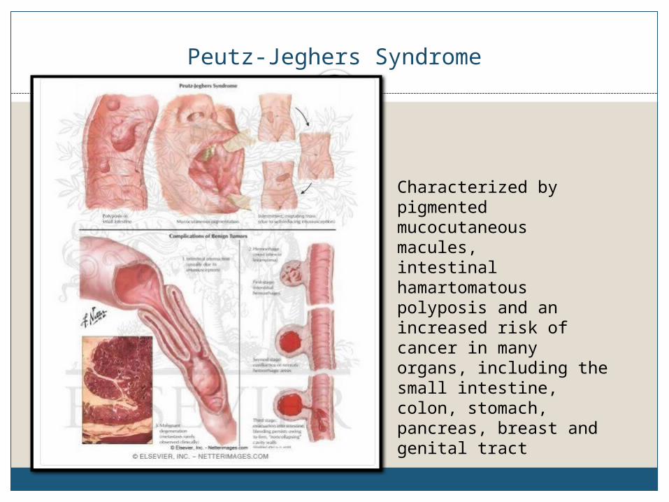
Characterized by pigmented mucocutaneous macules, intestinal hamartomatous polyposis and an increased risk of cancer in many organs, including the small intestine, colon, stomach, pancreas, breast and genital tract
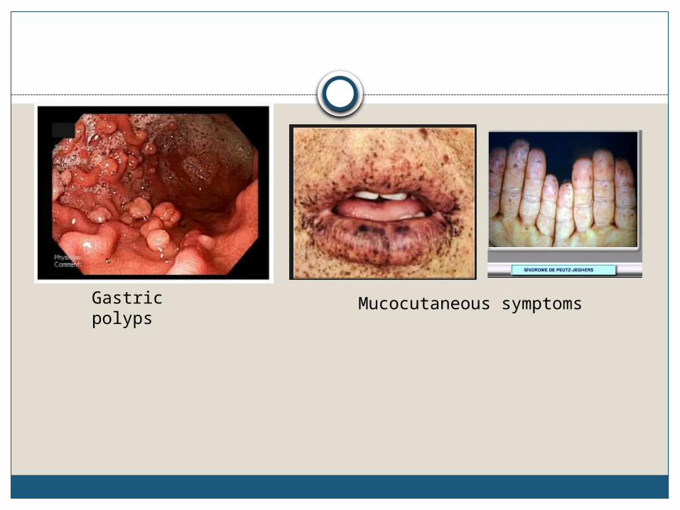
Gastric polyps Mucocutaneous symptoms
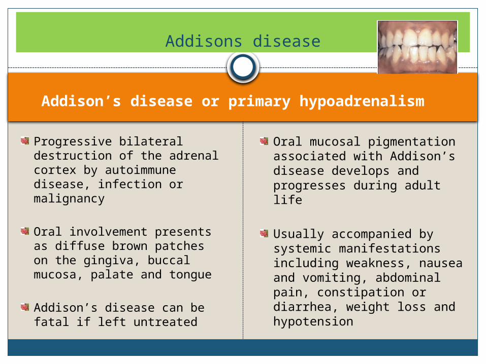
Addison’s disease or primary hypoadrenalism
Progressive bilateral destruction of the adrenal cortex by autoimmune disease, infection or malignancy
Oral involvement presents as diffuse brown patches on the gingiva, buccal mucosa, palate and tongue
Addison’s disease can be fatal if left untreated
Oral mucosal pigmentation associated with Addison’s disease develops and progresses during adult life
Usually accompanied by systemic manifestations including weakness, nausea and vomiting, abdominal pain, constipation or diarrhea, weight loss and hypotension
Addisons disease
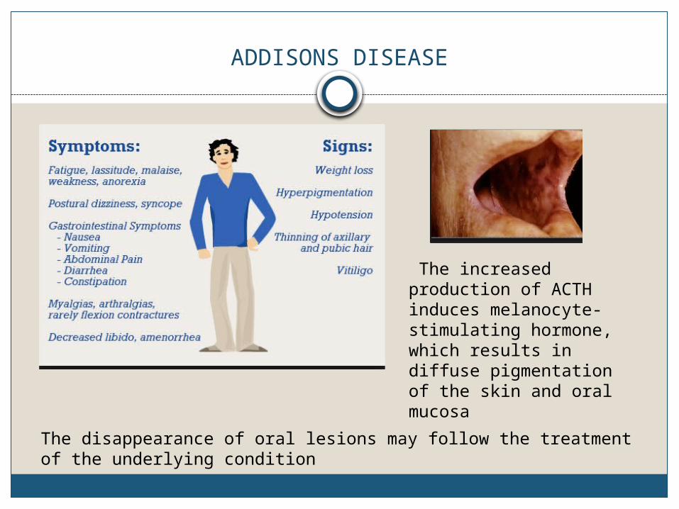
ADDISONS DISEASE
The increased production of ACTH induces melanocyte-stimulating hormone, which results in diffuse pigmentation of the skin and oral mucosa
The disappearance of oral lesions may follow the treatment of the underlying condition
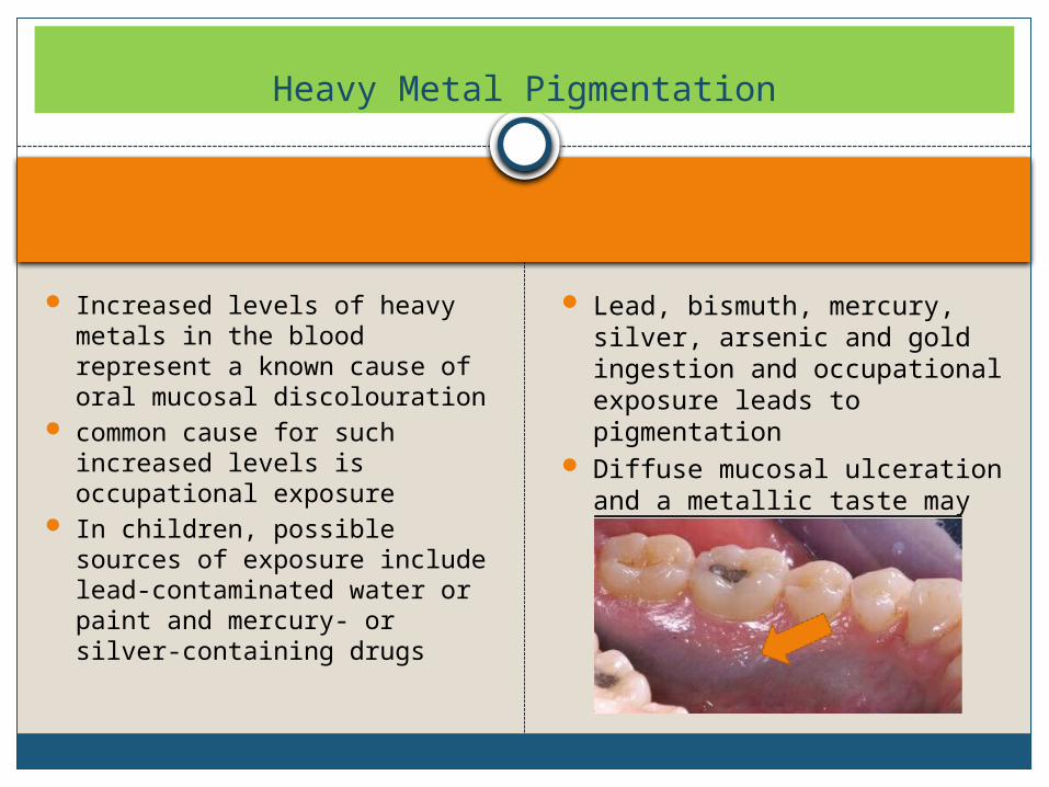
Increased levels of heavy metals in the blood represent a known cause of oral mucosal discolouration
common cause for such increased levels is occupational exposure
In children, possible sources of exposure include lead-contaminated water or paint and mercury- or silver-containing drugs
Lead, bismuth, mercury, silver, arsenic and gold ingestion and occupational exposure leads to pigmentation
Diffuse mucosal ulceration and a metallic taste may also be noted.
Heavy Metal Pigmentation
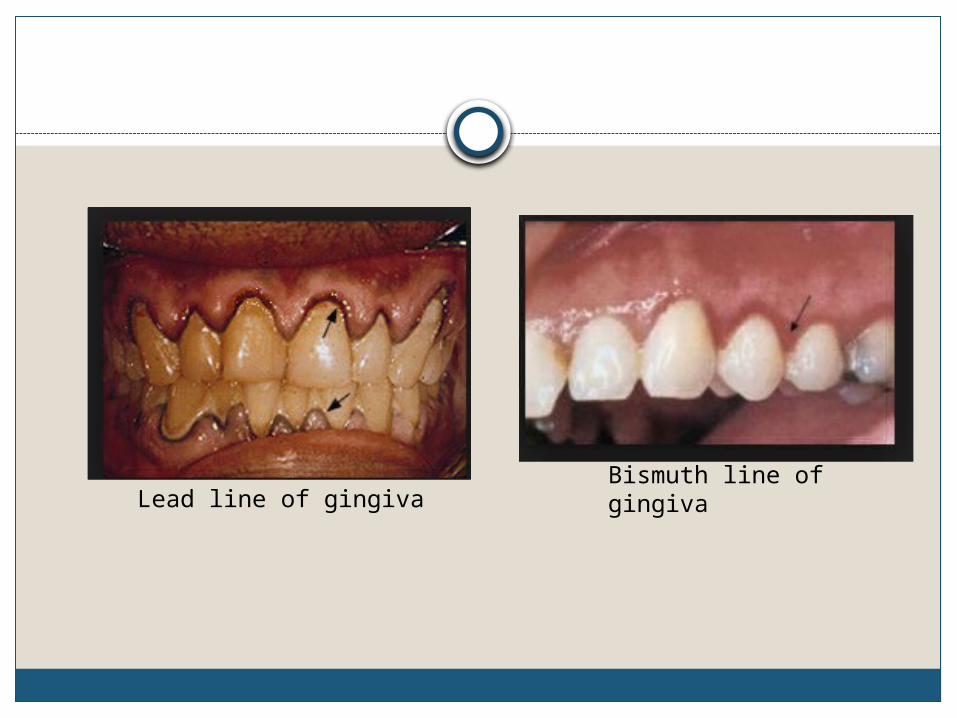
Lead line of gingivaBismuth line of gingiva
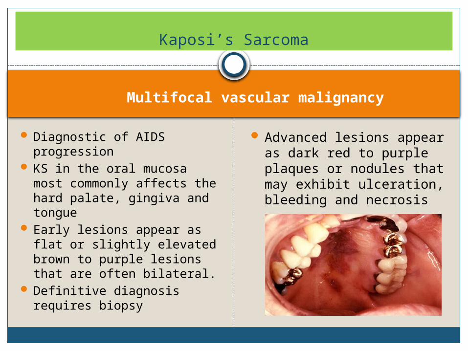
Multifocal vascular malignancy
Diagnostic of AIDS progression
KS in the oral mucosa most commonly affects the hard palate, gingiva and tongue
Early lesions appear as flat or slightly elevated brown to purple lesions that are often bilateral.
Definitive diagnosis requires biopsy
Advanced lesions appear as dark red to purple plaques or nodules that may exhibit ulceration, bleeding and necrosis
Kaposi’s Sarcoma
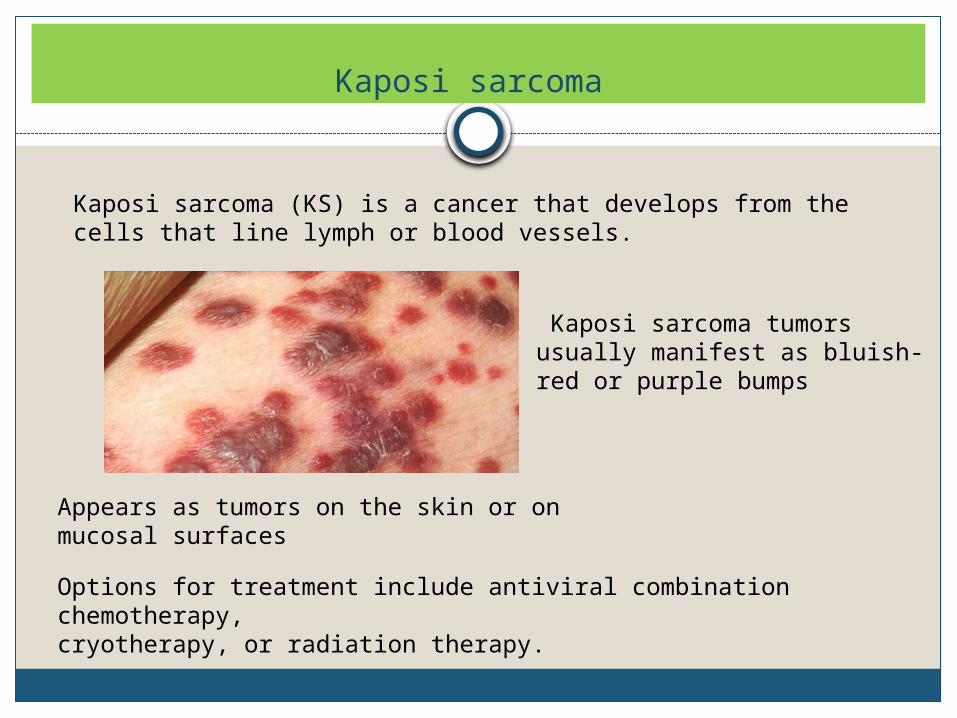
Kaposi sarcoma
Kaposi sarcoma (KS) is a cancer that develops from the cells that line lymph or blood vessels.
Appears as tumors on the skin or on mucosal surfaces
Kaposi sarcoma tumors usually manifest as bluish-red or purple bumps
Options for treatment include antiviral combination chemotherapy, cryotherapy, or radiation therapy.
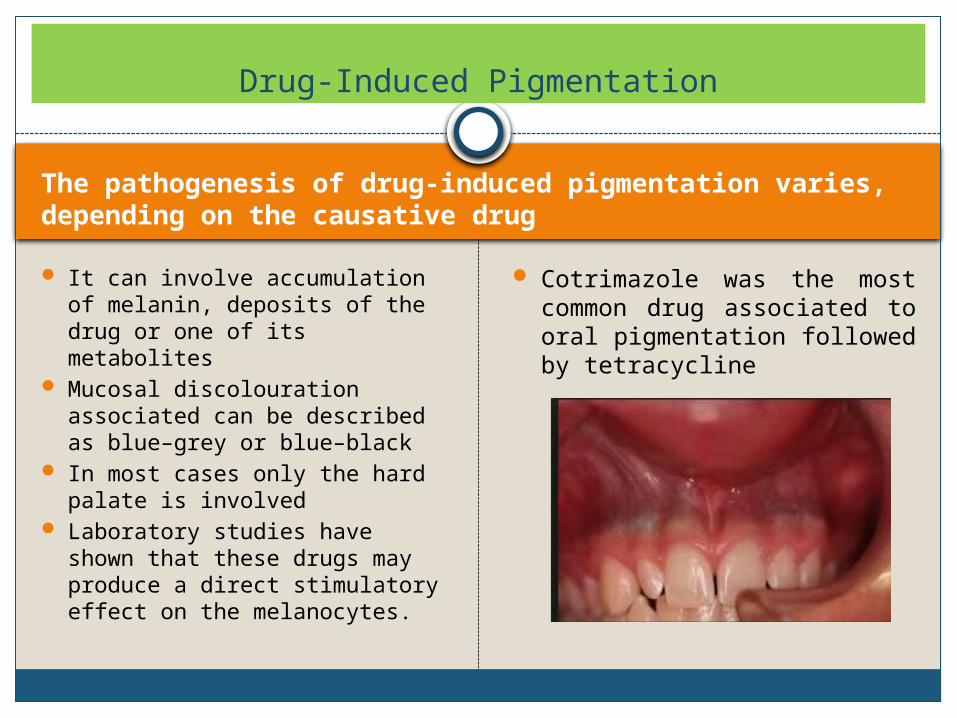
The pathogenesis of drug-induced pigmentation varies, depending on the causative drug
It can involve accumulation of melanin, deposits of the drug or one of its metabolites
Mucosal discolouration associated can be described as blue–grey or blue–black
In most cases only the hard palate is involved
Laboratory studies have shown that these drugs may produce a direct stimulatory effect on the melanocytes.
Cotrimazole was the most common drug associated to oral pigmentation followed by tetracycline
Drug-Induced Pigmentation
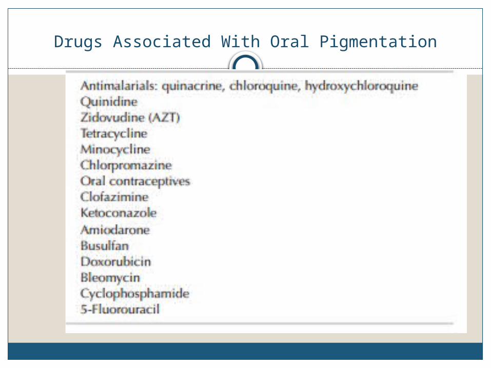
Drugs Associated With Oral Pigmentation
Postinflammatory Pigmentation
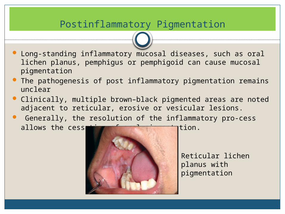
Long-standing inflammatory mucosal diseases, such as oral lichen planus, pemphigus or pemphigoid can cause mucosal pigmentation
The pathogenesis of post inflammatory pigmentation remains unclear
Clinically, multiple brown–black pigmented areas are noted adjacent to reticular, erosive or vesicular lesions.
Generally, the resolution of the inflammatory pro-cess allows the cessation of oral pigmentation.
Reticular lichen planus with pigmentation
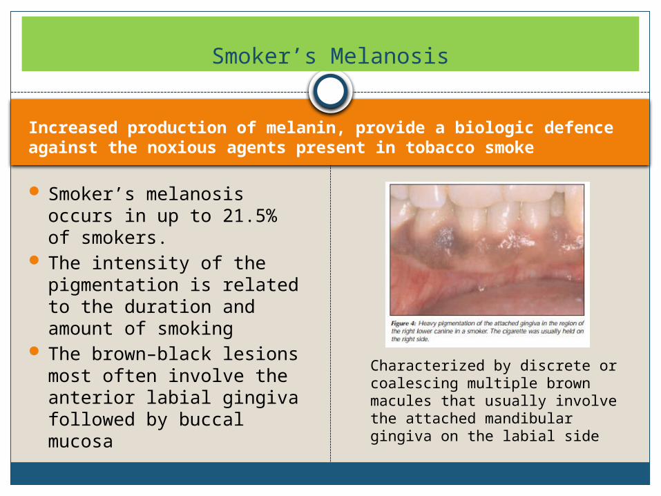
Increased production of melanin, provide a biologic defence against the noxious agents present in tobacco smoke
Smoker’s melanosis occurs in up to 21.5% of smokers.
The intensity of the pigmentation is related to the duration and amount of smoking
The brown–black lesions most often involve the anterior labial gingiva followed by buccal mucosa
Smoker’s Melanosis
Characterized by discrete or coalescing multiple brown macules that usually involve the attached mandibular gingiva on the labial side
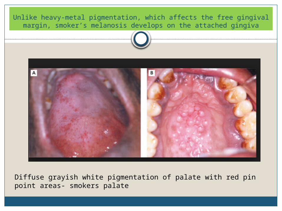
Unlike heavy-metal pigmentation, which affects the free gingival margin, smoker’s melanosis develops on the attached gingiva
Diffuse grayish white pigmentation of palate with red pin point areas- smokers palate
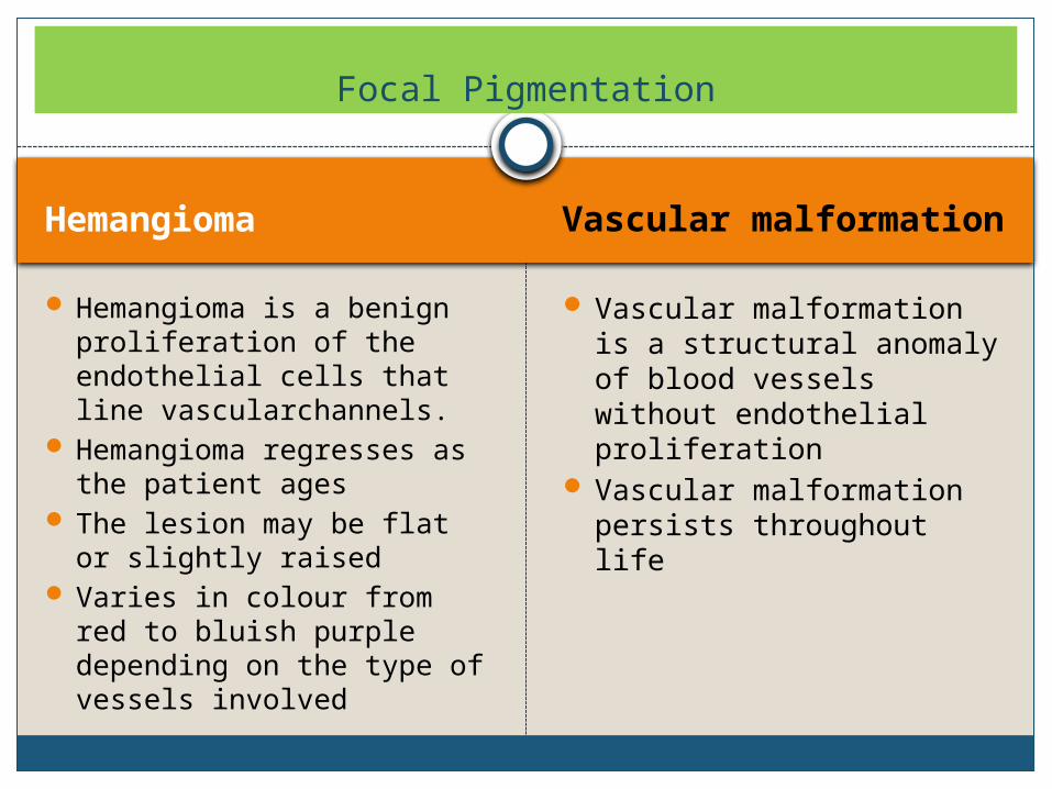
HemangiomaVascular malformation
Hemangioma is a benign proliferation of the endothelial cells that line vascularchannels.
Hemangioma regresses as the patient ages
The lesion may be flat or slightly raised
Varies in colour from red to bluish purple depending on the type of vessels involved
Vascular malformation is a structural anomaly of blood vessels without endothelial proliferation
Vascular malformation persists throughout life
Focal Pigmentation
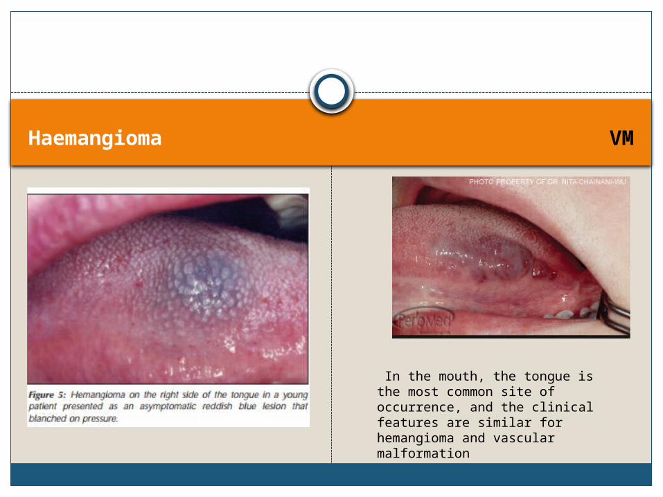
Haemangioma VM
In the mouth, the tongue is the most common site of occurrence, and the clinical features are similar for hemangioma and vascular malformation
Varix and Thrombus
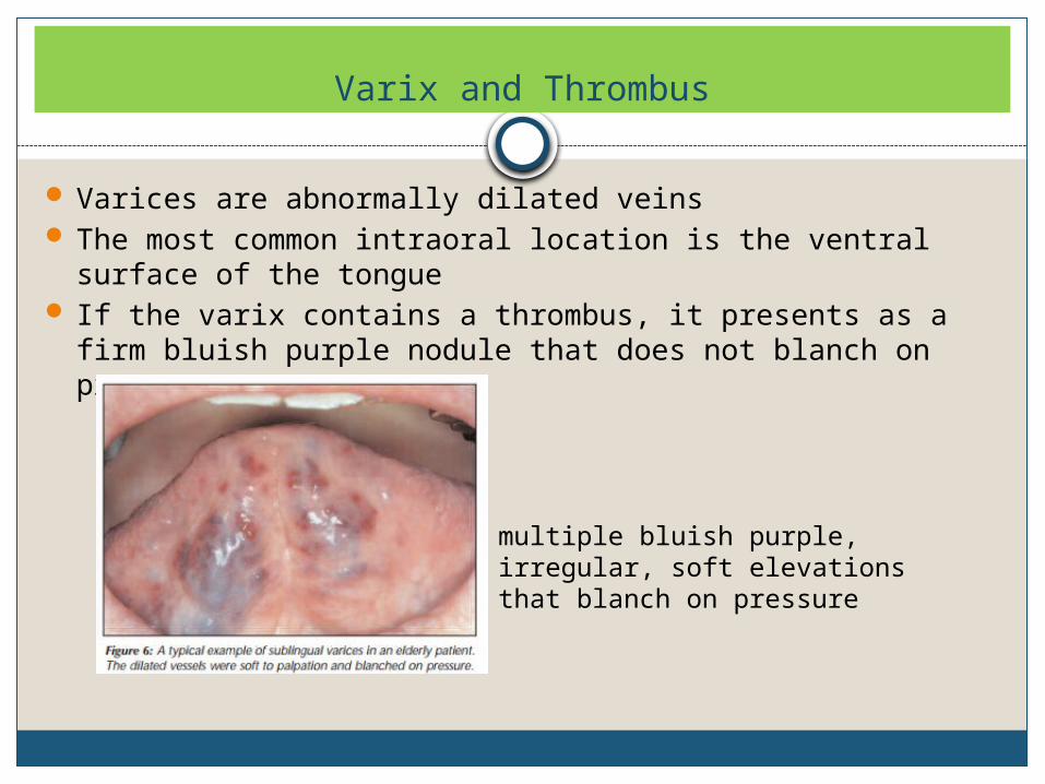
Varices are abnormally dilated veins The most common intraoral location is the ventral surface of
the tongue If the varix contains a thrombus, it presents as a firm bluish
purple nodule that does not blanch on pressure
multiple bluish purple, irregular, soft elevations that blanch on pressure
Amalgam Tattoo and Other Foreign-Body Pigmentation
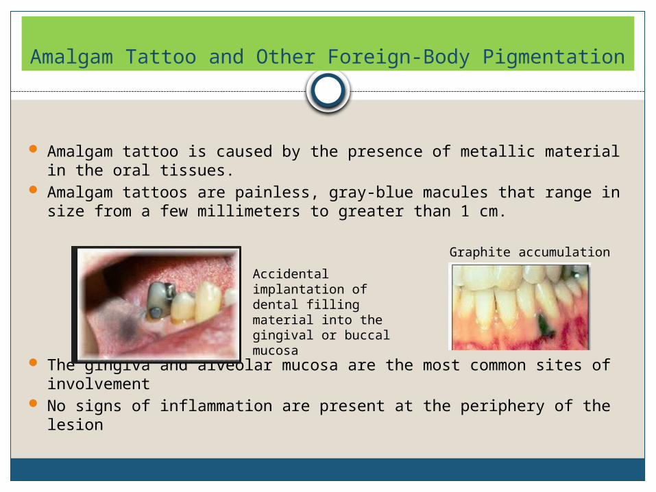
Amalgam tattoo is caused by the presence of metallic material in the oral tissues.
Amalgam tattoos are painless, gray-blue macules that range in size from a few millimeters to greater than 1 cm.
The gingiva and alveolar mucosa are the most common sites of involvement
No signs of inflammation are present at the periphery of the lesion
Accidental implantation of dental filling material into the gingival or buccal mucosa
Graphite accumulation
Melanotic Macules
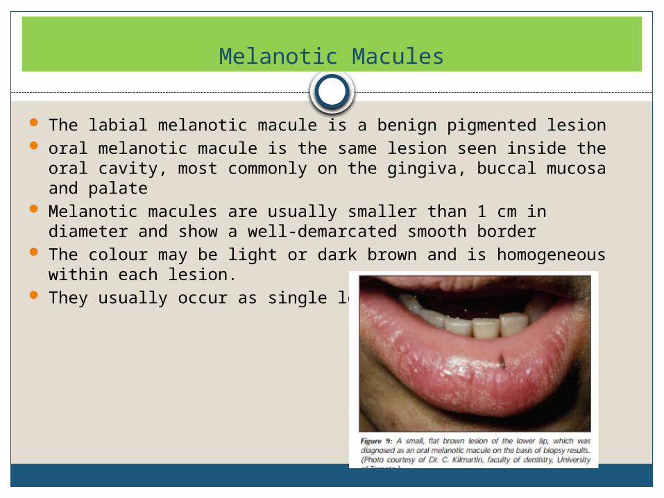
The labial melanotic macule is a benign pigmented lesion oral melanotic macule is the same lesion seen inside the oral cavity,
most commonly on the gingiva, buccal mucosa and palate Melanotic macules are usually smaller than 1 cm in diameter and
show a well-demarcated smooth border The colour may be light or dark brown and is homogeneous within
each lesion. They usually occur as single lesions
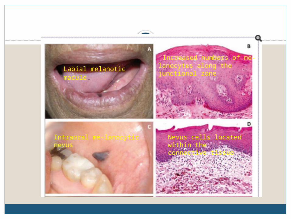
Labial melanotic macule.
Increased numbers of me-lanocytes along the junctional zone
Intraoral me-lanocytic nevus
Nevus cells located within the connective tissue
Pigmented Nevi
Pigmented Nevi
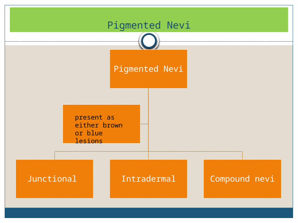
Junctional Intradermal Compound nevi
present as either brown or blue lesions
Junctional nevi are flat and dark brown in colour because the nevus cells proliferate at the tips of the rete pegs close to the surface. Intramucosal and compound nevi are typically light brown, dome-shaped lesions Melanocytic nevi constitute benign neoplasms of cutaneous melanocytesProliferation of benign neoplastic melanocytes along the submucosalmucosal junction -junctional nevusMigration of these cells to the underlying mesenchymal tissue compound nevusLoss of the junctional component of the nevi, so that all remaining nevomelanocytes are located within the subepithelial connective tissue stroma -subepithelial nevus
Blue nevus
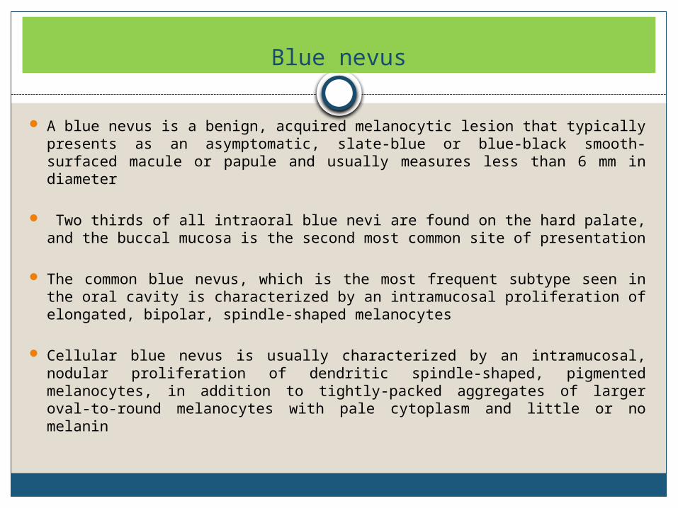
A blue nevus is a benign, acquired melanocytic lesion that typically presents as an asymptomatic, slate-blue or blue-black smooth-surfaced macule or papule and usually measures less than 6 mm in diameter
Two thirds of all intraoral blue nevi are found on the hard palate, and the buccal mucosa is the second most common site of presentation
The common blue nevus, which is the most frequent subtype seen in the oral cavity is characterized by an intramucosal proliferation of elongated, bipolar, spindle-shaped melanocytes
Cellular blue nevus is usually characterized by an intramucosal, nodular proliferation of dendritic spindle-shaped, pigmented melanocytes, in addition to tightly-packed aggregates of larger oval-to-round melanocytes with pale cytoplasm and little or no melanin
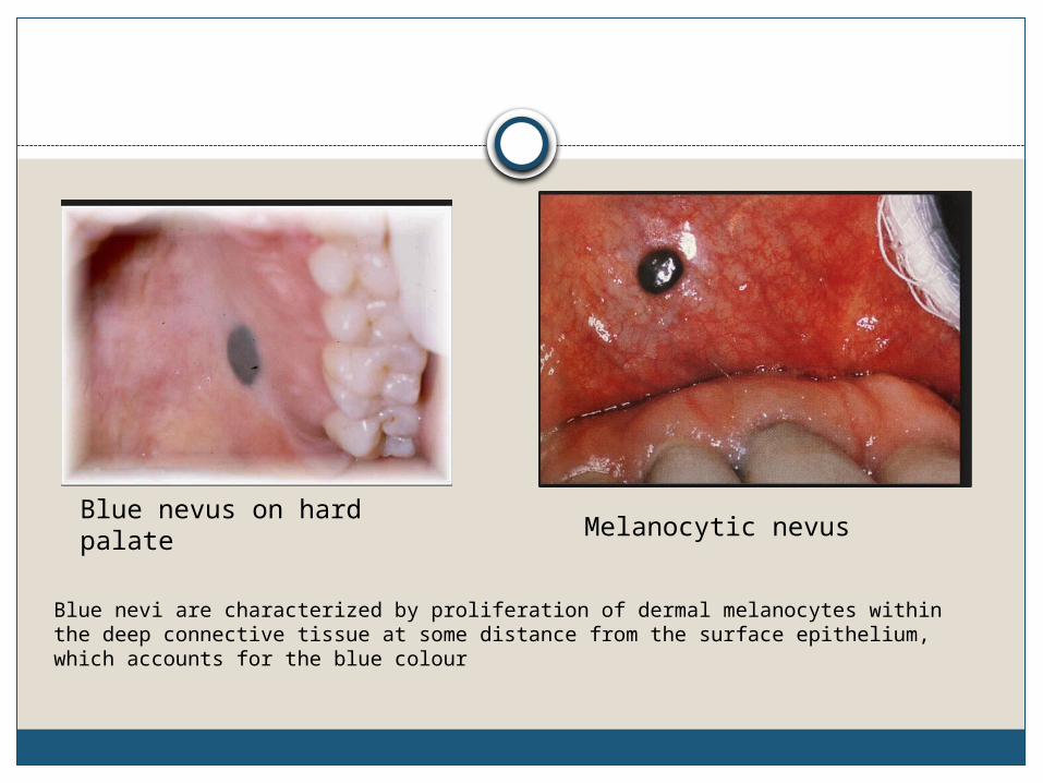
Blue nevus on hard palateMelanocytic nevus
Blue nevi are characterized by proliferation of dermal melanocytes within the deep connective tissue at some distance from the surface epithelium, which accounts for the blue colour
Oral Melanoacanthoma
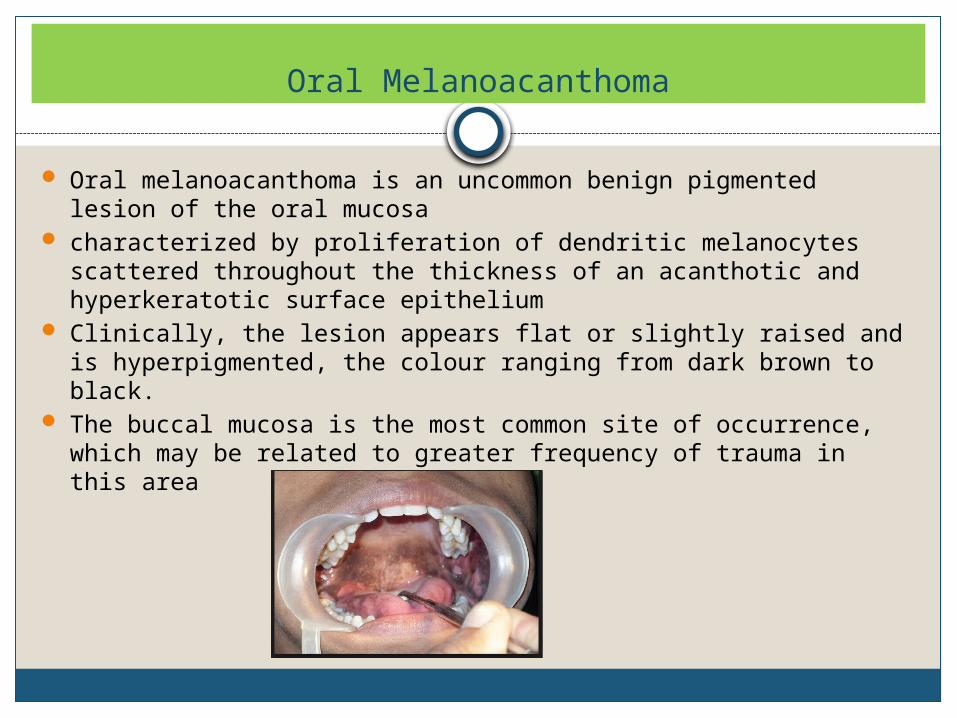
Oral melanoacanthoma is an uncommon benign pigmented lesion of the oral mucosa
characterized by proliferation of dendritic melanocytes scattered throughout the thickness of an acanthotic and hyperkeratotic surface epithelium
Clinically, the lesion appears flat or slightly raised and is hyperpigmented, the colour ranging from dark brown to black.
The buccal mucosa is the most common site of occurrence, which may be related to greater frequency of trauma in this area
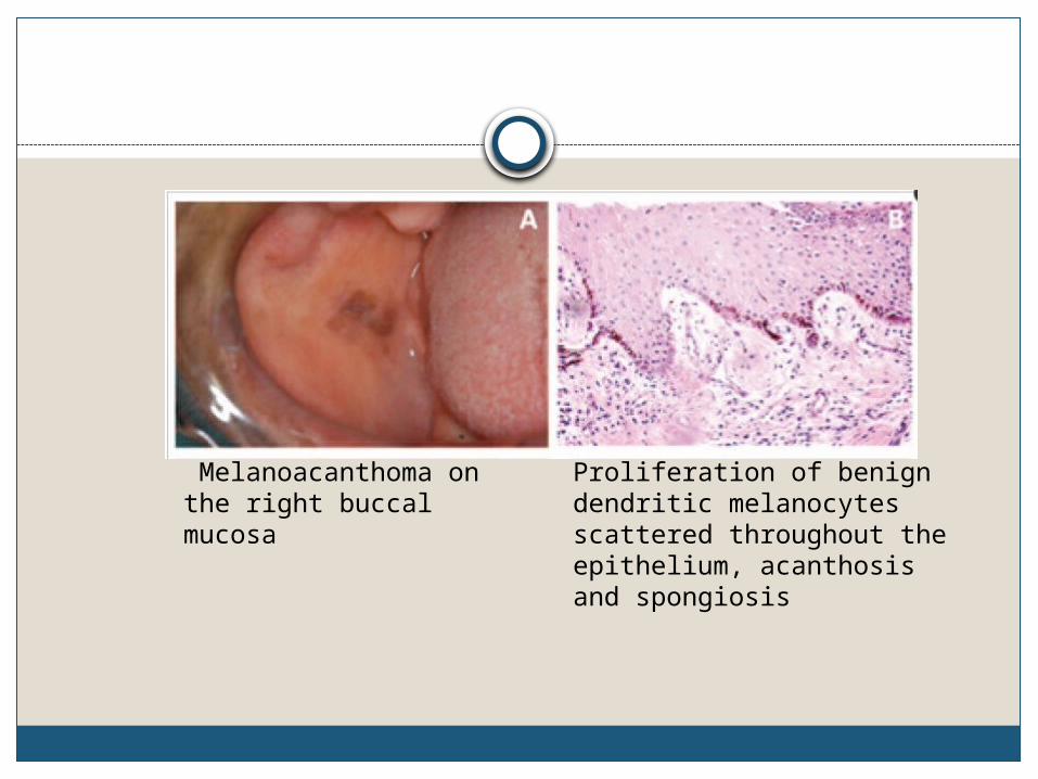
Melanoacanthoma on the right buccal mucosa
Proliferation of benign dendritic melanocytes scattered throughout the epithelium, acanthosis and spongiosis
Oral Melanoma
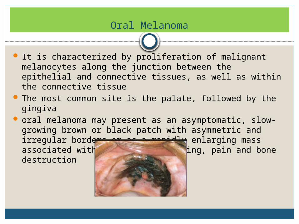
It is characterized by proliferation of malignant melanocytes along the junction between the epithelial and connective tissues, as well as within the connective tissue
The most common site is the palate, followed by the gingiva oral melanoma may present as an asymptomatic, slow-
growing brown or black patch with asymmetric and irregular borders or as a rapidly enlarging mass associated with ulceration, bleeding, pain and bone destruction
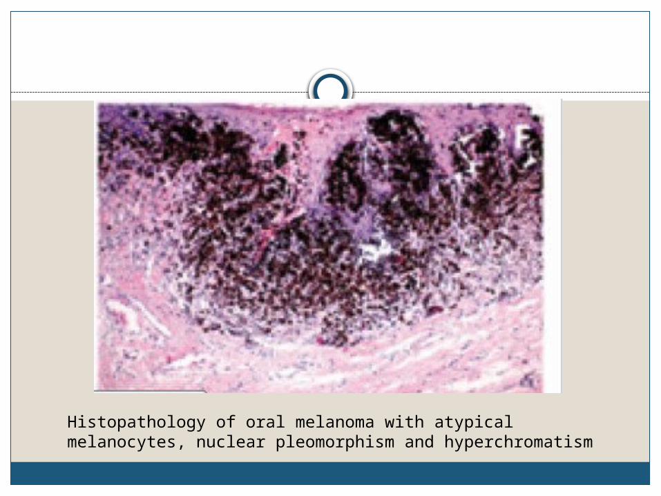
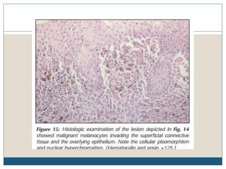
Histopathology of oral melanoma with atypical melanocytes, nuclear pleomorphism and hyperchromatism
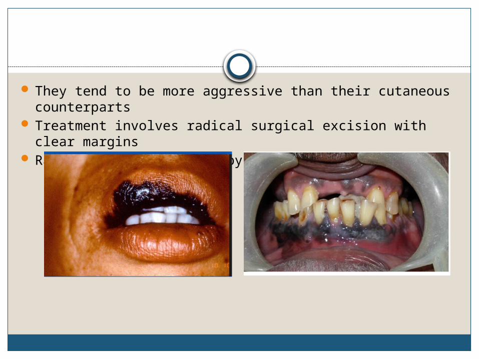
They tend to be more aggressive than their cutaneous counterparts
Treatment involves radical surgical excision with clear margins
Radiation and chemotherapy are ineffective
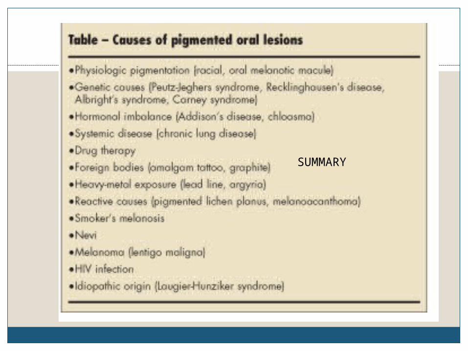
SUMMARY