Embed Size (px)
Citation preview

lable at ScienceDirect
Clinical Radiology 74 (2019) 410.e1e410.e9
Contents lists avai
Clinical Radiology
journal homepage: www.cl in icalradiologyonl ine.net
Comparison of cardiac MRI with PET forassessment of myocardial viability in patientswith coronary chronic total occlusionJ.N. Li a, Y. He b,*, W. Dong c, L.J. Zhang b, H.Z. Mi c, D.F. Zhang a,R.C. Huang d, X.T. Song a
aDepartment of Cardiology, Beijing Anzhen Hospital, Capital Medical University, Beijing, ChinabDepartment of Radiology, Beijing Anzhen Hospital, Capital Medical University, Beijing, ChinacDepartment of Nuclear Medicine, Beijing Anzhen Hospital, Capital Medical University, Beijing, ChinadDepartment of Cardiology, The First Affiliated Hospital of Dalian Medical University, Dalian, China
article information
Article history:Received 6 May 2018Accepted 24 January 2019
* Guarantor and correspondent: Y. He, DepartAnzhen Hospital, Capital Medical University, 2District, Beijing 100029, China. Tel.: þ86 1561111
E-mail address: [email protected] (Y. He).
https://doi.org/10.1016/j.crad.2019.01.0210009-9260/� 2019 The Royal College of Radiologists.
AIM: To compare cardiac magnetic resonance imaging (MRI) and positron-emission to-mography (PET) assessment of myocardial viability in patients with coronary chronic totalocclusion (CTO).MATERIALS AND METHODS: Eighty patients with coronary CTO underwent cardiac MRI and
PET. Cardiac MRI images were analysed using a 17-segment model, and late gadoliniumenhancement (LGE) and wall motion were scored. PET was used to classify myocardial viabilityvia myocardial perfusion and 18F-fluorodeoxyglucose, digital superscript uptake.RESULTS: With PET as the reference standard, the sensitivity of cardiac MRI in detecting
myocardial viability was 95.3%, specificity was 87.5%, positive predictive value was 96.8%,negative predictive value was 84.2%, and accuracy was 93.8% on a per patient basis. Thereceiver operator characteristic curve was used to analyse the performance of cardiac MRI inthe detection of myocardial viability on a per-patient basis and the area under the curve was0.910 (95% confidence interval 0.805 to 1). Cardiac MRI had the highest sensitivity and spec-ificity for differentiating viable and non-viable myocardium as defined by PET when the cut-offvalue of LGE was 50%. The motion consistency and correlation of cardiac MRI and PET wereanalysed and kappa was 0.788 (r¼0.825; p<0.001).CONCLUSION: Compared with PET, cardiac MRI assessment of myocardial viability in pa-
tients with coronary CTO has high sensitivity, specificity, and accuracy. Therefore, cardiac MRIcan be used as an important method for evaluating myocardial viability in coronary CTOpatients.
� 2019 The Royal College of Radiologists. Published by Elsevier Ltd. All rights reserved.
ment of Radiology, BeijingAnzhen Road, Chao Yang2775.
Published by Elsevier Ltd. All righ
Introduction
Coronary chronic total occlusion (CTO) is caused byatherosclerotic plaque rupture leading to thrombosis,thrombus regeneration leading to complete occlusion of the
ts reserved.

J.N. Li et al. / Clinical Radiology 74 (2019) 410.e1e410.e9410.e2
coronary artery with an occlusion time of >3 months.1e3
CTO lesions are found on coronary angiography in 20e30%of patients.4 The success rate of interventional treatment ofCTO lesions is low and the postoperative complications andthe requirements for the operators are high.5 CTO lesionsare even called “the last bastion” in the field of coronaryartery interventional therapy.
The American College of Cardiology (ACC)/AmericanHeart Association (AHA) and European Society of Cardiology(ESC) guidelines demonstrate the importance of myocardialviability analysis in patients with coronary CTO.6,7 Thepresence or absence and the degree of myocardial viabilityare important in determining whether patients will benefitfrom interventional treatment of CTO lesions.
Nowadays, positron-emission tomography (PET) is thereference standard for evaluating myocardial viability andcardiac magnetic resonance imaging (MRI) is one of themost commonly used methods for evaluating myocardialviability8; however, there are few studies comparing cardiacMRI and PET assessment of myocardial viability9e12 inparticular in patients with coronary CTO. Therefore, thepurpose of the present study was to compare cardiac MRIand PET assessment of myocardial viability in patients withcoronary CTO.
Materials and methods
Patients
A total of 80 consecutive patients with coronary CTOconfirmed by invasive coronary angiography (ICA) wererecruited and studied from January 2016 to December 2017.All patients scheduled for myocardial viability assessmentwere scanned with both cardiac MRI and PET within 1 weekof each other. All patients remained stable throughout theexamination period. This study excluded patients with leftmain coronary stenosis >50%, acute coronary syndromewithin 90 days, and decompensated heart failure, patientsundergoing coronary artery bypass grafting (CABG),implantable pacemakers or defibrillators, and patients witharrhythmias and claustrophobia. The study was approvedby the local Ethics Committee. All patients provided writteninformed consent.
Cardiac MRI protocol
Cardiac MRI was performed on the Siemens 3 T whole-body MRI system (MAGNETOM Verio, A Tim System;Siemens Healthcare, Erlangen, Germany). A 32-elementmatrix coil was activated for data collection, and all se-quences were electrocardiogram (ECG) gated. Total scanduration was approximately 1 hour. All images were ac-quired using phased array surface coils during mild expi-ration and electrocardiographic triggering. Eight millimetresections with no intersection gap were acquired in theshort-axis plane (from the base to the apex) and long-axisplane of the left ventricle to perform cine cardiac MRI andlate gadolinium enhancement (LGE) imaging. LGE imagingwas performed 15 minutes after the intravenous
administration of gadolinium (Magnevist, Bayer Healthcare,0.2 mmol/kg) by using a two-dimensional phase-sensitiveinversion recovery breath-hold sequence (parameters: 2.4ms repetition time [TR]/1.01 ms echo time [TE], 100 msinversion time [TI], 12� flip angle; 8 mm section thickness;99�160 matrix; 2.7�2.3 mm2 in-plane spatial resolution;651 Hz per pixel bandwidth). This acquisition wascompleted in �10 minutes (but no longer than 30 minutes)after the last administration of gadolinium.
Nuclear medicine protocol
Image acquisition was performed with fixed angle 90�
dual-head single-photon-emission computed tomography(SPECT)/computed tomography (CT) camera (Precedence 16,Philips, Amsterdam, Netherlands) equipped with low-energy high-resolution collimator. The heart was includedin the effective field of vision. Resting myocardial perfusionimaging was performed with 99mTc-MIBI (925 MBq, ChinaAtomic Hi-Tech, Beijing, China) and 20% window at 140 keV.Collecting over 6�104 myocardial count level, image recon-struction was used by Astonish iterative method and Han-ning filtering function, the cut-off frequency 0.62, order 10,iteration 4 times, subset number 8, without attenuationcorrection. 99mTc-MIBIwas injected intravenously and imagewas presented after 1e1.5 hours. The patient is supine, theacquisition mode is step-by-shot, the acquisition parame-ters: matrix 128�128,16-bit, magnification 1.5, rotation18 0 �
(32 frames, 25 seconds/frame). At each frame, eight ECG-gated frames according to per cardiac cycle were collected.
Myocardial 18F-fluorodeoxyglucose, digital superscript(18F-FDG) imaging was performed within 2 days of 99mTc-MIBI imaging. After an overnight fast for at least 12 hours, anoral glucose of 25e50 gwas given to the patients according totheir serum glucose level. In diabetes patients, acipimox wasadministrated (500 mg, oral dose) before glucose loading.Insulin was administrated intravenously if the blood glucoselevel was >9 mmol/l at 45 minutes after oral glucoseadministration with close monitoring of blood glucose.When the blood glucose level was appropriate, 18F-FDG (3MBq/kg) was administered intravenously. PET images wereacquired using PET/CT (Biograph mCT, Siemens, Pennsylva-nia, USA) equipped with a 52-ring PET that could accept 511KeV peak collimator and 128 slice spiral CT. The heart wasincluded in the effective field of vision and myocardialglucosemetabolism imagingwasperformedusing 18F-FDGata rotation of 360� (32 frames, 25 s/frame). The TrueX þ timeof flight (TOF) ultra-HD iteration method was used for short-axis reconstruction of the original image with two iterations,21 subsets, image size 200, zoom 1.0, and Gaussian filterwave half height 8. Filtered back projection was performedusing a Butterworth filter (cut-off frequency 0.17 cycles perpixel, order 8). The spatial resolution of the PET image was 2mm. Eight ECG-gated frames per cardiac cyclewere collected.
Segmental analysis
The left ventricle was divided into six basal, six mid-ventricular, four apical segments, and the apex using the

Table 1Basic patient characteristics.
Characteristics Data
SexMale 68 (85%)Female 12 (15%)
Age (years)Mean�SD 56.9�10.2Range 35e78
Previous infarction 25 (31.3%)Smoke 45 (56.3%)Hypertension 48 (60%)Diabetes 18 (22.5%)Hyperlipidaemia 25 (31.3%)History of PCI 29 (36.3%)CTO lesionsLAD CTO 32 (34.4%)LCX CTO 18 (19.4%)RCA CTO 43 (46.2%)
Cardiac MRI parameters (Mean�SD)Ejection fraction (%) 53.5�15.1End diastolic volume (ml) 121.8�65.3End diastolic volume (ml) 55.7�40.9Stroke volume (ml) 56.2�21.0Myocardial mass (g) 118.5�48.7
SD, standard deviation; CTO, chronic total occlusion; MRI, magnetic reso-nance imaging; PCI, percutaneous coronary intervention; LAD, left anteriordescending artery; LCX, left circumflex; RCA, right coronary artery.
J.N. Li et al. / Clinical Radiology 74 (2019) 410.e1e410.e9 410.e3
17-segmental model of the ACC/AHA.13 The basal, mid-ventricular, and apical segments were analysed in theshort-axis plane, the apex of the cardiac MRI image wasanalysed in the two-chamber long-axis plane, and the apexof the PET imagewas analysed in the horizontal and verticallong-axis plane. The myocardial regions supplied by the leftanterior descending (LAD), left circumflex (LCX), and rightcoronary arteries (RCA) were selected according to the ACC/AHA criteria.13,14
Cardiac MRI image analysis
Cardiac MRI image analysis was undertaken using aSiemens workstation. All short-axis sections were projectedon the two-chamber long-axis images and assigned todifferent positions according to their relationship with thepapillary muscles. The mid-ventricular segments corre-sponded to the papillary muscle and the basal and apicalsegments corresponded to above and below the papillarymuscle.13 Visual analysis of each myocardial segment wasperformed and the wall motion of each myocardial segmentwas scored as follows: 1 (normal), 2 (hypokinesia), 3 (aki-nesia), and 4 (dyskinesia). The wall motion score index(WMSI) was calculated by dividing the sum of the coronaryartery wall motion scores by the corresponding number ofmyocardial segments.13
All short-axis images of the left ventricle from the base tothe apex were analysed. The transmural extent of myocar-dial infarction or scarring was analysed by LGE, which wasdefined as the percentage of delayed myocardial thicknessto total myocardial thickness and was scored as 1 (0%), 2(1e25%), 3 (26e50%), 4 (51e75%), and 5 (76e100%). LGE�50% was considered to indicate a viable myocardium.15
PET image analysis
PET data analysis was performed using Cedars software.Myocardium with normal blood perfusion and normal orincreased 18F-FDG uptake (normal) and myocardium withreduced blood perfusion but normal or increased 18F-FDGuptake (mismatch) was considered to be viable myocar-dium. Myocardium with reduced blood perfusion andreduced 18F-FDG uptake (match) was considered to be non-viable myocardium.
Cardiac MRI and PET data were analysed by two expe-rienced radiologists and two experienced nuclear medicinephysicians, respectively, who were blinded to each patient’sclinical condition and the decision was made in consensus.
Global and regional left ventricular function analysis
Left ventricular volume, mass, ejection fraction (EF), end-diastolic volume (EDV), and end-systolic volume (ESV) werecalculated from cardiac MRI short-axis images.
Statistical analysis
Data are expressed as mean � standard deviation.Continuous data were compared using Student’s t test orone-way analysis of variance. Continuous data of non-
normal distribution were analysed using theKruskaleWallis test. Categorical variables were analysedusing the chi-square test. Spearman’s correlation analysiswas used to analyse the correlation between different mo-dalities, and Kappa analysis was used to compare the globalagreement between different modalities. PET was used asthe reference standard to calculate the sensitivity andspecificity of cardiac MRI in detecting myocardial viabilityand the receiver operator characteristic (ROC) curve wasused to analyse cardiac MRI performance for assessingmyocardial viability. A p-value of <0.05 was considered tobe statistically significant. All statistical analyses were per-formed using SPSS 23.
Results
In the present study, 1,360 myocardial segments wereanalysed in 80 patients. PET and cardiac MRI were per-formed in all patients within 1 week and there was nosignificant clinical change in any patient between the twoexaminations. The mean age of the patients was 56.9�10.2years with 68 (85%) male patients. Detailed patient clinicalcharacteristics are shown in Table 1.
Cardiac MRI and PET
Therewere 767myocardial segments with LGE of 0%, 223with LGE of 1e25%, 135 with LGE of 26e50%, 120 with LGEof 51e75%, and 115 with LGE of 76e100%. There were 892normal myocardial segments, 260 mismatched myocardialsegments, and 208 matched myocardial segments. Of the892 normal myocardial segments, 20 (2.2%) had LGE >50%,of the 260 mismatched myocardial segments, 228 (87.7%)

Figure 1 Distribution of LGE of myocardial segments in differenttypes of myocardial viability determined by PET.
Figure 2 In a patient with chronic total occlusion of LCX, short axis, horiz18F-FDG PET (bottom row) images were normal in all myocardial segmenshowed that the LGE of corresponding myocardial segments was 0%.
J.N. Li et al. / Clinical Radiology 74 (2019) 410.e1e410.e9410.e4
had LGE �50% and 32 (12.3%) had LGE>50%, and of the 208matched myocardial segments, 25 (12%) had LGE �50%(Fig 1). PET examination of normal, mismatched, andmatched myocardial segments with corresponding cardiacMRI examination results are shown in Figs 2e4. As shown inTable 2, there was a negative correlation between myocar-dial viability determined by PET and cardiac MRI-LGE(p<0.001; Table 2, Fig 5).
Comparison of cardiac MRI and PET in assessingmyocardial viability on a per-patient basis
With PET as the reference standard, the sensitivity ofcardiac MRI in assessing myocardial viability was 95.3%,specificity was 87.5%, positive predictive value was 96.8%,negative predictive value was 82.4%, and accuracy was93.8% (Table 3). Furthermore, with PET as the referencestandard for detection of myocardial viability, the ROC curve
ontal long axis, and vertical long axis SPECT perfusion (top row) andts (normal) and short axis and vertical long axis cardiac MRI images

Figure 3 In a patient with chronic total occlusion of the right coronary artery, short axis and vertical long axis SPECT perfusion (top row) imagesshowed a defect from the inferior wall to the posterolateral wall, and short axis and vertical long axis 18F-FDG PET (bottom row) images showeda mild hypometabolism in corresponding myocardial segments (mismatched), short axis and vertical long axis cardiac MRI images showed thatLGE of corresponding myocardial segments was 1e25%.
J.N. Li et al. / Clinical Radiology 74 (2019) 410.e1e410.e9 410.e5
was used to analyse the performance of cardiac MRI fordetection of myocardial viability (Fig 6) and the AUC was0.910 (95% confidence interval 0.805 to 1). A threshold of50% LGE was confirmed to yield optimal sensitivity andspecificity for the differentiation of viable and non-viablemyocardial segments defined by PET on a per-patient basis.
Comparison of cardiac MRI and PET in assessingmyocardial motion
The motion of each myocardial segment was analysedusing cardiac MRI and PET. The consistency and correlationof the two methods were analysed and the kappa value was0.788 (r¼0.825; p<0.001). According to the score of LGE, allmyocardial segments were divided into five groups and theWMSI of each group was 1.13�0.28, 1.23�0.34, 1.56�0.35,1.87�0.30 and 1.89�0.57 (p<0.0001; Fig 7). The WMSI of
normal, mismatched, and matched group was 1.22�0.35,1.40�0.40, and 2.21�0.58 (p<0.0001; Fig 8).
Discussion
Recently, studies have found that myocardial perfusion,systolic function, and prognosis of the correspondingmyocardium in coronary CTO can be significantly improvedafter revascularization.16,17 Gerber et al. and Allman et al.showed that patients with viable myocardium treated withrevascularization had a better prognosis than those treatedwith conservative drug therapy. Furthermore, in patientswithout viable myocardium regardless of treatment, theprognosis was worse compared to patients with viablemyocardium.18,19 Kim et al. found that increased delayedenhancement indicated decreased possibility of functionalrecovery of abnormal myocardial segments after
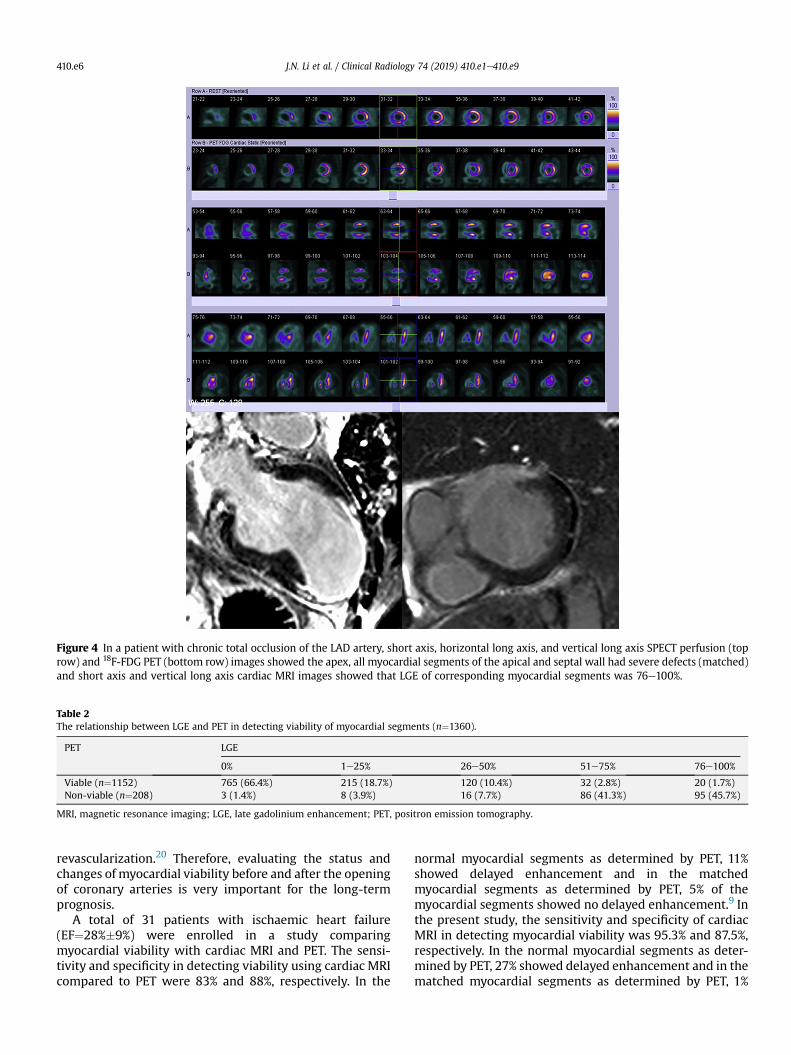
Figure 4 In a patient with chronic total occlusion of the LAD artery, short axis, horizontal long axis, and vertical long axis SPECT perfusion (toprow) and 18F-FDG PET (bottom row) images showed the apex, all myocardial segments of the apical and septal wall had severe defects (matched)and short axis and vertical long axis cardiac MRI images showed that LGE of corresponding myocardial segments was 76e100%.
Table 2The relationship between LGE and PET in detecting viability of myocardial segments (n¼1360).
PET LGE
0% 1e25% 26e50% 51e75% 76e100%
Viable (n¼1152) 765 (66.4%) 215 (18.7%) 120 (10.4%) 32 (2.8%) 20 (1.7%)Non-viable (n¼208) 3 (1.4%) 8 (3.9%) 16 (7.7%) 86 (41.3%) 95 (45.7%)
MRI, magnetic resonance imaging; LGE, late gadolinium enhancement; PET, positron emission tomography.
J.N. Li et al. / Clinical Radiology 74 (2019) 410.e1e410.e9410.e6
revascularization.20 Therefore, evaluating the status andchanges of myocardial viability before and after the openingof coronary arteries is very important for the long-termprognosis.
A total of 31 patients with ischaemic heart failure(EF¼28%�9%) were enrolled in a study comparingmyocardial viability with cardiac MRI and PET. The sensi-tivity and specificity in detecting viability using cardiac MRIcompared to PET were 83% and 88%, respectively. In the
normal myocardial segments as determined by PET, 11%showed delayed enhancement and in the matchedmyocardial segments as determined by PET, 5% of themyocardial segments showed no delayed enhancement.9 Inthe present study, the sensitivity and specificity of cardiacMRI in detecting myocardial viability was 95.3% and 87.5%,respectively. In the normal myocardial segments as deter-mined by PET, 27% showed delayed enhancement and in thematched myocardial segments as determined by PET, 1%
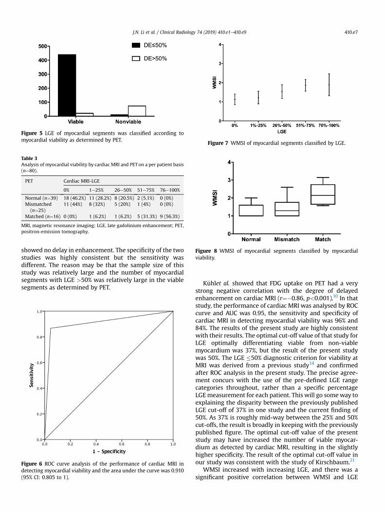
Table 3Analysis of myocardial viability by cardiac MRI and PET on a per patient basis(n¼80).
PET Cardiac MRI-LGE
0% 1e25% 26e50% 51e75% 76e100%
Normal (n¼39) 18 (46.2%) 11 (28.2%) 8 (20.5%) 2 (5.1%) 0 (0%)Mismatched
(n¼25)11 (44%) 8 (32%) 5 (20%) 1 (4%) 0 (0%)
Matched (n¼16) 0 (0%) 1 (6.2%) 1 (6.2%) 5 (31.3%) 9 (56.3%)
MRI, magnetic resonance imaging; LGE, late gadolinium enhancement; PET,positron-emission tomography.
Figure 5 LGE of myocardial segments was classified according tomyocardial viability as determined by PET. Figure 7 WMSI of myocardial segments classified by LGE.
Figure 8 WMSI of myocardial segments classified by myocardialviability.
J.N. Li et al. / Clinical Radiology 74 (2019) 410.e1e410.e9 410.e7
showed no delay in enhancement. The specificity of the twostudies was highly consistent but the sensitivity wasdifferent. The reason may be that the sample size of thisstudy was relatively large and the number of myocardialsegments with LGE >50% was relatively large in the viablesegments as determined by PET.
Figure 6 ROC curve analysis of the performance of cardiac MRI indetecting myocardial viability and the area under the curve was 0.910(95% CI: 0.805 to 1).
K€uhlet al. showed that FDG uptake on PET had a verystrong negative correlation with the degree of delayedenhancement on cardiac MRI (r¼�0.86, p<0.001).10 In thatstudy, the performance of cardiac MRI was analysed by ROCcurve and AUC was 0.95, the sensitivity and specificity ofcardiac MRI in detecting myocardial viability was 96% and84%. The results of the present study are highly consistentwith their results. The optimal cut-off value of that study forLGE optimally differentiating viable from non-viablemyocardium was 37%, but the result of the present studywas 50%. The LGE �50% diagnostic criterion for viability atMRI was derived from a previous study14 and confirmedafter ROC analysis in the present study. The precise agree-ment concurs with the use of the pre-defined LGE rangecategories throughout, rather than a specific percentageLGEmeasurement for each patient. This will go someway toexplaining the disparity between the previously publishedLGE cut-off of 37% in one study and the current finding of50%. As 37% is roughly mid-way between the 25% and 50%cut-offs, the result is broadly in keeping with the previouslypublished figure. The optimal cut-off value of the presentstudy may have increased the number of viable myocar-dium as detected by cardiac MRI, resulting in the slightlyhigher specificity. The result of the optimal cut-off value inour study was consistent with the study of Kirschbaum.21
WMSI increased with increasing LGE, and there was asignificant positive correlation between WMSI and LGE

J.N. Li et al. / Clinical Radiology 74 (2019) 410.e1e410.e9410.e8
(p<0.001).22 In a previous study, WMSI of the correspond-ing myocardial segment of CTO was higher compared to themyocardial segment corresponding of non-CTO and WMSIwas higher in the myocardial segment with high LGEcompared to the myocardial segment with low LGE.23 In thepresent study, WMSI was highest in the group with an LGEof 76e100% andWMSI was lowest in the group with an LGEof 0%. It was concluded that WMSI was positively correlatedwith LGE, consistent with previous studies.
Due to differences in spatial resolution, cardiac MRIproved to be superior compared to nuclear imaging in thedetection of small scars. Wagner et al. showed that thetransmural infarction segments defined by the cardiac MRIcould be detected by SPECT, but in 181 subendocardialinfarction segments, 85 segments were not detected bySPECT.24 Another study also showed that cardiac MRI had abetter sensitivity in detecting myocardial infarction due tothe higher spatial resolution.25 Cardiac MRI can also provideoverall and local cardiac function, myocardial perfusion,tissue characteristics, and other information in a singleexamination.26 Therefore, the clinical application of cardiacMRI in evaluating myocardial viability is more promising.
The limitations of this study include the following as-pects. Analysis of myocardial systolic function by low-dosedobutamine stress cardiac MRI may improve the diag-nostic accuracy of myocardial function recovery afterrevascularization.27 WMSI in the present study was calcu-lated based on the entire myocardium dominated by eachvessel and was not specific to each myocardial segment.Some studies have analysed the changes of left ventricularfunction after revascularization,28e30 and further analysis ofthe changes in left ventricular function is needed in futurestudies. In addition, large-scale randomized controlledstudies are needed to confirm the necessity of evaluatingmyocardial viability before interventional treatment ofcoronary CTO lesions.28
In conclusion, cardiac MRI assessment of myocardialviability in patients with coronary CTO has very good con-sistency and high accuracy compared to PET. Therefore,cardiac MRI can be used as a very important method toevaluate myocardial viability in patients with coronary CTOand it has important clinical significance for treatmentchoice in patients with coronary CTO.
Conflict of interest
There are no conflicts of interest.
Acknowledgements
This study was supported by the Beijing Municipal Sci-ence and Technology Project (no. Z161100000516139),Beijing Lisheng Cardiovascular Health Foundation PilotFund Project (no. LHJJ20158521), Translational MedicineProject of Dalian Medical University (no. 2015003), BeijingLab for Cardiovascular Precision Medicine (no.PXM2017_014226_000037) and National Natural Science
Foundation (no. 81671650). This study is part of the regis-tration trial (ClinicalTrials.gov NCT02767401).
References
1. Sianos G, Werner GS, Galassi AR, et al. Recanalisation of chronic totalcoronary occlusions: 2012 consensus document from theEuroCTO club.EuroIntervention 2012;8(1):139e45.
2. Stone GW, Kandzari DE, Mehran R, et al. Percutaneous recanalization ofchronically occluded coronary arteries: a consensus document: part I.Circulation 2005;112(15):2364e72.
3. Zidar FJ, Kaplan BM, O0Neill WW, et al. Prospective, randomized trial ofprolonged intracoronary urokinase infusion for chronic total occlusionsin native coronary arteries. J Am Coll Cardiol 1996;27(6):1406e12.
4. Christofferson RD, Lehmann KG, Martin GV, et al. Effect of chronic totalcoronary occlusion on treatment strategy. Am J Cardiol 2005;95(9):1088e91.
5. Galassi AR, Tomasello SD, Reifart N, et al. In-hospital outcomes ofpercutaneous coronary intervention in patients with chronic total oc-clusion: insights from the ERCTO (European Registry of Chronic TotalOcclusion) registry. EuroIntervention 2011;7(4):472e9.
6. Levine GN, Bates ER, Blankenship JC, et al. 2011 ACCF/AHA/SCAI guide-line for percutaneous coronary intervention: a report of the AmericanCollege of Cardiology Foundation/American Heart Association task forceon practice guidelines and the society for cardiovascular angiographyand interventions. Circulation 2011;124(23):574e651.
7. Windecker S, Kolh P, Alfonso F, et al. 2014 ESC/EACTS guidelines onmyocardial revascularization: the task force on myocardial revasculari-zation of the european society of Cardiology (ESC) and the europeanassociation for cardio-thoracic surgery (EACTS) developed with thespecial contribution of the european association of percutaneous car-diovascular interventions (EAPCI). Eur Heart J 2014;35(37):2541e619.
8. Allman KC. Noninvasive assessment myocardial viability: current statusand future directions. J Nucl Cardiol 2013;20(4):618e37.
9. Klein C, Nekolla SG, Bengel FM, et al. Assessment of myocardial viabilitywith contrast-enhanced magnetic resonance imaging imaging: compar-isonwith positron emission tomography. Circulation 2002;105(2):162e7.
10. K€uhl HP, Beek AM, van der Weerdt AP, et al. Myocardial viability inchronic ischemic heart disease: comparison of contrast-enhancedmagnetic resonance imaging imaging with (18)F-fluorodeoxyglucosepositron emission tomography. J Am Coll Cardiol 2003;41(8):1341e8.
11. Wu YW, Tadamura E, Yamamuro M, et al. Comparison of contrast-enhanced MRI with (18)F-FDG PET/201Tl SPECT in dysfunctional myocar-dium: relation to early functional outcome after surgical revascularizationin chronic ischemic heart disease. J Nucl Med 2007;48(7):1096e103.
12. Roes SD, Kaandorp TA, Marsan NA, et al. Agreement and disagreementbetween contrast-enhanced magnetic resonance imaging imaging andnuclear imaging for assessment of myocardial viability. Eur J Nucl MedMol Imaging 2009;36(4):594e601.
13. Cerqueira MD, Weissman NJ, Dilsizian V, et al. Standardized myocardialsegmentation and nomenclature for tomographic imaging of the heart:a statement for healthcare professionals from the Cardiac ImagingCommittee of the Council on Clinical Cardiology of the American HeartAssociation. Circulation 2002;105(4):539e42.
14. Pereztol-Vald�es O, Candell-Riera J, Santana-Boado C, et al. Correspon-dence between left ventricular 17 myocardial segments and coronaryarteries. Eur Heart J 2005;26(24):2637e43.
15. Kim RJ, Fieno DS, Parrish TB, et al. Relationship of MRI delayed contrastenhancement to irreversible injury, infarct age, and contractile function.Circulation 1999;100(19):1992e2002.
16. Pujadas S, Martin V, Rossell�o X, et al. Improvement of myocardialfunction and perfusion after successful percutaneous revascularizationin patients with chronic total coronary occlusion. Int J Cardiol 2013;169(2):147e52.
17. Olivari Z, Rubartelli P, Piscione F, et al. Immediate results and one-yearclinical outcome after percutaneous coronary interventions in chronictotal occlusions: data from a multicenter, prospective, observationalstudy (TOAST-GISE). J Am Coll Cardiol 2003;41(10):1672e8.
18. Gerber BL, Rousseau MF, Ahn SA, et al. Prognostic value of myocardialviability by delayed-enhanced magnetic resonance imaging in patients

J.N. Li et al. / Clinical Radiology 74 (2019) 410.e1e410.e9 410.e9
with coronary artery disease and low ejection fraction: impact ofrevascularization therapy. J Am Coll Cardiol 2012;59(9):825e35.
19. Allman KC, Shaw LJ, Hachamovitch R, et al. Myocardial viability testingand impact of revascularization on prognosis in patients with coronaryartery disease and left ventricular dysfunction: a meta-analysis. J AmColl Cardiol 2002;39(7):1151e8.
20. Kim RJ, Wu E, Rafael A, et al. The use of contrast-enhanced magneticresonance imaging imaging to identify reversible myocardial dysfunc-tion. N Engl J Med 2000;343(20):1445e53.
21. Kirschbaum SW, Rossi A, Boersma E, et al. Combining magnetic reso-nance imaging viability variables better predicts improvement ofmyocardial function prior to percutaneous coronary intervention. Int JCardiol 2012;159(3):192e7.
22. Ripley DP, Gosling OE, Bhatia L, et al. The relationship between thecontralateral collateral supply and myocardial viability on cardiovas-cular magnetic resonance imaging: can the angiogram predict func-tional recovery? Int J Cardiol 2014;177(2):362e7.
23. Choi JH, Chang SA, Choi JO, et al. Frequency of myocardial infarction andits relationship to angiographic collateral flow in territories supplied bychronically occluded coronary arteries. Circulation 2013;127(6):703e9.
24. Wagner A, Mahrholdt H, Holly TA, et al. Contrast-enhanced MRI androutine single photon emission computed tomography (SPECT)
perfusion imaging for detection of subendocardial myocardial infarcts:an imaging study. Lancet 2003;361(9355):374e9.
25. Ordovas KG, Higgins CB. Delayed contrast enhancement on MRI imagesof myocardium: past, present, future. Radiology 2011;261(2):358e74.
26. Saeed M, Hetts SW, Do L, et al. MRI study on volume effects of coronaryemboli on myocardial function, perfusion and viability. Int J Cardiol2013;165(1):93e9.
27. Baer FM, Theissen P, Schneider CA, et al. Dobutamine magnetic reso-nance imaging imaging predicts contractile recovery of chronicallydysfunctional myocardium after successful revascularization. J Am CollCardiol 1998;31(5):1040e8.
28. Baks T, van Geuns RJ, Duncker DJ, et al. Prediction of left ventricularfunction after drug-eluting stent implantation for chronic total coronaryocclusions. J Am Coll Cardiol 2006;47(4):721e5.
29. K€uhl HP, Lipke CS, Krombach GA, et al. Assessment of reversiblemyocardial dysfunction in chronic ischaemic heart disease: comparisonof contrast-enhanced cardiovascular magnetic resonance and a com-bined positron emission tomography-single photon emission computedtomography imaging protocol. Eur Heart J 2006;27(7):846e53.
30. Kirschbaum SW, Baks T, van den Ent M, et al. Evaluation of left ven-tricular function three years after percutaneous recanalization ofchronic total coronary occlusions. Am J Cardiol 2008;101(2):179e85.