Embed Size (px)
Citation preview
THE JOURNAL OF BIOLOGICAL CHEMIWRY Vol. 249, No. 20, Issue of October 25, pp. 6416-6423, 1974
Printed in U.S.A.
Properties and Reaction Mechanism of DT
Diaphorase from Rat Liver
(Received for publication, December 20, 1973)
SYUN HOSODA, WATARU KAKAMURA, AND KAZUKO HAYASHI
From the Laboratory of Pathology, Aichi Cancer Center Research Institute, ni’agoya, Japan
SUMMARY properties. These consist of an immediate disappearance of
DT diaphorase was purified to homogeneity from rat liver and characterized. The molecular weight of the enzyme was calculated to be 5.0 h 0.06 x lo4 from sedimentation equilib- rium experiments and to be 4.8 x IO4 by thin layer gel filtra- tion method using Sephadex G-200. The identity of FAD as a prosthetic group was confirmed by o-amino acid oxidase test. It was found that 1 mole of FAD was present per mole of enzyme.
The interaction of the enzyme with NADPH and K3 in the presence and absence of bovine serum albumin and di- cumarol were also studied by steady state and stopped flow kinetic methods, together with binding experiments using f14C]Ks and [14C]dicumarol. The probable reaction sequences of the enzyme, including the pyridine nucleotide-dependent reduction of K3 mainly by a ping-pong type mechanism with a rate-limiting step at the point of dissociation of the enzyme-
the yellow color in oxidized Aavoprotein after adding excess NAD(P)H, a marked increase in enzyme activity produced by bovine serum albumin, and a powerful specific inhibition by dicumarol (2, 4, 6). Further clarification of these aspects, how- ever, has not been accomplished, since no precise react’ion mechanism of this enzyme had been established.
In this paper we report some physicochemical properties of DT diaphorase purified from rat liver, together with steady state and stopped flow kinetic studies of the interaction of the enzyme with NADPH and Ka in the presence and absence of serum albumin as well as of dicumarol. It is proposed that DT diaphorase catalyzes the pyridine nucleotide-dependent reduc- tion of Ka mainly by a ping-pong type mechanism in which the oxidized enzyme is reduced by NADPH to produce the free reduced species. This type of mechanism is indicated sche- matically in Equations 1, 2, 3, and 4.
reduced EC3 complex, are proposed. There is evidence that the native form of enzyme has two independent binding sites k+l
for Ks and dicumarol in its active center, forming the partially WAD + NAOPH \= E l FAD& - NAD p ( 1 1
L
active enzyme-& complex reducible by NADPH but not R -1
oxidizable by Ka and the fully inactive enzyme-dicumarol complex not reducible by NADPH. The reduced species is k f2 capable of binding only K3. It was also demonstrated that E*FADH,-NADP e E+ADH, + NADP ( 1 2
bovine serum albumin competes with the native enzyme for KS and dicumarol, probably through the ability of this simple protein to associate with these substances, preventing the formation of enzyme& and enzyme-dicumarol complexes, and leading to the apparent increase in enzyme activity.
k -2
EeFAD- K,H2
k +4
EaFAD-K,H, ’ CFAD + K,H, ,
In 1958, a flavoprotein which catalyzes the oxidation of NADH k -4
and NADPH by various dyes and quinones was found in rat It will be shown that in the reaction sequences of DT liver by Ernster and Navazio (1) who named the enzyme DT diaphorase (EC 1.6.99.2, reduced NAD(P) : (acceptor) oxido- reductase) from its almost equal activity with NADH(DPNH) and NADPH (TPNH). Subsequent studies (2, 3) disclosed that this enzyme closely resembles “vitamin Ka reductase” from ox liver and ox “brain diaphorase,” described by M&rki and Martius (4, 5) and Giuditta and Strecker (6), respectively.
It has been shown that the enzyme purified from several mammalian sources is characterized by three distinctive enaymic
di- aphorase, the step in Equation 4 is rate-limiting. In addition, we present some evidence that both Ka and dicumarol can bind to the native enzyme at equimolar concentration through two different binding sites to form the enzyme-KS complex and the enzyme-dicumarol complex, respectively, whereas the reduced species is capable of binding only Ka. It was also demonstrated that bovine serum albumin appears to lead to an increase in enzyme activity by facilitating the dissociation of these com- plexes.
6416
by guest on March 2, 2019
http://ww
w.jbc.org/
Dow
nloaded from
EXPERIMEKTAL PROCEDURE
Malerials-NADP, NADPH, FAD, cytochrome c, and D-amino acid oxidase were purchased from Boehringer, vitamin KS (mena- dione, 2-methyl-l,4-naphthoquinone), dicumarol, and crystallized bovine serum albumin from Sigma, crystallized egg albumin from Nutritional Biochemicals Corporation, 2-[14C]methyl-1 ,4-naph- thoquinone (8.8 mCi per mole) from The Radiochemical Centre, and [methylene- 14C]dicumarol (3.64 mCi per mole) from New Eng- land Nuclear. CM-cellulose and DEAE-cellulose were obtained from Brown, hydroxylapatite from Bio-Rad, cellulose acetate in a gel form (Cellogel) from Chemet,ron (Milan, Italy), and Sepha- dex G-200 from Pharmacia. The reduced vitamin KS (KZHz) was prepared according to the procedure of Fieser (7).
Enzyme Assays-DT diaphorase activity was determined spec- trophotometrically by following the decrease in absorbance of NADPH at 340 nm. All determinations were made at room tem- perature with cuvettes having a l-cm light path. The assay solu- tion contained 100 pmoles of Tris-HCl buffer, pH 7.4, 0,04 to 0.2 pmole of NADPH, 0.0008 to 0.16 pmole of KB, 0 to 2 mg of bovine serum albumin, 1 mg of cytochrome c, and 5 to 20 ~1 of the enzyme in a total volume of 2 ml. A unit of activity is the amount of enzyme that catalyzes the oxidation of 1 pmole of NAl>PH per min. Specific activity is expressed as units of enzymic activity per mg of protein. The protein concentrations were determined by the Lowry-Folin method (8).
Ultracentrifugal Measurements-Sedimentation velocity and diffusion and sedimentation equilibrium experiments were per- formed in the Beckman-Spinco model X analytical ultracentrifuge equipped with phase plate schlieren system and ItTIC tempera- ture-control system. The diffusion coefficient was estimated from the spreading of the boundary of 8 mg of protein per ml at 12,590 rpm according to the method of Ehrenberg (9). For sedimenta- tion equilibrium experiments a schlieren optical system was used with 3-mm column in a filled Upon double-sector cell. The schlie- ren patterns were photographed with the phase plate angle at 85”, 80”, 75’ and 65”.
Thin Layer GeZ Filtralion-The thin layer gel filtration for esti- mation of approximate molecular weight of purified 1)‘~ diaphorase was performed according to Itadola (10) using Sephadex G-200 (superfine) and Ph armacia TLG apparatus.
Cellogel Eleclrophoresis-Cellogel was equilibrated with 20 mM Tris-HCl buffer, pH 8.0. After applying each 5-J sanlple on the original line of the gel, electrophoresis was performed horizontally at, 4” with a constant voltage of 200 volts. The gel was s!-ained with a freshly prepared mixture composed of 1.3 mM NADH? 0.24 mMZ,Z’-di-p-nitropheny1-5,5’-diphenyl-3,3’-(3,3’-dimethoxy-4,4’- dephenylene) ditetrazolium chloride, 0.1 mM Z,(i-dichloroindo- phenol, and 2 PM bovine serum albumin in 0.1. M Tris-HCl buffer, pH 8.0. The gel was developed at room temperature after apply-
6417
ing the staining mixture. The enzyme activity appeared as a reddish blue band.
Stopped Flow Kinetic Experiments-Stopped flow experiments were performed with a Yanaco SPS-I stopped flow spectrophotom- eter. The details of this apparatus have been described elsewhere (11). The light path length of the observation chamber was 10 mm and the dead time was about 2.0 ms. The temperature was con- trolled at 15”.
Binding Experiments---DT diaphorase at I PM concentration in terms of the enzyme-bound FAD was incubated for 20 min at 30” in the dark with 0.2 mM [l”C]K,, 10 PM [l4C]dicumarol, or both in 10 mM potassium phosphate buffer, pH 7.0. After incubation, each 0.5 ml of sample chilled in ice was directly applied to a col- umn, 0.9 X 15 cm, of Sephadex G-25 (coarse) equilibrated with 10 mM potassium phosphate buffer, pII 7.0, and was eluted with the same buffer. The gel filtration was carried out at 2” and fractions of 1 ml each were collected. To another 0.5 ml of sample at room temperature was added 1 mg of NADPH. After 1 min the.reaction mixtures were quickly chilled in ice and applied to the Sephadex column in the same manner as in the former gel filtration. Under these conditions the enzyme was eluted between fractions 3 and 5 with the peak at fraction 4 and KS between fractions 7 and 20 with the peak at fraction 11, but no dicumarol was eluted because of its strong affinity for Sephadex. However enzyme-bound dicumarol was eluted in the void volume of a column. A 0.5-ml aliquot of eluate from each fraction was transferred to a vial containing 4.5 ml of the dioxane scintillator, composed of 300 mg of 1,4-bis(4- methyl-5-phenyloxazolyl)benzene, 0.7 g of 2,5-diphenyloxa- zole, 100 g of naphthalene, and 1 liter of dioxane, The radioactiv- ity was measured in a liquid scintillation counter. The amounts of [14C]K3 and [14C]dicumarol bound to enzyme were calculated from the radioactivity of authentic samples at known concentra- tion. For comparison bovine serum albumin and egg albumin were used instead of the enzyme.
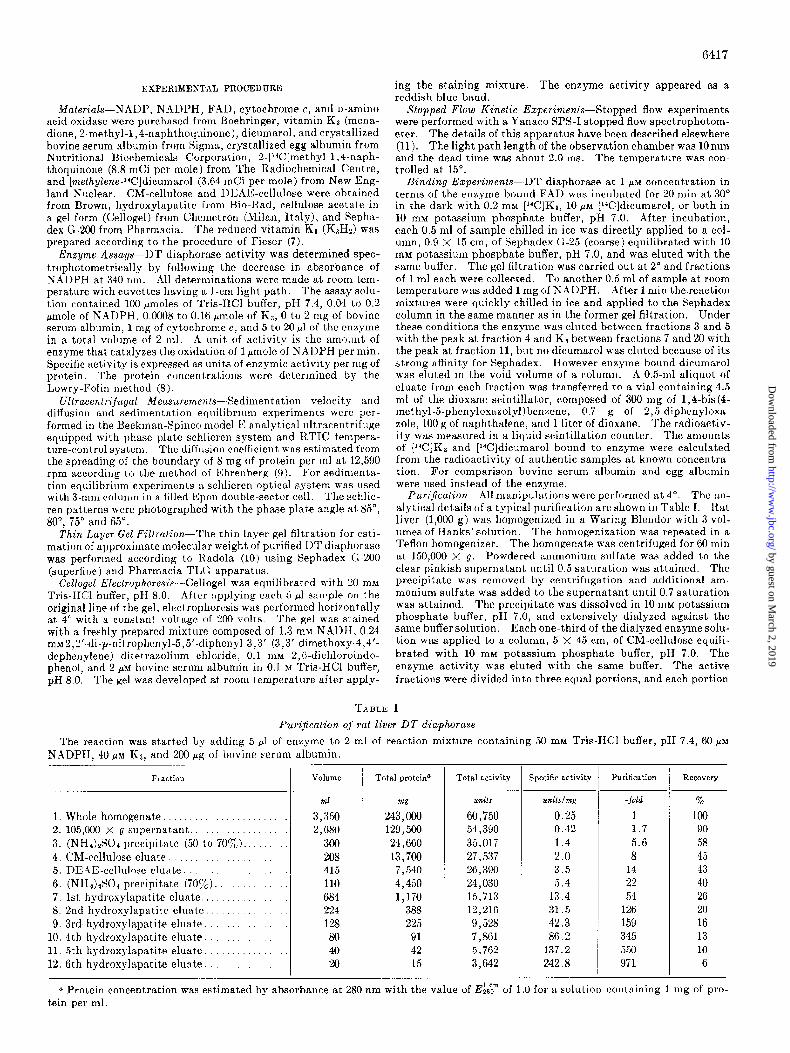
Pur$caiion-All manipulations were performed at 4’. The an- alytical details of a typical purification are shown in Table I. Rat liver (1,000 g) was homogenized in a Waring Blendor with 3 vol- umes of Hanks’ solution. The homogenization was repeated in a Teflon homogenizer. The homogenate was centrifuged for 60 min at 150,000 X g. Powdered ammonium sulfate was added to the clear pinkish supernatant until 0.5 saturation was attained. The precipitate was removed by centrifugation and additional am- monium sulfate was added to the supernatant until 0.7 saturation was attained. The precipitate was dissolved in 10 mM potassium phosphate buffer, pH 7.0, and extensively dialyzed against the same buffer solution. Each one-third of the dialyzed enzyme solu- tion was applied to a column, 5 x 45 cm, of CM-cellulose equili- brated with 10 mM potassium phosphate buffer, pH 7.0. The enzyme activity was eluted with the same buffer. The active fractions were divided into three equal portions, and each portion
TABLE: I
Purijkatio?l o.f rat lirer DT diaphoraae
The reaction was started by adding 5 ~1 of enzyme to 2 ml of reaction mixture containing 50 mM Tris-HC1 buffer, pH 7.4, 60 FM
NADPH, 40 ~.L;ZI Ka, and 200 pg of bovine serum albumin,
1. Whole homogenate. ....................... 2. 105,000 X g supernatant. .................. 3. (NH&S04 precipitat.e (50 to 70yc). ........ 4. i:M-cellulose eluate ....................... 5. DEAE-cellulose eluate .................... 6. (NH&SO* precipitate (707,). ............. 7. 1st hydroxylapatite eluate ................. 8. 2nd hydroxylapatit,e eluate. ............... 9, 3rd. hydroxylapatite eluate ................
10. 4th hydroxylapatite eluate. ............... 11. 5th hydroxylapatite eluate. ............... 12. 6th hydrosylapatite eluate. ...............
Fraction Volume Total proteina Total activity Specific activity Purification Recovery _
mg units units/??tg -fold %
3,350 243,OW 60,750 0.25 1 100 2,680 129,500 54,390 0.42 1.7 90
300 24,660 35,017 1.4 5.6 58 208 13,700 27,537 2.0 8 45 415 7,540 26,390 3.5 14 43 110 4,450 24,030 5.4 22 40 684 1,170 15,713 13.4 54 26 224 388 12,216 31.5 126 20 128 225 9,528 42.3 159 16
80 91 7,861 86.2 1 345 13 40 42 5,762 137.2 550 10 20 15 3,642 242.8 971 6
a Prot’ein concentration was estimated by absorbance at 280 nm with the value of Eigm of 1.0 for a solution containing 1 mg of pro- tein per ml.
by guest on March 2, 2019
http://ww
w.jbc.org/
Dow
nloaded from
6418
was applied to a column, 5 X 45 cm, of DEAE-cellulose equili- brated with the same phosphate buffer. The column was eluted bv a linear gradient from 0.01 to 0.3 M Dhosphate buffer, pH 7.0. T”hk enzyme fractions with a higher activity were collected and powdered ammonium sulfate was added to the enzyme solution until 0.7 saturation was attained. The precipitate redissolved in 10 rnM phosphate buffer, pH 7.0, was dialyzed against the same buffer solution. Thereafter, each xth volume of the enzyme solu- tion was applied to a column, 1 X 12 cm, of hydroxylapatite (5 g) equilibrated with 10 mM phosphate buffer, pH 7.0, and the column was developed with a linear gradient made from 200 ml of the equilibration buffer and 200 ml of 0.3 M phosphate buffer, pH 7.0. Fractions of 4 ml each were collected at a flow rate of 20 ml per hour. The active fractions from 8 columns were combined and concentrated by ultrafiltration. The concentrated enzyme solu- tion was divided into four equal portions. Each portion was loaded again onto the same size column and a similar run was per- formed. The active fractions were combined and concentrated. The third hydroxylapatite chromatography using two columns was carried out in exactly the same manner as the previous run. When this procedure was repeated once, the protein and enzyme activity were eluted almost concurrently between fractions 35 and 45. After this stage the chromatography was performed by load- ing the enzyme onto one column of the same size.
RESULTS
Properties
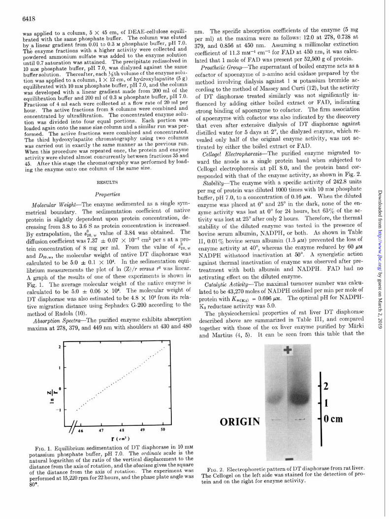
Molecular Weight-The enzyme sedimented as a single sym- metrical boundary. The sedimentation coefficient of native protein is slightly dependent upon protein concentration, de- creasing from 3.8 to 3.6 S as protein concentration is increased. By extrapolation, the &, w value of 3.84 was obtained. The diffusion coefficient was 7.37 f 0.07 x 10’ cm2 per s at a pro- tein concentration of 8 mg per ml. From the value of s$,~ and D2~.w, the molecular weight of native DT diaphorase was calculated to be 5.0 f 0.1 x 10⁴. In the sedimentation equi- librium measurements the plot of 1x1 (Z)/r verSuS r2 nas linear. A graph of the results of one of these experiments is shown in Fig. 1. The average molecular weight of the native enzyme is calculated to be 5.0 f 0.06 x 10⁴. The molecular weight of DT diaphorase was also estimated to be 4.8 x lo4 from its rela- tive migration distance using Sephadex G-200 according to the method of Radola (10).
Absorption Spectra-The purified enzyme exhibits absorption maxima at 278, 379, and 449 nm with shoulders at 430 and 480
// 46 41 48 49 50
f (cm’)
FIG. 1. Equilibrium sedimentation of DT diaphorase in 10 mM potassium phosphate buffer, pH 7.0. The ordinate scale is the natural logarithm of the ratio of the vertical displacement to the distance from the axis of rotation, and the abscissa gives the square of the distance from the axis of rotation. The experiment was performed at 15,220 rpm for 22 hours, and the phase plate angle was 80°.
nm. The specific absorption coefficients of the enzyme (5 mg per ml) at the maxima were as follows: 12.0 at 278, 0.738 at 379, and 0.856 at 450 nm. Assuming a millimolar extinction coefficient of 11.3 rnM-l cm-l for FAD at 450 nm, it was calcu- lated that 1 mole of FAD was present per 52,500 g of protein.
Prosthetic Group-The supernatant of boiled enzyme acts as a cofactor of apoenzyme of n-amino acid oxidase prepared by the method involving dialysis against 1 M potassium bromide ac- cording to the method of Massey and Curti (12)) but the activity of DT diaphorase treated similarly was not significantly in- fluenced by adding either boiled extract or FAD, indicating strong binding of apoenzyme to cofactor. The firm association of apoenzyme with cofactor was also indicated by the discovery that even after extensive dialysis of DT diaphorase against distilled water for 5 days at 2’, the dialyzed enzyme, which re- vealed only half of the original enzyme activity, was not ac- tivated by either the boiled extract or FAD.
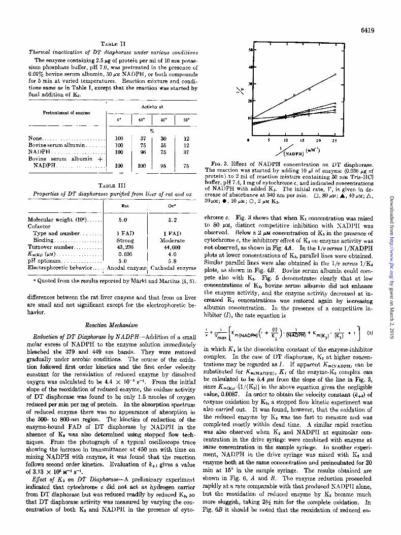
Cellogel Electrophoresis-The purified enzyme migrated to- ward the anode as a single protein band when subjected to Cellogel electrophoresis at pH 8.0, and the protein band cor- responded with that of the enzyme activity, as shown in Fig. 2.
Stability-The enzyme with a specific activity of 242.8 units per mg of protein was diluted 1000 times with 10 mM phosphate buffer, pH 7.0, to a concentration of 0.16 PM. When the diluted enzyme was placed at 0’ and 25’ in the dark, none of the en- zyme activity was lost at 0” for 24 hours, but 63% of the ac- tivity was lost at 25” after only 2 hours. Therefore, the thermal stability of the diluted enzyme was tested in the presence of bovine serum albumin, NADPH, or both. As shown in Table II, 0.01% bovine serum albumin (1.5 PM) prevented the loss of enzyme activity at 40”, whereas the enzyme reduced by 60 pM NADPH withstood inactivation at 50”. A synergistic action against thermal inactivation of enzyme was observed after pre- treatment with both albumin and NADPH. FAD had no activating effect on the diluted enzyme.
Catalytic Actitity-The maximal turnover number was calcu- lated to be 43,270 moles of NADPH oxidized per min per mole of protein with KmcK3) = 0.696 pM. The optimal pH for NADPH- KS reductase activity was 5.0.
The physicochemical properties of rat liver DT diaphorase described above are summarized in Table III, and compared together with those of the ox liver enzyme purified by MLrki and Martius (4, 5). It can be seen from this table that the
ORIGIN
2
1
Ocm
FIG. 2. Electrophoretic pattern of DT diaphorase from rat liver. The Cellogel on the left side was stained for the detection of pro- tein and on the right for enzyme activity.
by guest on March 2, 2019
http://ww
w.jbc.org/
Dow
nloaded from
64.19
TABLE II t 4 1 1 1 I
Thermal inactivation of DT diaphorase under various conditions The enzyme containing 2.5 pg of protein per ml of 10 mM potas-
sium phosphate buffer, pH 7.0, was pretreated in the presence of 0.01% bovine serum albumin, 50 PM NADPH, or both compounds for 5 min at varied temperatures. Reaction mixture and condi- tions same as in Table I, except that the reaction was started by final addition of KS.
Pretreatment of enzyme
Activity at
00 [ 400 1 450 1 500
% None........................ 100 Bovine serum albumin. 100 NADPH. . . 100 Bovine serum albumin +
NADPH. . ‘. 100
37 30 12 75 55 12 96 75 37
100 95 75
TABLE III Properties of DT diaphorases puri$ed from liver of rat and oz
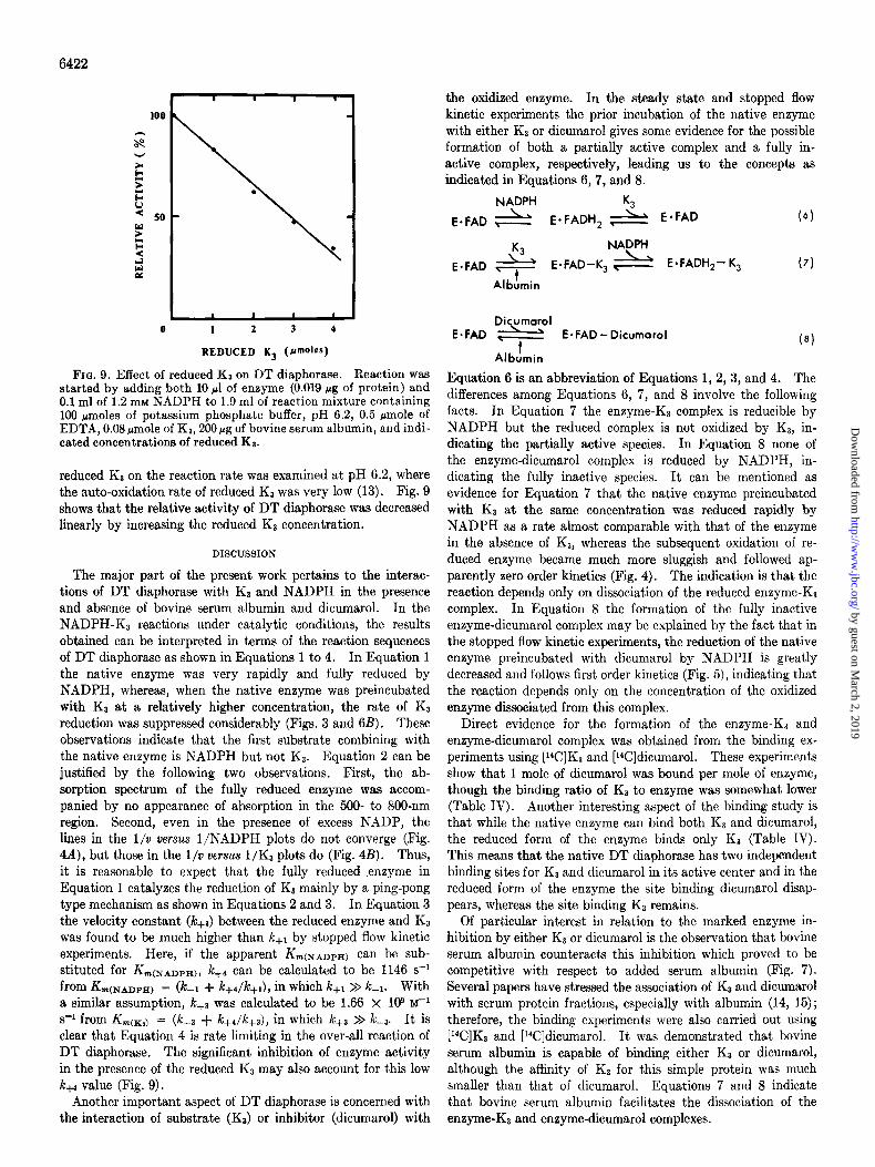
FIG. 3. Effect of NADPH concentration on DT diaphorase. The reaction was started by adding 10 ~1 of enzyme (0.036 pg of protein) to 2 ml of reaction mixture containing 50 mM Tris-HCl buffer, pH 7.4,l mg of cytochrome c, and indicated concentrations of NADPH with added Ka. The initial rate, V, is given in de- crease of absorbance at 340 nm per min. 20j~M;O,lO@d;0,2fi~Ka.
El, 80 MM; A, 40 PM; A,
Rat OXQ
Molecular weight (104). . 5.0 5.2 Cofactor
Type and number. ~ . . 1 FAD 1 FAD Binding. . Strong Moderate
Turnover number.. . 43,270 44,000 Km,(&) (PM) 0.696 4.0 pH optimum. 5.0 5.8 Electrophoretic behavior, . Anodal enzyme Cathodal enzyme
a Quoted from the results reported by Marki and Martius (4,5).
differences between the rat liver enzyme and that from ox liver are small and not significant except for the electrophoretic be- havior.
Reaction Mechanism
chrome c. Fig. 3 shows that when Ka concentration was raised to 80 PM, distinct competitive inhibition with NADPH was observed. Below a 2 PM concentration of KS in the presence of cytochrome c, the inhibitory effect of K3 on enzyme activity was not observed, as shown in Fig. 4A. In the l/o versus l/NADPH plots at lower concentrations of Ka, parallel lines were obtained. Similar parallel lines were also obtained in the I/v versus l/K3 plots, as shown in Fig. 4B. Bovine serum albumin could com- pete also with KI. Fig. 5 demonstrates clearly that at low concentrations of Ka, bovine serum albumin did not enhance the enzyme activity, and the enzyme activity decreased at in- creased Kt concentrations was restored again by increasing albumin concentration. In the presence of a competitive in- hibitor (I), the rate equation is
Reduction of DT Diaphorase by NADPH-Addition of a small molar excess of NADPH to the enzyme solution immediately bleached the 379 and 449 nm bands. They were restored gradually under aerobic conditions. The course of the oxida- tion followed first order kinetics and the first order velocity constant for the reoxidation of reduced enzyme by dissolved oxygen was calculated to be 4.4 X 1OW s-i. From the initial slope of the reoxidation of reduced enzyme, the oxidase activity of DT diaphorase was found to be only 1.5 nmoles of oxygen reduced per min per mg of protein. In the absorption spectrum of reduced enzyme there was no appearance of absorption in the 500- to 800-nm region. The kinetics of reduction of the enzyme-bound FAD of DT diaphorase by NADPH in the absence of K3 was also determined using stopped flow tech- niques. From the photograph of a typical oscilloscope trace showing the increase in transmittance at 450 nm with time on mixing NADPH with enzyme, it was found that the reaction follows second order kinetics. Evaluation of le+i gives a value of 3.13 x 10⁸ M-1 s-1.
E$ect of KS on DT Diaphorase-A preliminary experiment indicated that cytochrome c did not act as hydrogen carrier from DT diaphorase but was reduced readily by reduced Ka, so that DT diaphorase activity was measured by varying the con- centration of both KJ and NADPH in the presence of cyto-
in which Ki is the dissociation constant of the enzyme-inhibitor complex. In the case of DT diaphorase, KS at higher concen- trations may be regarded as 1. If apparent Krn(~ADPB) can be substituted for Km(NADPH), Kc of the enzyme-K3 complex can be calculated to be 5.4 C(M from the slope of the line in Fig. 3, since KrncKI) .[ l/(Ks)] in the above equation gives the negligible value, 0.0087. In order to obtain the velocity constant (le+,) of enzyme oxidation by KS, a stopped flow kinetic experiment was also carried out. It was found, however, that the oxidation of the reduced enzyme by Ka was too fast to measure and was completed mostly within dead time. A similar rapid reaction was also observed when K3 and NADPH at equimolar con- centration in the drive syringe were combined with enzyme at same concentration in the sample syringe. In another experi- ment, NADPH in the drive syringe was mixed with KS and enzyme both at the same concentration and preincubated for 20 min at 15” in the sample syringe. The results obtained are shown in Fig. 6, A and B. The enzyme reduction proceeded rapidly at a rate comparable with that produced NADPH alone, but the reoxidation of reduced enzyme by KS became much more sluggish, taking 2% min for the complete oxidation. In Fig. 6B it should be noted that the reoxidation of reduced en-
by guest on March 2, 2019
http://ww
w.jbc.org/
Dow
nloaded from

6420
0
FIG. 4. A, effect of NAr)PH concentration on x)T diaphorase in rase in the absence and presence of NAZ)P. Reaction mixture the absence and presence of NAIIP. Reaction mixture and condi- and conditions are the same as in A except that indicated concen- tions are the same as in Fig+ 3, except that 0.026 pg of enzyme pro- trations of Kz were used, 0,100 pM NAIIPH ; & 50 FM NAX)PH ; tein was used. l , 2 PM Ka; 0, 2 PM Ka in the presence of 1 mM A, 50 pM NADPH in the presence of 1 mM NAIIP; 0, 20 NADP; w, 0.4pM Ks, B, effect of K3 concentration on DT diapho- PM NAII>PH.
1 1 10 to
BOVINE SERUM ALBUMIN ( Pg/mi Incubatiaa mixture )
I 1 30
FIG. 5. Effect of bovine serum albumin on DT diaphorase ac- tivity at lower Ka concentrations. Reaction was started by add- ing 0.1 ml of 1.2 mM NADPH to 1.9 ml of reaction mixture con- taining 100 pmoles of Tris-HCI buffer, pH 7.4, 0.036 pg of enzyme protein, I mg of cytochrome c, and ‘indicated concentrations of bovine serum albumin with added K3. l , 10 PM KS; A, 8 FM I&;
l , 6~ K3; 0, ~PM&-
eyme by K3 follows approximately zero order kinetics for at least 80% of the total reaction,
Eflect of NADP on DT Diaphorase Activity-Fig. 4A shows that in the l/v versus l/NADPH plots the lines do not converge even in the presence of excess NADP, indicating that NADP does not compete with NADPH for the oxidized enzyme. As shown in Fig. 4B, however, in the l/v versus l/K3 plots, the line in the presence of added NADP crosses that in the absence of added NADP, suggesting the possible competition of NADP with Ka for the reduced enzyme.
Effect of Dicumarol on DT Diaphorase-The inhibition by dicumarol was competitive with respect to NADPH, as has been reported (Z), and Ki was found to be 47.5 PM. Fig. 7 shows that bovine serum albumin also counteracts the dicumarol in- hibition which proved to be competitive with respect to serum albumin added at 0.15 to 1.5 PM concentration. In order to ascertain the inhibition step of dicumarol, stopped flow kinetic experiments were performed. Fig. 8 shows that the rate of enzyme reduction by NADPH in the presence of dicumarol was slowed down considerably and followed first order kinetics. After complete enzyme reduction, slow reoxidation of the re- duced enzyme by dissolved oxygen was also observed.
Binding of [V]K3 and [V]Dicumarol by DT Diaphorase-In order to demonstrate the actual formation of the enzyme-K3 complex and the enzyme-dicumarol complex, binding experi- ments using [14C]KB and [14C]d icumarol were carried out by gel filtration through a column of Sephadex G-25. The results of these experiments are given in Table IV. It can be seen that almost exactly 1 mole of dicumarol was bound per mole of en-
by guest on March 2, 2019
http://ww
w.jbc.org/
Dow
nloaded from

AA= 0.0446
A I 2 3 n
-1 I- IO m set TIME (min)
FIG. 6. A, photograph of stopped flow oscilloscope trace at 450 nm on mixing DT diaphorase with NADPH in the presence of KJ. Enzyme (15.2 PM) preincubated with Ka at same concentration at 15” for 20 min in sample syringe was combined with same concentra- tion of NADPH in drive syringe. B, reoxidation of reduced enzyme in A by Ka. Changes in transmittance at 450 nm were followed with a recorder connected with a Yanaco SPS-I stopped flow spectrophotometer.
I I I I I I I I
0 1 2 3 4 5 6 : 1
1 / [*mJM*Nl(““-‘)
FIG. 7. Effect of bovine serum albumin on DT diaphorase in the absence (0) and presence (0 ) of dicumarol. Reaction was started by adding 0.1 ml of 0.8 mM Kt to 1.9 ml of reaction mixture containing 100 pmoles of Tris-HCl buffer, pH 7.4, 0.051 pg of en- zyme protein, 60 pu NADPH, and indicated concentrations of albumin used.
zyme and the enzyme reduced by excess NADPH revealed no radioactivity. Table IV also shows that 1 mole of the native enzyme was able to bind 0.78 mole of [W]K3, which was re- tained by the reduced form of the enzyme. Of particular interest is the observation that in the presence of both [i4C]K0 and [Wldicumarol the enzyme is capable of binding both sub- stances, since the enzyme eluted contained radioactivity cor- responding to the sum of the protein-bound KS and dicumarol. When the enzyme incubated with both [14C]Ks and [“C]di- cumarol was reduced by excess of NADPH, the radioactivity of the enzyme was decreased in proportion to the amount of pro- tein-bound dicumarol. Bovine serum albumin could also bind either [i4C]Kp or [i4C]dicumarol (9.4 mmoles of Ka and 528 mmoles of dicumarol per mole of albumin). Egg albumin had no affinity for these reagents.
E$ect of Reduced Ka on DT Diaphorase Activity-The effect of
TIME (dn )
FIG. 8. Reduction of DT diaphorase preincubated with dicu- marol by NADPH. Enzyme (13 PM) preincubated with equimolar dicumarol at 15” for 20 min in a sample syringe was combined with same concentration of NADPH in a drive syringe. Changes in transmittance at 450 nm were recorded as illustrated in Fig. 6B.
TABLE IV Binding of [W]K1 and [W]dicumarol by DT diaphorase
The details of enzyme treatments and experimental conditions are described under “Experimental Procedure.”
Enzyme treatment
[“C]K, . NADPH after treatment with [14C]Ka.. [14C]Dicumarol. NADPH after treatment with [“Cl-
dicumarol.......................... [i4C]K3 and [W]dicumarol. . . . . . . NADPH after treatment with [W]Ka
and [W]dicumarol.. . .
[“ClKs or Radioactivity [Wldicumarol
bound/mole protein
:ounts in I r&in mote
3040 0.78 3160 0.81 1980 0.99
0 0 5080
3100
by guest on March 2, 2019
http://ww
w.jbc.org/
Dow
nloaded from
6422
1 2 3 4
REDUCED K3 (P”‘o’-)
Fro. 9. Effect of reduced KS on DT diaphorase. Reaction was started by adding both 10 ~1 of enzyme (0.019 pg of protein) and 0.1 ml of 1.2 mM NADPH to 1.9 ml of reaction mixture containing 100 Hmoles of potassium phosphate buffer, pH 6.2, 0.5 rmole of EDTA, 0.08 rmole of Kz, 200rg of bovine serum albumin, and indi- cated concentrations of reduced Ka.
reduced KI on the reaction rate was examined at pH 6.2, where the auto-oxidation rate of reduced KP was very low (13). Fig. 9 shows that the relative activity of DT diaphorase was decreased linearly by increasing the reduced KS concentration.
DISCUSSION
The major part of the present work pertains to the interac- tions of DT diaphorase with KS and NADPH in the presence and absence of bovine serum albumin and dicumarol. In the NADPH-KI reactions under catalytic conditions, the results obtained can be interpreted in terms of the reaction sequences of DT diaphorase as shown in Equations 1 to 4. In Equation 1 the native enzyme was very rapidly and fully reduced by NADPH, whereas, when the native enzyme was preincubated with KS at a relatively higher concentration, the rate of KI reduction was suppressed considerably (Figs. 3 and 6B). These observations indicate that the first substrate combining with the native enzyme is NADPH but not KS. Equation 2 can be justified by the following two observations. First, the ab- sorption spectrum of the fully reduced enzyme was accom- panied by no appearance of absorption in the 500- to 800~nm region, Second, even in the presence of excess NADP, the lines in the l/u versus l/NADPH plots do not converge (Fig. 4A), but those in the l/v versus l/K8 plots do (Fig. 4B). Thus, it is reasonable to expect that the fully reduced .enzyme in Equation 1 catalyzes the reduction of Ka mainly by a ping-pong type mechanism as shown in Equations 2 and 3. In Equation 3 the velocity constant (/c+s) between the reduced enzyme and Ka was found to be much higher than lc+r by stopped flow kinetic experiments. Here, if the apparent Km(NADPH) can be sub- stituted for Km(NADPH)r kf4 can be calculated to be 1146 s-l from L(NAD~H) = (lc-I + Ic+&+i), in which k+r >> k-1. With a similar assumption, kfS was calculated to be 1.66 X 10⁹ M-’
s-i from Krn(xl) = (k-a + k+Jc+,), in which kfl >> L-3. It is clear that Equation 4 is rate limiting in the over-all reaction of DT diaphorase. The significant inhibition of enzyme activity in the presence of the reduced KS may also account for this low kw value (Fig. 9).
Another important aspect of DT diaphorase is concerned with the interaction of substrate (Ka) or inhibitor (dicumarol) with
the oxidized enzyme. In the steady state and stopped flow kinetic experiments the prior incubation of the native enzyme with either K3 or dicumarol gives some evidence for the possible formation of both a partially active complex and a fully in- active complex, respectively, leading us to the concepts as indicated in Equations 6,7, and 8.
NADPH
E.FAD &
K3
E.FADH, & E*FAD (6)
E-FAD +
NADPH
E-FAD-K, ,\ E*FADH,- K, (7) t
Albumin
E.FAD Diymyl
’ t E-FAD- Dicumarol
(8)
Albumin
Equation 6 is an abbreviation of Equations 1, 2, 3, and 4. The differences among Equations 6, 7, and 8 involve the following facts. In Equation 7 the enzyme-KS complex is reducible by NADPH but the reduced complex is not oxidized by KS, in- dicating the partially active species. In Equation 8 none of the enzyme-dicumarol complex is reduced by NADPH, in- dicating the fully inactive species. It can be mentioned as evidence for Equation 7 that the native enzyme preincubated with KS at the same concentration was reduced rapidly by NADPH as a rate almost comparable with that of the enzyme in the absence of Kt, whereas the subsequent oxidation of re- duced enzyme became much more sluggish and followed ap- parently zero order kinetics (Fig. 4). The indication is that the reaction depends only on dissociation of the reduced enzyme-K, complex. In Equation 8 the formation of the fully inactive enzyme-dicumarol complex may be explained by the fact that in the stopped flow kinetic experiments, the reduction of the native enzyme preincubated with dicumarol by NADPH is greatly decreased and follows first order kinetics (Fig. 5), indicating that the reaction depends only on the concentration of the oxidized enzyme dissociated from this complex.
Direct evidence for the formation of the enzyme-K3 and enzyme-dicumarol complex was obtained from the binding ex- periments using [i4C]K3 and [l%]dicumarol. These experiments show that 1 mole of dicumarol was bound per mole of enzyme, though the binding ratio of KB to enzyme was somewhat lower (Table IV). Another interesting aspect of the binding study is that while the native enzyme can bind both Kz and dicumarol, the reduced form of the enzyme binds only Ka (Table IV). This means that the native DT diaphorase has two independent binding sites for Ka and dicumarol in its active center and in the reduced form of the enzyme the site binding dicumarol disap- pears, whereas the site binding K3 remains.
Of particular interest in relation to the marked enzyme in- hibition by either K1 or dicumarol is the observation that bovine serum albumin counteracts this inhibition which proved to be competitive with respect to added serum albumin (Fig. 7). Several papers have stressed the association of K3 and dicumarol with serum protein fractions, especially with albumin (14, 15) ; therefore, the binding experiments were also carried out using [r4C]Ka and [%]dicumarol. It was demonstrated that bovine serum albumin is capable of binding either Ka or dicumarol, although the affinity of K1 for this simple protein was much smaller than that of dicumarol. Equations 7 and 8 indicate that bovine serum albumin facilitates the dissociation of the enzyme-K3 and enzyme-dicumarol complexes.
by guest on March 2, 2019
http://ww
w.jbc.org/
Dow
nloaded from
6423
remain unclear. We feel that there may be significant con- formational differences between the native enzyme and the re- duced one since the reduced enzyme is more stable than the oxidized enzyme at the elevated temperatures (Table II) and reveals higher fluorescence polarization (0.130) than native en- zyme (O.lOS).l
Finally, it should be pointed out that further studies should include the interaction of NADH with DT diaphorase in the presence of the various compounds mentioned above, since jNAopH a this enz;me. NADH is also a hydrogen donor, albeit a rather poor one, for
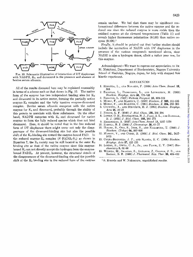
FIG. 10. Schematic illustration of interaction of DT diaphorase with NADPH, KS, and dicumarol in the presence and absence of bovine serum albumin.
All of the results discussed here may be explained reasonably in terms of a scheme such as that shown in Fig. 10. The native form of the enzyme has two independent binding sites for KS and dicumarol in its active center, forming the partially active enzyme-K3 complex and the fully inactive enzyme-dicumarol complex. Bovine serum albumin competes with the native enzyme for Ka and dicumarol, probably through the ability of this protein to associate with these substances. On the other hand, NADPH competes with KS and dicumarol for native enzyme to form the fully reduced species which does not bind dicumarol. Here, it should be noted that in the free reduced form of DT diaphorase there might occur not only the disap- pearance of the dicumarol-binding site but also the possible shift of the Ks-binding site toward the enzyme-bound FAD. In the reduced enzyme-KS complex (E.FADH2-KS) as shown in Equation 7, the KS moiety may be still located in the same KS binding site as that of the native enzyme since this enzyme- bound K3 can not directly accept the hydrogen from the enzyme. bound FADHz. At present, however, the structural details of the disappearance of the dicumarol-binding site and the possible
Acknowledgment-We want to express our appreciation to Dr. M. Nishikimi, Department of Biochemistry, Nagoya University School of Medicine, Nagoya, Japan, for help with stopped flow kinetic experiment.
REFERENCES
1. ERNSTER, L., AND NAVAZIO, F. (1958) Acta Chem. Stand. 12, 595
2. ERNSTKR, L., DANIELSON, L., AND LJUNGGRKN, M. (1962) Biochim. Biophys. Acta 68, 171-188
3. ERNSTER, L. (1967) Methods Enzymol. 10, 309-319 4. MHRKI, F., AND MARTIUS, C. (1960) Biochem. 2. 333, 111-135 5. MXI~KI, F., AND MARTIUS, C. (1961) Biochem. 2. 334,293-303 6. GIUDITTA, A., AND STRECKER, H. J. (1961) Biochim. Biophys.
Acta 48, lo-19 7. FIESI”R, L. F. (1940) J. Biol. Chem. 133, 391-396 8. LOWRY, 0. H., ROSEBROUGH, N. J., FARR, A. L., AND RANDALL,
R. J. (1951) J. Biol. Chem. 193, 265-275 9. EHRENBERG, A. (1957) Actu Chem. Scund. 11, 1257-1270
10. RADOLA, B. J. (1968) J. Chromutogr. 38, 61-77 11. HIROMI, S., ONO, S., ITOH, S., AND NAGAMURA, T. (1968) J.
Biochem. (Tokyo) 64, 897-900 12. MASSF:Y, V., AND CURTI, B. (1966) J. Biol. Chem. 241, 3417-
3423 13. COLPA-BOONSTRA, J. P., AND SLATER, E. C. (1958) Biochim.
Biophys. Acta i7, 122-133 14. LOSITO. R.. OWEN. C. A.. JR.. AND FLOCK. E. V. (1967) Bio-
chemktry’6, 61-8s ’ ’ ~ I
15. WEINER, M., SHAPIRO, S., AXELROD, J., COOPER, J. R., AND BRODIE, B. B. (1950) J. Pharmacol. Exp. Ther. 99, 409-420
shift of the KS binding site in the reduced form of the enzyme 1 S. Hosoda and W. Nakamura, unpublished results
by guest on March 2, 2019
http://ww
w.jbc.org/
Dow
nloaded from
Syun Hosoda, Wataru Nakamura and Kazuko HayashiProperties and Reaction Mechanism of DT Diaphorase from Rat Liver
1974, 249:6416-6423.J. Biol. Chem.
http://www.jbc.org/content/249/20/6416Access the most updated version of this article at
Alerts:
When a correction for this article is posted•
When this article is cited•
to choose from all of JBC's e-mail alertsClick here
http://www.jbc.org/content/249/20/6416.full.html#ref-list-1
This article cites 0 references, 0 of which can be accessed free at
by guest on March 2, 2019
http://ww
w.jbc.org/
Dow
nloaded from