Embed Size (px)
Citation preview

Kidney International, Vol. 62 (2002), pp. 1650–1658
Compensatory renal hypertrophy is mediated by acell cycle-dependent mechanism
BAOLIAN LIU and PATRICIA A. PREISIG
Department of Internal Medicine, University of Texas Southwestern Medical Center, Dallas, Texas, USA
Compensatory renal hypertrophy is mediated by a cell cycle- The majority of the growth occurs in the cortex, particu-dependent mechanism. larly in the proximal convoluted tubules, and appears to
Background. Two mechanisms exist for inducing renal prox- be of a hypertrophic, rather than hyperplastic nature.imal tubule hypertrophy. One is characterized by regulationThe mechanism(s) responsible for initiating the hyper-of the G1 cell cycle kinase (cell cycle-dependent mechanism),trophic growth response are not well understood.while the other mechanism involves an imbalance between
rates of protein synthesis and degradation, and occurs indepen- We have characterized two mechanisms that can causedently of cell cycle kinase regulation (cell cycle-independent renal epithelial cells to hypertrophy [1–7]. One of these,mechanism). The present studies examined whether the com- referred to as a cell cycle-dependent hypertrophy mecha-pensatory proximal tubule growth following uninephrectomy
nism, involves the coordinated effects of a mitogen andis mediated by the cell cycle-dependent or -independent mech-antiproliferative agent. In this model, a mitogenic stimu-anism.
Methods. Studies were done in both rats and C57Bl6 mice lus “moves” quiescent renal epithelial cells into the G1on tissue harvested from sham-operated or uninephrectomized phase of the cell cycle, where they initiate events typicalanimals. The magnitude of BrdU incorporation was used as
of early G1, including activation of cdk4(6)/cyclin D ki-the hyperplasia marker, while the proximal tubule protein:nase and stimulation of protein synthesis. The antiprolif-DNA ratio was used as the hypertrophy marker. Cdk4/cyclin
D and cdk2/cyclin E kinase activities were assayed on renal erative agent, transforming growth factor-� (TGF-�) incortex (rat studies) or isolated proximal tubules (mouse stud- the systems we have used, arrests cell cycle progressionies) using an in vitro kinase assay. prior to the restriction point, preventing activation of
Results. In both rats and mice, compensatory proximal tu-cdk2/cyclin E kinase. Failure to activate cdk2/cyclin Ebule growth was hypertrophic, not hyperplastic, evidenced bykinase results in the retinoblastoma protein, pRB, remain-an increase in the protein:DNA ratio without a change in BrdU
incorporation. In mice, cdk4/cyclin D kinase activity progres- ing activated, E2F-induced transcription being blocked,sively increased between days 4 and 7, while cdk2/cyclin E and failure of the cells to cross the restriction point andkinase activity was decreased at both 4 and 7 days. In rats the move into S phase. However, having undergone the in-development of hypertrophy was associated with an increase
crease in physical growth associated with the early G1in cdk4/cyclin D kinase at days 4, 7, and 10, and an increasephase, the arrested cells are now “trapped” in a physi-in cdk2/cyclin E kinase activity at days 2, 4, and 7. Roscovitine,
a cdk2/cyclin E kinase inhibitor, inhibited cdk2/cyclin E kinase cally enlarged or hypertrophied state.activity in both sham and nephrectomized rats; however, it did The second mechanism involves an imbalance be-not prevent the development of proximal tubule hypertrophy. tween rates of protein synthesis and degradation. ThisConclusions. Uninephrectomy-induced compensatory prox-
form of hypertrophy is induced by exposing cells to com-imal tubule growth is a hypertrophic form of growth that ispounds, such as NH4Cl, that alkalinize acidic intracellularmediated by a cell cycle-dependent mechanism.vesicles, such as lysosomes. Increases in ambient NH4Clconcentration in media have no effect on rates of protein
Kidney size increases in a number of physiological synthesis, yet total cellular protein content increases,and pathological situations, including uninephrectomy, suggesting that rates of protein degradation have de-diabetes mellitus, and chronic electrolyte imbalances. clined, leading to an “accumulation” of cellular protein
and the development of an enlarged cell. This scenariois supported by studies showing that alkalinization ofKey words: uninephrectomy, proximal tubule, G1 cell cycle kinases,
roscovitine, kidney size, renal mass. acidic intravesicular compartments inhibits the activityof lysosomal enzymes involved in protein degradationReceived for publication August 3, 2001[8, 9]. A characteristic of this form of hypertrophy is thatand in revised form June 11, 2002
Accepted for publication June 13, 2002 it occurs without regulation or involvement of cells cycleprocesses. Thus, the development of hypertrophy is not 2002 by the International Society of Nephrology
1650
Liu and Preisig: Cell cycle-dependent renal hypertrophy 1651
associated with cdk4/6:cyclin D kinase activation, and antibody (primary antibody), demonstrated the absenceof non-specific staining.occurs in the absence of the activated form of pRB. This
form of hypertrophy is referred to as being cell cycle-Isolation of intact proximal tubulesindependent.
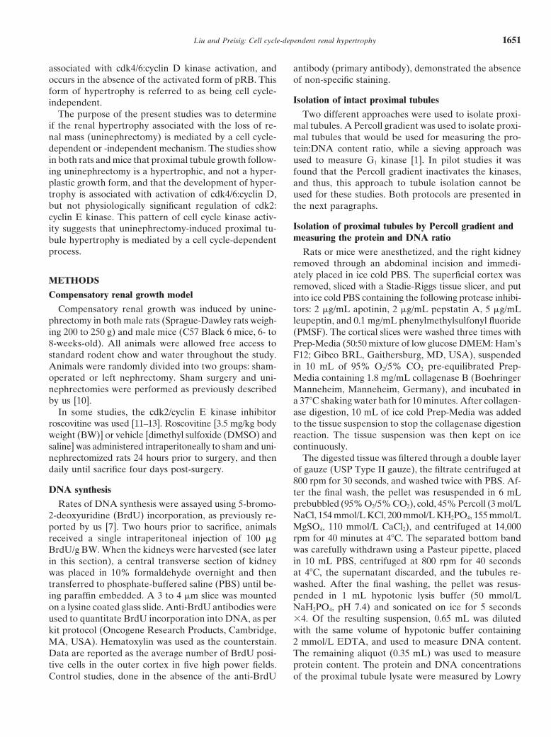
The purpose of the present studies was to determine Two different approaches were used to isolate proxi-if the renal hypertrophy associated with the loss of re- mal tubules. A Percoll gradient was used to isolate proxi-nal mass (uninephrectomy) is mediated by a cell cycle- mal tubules that would be used for measuring the pro-dependent or -independent mechanism. The studies show tein:DNA content ratio, while a sieving approach wasin both rats and mice that proximal tubule growth follow- used to measure G1 kinase [1]. In pilot studies it wasing uninephrectomy is a hypertrophic, and not a hyper- found that the Percoll gradient inactivates the kinases,plastic growth form, and that the development of hyper- and thus, this approach to tubule isolation cannot betrophy is associated with activation of cdk4/6:cyclin D, used for these studies. Both protocols are presented inbut not physiologically significant regulation of cdk2: the next paragraphs.cyclin E kinase. This pattern of cell cycle kinase activ-
Isolation of proximal tubules by Percoll gradient andity suggests that uninephrectomy-induced proximal tu-measuring the protein and DNA ratiobule hypertrophy is mediated by a cell cycle-dependent
process. Rats or mice were anesthetized, and the right kidneyremoved through an abdominal incision and immedi-ately placed in ice cold PBS. The superficial cortex was
METHODSremoved, sliced with a Stadie-Riggs tissue slicer, and put
Compensatory renal growth model into ice cold PBS containing the following protease inhibi-Compensatory renal growth was induced by unine- tors: 2 �g/mL apotinin, 2 �g/mL pepstatin A, 5 �g/mL
phrectomy in both male rats (Sprague-Dawley rats weigh- leupeptin, and 0.1 mg/mL phenylmethylsulfonyl fluorideing 200 to 250 g) and male mice (C57 Black 6 mice, 6- to (PMSF). The cortical slices were washed three times with8-weeks-old). All animals were allowed free access to Prep-Media (50:50 mixture of low glucose DMEM: Ham’sstandard rodent chow and water throughout the study. F12; Gibco BRL, Gaithersburg, MD, USA), suspendedAnimals were randomly divided into two groups: sham- in 10 mL of 95% O2/5% CO2 pre-equilibrated Prep-operated or left nephrectomy. Sham surgery and uni- Media containing 1.8 mg/mL collagenase B (Boehringernephrectomies were performed as previously described Manneheim, Manneheim, Germany), and incubated inby us [10]. a 37�C shaking water bath for 10 minutes. After collagen-
In some studies, the cdk2/cyclin E kinase inhibitor ase digestion, 10 mL of ice cold Prep-Media was addedroscovitine was used [11–13]. Roscovitine [3.5 mg/kg body to the tissue suspension to stop the collagenase digestionweight (BW)] or vehicle [dimethyl sulfoxide (DMSO) and reaction. The tissue suspension was then kept on icesaline] was administered intraperitoneally to sham and uni- continuously.nephrectomized rats 24 hours prior to surgery, and then The digested tissue was filtered through a double layerdaily until sacrifice four days post-surgery. of gauze (USP Type II gauze), the filtrate centrifuged at
800 rpm for 30 seconds, and washed twice with PBS. Af-DNA synthesis ter the final wash, the pellet was resuspended in 6 mL
prebubbled (95% O2/5% CO2), cold, 45% Percoll (3 mol/LRates of DNA synthesis were assayed using 5-bromo-NaCl, 154 mmol/L KCl, 200 mmol/L KH2PO4, 155 mmol/L2-deoxyuridine (BrdU) incorporation, as previously re-MgSO4, 110 mmol/L CaCl2), and centrifuged at 14,000ported by us [7]. Two hours prior to sacrifice, animalsrpm for 40 minutes at 4�C. The separated bottom bandreceived a single intraperitoneal injection of 100 �gwas carefully withdrawn using a Pasteur pipette, placedBrdU/g BW. When the kidneys were harvested (see laterin 10 mL PBS, centrifuged at 800 rpm for 40 secondsin this section), a central transverse section of kidneyat 4�C, the supernatant discarded, and the tubules re-was placed in 10% formaldehyde overnight and thenwashed. After the final washing, the pellet was resus-transferred to phosphate-buffered saline (PBS) until be-
ing paraffin embedded. A 3 to 4 �m slice was mounted pended in 1 mL hypotonic lysis buffer (50 mmol/LNaH2PO4, pH 7.4) and sonicated on ice for 5 secondson a lysine coated glass slide. Anti-BrdU antibodies were
used to quantitate BrdU incorporation into DNA, as per �4. Of the resulting suspension, 0.65 mL was dilutedwith the same volume of hypotonic buffer containingkit protocol (Oncogene Research Products, Cambridge,
MA, USA). Hematoxylin was used as the counterstain. 2 mmol/L EDTA, and used to measure DNA content.The remaining aliquot (0.35 mL) was used to measureData are reported as the average number of BrdU posi-
tive cells in the outer cortex in five high power fields. protein content. The protein and DNA concentrationsof the proximal tubule lysate were measured by LowryControl studies, done in the absence of the anti-BrdU

Liu and Preisig: Cell cycle-dependent renal hypertrophy1652
(protein) and the fluorescent compound Hoechst H33258 phate (ATP) or kinase substrate (see below). After thelast wash, the bead pellet was resuspended in 30 �L of(DNA), respectively, as previously reported by us [7].kinase reaction buffer containing ATP and kinase sub-
Isolation of proximal tubules by sieving strate [50 mmol/L Tris-HCl, pH 7.4, 10 mmol/L MgCl2,
1 mmol/L DTT, 70 mmol/L NaCl, 0.1 mmol/L ATP,Proximal tubules also were collected using a sievingtechnique [14, 15]. The collagenase-digested tissue de- 0.2 �g/�L histone H1, and 0.2 �Ci/�L [�-32P]ATP (6000
Ci/mmol)]. The reaction was carried out by gently shak-scribed above was first passed through an 80-mesh sizemini-sieve (Fisher Scientific, Pittsburgh, PA, USA). The ing the sample in a 30�C water bath � 30 minutes. The
kinase reaction was stopped by the addition of 7.5 �Lflow through containing the proximal tubules was thenpassed through a 170-mesh size sieve. The proximal tu- Laemmli sample buffer. The samples were then boiled
for three minutes and size separated by SDS-PAGE onbules, which now remained on the sieve filter, were col-lected and suspended in 20 mL Prep-Media, and the a 12% gel. The gel was stained with Coomassie blue stain
to confirm equal amounts of kinase substrate in eachtubules pelleted by gravity sedimentation �5 minutes.To insure removal of any remaining cellular debris, the sample, destained, and dried. Phosphorylated substrate
was visualized by autoradiography and quantitated bysedimentation process was repeated �2 after resus-pending the tubules in ice cold PBS containing the prote- densitometry.
cdk4/cyclin D kinase activity. cdk4/cyclin D kinase ac-ase inhibitors described earlier. The final sedimentedpellet was kept on ice until used. tivity was measured using the same protocol described
above with the following exceptions, as previously re-G1 kinase activity ported by us [5, 7]. The lysis buffer contained 150 mmol/L
NaCl, 1 mmol/L EDTA, 2.5 mmol/L egtazic acid (EGTA),cdk2/cyclin E kinase activity. cdk2/cyclin E kinase ac-tivity was measured on whole cortex in the rat studies 10% glycerol, 0.1% Tween 20, 1 mmol/L DTT, 0.1 mmol/L
Na3VO4, 1 mmol/L NaF, 10 mmol/L �-glycerophosphate,and on isolated proximal tubules in the mouse studiesusing a protocol similar to that which we have previously 20 �g/mL aprotinin, 10 �g/mL pepstatin A, 10 �g/mL
leupeptin, and 0.1 mg/mL PMSF. The immunoprecipitat-published [5, 7]. For the rat studies, outer cortex was ho-mogenized in PBS containing protease inhibitors (2 �g/mL ing antibody was a rabbit anti-cdk4 antibody (Santa Cruz
Biotechnology, Santa Cruz, CA, USA). The cdk4 kinaseapotinin, 2 �g/mL pepstatin A, 5 �g/mL leupeptin, and0.1 mg/mL PMSF) and centrifuged at 10,000 rpm for reaction buffer contained 10 mmol/L MgCl2, 50 mmol/L
HEPES pH 7.5, 2.5 mmol/L EGTA, 10 mmol/L �-glycer-five minutes at 4�C. For the mouse studies, isolatedproximal tubules were obtained by the sieving method ophosphate, 0.1 mmol/L Na3VO4, 1 mmol/L NaF, 1
mmol/L DTT, 20 �mol/L ATP, 0.2 �Ci/�L [�-32P] ATPdescribed above. The homogenized cortical pellet (ratstudies) or isolated proximal tubules (mouse studies) was (6000 Ci/mmol), and 0.01 �g/�L GST-pRB [pRB amino
acids 769-921 fused to glutathione S-transferase (GST);resuspended in 1000 �L lysis buffer with protease inhib-itors [150 mmol/L NaCl, 50 mmol/L Tris-HCl pH 7.4, Santa Cruz Biotechnology].5 mmol/L EDTA, 0.5% Na-deoxycholate (DOC), 1%
StatisticsNP-40, 1 mmol/L Na3VO4, 1 mmol/L NaF, 1 mmol/Ldithiothreitol (DTT), 20 �g/mL apotinin, 10 �g/mL pep- Statistical significance was determined by the appro-
priate t test. Differences between means were consideredstatin A, 10 �g/mL leupeptin, and 0.1 mg/mL PMSF]and rotated for 60 minutes at 4�C. After centrifugation significant if P � 0.05 or less.at 10,000 rpm for 20 minutes at 4�C, the supernatantwas used to measure the protein concentration of the
RESULTSresulting lysate, using a Bio-Rad protein assay kit (Bio-
Compensatory renal growth in the rat followingRad Laboratories, Hercules, CA, USA). Cyclin E wasuninephrectomyimmunoprecipitated by adding 1 �g rabbit anti-cyclin E
antibody (Santa Cruz Biotechnology, Santa Cruz, CA, As has been shown by other laboratories, following aleft uninephrectomy there is a progressive increase inUSA) to 500 �g of cortical or tubular protein. The immu-
noprecipitation reaction was carried out overnight at 4�C right kidney weight (Fig. 1). The first studies done todetermine the type of growth responsible for this kidneywith gentle rotation. The next day 100 �L protein-G
agarose beads were added, and the sample incubated at enlargement were to examine right kidney proximal tu-bule BrdU incorporation and the protein:DNA ratio of4�C with gentle rotation for one hour. The beads were
collected by centrifugation at 10,000 rpm for 30 seconds isolated proximal tubules harvested from the right kid-ney during the growth period (Table 1 and Fig. 2, respec-at 4�C, the supernatant discarded, and the beads washed
four times with 1 mL lysis buffer, followed by three ad- tively). As shown, there was no increase in BrdU incor-poration at days 2, 4, and 14, but there was a progressiveditional washings with 1 mL kinase reaction buffer that
did not contain either cold or hot adenosine 5�-triphos- increase in the protein:DNA ratio. These data demon-

Liu and Preisig: Cell cycle-dependent renal hypertrophy 1653
Fig. 2. Protein:DNA ratio in rat isolated proximal tubules post-uni-nephrectomy. Proximal tubules were isolated and the Protein:DNA
Fig. 1. Right kidney weight in rats post-uninephrectomy. A left ne- ratio measured as described in the Methods section. Number of sham/phrectomy ( ) or sham surgery (�) was performed and the right kidney uninephrectomy pairs � 5 (2, 4, and 7 days) and 8 (14 days). *P � 0.05harvested and weighed as described in the Methods section. Number vs. sham-operated animals.of sham/uninephrectomy pairs � 6 (2 days), 14 (4 days), 10 (7 days),and 7 (14 days). *P � 0.05 vs. sham-operated animals.
(3/5), N � 5.* cdk2/cyclin E kinase activity was increasedat 2, 4, and 7 days, and slightly inhibited at 14 days [dayTable 1. Compensatory renal activity in the rat and
mouse models after surgery 2, 74% increase (5/5), N � 5; day 4, 56% increase (4/4),N � 4; day 7, 66% increase (5/5), N � 5; day 14, 17%BrdU incorporationa
decrease (4/5), N � 5]. The observation that the increase2 Days 4 Days 7 Days 14 Days
in cdk4/cyclin D kinase activity correlates with the begin-Rat ning of the increase in protein:DNA ratio suggests thatSham 2.20.3 2.30.8 0.80.3 4.91.3
movement into the G1 phase of the cell cycle is needed toUninephrectomy 5.31.6 4.11.6 4.31.6 6.91.8Mice initiate the hypertrophic growth process. Although cdk2/
Sham 1.80.5 1.20.4 cyclin E kinase activity also increased, there was no in-Uninephrectomy 1.70.4 2.00.5crease in BrdU incorporation (Table 1), suggesting thata Days post-nephrectomy; average number of BrdU positive cells/high powerthe increase in cdk2/cyclin E kinase activity was insuffi-field; N � 5–9/group; P � NS, except for the 7 day rat study (see text for
explanation) cient to inactivate pRB and move the cell into the S phase.By comparison, following a transient renal ischemic pe-riod, the hyperplastic growth response is marked byBrdU incorporation increasing from an average of 3
strate that compensatory renal growth is a hypertrophic, (sham) to 100 (renal ischemia) BrdU-positive cells/and not a hyperplastic growth process. There was, at 7 high powered field. This increase in BrdU incorporationdays post-nephrectomy, a significant increase in BrdU was associated with a 700% increase in cdk2/cyclin Eincorporation due to a smaller magnitude of incorpora- kinase activity (unpublished observations).tion in the sham-operated rats compared to the othertime points. Since the magnitude of BrdU incorporation Compensatory renal growth following uninephrectomyin the nephrectomized rats was similar to the other time in the mousepoints, we do not believe that this represents a prolifera- To determine if there is a species differences in thetive response at 7 days. renal growth pattern following uninephrectomy, studies
were performed in C57Black 6 mice (Fig. 4). As shown,G1 kinase activity following uninephrectomy in the rat at both 4 and 7 days post-nephrectomy, there is an in-
crease in right kidney weight and the protein:DNA ratioFigure 3 profiles the time course of cdk4/cyclin D andin isolated proximal tubules, but no change in BrdUcdk2/cyclin E kinase activity following uninephrectomy.incorporation (Table 1), demonstrating that the kidneycdk4/cyclin D kinase activity was increased at 4 days
post-nephrectomy, and remained elevated but then de-creased toward baseline at 7 and 14 days [day 2, 6%
*The number in parentheses indicates the number of experiments indecrease (3/5), N � 5; day 4, 42% increase (3/4), N � which the individual experimental result was in the same direction asthe mean divided by the total number of experiments for that protocol.4; day 7, 14% increase (4/5), N � 5; day 14, 27% increase

Liu and Preisig: Cell cycle-dependent renal hypertrophy1654
Fig. 3. G1 kinase activity in rats post-uninephrectomy. Typical blots of cdk4/cyclin D and cdk2/cyclin E kinase activities, assayed as described inthe Methods section.
Fig. 5. G1 kinase activity in C57Black 6 mice following uninephrec-tomy. Typical blots of cdk4/cyclin D and cdk2/cyclin E kinase activities,assayed as described in the Methods section.
Fig. 4. Renal growth in C57Black 6 mice at 4 (�) and 7 ( ) daysfollowing uninephrectomy. Protein:DNA ratio was measured as de-
kinase activity progressively increased at 4 and 7 daysscribed in the Methods section. N � 6 sham/uninephrectomy pairs forboth studies at 4 days, 8 sham/uninephrectomy pairs for kidney weight [day 4, 30% increase (5/6), N � 6; day 7, 99% increaseat 7 days, and 4 sham/uninephrectomy pairs for protein:DNA ratio at (8/8), N � 8], whereas cdk2/cyclin E kinase activity was7 days. *P � 0.05 vs. sham-operated animals.
suppressed at both time points [day 4, 38% decrease(6/6), N � 6; day 7: 30% decrease (7/8), N � 8] (Fig. 5).
Inhibition of cdk2/cyclin E kinase in rats does notenlargement is due to a hypertrophic growth process, asprevent compensatory renal hypertrophyin the rat. However, unlike the rat, the pattern of G1
kinase activity is more similar to that observed in diabe- In the cell cycle-dependent hypertrophy model cdk2/cyclin E kinase activity defines the growth pattern. Whentes mellitus and in the in vitro cell culture model of cell
cycle-dependent hypertrophy [1, 5, 7]. Cdk4/cyclin D cdk4/cyclin D kinase activity is increased, but cdk2/cyclin
Liu and Preisig: Cell cycle-dependent renal hypertrophy 1655
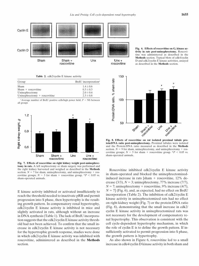
Fig. 6. Effects of roscovitine on G1 kinase ac-tivity in rats post-uninephrectomy. Roscovi-tine was administered as described in theMethods section. Typical blots of cdk4/cyclinD and cdk2/cyclin E kinase activities, assayedas described in the Methods section.
Table 2. cdk2/cyclin E kinase activity
Group BrdU incorporationa
Sham 1.40.6Sham � roscovitine 0.30.3Uninephrectomy 2.00.4Uninephrectomy � roscovitine 2.30.8
a Average number of BrdU positive cells/high power field; P � NS betweenall groups
Fig. 8. Effects of roscovitine on rat isolated proximal tubule pro-tein:DNA ratio post-uninephrectomy. Proximal tubules were isolatedand the Protein:DNA ratio measured as described in the Methodssection. N � 9 for sham, uninephrectomy, and uninephrectomy � ros-covitine groups; N � 5 for sham � roscovitine group. *P � 0.05 vs.sham-operated animals.
Fig. 7. Effects of roscovitine on right kidney weight post-uninephrec-tomy in rats. A left nephrectomy or sham surgery was performed andthe right kidney harvested and weighed as described in the Methods Roscovitine inhibited cdk2/cyclin E kinase activitysection. N � 7 for sham, uninephrectomy, and uninephrectomy � ros-
in sham-operated and blocked the uninephrectomized-covitine groups; N � 3 for sham � roscovitine group. *P � 0.05 vs.sham-operated animals. induced increase in rats [sham � roscovitine, 12% de-
crease (3/3), N � 3; uninephrectomy, 77% increase (7/7),N � 7; uninephrectomy � roscovitine, 9% increase (4/7),N � 7] (Fig. 6), and, as expected, had no effect on BrdUE kinase activity inhibited or activated insufficiently toincorporation (Table 2). The inhibition of cdk2/cyclin Ereach the threshold needed to inactivate pRB and permitkinase activity in uninephrectomized rats had no effectprogression into S phase, then hypertrophy is the result-on right kidney weight (Fig. 7) or the protein:DNA ratioing growth pattern. In compensatory renal hypertrophy,(Fig. 8), demonstrating that the small increase in cdk2/cdk2/cyclin E kinase activity is inhibited in mice andcyclin E kinase activity in uninephrectomized rats wasslightly activated in rats, although without an increasenot necessary for the development of compensatory re-in DNA synthesis (Table 1). The lack of BrdU incorpora-nal hypertrophy. This observation is consistent with thetion suggests that the cdk2/cyclin E kinase activity thresh-cell cycle-dependent hypertrophy mechanism, in whichold had not been achieved. To confirm that the small in-the role of cyclin E is to define the growth pattern. If in-crease in cdk2/cyclin E kinase activity is not necessarysufficiently activated to permit progression into S phase,for the hypertrophic growth response, studies were donethe growth pattern is hypertrophy.in which cdk2/cyclin E kinase activity was inhibited with
As also shown in Figure 6, roscovitine led to a smallroscovitine, administered as described in the Methodssection. increase in cdk4/cyclin D kinase activity in both sham and

Liu and Preisig: Cell cycle-dependent renal hypertrophy1656
nephrectomized rats [sham � roscovitine, 43% increase(3/3), N � 3; uninephrectomy, 65% increase (6/6), N �6; uninephrectomy � roscovitine, 90% increase (6/6),N � 6] and to a small increase in the proximal tubuleprotein:DNA ratio in sham-operated rats. These obser-vations suggest that cdk2/cyclin E kinase activity mayplay a modulating role for basal and induced cdk4/cyclinD kinase activity.
DISCUSSION
Compensatory renal growth is an adaptive responseto the loss of renal mass. The present studies demonstratethat in both rats and mice compensatory proximal tubulegrowth following uninephrectomy is hypertrophic, nothyperplastic, and associated with regulation of the G1
kinases in a pattern consistent with a cell cycle-depen-dent hypertrophy mechanism [1–6]. In this model, thedevelopment of hypertrophy requires that the cell enterG1 and initiate events of this phase (increased protein
Fig. 9. A model for cell cycle-dependent compensatory renal hyper-synthesis, increased cdk4/cyclin D kinase activity, main-trophy.
tain pRB in its hypophosphorylated state), yet fail toprogress onto S phase (absence of DNA synthesis).
The kinase activity patterns in the mice are essentiallyidentical to the in vitro studies that characterized the sent a species difference, with a tendency in the rat tomechanism, with the development of hypertrophy tem- a small amount of hyperplasia post-nephrectomy.porally associated with a progressive increase in cdk4/ Hypertrophic growth results in a physically enlargedcyclin D kinase activity, and failure to activate cdk2/ cell. Cells that undergo a hyperplastic growth responsecyclin E kinase. This kinase profile is also the same as that also enlarge during the G1 phase of the cell cycle. Theobserved in diabetes-induced proximal tubule hyper- purpose of this size increase is to ensure that at the end
of mitosis each daughter cell has a full complement oftrophy, as is the magnitude of the kinase changes relatedto the hypertrophic growth [7]. In rats, the uninephrec- the cellular material needed for survival. In hyperplasia,
physical size is thought to be one of the markers indicat-tomy-induced cell cycle kinase regulation is qualitativelythe same. cdk4/cyclin D kinase activity is increased in ing that the cell has successfully completed the events of
G1, and is ready to move onto the S phase [16, 17]. Hyper-temporal association with the development of hypertro-phy. However, cdk2/cyclin E kinase activity is not inhib- trophied cells can achieve a physical size larger than that
achieved by cells undergoing hyperplasia. This suggestsited, but rather slightly increased. However, the fact that(1) inhibition of cdk2/cyclin E kinase activity with ros- that in cell cycle-dependent hypertrophy the normal
mechanisms permitting size-signaled cell cycle progres-covitine does not prevent the development of hyper-trophy and (2) BrdU incorporation is not significantly sion have been interrupted by the hypertrophy signal.
Thus, as schematically diagramed in Figure 9, the de-increased in nephrectomized rats compared to sham-operated controls demonstrates that the slightly in- velopment of cell cycle-dependent hypertrophy requires
two events to occur. First, a mitogenic signal must becreased cdk2/cyclin E kinase activity is not required forthe development of hypertrophy and does not induce a activated that will move cells into the G1 phase of the cell
cycle and initiate the events of the early G1 phase. Second,hyperplastic growth response. These results confirm thehypothesis that the development of hypertrophy requires since hypertrophied cells have a single copy of DNA, cell
cycle progression must be halted prior to the Restrictionthat cells remain in the G1 phase, and thus, that cdk2/cyclin E kinase activity be inhibited, remain unchanged, Point, so that the retinoblastoma protein remains active
and progression into S phase is prohibited [1, 4, 6]. Theor not increase sufficiently to permit the cells to moveinto S phase. cdk2/cyclin E kinase is necessary for inactivation of the
retinoblastoma protein [18–20]. Thus, regulation of cdk2/In the rat there was a small increase in BrdU incorpo-ration that was not statistically significant, and a small cyclin E kinase activity, either by inhibition of or failure
to sufficiently activate the kinase, appears to be the pointincrease in cdk2/cyclin E kinase activity, neither of whichwere present in the mouse. This observation may repre- at which cell cycle progression must be blocked.
Liu and Preisig: Cell cycle-dependent renal hypertrophy 1657
Reprint requests to Patricia Preisig, Ph.D., Department of InternalIn both in vitro studies and diabetes mellitus (an inMedicine, University of Texas Southwestern Medical Center, 5323 Harry
vivo proximal tubule cell cycle-dependent hypertrophy Hines Blvd., Rm H5.112, Dallas, Texas 75390-8856, USA.E-mail: [email protected]) preventing sustained cdk2/cyclin E kinase activa-
tion marks the point of interruption of the cell cycle [1, 5,REFERENCES7]. The same appears to be true for uninephrectomy-
induced proximal tubule hypertrophy. In models where 1. Franch HA, Shay JW, Alpern RJ, Preisig PA: Involvement ofpRB family in TGF�-dependent epithelial cell hypertrophy. J Cellit has been studied, failure to maintain activation of cdk2/Biol 129:245–254, 1995cyclin E kinase is due to both a decrease in the number 2. Franch HA, Preisig PA: NH4Cl-induced hypertrophy is mediated
of kinase complexes that form and an increase in the by weak base effects and is independent of cell cycle processes.Am J Physiol 270:C932–C938, 1996abundance of cyclin kinase inhibitors (CKIs) associated
3. Preisig PA, Franch HA: Renal epithelial cell hyperplasia andwith complexes that do form. Regulation of cdk2/cyclin hypertrophy. Semin Nephrol 15:327–340, 1995E kinase activity by cyclin kinase inhibitors in hypertro- 4. Preisig PA: Renal hypertrophy and hyperplasia (chapt 27), in The
Kidney: Physiology and Pathophysiology (vol 1, 3rd ed), editedphy has been shown in a number of renal cell typesby Selden DW, Giebisch G, Philadelphia, Lippincott Williams &and conditions [4, 6, 21]. This has been most thoroughly Wilkins, 2000, pp 727–748
5. Liu B, Preisig PA: TGF�-mediated hypertrophy in rat renal epi-studied in both in vitro and in vivo models of hyperglyce-thelial cells involves inhibiting pRB phosphorylation by prevent-mia and diabetes mellitus, respectively. In these systems,ing activation of cdk2/cyclin E kinase. Am J Physiol 277:F186–
an increase in both p27 and p21, regulators of cyclin E F194, 19996. Preisig PA: What makes cells grow larger and how do they do it:kinase, but not p16, a regulator of cdk4/cyclin D kinase,
Hypertrophy revisited. Exper Nephrol 7:273–283, 1999expression is associated with mesangial cell and tubular7. Huang H-C, Preisig PA: G1 kinases and transforming growth
epithelial cell hypertrophy [7, 21–27]. A role for both p27 factor-� signaling are associated with a growth pattern switch indiabetes-induced renal growth. Kidney Int 58:162–172, 2000and p21 is supported by transgenic mouse studies in
8. Jurkovitz CT, England BK, Ebb RG, Mitch WE: Influence ofwhich either p27 or p21 has been knocked out. In these ammonia and pH on protein and amino acid metabolism in LLC-mice, the lack of either p27 or p21 prevents mesangial PK1 cells. Kidney Int 42:595–601, 1992
9. Ling H, Vamvakas S, Gekle M, et al: Role of lysosomal cathepsincell hypertrophy [21, 28].activities in cellular hypertrophy induced by NH4Cl in cultured
As mentioned, cell cycle-dependent hypertrophy re- renal proximal tubule cells. J Am Soc Nephrol 7:73–80, 199610. Preisig PA, Alpern RJ: Increased Na/H antiporter and Na/3HCO3quires not only a mechanism for interrupting progression
symporter activties in chronic hyperfiltration: A model of cell hy-through the cell cycle, but also a mitogenic signal thatpertrophy. J Gen Physiol 97:195–217, 1991
drives the cell into the G1 phase, activates cyclin D kinase, 11. De Azevedo WF, Leclerc S, Meijer L, et al: Inhibition of cyclin-dependent kinases by purine analogues: Crystal structure of humanand permits the growth process to begin. Preliminarycdk2 complexed with roscovitine. Eur J Biochem 243:518–526, 1997studies from our laboratory suggest that the endothelin 12. Meijer L, Borgne A, Mulner O, et al: Biochemical and cellular
signaling system, particularly signaling through the endo- effects of roscovitine, a potent and selective inhibitor of the cyclin-dependent kinases cdc2, cdk2 and cdk5. Eur J Biochem 243:527–thelin B receptor provides that mitogenic signal in uni-536, 1997nephrectomy-induced, but not diabetes mellitus-induced 13. Pippin J, Qu Q, Meijer L, Shankland SJ: Direct in vivo inhibition
proximal tubule hypertrophy (abstract; Liu B et al, J Am of the nuclear cell cycle cascade in experimental mesangial prolifer-ative glomerulonephritis with roscovitine, a novel cyclin-dependentSoc Nephrol 10:496A, 1999; and unpublished observa-kinase antagonist. J Clin Invest 100:2512–2520, 1997
tions). Our current study shows, in contrast to wild-type 14. Kumar AM, Spitzer A, Gupta RK: 23Na NMR spectroscopy ofproximal tubule suspensions. Kidney Int 29:747–751, 1986mice, that in endothelin B receptor-deficient mice uni-
15. Schaefer L, Schaefer RM, Teschner M, Heidland A: Renalnephrectomy is not associated with an increase in cyclinproteinases and kidney hypertrophy in experimental diabetes. Dia-
D kinase activity, does not result in an increase in the betologia 37:567–571, 199416. Piatti S: Cell cycle regulation of S phase entry in Saccharomycesproximal tubule protein:DNA ratio, and thus, does not
cerevisiae. Prog Cell Cycle Res 3:143–156, 1997result in proximal tubule hypertrophy. 17. Zetterberg A, Larsson O, Wiman KG: What is the restrictionIn summary, compensatory renal growth following point? Curr Biol 7:835–842, 1995
18. Kitagawa M, Higashi H, Jung H-K, et al: The consensus motifuninephrectomy involves hypertrophy of the proximalfor phosphorylation by cyclin D1-Cdk4 is different from that fortubule in both rats and mice. The mechanism responsible phosphorylation by cyclin A/E-Cdk2. EMBO J 15:7060–7069, 1997
19. Lundberg AS, Weinberg RA: Functional inactivation of the reti-for regulating the growth process exhibits characteristicsnoblastoma protein requires sequential modification by at leastof a cell cycle-dependent mechanism (activation of cdk4/two distinct cyclin-cdk complexes. Mol Cell Biol 18:753–761, 1998
cyclin D kinase and failure to activate cdk2/cyclin E suffi- 20. Weintraub SJ, Prater CA, Dean DC: Retinoblastoma proteinswitches the E2F site from a positive to negative element. Natureciently to enter S phase), and is similar to that responsible358:259–261, 1992for proximal tubule hypertrophy in diabetes mellitus.
21. Shankland SJ, Wolf G: Cell cycle regulatory proteins in renaldisease: role in hypertrophy, proliferation, and apoptosis. Am JPhysiol 278:F515–F529, 2000ACKNOWLEDGMENTS
22. Wolf G, Schroeder R, Thaiss F, et al: Glomerular expression ofThese studies were supported by a grant DK54444 from the National p27Kip1 in diabetic db/db mouse: Role of hyperglycemia. Kidney
Institutes of Health. The authors thank E. Abdel-Salam and K. Mahti Intern 53:869–879, 199823. Wolf G, Schroeder R, Ziyadeh FN, et al: High glucose stimulatesfor technical assistance with these studies.
Liu and Preisig: Cell cycle-dependent renal hypertrophy1658
expression of p27Kip1 in cultured mouse mesangial cells: Relation- 26. Wolf G, Stahl RAK: Angiotensin II-stimulated hypertrophy ofLLC-PK1 cells depends on the induction of the cyclin-dependentship to hypertrophy. Am J Physiol 273:F348–F356, 1997kinase inhibitor p27Kip1. Kidney Int 50:2112–2119, 199624. Kuan C-J, Al-Douahji M, Shankland SJ: The cyclin kinase inhib-
27. Wolf G, Wenzel U, Ziyadeh FN, Stahl RAK: Angiotensin con-itor p21WAF1, CIP1 is increased in experimental diabetic nephropathy:verting-enzyme inhibitor treatment reduces glomerular p16INK4 andPotential role in glomerular hypertrophy. J Am Soc Nephrol 9:986– p27Kip1 expression in diabetic BBdp rats. Diabetologia 42:1425–
993, 1998 1432, 199925. Wolf G, Schroeder R, Zahner G, et al: High glucose-induced 28. Al’Douahji M, Brugarolis J, Brown PAJ, et al: The cyclin kinase
hypertrophy of mesangial cells requires p27 (Kip1), an inhibitor inhibitor p21WAF/CIP1 is required for glomerular hypertrophy in ex-perimental diabetic nephropathy. Kidney Int 56:1691–1699, 1999of cyclin-dependent kinases. Am J Pathol 158:1091–1100, 2001