Embed Size (px)
Citation preview
Complex
Sleep Apnea Can we do better?
David Weed D.O.,FCCP,FAASM
September 11,2014
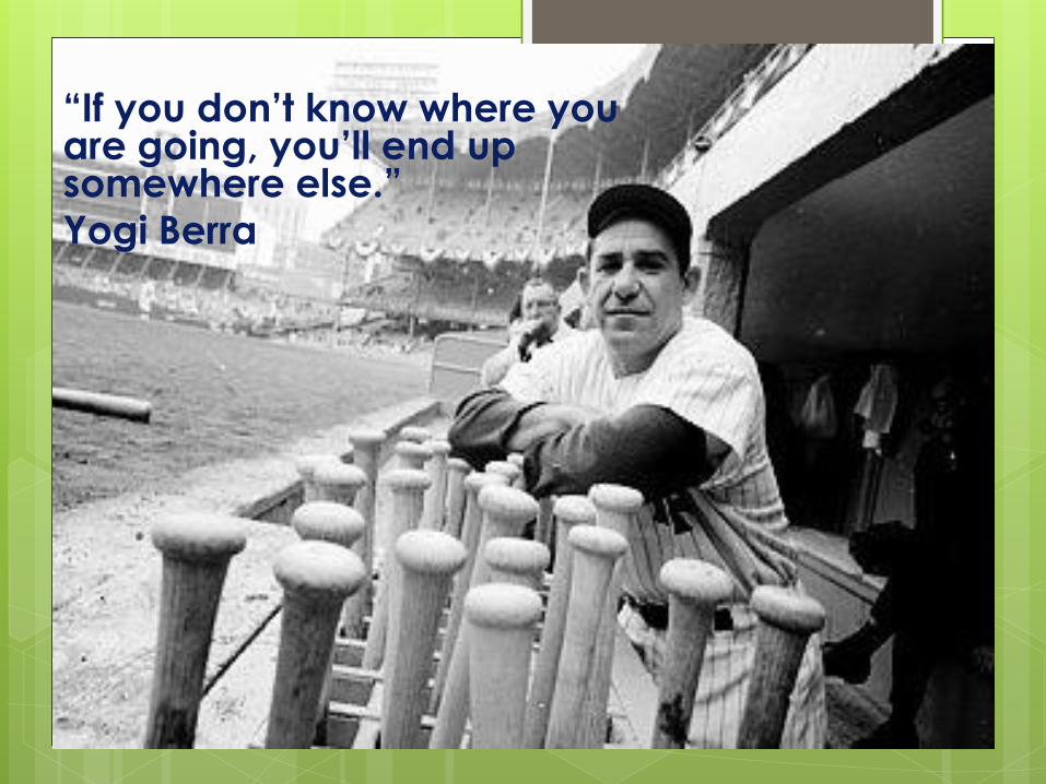
“If you don’t know where you are going, you’ll end up somewhere else.”
Yogi Berra
Objectives
Discuss what syndromes comprise mixed
sleep disordered breathing pathology
Discuss treatment options
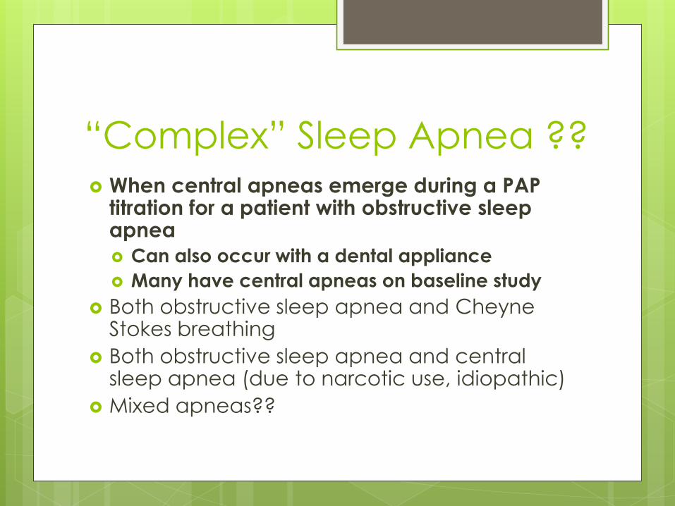
“Complex” Sleep Apnea ?? When central apneas emerge during a PAP
titration for a patient with obstructive sleep apnea
Can also occur with a dental appliance
Many have central apneas on baseline study
Both obstructive sleep apnea and Cheyne Stokes breathing
Both obstructive sleep apnea and central sleep apnea (due to narcotic use, idiopathic)
Mixed apneas??

“Complex” Sleep Apnea
Have obstructive sleep apnea on baseline
Most have a hint that centrals may appear
with PAP
scattered mixed and central apneas
Cheyne Stokes pattern
As obstructive events are eliminated, more
central events occur; note differences
between events in REM vs NREM
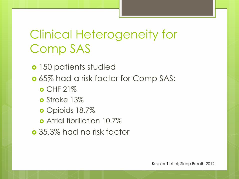
Clinical Heterogeneity for
Comp SAS
150 patients studied
65% had a risk factor for Comp SAS:
CHF 21%
Stroke 13%
Opioids 18.7%
Atrial fibrillation 10.7%
35.3% had no risk factor
Kuzniar T et al; Sleep Breath 2012

Sleep Disordered Breathing
Syndromes
Obstructive sleep apnea disorders
Adult; pediatric
Central sleep apnea disorders
Sleep related hypoventilation syndromes
Sleep related hypoxemia disorders

From ICSD‐3:

Central Sleep Apnea
A diagnosis of central sleep apnea (CSA)
requires all of the following:
An apnea hypopnea index > 5
Central apneas/hypopneas > 50% of the
total apneas/hypopneas
Central apneas or hypopneas ≥ 5 times per
hour
Symptoms of either excessive sleepiness or
disrupted sleep
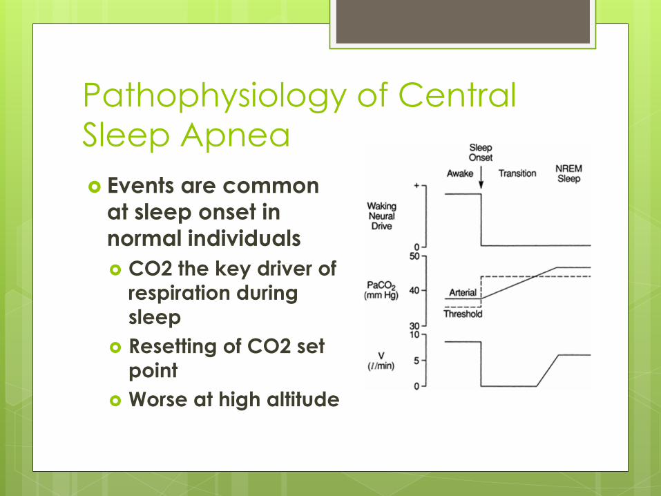
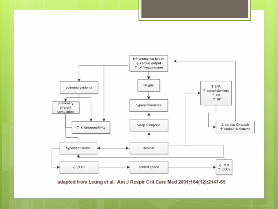
Pathophysiology of Central
Sleep Apnea
Events are common
at sleep onset in
normal individuals
CO2 the key driver of
respiration during
sleep
Resetting of CO2 set
point
Worse at high altitude
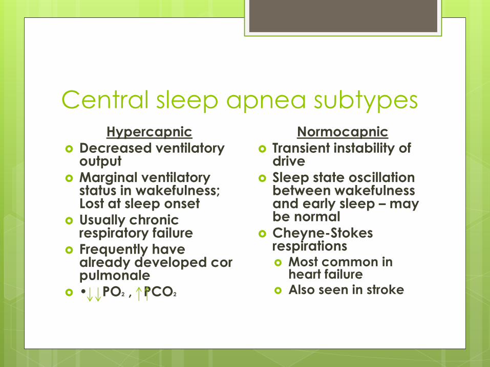
Central sleep apnea subtypes Hypercapnic
Decreased ventilatory output
Marginal ventilatory status in wakefulness; Lost at sleep onset
Usually chronic respiratory failure
Frequently have already developed cor pulmonale
• PO2 , PCO2
Normocapnic
Transient instability of drive
Sleep state oscillation between wakefulness and early sleep – may be normal
Cheyne-Stokes respirations Most common in
heart failure Also seen in stroke

Central Sleep Apnea
Types:
Primary – idiopathic
Cheyne Stokes (CS)
High altitude periodic breathing
Due to neurologic or medical condition
(not CS) – usually secondary to a structural
CNS lesion
Due to drug ‐ opioids

Central Sleep Apnea with Cheyne‐Stokes Breathing (CS)
ICD-9CM
780-04 – Cheyne-Stokes respiration
ICD-10CM
R06.3 – Periodic breathing

Cheyne‐Stokes Respiration
(CS)
First described by Cheyne (1818) then Stokes (1854)
Best studied in relation to CHF Found in 25‐40%
Risk factors for CSR in CHF male gender
atrial fibrillation
age > 60
hypocapnia
Associated with increased mortality in CHF
Seen following acute stroke (26‐50%)

Transplant free survival:
CSR vs No CSR
Sin et al, Circulation 2000;102:61‐6

Cheyne Stokes: Diagnostic
Criteria (A or B) + C + D satisfy the criteria
A. The presence of one or more of the following:
Sleepiness
Difficulty initiating or maintaining sleep, frequent awakenings, or nonrestorative sleep
Awakening short of breath
Snoring
Witnessed apneas
B. The presence of atrial fib/flutter, congestive heart failure, or a neurological disorder
C. PSG (during diagnostic or PAP titration) shows all of the following:
Five or more central apneas and/or central hypopneas per hour of sleep
The total number of central apneas and/or central hypopneas is > 50% of the total number of apneas and hypopneas
The pattern of ventilation meets criteria for Cheyne‐Stokes breathing (CSB)
D. The disorder is not better explained by another current sleep
disorder, medication use (e.g.,opioids), or substance use disorder

Scoring Cheyne Stokes Score a respiratory event as Cheyne‐Stokes
breathing if BOTH of the following are met:
There are episodes of ≥ 3 consecutive central apneas and/or central hypopneas separated by a crescendo and decrescendo change in breathing amplitude with a cycle length of ≥ 40 seconds.
There are ≥ 5 central apneas and/or central hypopneas per hour of sleep associated with the crescendo/decrescendo breathing pattern recorded over ≥ 2 hours of monitoring.


Mechanisms thought to cause CSR in CHF
Increased awake ventilation
Increased sensitivity of respiratory drive
Circulation delay – reduced CO
Heart failure
Sleep onset
Hypoxemia from CSR
(increased PAP RV dec LV filling)
Supine position

CSA due to a medical or neurologic
condition without
CS breathing ICD-9CM
327.27 – Central sleep apnea in conditions
classified elsewhere
ICD-10CM
G47.37 – Central sleep apnea in conditions
classified elsewhere
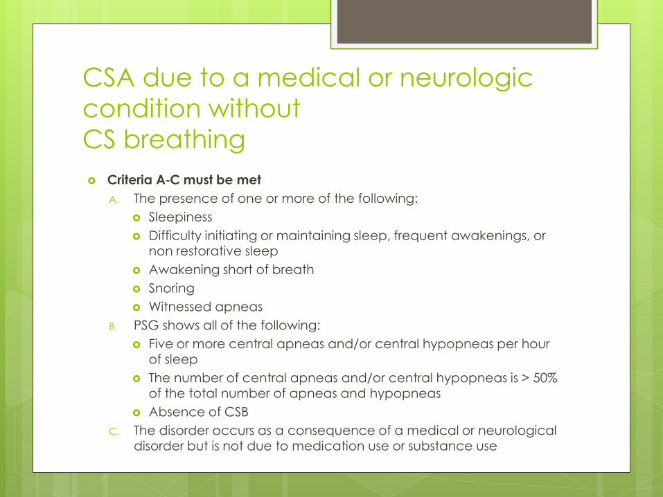
CSA due to a medical or neurologic
condition without
CS breathing
Criteria A‐C must be met
A. The presence of one or more of the following:
Sleepiness
Difficulty initiating or maintaining sleep, frequent awakenings, or non restorative sleep
Awakening short of breath
Snoring
Witnessed apneas
B. PSG shows all of the following:
Five or more central apneas and/or central hypopneas per hour
of sleep
The number of central apneas and/or central hypopneas is > 50% of the total number of apneas and hypopneas
Absence of CSB
C. The disorder occurs as a consequence of a medical or neurological
disorder but is not due to medication use or substance use

CSA due to a medical or neurologic
condition without
CS breathing
Brainstem lesions of developmental, vascular, neoplastic, degenerative, demyelinating, or traumatic origin
Chiari malformation
Post stroke
Brain neoplasm
Multiple system atrophy
due to dysfunction of central ventilatory control centers to initiate ventilatory effort

Primary Central Sleep Apnea
ICD-9CM
327.21 – Primary central sleep apnea
ICD10-CM
G47.31 – Primary central sleep apnea
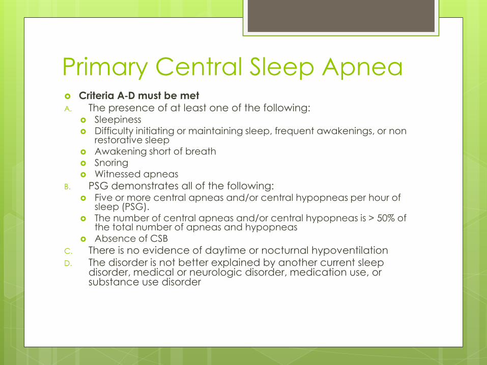
Primary Central Sleep Apnea Criteria A‐D must be met
A. The presence of at least one of the following: Sleepiness Difficulty initiating or maintaining sleep, frequent awakenings, or non
restorative sleep Awakening short of breath
Snoring Witnessed apneas
B. PSG demonstrates all of the following: Five or more central apneas and/or central hypopneas per hour of
sleep (PSG). The number of central apneas and/or central hypopneas is > 50% of
the total number of apneas and hypopneas Absence of CSB
C. There is no evidence of daytime or nocturnal hypoventilation
D. The disorder is not better explained by another current sleep disorder, medical or neurologic disorder, medication use, or substance use disorder

Central sleep apnea due to
drug or substance
ICD-9CM
327.29 – Other organic sleep apnea
ICD10-CM
G47.39 – Other sleep apnea
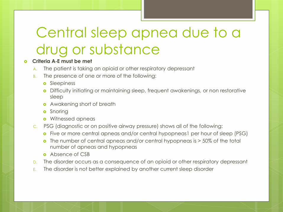
Central sleep apnea due to a
drug or substance Criteria A‐E must be met
A. The patient is taking an opioid or other respiratory depressant
B. The presence of one or more of the following:
Sleepiness
Difficulty initiating or maintaining sleep, frequent awakenings, or non restorative
sleep
Awakening short of breath
Snoring
Witnessed apneas
C. PSG (diagnostic or on positive airway pressure) shows all of the following:
Five or more central apneas and/or central hypopneas1 per hour of sleep (PSG)
The number of central apneas and/or central hypopneas is > 50% of the total
number of apneas and hypopneas
Absence of CSB
D. The disorder occurs as a consequence of an opioid or other respiratory depressant
E. The disorder is not better explained by another current sleep disorder

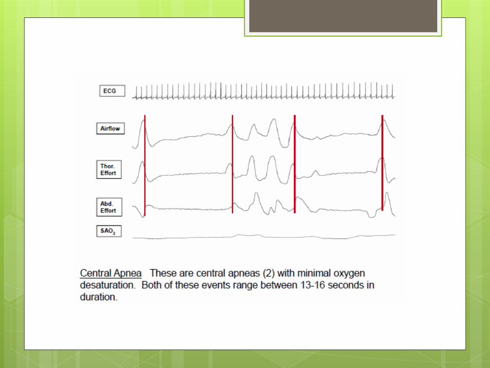
Ataxic Breathing Pattern
Methadone
• Oxycontin
• Fentanyl patch
• Suboxone

Treatment Emergent Central
Sleep Apnea
ICD-9CM
327.29 – Other organic sleep apnea
ICD-10CM
G47.39 – Other sleep apnea
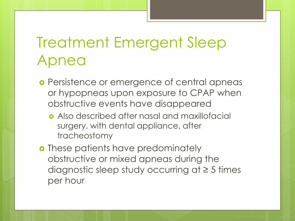
Treatment Emergent Sleep
Apnea
Persistence or emergence of central apneas
or hypopneas upon exposure to CPAP when
obstructive events have disappeared
Also described after nasal and maxillofacial
surgery, with dental appliance, after
tracheostomy
These patients have predominately
obstructive or mixed apneas during the
diagnostic sleep study occurring at ≥ 5 times
per hour
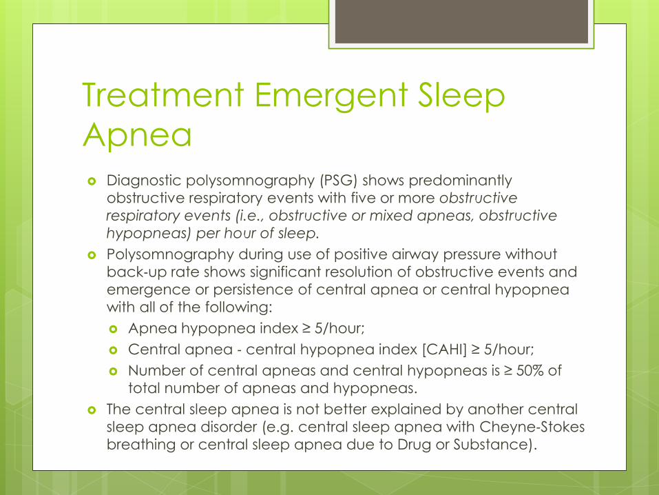
Treatment Emergent Sleep
Apnea Diagnostic polysomnography (PSG) shows predominantly
obstructive respiratory events with five or more obstructive
respiratory events (i.e., obstructive or mixed apneas, obstructive
hypopneas) per hour of sleep.
Polysomnography during use of positive airway pressure without
back‐up rate shows significant resolution of obstructive events and
emergence or persistence of central apnea or central hypopnea
with all of the following:
Apnea hypopnea index ≥ 5/hour;
Central apnea ‐ central hypopnea index [CAHI] ≥ 5/hour;
Number of central apneas and central hypopneas is ≥ 50% of
total number of apneas and hypopneas.
The central sleep apnea is not better explained by another central
sleep apnea disorder (e.g. central sleep apnea with Cheyne‐Stokes
breathing or central sleep apnea due to Drug or Substance).


Characteristics of TE sleep
apnea A high number of arousals persist on PAP treatment
and the AHI is often higher during NREM than REM sleep
Treatment emergent central apneas invariably occur during NREM sleep
Pressures that are effective in controlling obstructive events during REM sleep prove ineffective during NREM sleep due to emergence of central apneas
In N3, central apneas often decrease until interrupted by an arousal which precipitates another run of central events
Persistent sleep fragmentation on CPAP treatment and may report little benefit from therapy
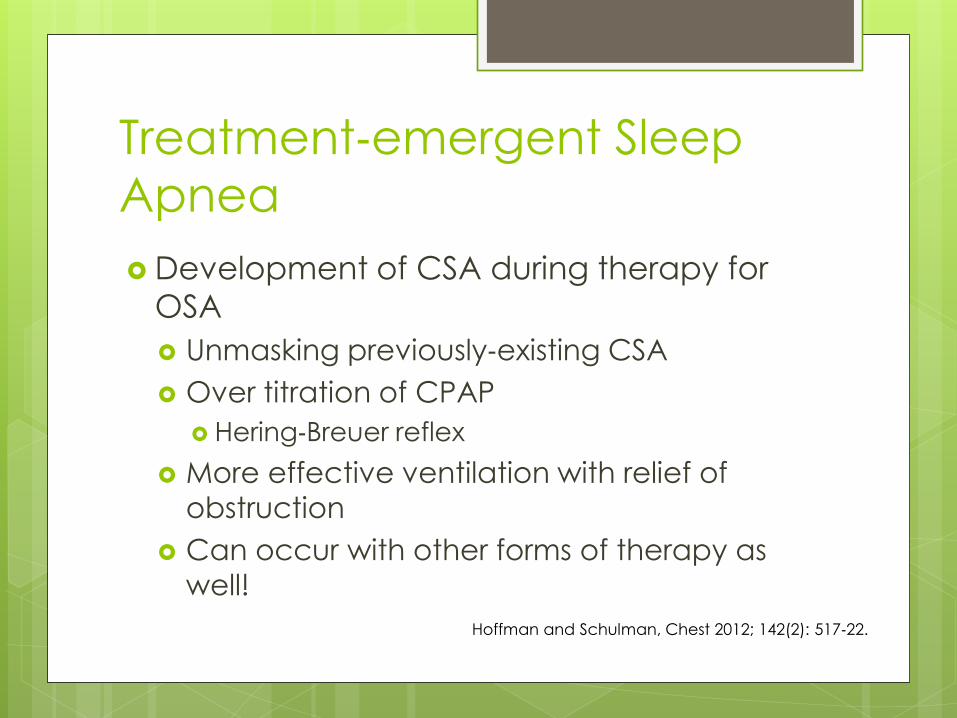
Treatment‐emergent Sleep
Apnea
Development of CSA during therapy for
OSA
Unmasking previously‐existing CSA
Over titration of CPAP
Hering‐Breuer reflex
More effective ventilation with relief of
obstruction
Can occur with other forms of therapy as
well!
Hoffman and Schulman, Chest 2012; 142(2): 517‐22.

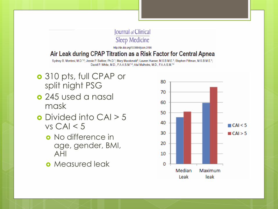
310 pts, full CPAP or split night PSG
245 used a nasal mask
Divided into CAI > 5 vs CAI < 5
No difference in age, gender, BMI, AHI
Measured leak


Therapy Options
Determine if there is an etiology for
centrals
Atrial fibrillation
CHF
Opioids or other depressant medication
Stroke/TIA in history
If no obvious reason, consider CNS
imaging
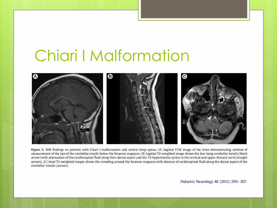
Chiari I Malformation

Chiari I Malformation
1:1000‐5000
Downward displacement of cerebellar tonsils through foramen magnum
Both central and obstructive apneas described
SDB may be related to compression of brainstem structures
Decompressive surgery has been shown to reduce both OSA and CSA
Etiology of CSA
Atrial fibrillation
Cardioversion
Ablation
CHF
Maximize therapy
Pacemaker
Opioids – reduce dose
Chiari malformation ‐ surgery

Therapy Options
Determine if there is an etiology for
centrals
If specific etiology found, may target that
initially
Consider drug trial
Hypnotics
Acetazolamide
Acetazolamide for CSA 2 non‐randomized treatment studies reported on
the use of acetazolamide for primary CSA
250 mg/day decreased the AHI from 37.2 ± 23.2 to 12.8 ± 10.8 in 14 patients at 1‐month follow‐up
1000 mg/day ‐ CAI decreased 54 ± 29 to 12 ± 20 in 6 patients after 1 week of therapy
1 study in CS/CHF
Randomized crossover with reduction in AHI
Considered a low evidence level option
Side effects: paresthesias, tinnitus, GI symptoms, metabolic acidosis, electrolyte imbalance
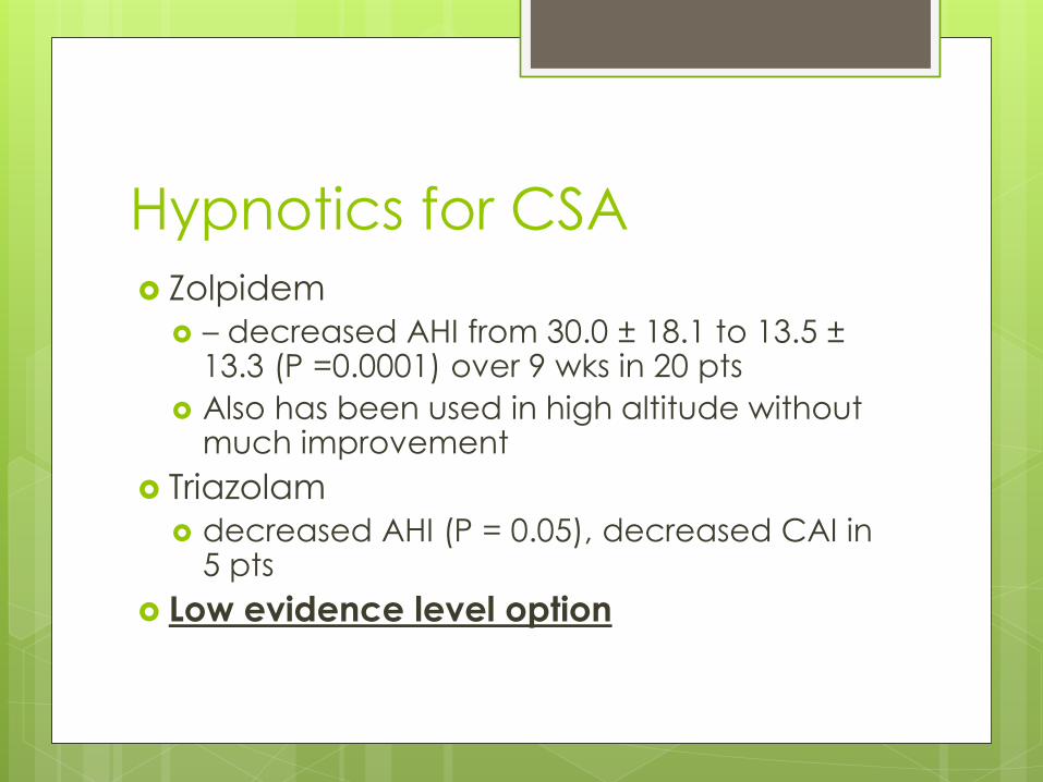
Hypnotics for CSA
Zolpidem
– decreased AHI from 30.0 ± 18.1 to 13.5 ± 13.3 (P =0.0001) over 9 wks in 20 pts
Also has been used in high altitude without much improvement
Triazolam
decreased AHI (P = 0.05), decreased CAI in 5 pts
Low evidence level option

Therapy Options
Determine if there is a potential etiology
for centrals
If specific etiology found, may target that
initially
Consider drug trial (low level evidence)
Gases
CO2/dead space
Oxygen

Gases – Carbon Dioxide Low CO2 levels (hypocapnia) drive most CSA
1‐4% inhalation of CO2 resolves CSA Lorenzi‐Filho G, et al. Effects of inhaled carbon dioxide and
oxygen on cheyne‐stokes respiration in patients with heart failure. Am J Respir Crit Care Med 1999;159:1490‐1498
Steens RD, et al. Effect of inhaled 3% CO2 on Cheyne‐Stokes respiration in congestive heart failure Sleep. 1994;17(1):61‐68.
Szollosi I, et al. Effect of CO2 inhalation on central sleep apnea and arousals from sleep. Respiration 2004;71(5):493‐498.
PAP gas modulator ‐ Sleep 2005;28(1):69‐77 6 pts Comp SAS ‐ quick abolition of sleep‐disordered breathing cost of device, the need for a continuous supply of
medical‐grade CO2, and potential adverse effects of CO2 therapy limit applicability
Addition of dead space also been shown to help but cannot be individualized

Gases – Oxygen
Stabilizes respiratory drive
CPAP + Oxygen reduces SDB
Oxygen can help CSB
Problem:
Usually cannot justify payment
Not as effective as ASV
No long term outcome studies

Therapy Options
Determine if there is a potential etiology for centrals
If specific etiology found, may target that initially
Consider drug trial (low level evidence)
Gases
PAP therapy
Best CPAP and re‐evaluate; monitor leak
ASV
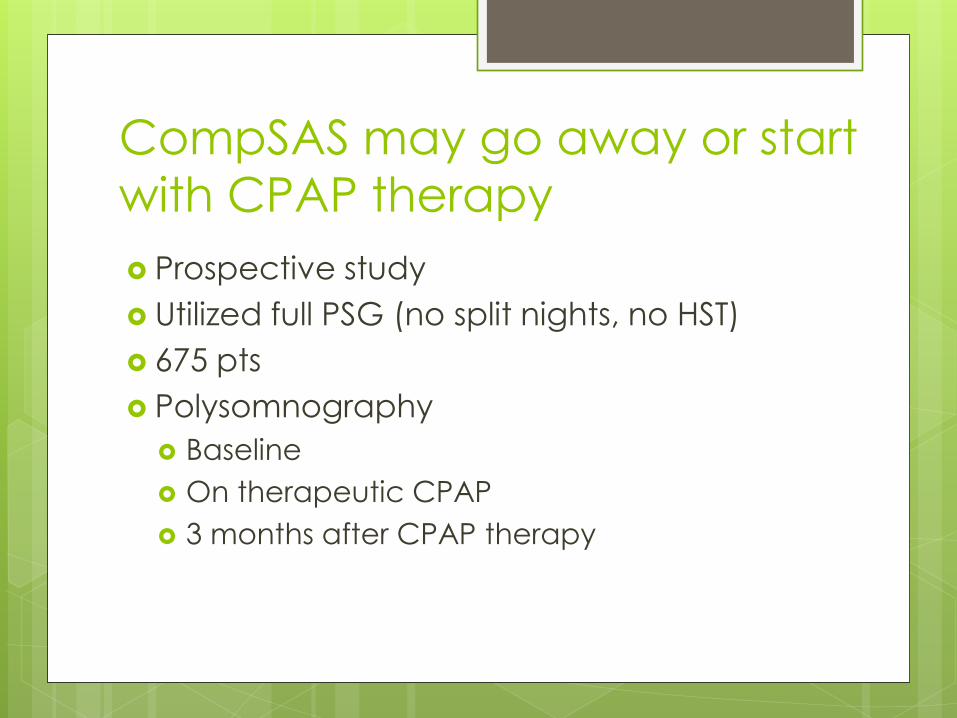
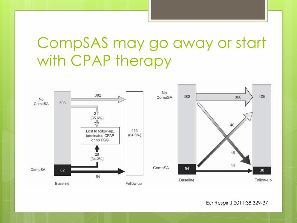
CompSAS may go away or start
with CPAP therapy
Prospective study
Utilized full PSG (no split nights, no HST)
675 pts
Polysomnography
Baseline
On therapeutic CPAP
3 months after CPAP therapy
CompSAS may go away or start
with CPAP therapy
Eur Respir J 2011;38:329‐37

Adaptive Servo‐ventilation bilevel positive airway pressure that introduced for
treatment of central / Comp SAS
provides variable pressure support (PS) in response to a servo mechanism‐based assessment
Measures or estimates patient’s respiratory output (tidal exhalation, flow, minute ventilation)
Increase PS during hypopnea, reduce PS during hyperpnea, timed breaths during central apnea
In US, 2 companies provide:
ResMed – VPAP Adapt SV
Phillips Respironics – BIPAP Auto SV (advanced)
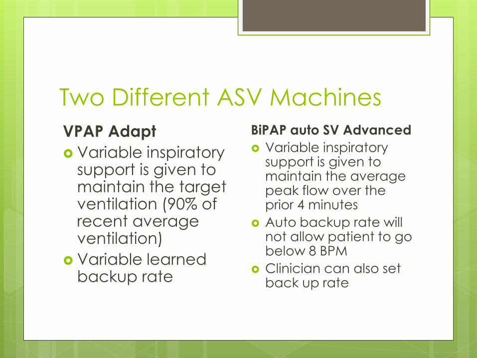
Two Different ASV Machines
VPAP Adapt
Variable inspiratory support is given to maintain the target ventilation (90% of recent average ventilation)
Variable learned backup rate
BiPAP auto SV Advanced
Variable inspiratory support is given to maintain the average peak flow over the prior 4 minutes
Auto backup rate will not allow patient to go below 8 BPM
Clinician can also set back up rate

Adaptive Servo Ventilation


Average Volume Assured
Pressure Support
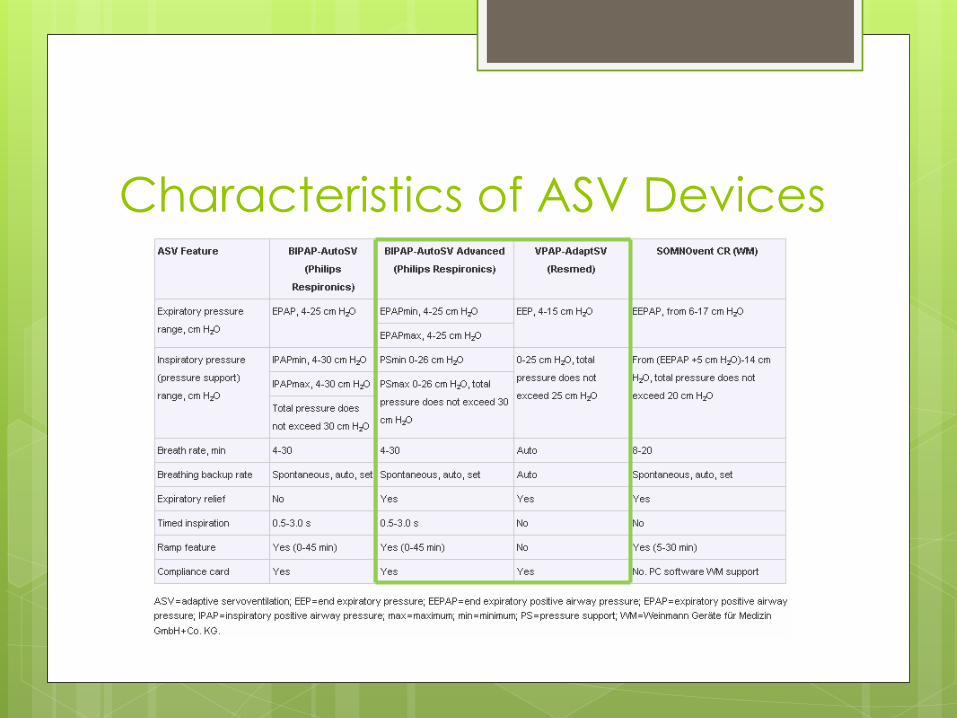
Characteristics of ASV Devices

Prescription Settings VPAP Adapt (ASV mode) EPAP PS min (lowest 3cmH2O)
PS max SPAP Adapt (ASVauto mode) EPAP min/max PS min (lowest 0 cmH2O) PS max
PS=pressure above APAP min at all times in VPAP Adaprt ASVauto mode
BiPAP auto SV Advanced
EPAP min/max
PS min (lowest 0 cmH2O)
PS max
Max Pressure
Rate: Auto or BPM
I-Time (with fixed rate)
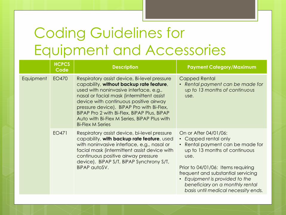
Coding Guidelines for
Equipment and Accessories HCPCS
Code Description Payment Category/Maximum
Equipment EO470 Respiratory assist device, Bi-level pressure
capability, without backup rate feature,
used with noninvasive interface, e.g.,
nasal or facial mask (intermittent assist
device with continuous positive airway
pressure device). BiPAP Pro with Bi-Flex,
BiPAP Pro 2 with Bi-Flex, BiPAP Plus, BiPAP
Auto with Bi-Flex M Series, BiPAP Plus with
Bi-Flex M Series
Capped Rental
• Rental payment can be made for
up to 13 months of continuous
use.
EO471 Respiratory assist device, bi-level pressure
capability, with backup rate feature, used
with noninvasive interface, e.g., nasal or
facial mask (intermittent assist device with
continuous positive airway pressure
device). BiPAP S/T, BiPAP Synchrony S/T,
BiPAP autoSV.
On or After 04/01/06:
• Capped rental only
• Rental payment can be made for
up to 13 months of continuous
use.
Prior to 04/01/06: Items requiring
frequent and substantial servicing
• Equipment is provided to the
beneficiary on a monthly rental
basis until medical necessity ends.


ASV in CSR patients
29 male patients, LVEF < 40%
Tx’d ~ 6 months with ASV; no control group
AHI at baseline = 37.4; min SpO2 80.9%
Used Embletta PM device
CSR defined as >80% of respiratory events were central apneas with CSR
AHI > 15 to be enrolled
Avg ASV setting: IPAP 8‐12; EPAP 4‐7
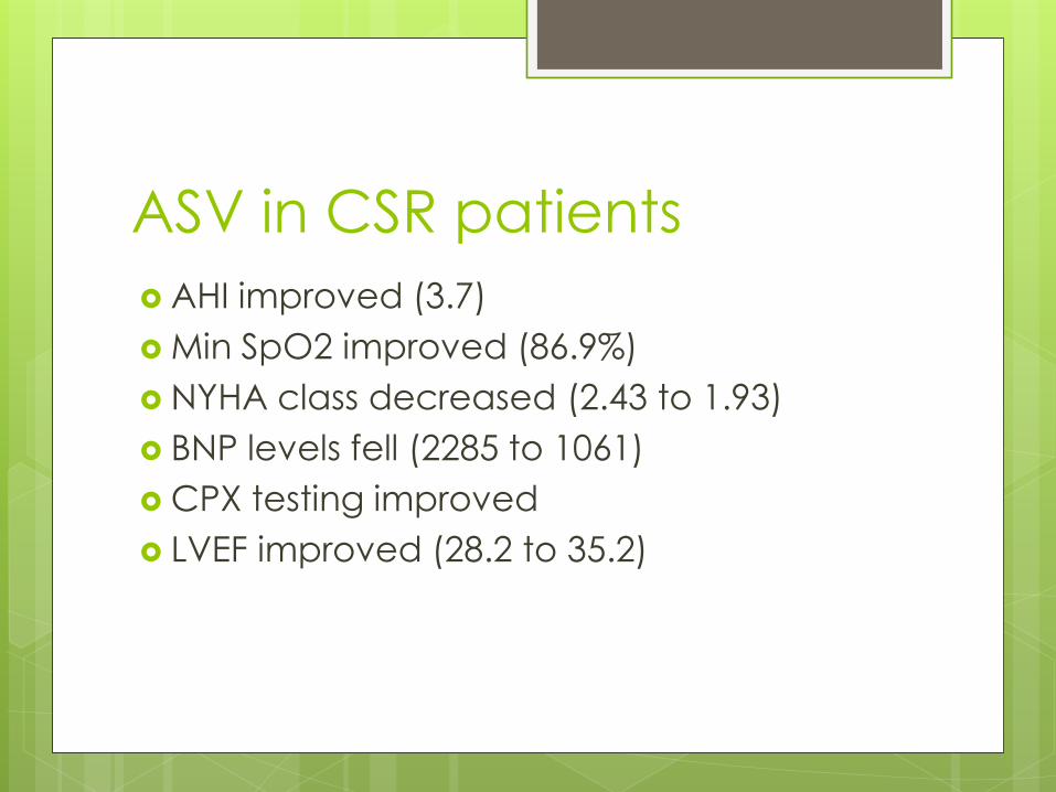
ASV in CSR patients
AHI improved (3.7)
Min SpO2 improved (86.9%)
NYHA class decreased (2.43 to 1.93)
BNP levels fell (2285 to 1061)
CPX testing improved
LVEF improved (28.2 to 35.2)

NPPV vs ASV
Somnovent
Bilevel PAP with backup rate; EPAP to
eliminate OA, IPAP to optimal treatment of
CA
ASV to maintain flow and minute ventilation
30 patients completed study

NPPV vs ASV

ASV to treat Opioid induced CSA 5 articles, 127 patients; on opioids > 6 months
Dosage ranged from 10 ‐ 450 mg daily
(morphine equivalent)
CPAP mostly ineffective
Bilevel PAP with and without supplemental
oxygen achieved elimination of central
apneas in 62 %
ASV: conflicting results ‐ 58% central apnea
index <10/hour
Ataxic breathing predicted poor response to
PAP

ASV caveats
No long term trials to show a mortality reduction
Cost is much higher than CPAP
Excellent choice for CS alone or CS with OSA
Not as effective for CSA secondary to opioids or without CS
Acceptable AHI is higher than with CPAP – 10?; converts apneas to hypopneas

Take Home Points
Complex sleep apnea is a confusing term
Look for etiology for central events and
address those if possible
Many patients with treatment emergent
CSA will improve with time
ASV appears to be better than NPPV
(bilevel PAP) for persistent CSA/CS


![Home [registers.centralbank.ie]registers.centralbank.ie/ICAVDocuments/C148578/Director... · 2016-01-08 · Cheyne Select Funds Public Limited Company 07-12-2010 Ireland 492329 Cheyne](https://img.pdfslide.net/doc/110x75/5f342dfc40076e748c3bba71/home-2016-01-08-cheyne-select-funds-public-limited-company-07-12-2010-ireland.jpg)