Embed Size (px)
Citation preview

Computed Tomography to Detect Coronary Artery Calcification Policy # 00031 Original Effective Date: 10/21/2002 Current Effective Date: 01/02/2018
Page 1 of 22
Applies to all products administered or underwritten by Blue Cross and Blue Shield of Louisiana and its subsidiary, HMO Louisiana, Inc.(collectively referred to as the “Company”), unless otherwise provided in the applicable contract. Medical technology is constantly evolving, and we reserve the right to review and update Medical Policy periodically.
Note: Contrast-Enhanced Coronary Computed Tomography Angiography (CCTA) for Coronary Artery Evaluation is addressed separately in medical policy 00153. Services Are Considered Investigational Coverage is not available for investigational medical treatments or procedures, drugs, devices or biological products. Based on review of available data, the Company considers the use of electron beam computed tomography (EBCT) or spiral computed tomography (CT) to detect coronary artery calcification to be investigational.*
Background/Overview CORONARY ARTERY CALCIUM Coronary artery calcium (CAC) is associated with coronary artery disease (CAD) based anatomic studies. The development of fast CT scanners has allowed the measurement of CAC in clinical practice. CAC has been evaluated in several clinical settings. The most widely studied indication is for the use of CAC in the prediction of future risk of CAD in patients with subclinical disease, with the goal of instituting appropriate risk-reducing therapy (e.g., statin treatment, lifestyle modifications) to improve outcomes. Also, CAC has been evaluated in patients with symptoms potentially consistent with CAD, but in whom a diagnosis is unclear. Detection EBCT, also known as ultrafast CT and spiral CT (or helical CT) may be used as an alternative to conventional CT scanning due to faster throughput. In both methods, speed of image acquisition gives them unique value for imaging a moving heart. The rapid image acquisition time virtually eliminates motion artifact related to cardiac contraction, permitting visualization of the calcium in the epicardial coronary arteries. EBCT software permits quantification of calcium area and density, which are translated into calcium scores. Calcium scores have been investigated as a technique for detecting CAC, both as a diagnostic technique in symptomatic patients to rule out an atherosclerotic etiology of symptoms or, in asymptomatic patients, as an adjunctive method for risk stratification for CAD. EBCT and multidetector CT were initially the primary fast CT methods for measurement of CAC. A fast CT study for CAC measurement takes 10 to 15 minutes and requires only a few seconds of scanning time. More recently, CT angiography has been used to assess coronary calcium. Because of the basic similarity between EBCT and CT angiography in measuring coronary calcium, it is expected that CT angiography provides information on coronary calcium that is similar to EBCT.
©2017 Blue Cross and Blue Shield of Louisiana
Blue Cross and Blue Shield of Louisiana is an independent licensee of the Blue Cross and Blue Shield Association and incorporated
as Louisiana Health Service & Indemnity Company.
No part of this publication may be reproduced, stored in a retrieval system, or transmitted, in any form or by any means, electronic, mechanical, photocopying, or otherwise, without permission from Blue Cross and Blue Shield of Louisiana.

Computed Tomography to Detect Coronary Artery Calcification Policy # 00031 Original Effective Date: 10/21/2002 Current Effective Date: 01/02/2018
©2017 Blue Cross and Blue Shield of Louisiana
Blue Cross and Blue Shield of Louisiana is an independent licensee of the Blue Cross and Blue Shield Association and incorporated
as Louisiana Health Service & Indemnity Company.
No part of this publication may be reproduced, stored in a retrieval system, or transmitted, in any form or by any means, electronic, mechanical, photocopying, or otherwise, without permission from Blue Cross and Blue Shield of Louisiana.
Page 2 of 22
CT scan‒derived coronary calcium measures have been used to evaluate coronary atherosclerosis. Coronary calcium is present in coronary atherosclerosis, but the atherosclerosis detected may or may not be causing ischemia or symptoms. Coronary calcium measures may be correlated with the presence of critical coronary stenoses or serve as a measure of the patient’s proclivity toward atherosclerosis and future coronary disease. Thus, coronary calcium could serve as a variable to be used in a risk assessment calculation to determine appropriate preventive treatment in asymptomatic patients. Alternatively, in other clinical scenarios, coronary calcium scores might help determine whether there is an atherosclerotic etiology or component to the presenting clinical problem in symptomatic patients, thus helping to direct further workup for the clinical problem. In this second scenario, a calcium score of 0 usually indicates that the patient’s clinical problem is unlikely to be due to atherosclerosis and that other etiologies should be more strongly considered. In neither case does the test determine a specific diagnosis. Most clinical studies have examined the use of coronary calcium for its potential use in estimating the risk of future coronary heart disease (CHD) events. Nomenclature Coronary calcium levels can be expressed in many ways. The most common method is the Agatston score, which is a weighted summed total of calcified coronary artery area observed on CT. This value can be expressed as an absolute number, commonly ranging from 0 (low risk) to 400 (high risk). These values can be translated into age- and sex-specific percentile values. Different imaging methods and protocols will produce different values based on the specific algorithm used to create the score, but the correlation between any 2 methods appears to be high, and scores from 1 method can be translated into scores from a different method.
FDA or Other Governmental Regulatory Approval U.S. Food and Drug Administration (FDA) Many models of CT devices, including EBCT and other ultrafast CT devices, have been cleared for marketing by the U.S. FDA through the 510(k) process. FDA product code: JAK. Centers for Medicare and Medicaid Services (CMS) There is no national coverage determination. In the absence of a national coverage determination, coverage decisions are left to the discretion of local Medicare carriers.
Rationale/Source Assessment of a diagnostic technology typically focuses on 3 categories of evidence: (1) its technical reliability (test-retest reliability or interrater reliability); (2) clinical validity (sensitivity, specificity, and positive and negative predictive value) in relevant populations of patients; and (3) clinical utility (i.e., demonstration that the diagnostic information can be used to improve patient outcomes). This review was informed, in part, by a 1998 TEC Assessment. The Assessment concluded that the evidence available was sufficient to permit conclusions about the technology’s performance, but not the

Computed Tomography to Detect Coronary Artery Calcification Policy # 00031 Original Effective Date: 10/21/2002 Current Effective Date: 01/02/2018
©2017 Blue Cross and Blue Shield of Louisiana
Blue Cross and Blue Shield of Louisiana is an independent licensee of the Blue Cross and Blue Shield Association and incorporated
as Louisiana Health Service & Indemnity Company.
No part of this publication may be reproduced, stored in a retrieval system, or transmitted, in any form or by any means, electronic, mechanical, photocopying, or otherwise, without permission from Blue Cross and Blue Shield of Louisiana.
Page 3 of 22
effect of the technology on health outcomes, especially when compared with other noninvasive methods of assessing CAD. CORONARY ARTERY CALCIUM SCORING IN ASYMPTOMATIC INDIVIDUALS Clinical Context and Test Purpose The purpose of CAC scoring using CT in asymptomatic patients is to assess who may benefit from preventive interventions targeted to minimize the risk of atherosclerotic cardiovascular disease (CVD). The question addressed in this evidence review is: Does CAC scoring result in an improved health outcome compared with CAD risk stratification based on standard risk factors among asymptomatic patients? The following PICOTS were used to select literature to inform this review. Patients The population of interest includes individuals who are asymptomatic with the risk of CAD.
Interventions The intervention of interest is CAC scoring using fast CT imaging, including EBCT and spiral CT. Comparators The comparator of interest is CAD risk factor stratification based on standard risks, such as Framingham risk scores (FRS). Outcomes The outcomes of interest include overall survival, test accuracy, test validity, morbid events (e.g., major adverse cardiac events [MACEs]), need for invasive coronary angiography (ICA), and revascularization. Additional intermediate or surrogate outcomes of interest are changes in cardiac risk profile indicators such as smoking, hyperlipidemia, or hypertension. Timing CAC scoring is usually initiated or used to modify cardiac risk-reduction interventions in individuals asymptomatic for CAD. Setting The setting is a primary care or general cardiology practice setting to assess the risk of CAD. Technical Reliability Data supporting technical reliability derive from the test-retest reliability of CAC scoring measured by CT. The 1998 TEC Assessment reported that there was sufficient evidence to permit conclusions concerning the technical reliability of CAC scoring. Current review includes more recent evidence on the technical reliability of CAC scoring.

Computed Tomography to Detect Coronary Artery Calcification Policy # 00031 Original Effective Date: 10/21/2002 Current Effective Date: 01/02/2018
©2017 Blue Cross and Blue Shield of Louisiana
Blue Cross and Blue Shield of Louisiana is an independent licensee of the Blue Cross and Blue Shield Association and incorporated
as Louisiana Health Service & Indemnity Company.
No part of this publication may be reproduced, stored in a retrieval system, or transmitted, in any form or by any means, electronic, mechanical, photocopying, or otherwise, without permission from Blue Cross and Blue Shield of Louisiana.
Page 4 of 22
Systematic Reviews Xie et al (2013) conducted a systematic review and meta-analysis to determine the correlation in calcium score between nontriggered and electrocardiography-triggered CT. The pooled correlation coefficient for calcium score from the meta-analysis of 3 studies (661 participants) was 0.94 (95% confidence interval [CI], 0.89 to 0.97). The pooled Cohen’s κ from 2 studies (533 participants) was 0.89 (95% CI, 0.83 to 0.95) for 4 categories of calcium scores (0, 1-99, 100-399, ≥400). Heterogeneity was observed in the pooling calculation of the calcium score (p<0.001 for Q statistic, I
2>50%).
Observational Studies We identifed 3 studies relevant to discussion of the technical reliability of the CAC scoring in asymptomatic patients (see Tables 1-2). Choi et al (2016) conducted a prospective study to assess the interscan variability of CT for coronary calcium quantification using image acquisition with standard and reduced radiation dose protocols. A total of 200 consecutive patients underwent nonenhanced CT for coronary calcium quantification twice at a standard radiation dose and twice at a reduced radiation dose in randomized order. Each scan underwent reconstruction with both filtered back projection (FBP) and iterative reconstruction (IR). Interscan agreement with respect to Agatston categories for reduced-dose/IR protocol was 91% (95% CI, 87% to 94%), with a κ value of 0.87 (95% CI, 0.83 to 0.93). For standard-dose/FBP protocol, the agreement was 93% (95% CI, 89% to 96%) with a κ value of 0.91 (95% CI, 0.86 to 0.95), for standard-dose/IR protocol, the agreement was 92% (95% CI, 87% to 94%), with a κ value of 0.89 (95% CI, 0.84 to 0.94); and for reduced-dose/FBP protocol, the agreement was 90% (95% CI, 86% to 94%), with a κ value of 0.88 (95% CI, 0.82 to 0.93). Williams et al (2015) assessed results from 210 computed tomographic coronary angiographies (CTCAs) from the Scottish Computed Tomography of the Heart (SCOT-HEART) trial to examine intraobserver and interobserver variability in determining CAC score. There were no differences in Agatston calcium score on intraobserver assessment (373 [95% CI, 224 to 505] Agatston units vs 278 [95% CI, 202 to 354] Agatston units; p=0.138) or interobserver assessment (290 [95% CI, 210 to 370] Agatston units; p=0.191).The authors used Bland-Altman plots to examine intraobserver and interobserver agreement. Excellent intraobserver and interobserver agreement was identified for CAC scores below 1000. Sabour et al (2007) conducted a cross-sectional study with repeated measurements to assess interscan reproducibility of CAC measurements obtained from multidetector computer tomography (MDCT) images. The authors assessed coronary calcium in 76 healthy women participants twice in 1 session. One scan reader blinded to the scores of the first scan scored the second scan of the participants. While using a slice thickness of 1.5 mm, there was strong interscan correlation (intraclass correlation coefficient [ICC], 0.98) in Agatston score between scans. When quartiles of Agatston scores between scans were compared, high interscan agreement was observed (κ=0.88). Similar interscan correlation was observed with slice thickness of 3.0 mm, but interscan agreement was slightly lower (κ=0.84). Table 1. Summary of Key Technical Reliability Study Characteristics for CT CAC Scoring
Study (Year) Test-Retest Method Agreement Method
Choi et al (2016) Interscan agreement

Computed Tomography to Detect Coronary Artery Calcification Policy # 00031 Original Effective Date: 10/21/2002 Current Effective Date: 01/02/2018
©2017 Blue Cross and Blue Shield of Louisiana
Blue Cross and Blue Shield of Louisiana is an independent licensee of the Blue Cross and Blue Shield Association and incorporated
as Louisiana Health Service & Indemnity Company.
No part of this publication may be reproduced, stored in a retrieval system, or transmitted, in any form or by any means, electronic, mechanical, photocopying, or otherwise, without permission from Blue Cross and Blue Shield of Louisiana.
Page 5 of 22
κ Williams et al (2015) Bland-Altman plots Sabour et al (2007) Intraclass correlation coefficient
κ CAC: coronary artery calcium; CT: computed tomography.
Table 2. Summary of Key Technical Reliability Study Results for CT CAC Scoring
Study (Year) Initial N
Final N Excluded Samplesa Agreement (95% CI)
b
Choi et al (2016)
Standard-dose/FBP 200 200 0 ICA=92% (87% to 94%)
κ=0.89 (0.84 to 0.94) Standard-dose/IR 200 200 0 ICA=92% (87% to 94%)
κ=0.89 (0.84 to 0.94) Reduced-dose/FBP 200 200 0 ICA=90% (86% to 94%)
κ=0.88 (0.82 to 0.93) Reduced-dose/IR 200 200 0 ICA=91% (87% to 94%)
κ=0.87 (0.83 to 0.93) Williams et al (2015)
64- or 320-MDCT 210 210 0 Excellent intra- and interobserver agreement for CAC score <1000
Sabour et al (2007) Slice thickness 1.5 mm 76 76 0 ICC=0.98
κ=0.88 Slice thickness 3.0 mm 76 76 0 ICC=0.98
κ=0.84
CAC: coronary artery calcium; CI: confidence interval; CT: computed tomography; FBP: filtered back projection; IR: iterative reconstruction; ICA: interscan agreement; ICC: intraclass correlation coefficient, MDCT: multidetector computed tomography.
a Discarded, not run, invalid, or failed.
b Across sites or users.
Section Summary: Technical Reliability Excellent intra- and interobserver agreement in the estimation of CAC score was observed in studies using varying designs and with variations in calcium score measuring techniques. Clinical Validity Nakanishi et al (2016) conducted a study among 13,092 consecutive asymptomatic individuals without known CAD (mean age, 58 years) clinically referred for a CAC scan between 1997 and 2011 at a university medical center; the study examined the predictive value of CAC for 5- and 15-year mortality rates among men and women. CAC showed an incremental prognostic value over traditional risk factors among men at 5 years (area under curve [AUC], 0.702 vs 0.655; p=0.002) as well as at 15 years (AUC, 0.723 vs 0.656; p<0.001). In women, the incremental prognostic value of CAC was not statistically significant at 5 years (AUC, 0.650 vs 0.612; p=0.065) but was statistically significant at 15 years (AUC, 0.690 vs 0.624; p<0.001).

Computed Tomography to Detect Coronary Artery Calcification Policy # 00031 Original Effective Date: 10/21/2002 Current Effective Date: 01/02/2018
©2017 Blue Cross and Blue Shield of Louisiana
Blue Cross and Blue Shield of Louisiana is an independent licensee of the Blue Cross and Blue Shield Association and incorporated
as Louisiana Health Service & Indemnity Company.
No part of this publication may be reproduced, stored in a retrieval system, or transmitted, in any form or by any means, electronic, mechanical, photocopying, or otherwise, without permission from Blue Cross and Blue Shield of Louisiana.
Page 6 of 22
Gepner et al (2017) prospectively evaluated CVD, CHD, and stroke or transient ischemic attack (TIA) events to compare the use of CAC with carotid plaque scores to predict CVD events; the study used data from the Multi-Ethnic Study of Atherosclerosis (MESA), a population-based cohort of individuals without known CVD. After 11.3 years of follow-up among 4955 participants (mean age, 61.6 years), 709 CVD, 498 CHD, and 262 stroke/TIA events had occurred. CAC score significantly reclassified non-CVD events (3%; 95% CI, 2% to 5%) and CHD events (13%; 95% CI, 5% to 18%). Carotid plaque score did not consistently reclassify CVD or CHD events or nonevents. Blaha et al (2016) conducted a study using data from MESA to compare the value of various negative risk markers. The authors evaluated the accuracy of change in risk classification by calculating the net reclassification improvement (NRI) for each of the 13 negative risk markers. During a median of 10.3 years of follow-up among a cohort of 6814, 710 CVD events occurred. Among all negative risk markers, a CAC score of 0 was the strongest, with an adjusted mean diagnostic likelihood ratio of 0.41 (SD=0.12) for all CHD. NRI for downward reclassification (10-year CVD risk, <7.5%) of CVD events with CAC scores of 0 in participants with a pretest 10-year CVD risk of 7.5% or higher (n=3833 [3227 participants without events and 606 with events]) was 0.14, higher than other negative risk markers included in the study. Polonsky et al (2010) also used data from MESA to determine whether incorporation of calcium score into a risk model based on traditional risk factors improve classification of risk. During a median of 5.8 years of follow-up among a final cohort of 5878, 209 CHD events occurred, of which 122 were myocardial infarction, death from CHD, or resuscitated cardiac arrest. Addition of CAC score in the model resulted in significant improvements in risk prediction compared with the model without CAC score (NRI=0.25; 95% CI, 0.16 to 0.34; p<0.001). Subjects reclassified to high risk had a similar risk of CHD events as those originally classified as high risk. Elias-Smale et al (2011) conducted a study among 2153 asymptomatic participants (69.6 years) who underwent an MDCT scan. During a median follow-up of 3.5 years, 58 CHD events (myocardial infarction or death) occurred. Participants were classified into low (<5%), intermediate (5%-10%), and high (>10%) 5-year risk categories based on a refitted Framingham risk model. For the outcome of CHD, the C statistic improved from 0.693 for the Framingham refitted model to 0.743 by addition of coronary calcium. Reclassification of subjects occurred most substantially in the intermediate-risk group (5-year risk, 5%-10%) where 56% of persons were reclassified. Addition of CAC scoring reclassified 56% of persons: 36% moved to low risk while 20% moved to high risk, leading to a net gain in reclassification of 18% in persons with an event and a net decline in reclassification of 3% in persons without event, resulting in an NRI of 15% (p<0.01). Won et al (2015) conducted a single-center cross-sectional study among 328 consecutive asymptomatic patients with type 2 diabetes who underwent CTCA between 2008 and 2009 in a hospital in South Korea to evaluate the predictive value of the CAC score for obstructive coronary plaques (OCP) assessed by CTCA. On the basis of a CAC score of 0, 1 to 10, 11 to 100, or greater than 100, OCPs were found in 2%, 5%, 15%, and 36% of patients, respectively. On receiver operating characteristic curve analysis, the optimal cutoff CAC score for predicting OCPs was found to be 33, with 83% sensitivity and 81% specificity

Computed Tomography to Detect Coronary Artery Calcification Policy # 00031 Original Effective Date: 10/21/2002 Current Effective Date: 01/02/2018
©2017 Blue Cross and Blue Shield of Louisiana
Blue Cross and Blue Shield of Louisiana is an independent licensee of the Blue Cross and Blue Shield Association and incorporated
as Louisiana Health Service & Indemnity Company.
No part of this publication may be reproduced, stored in a retrieval system, or transmitted, in any form or by any means, electronic, mechanical, photocopying, or otherwise, without permission from Blue Cross and Blue Shield of Louisiana.
Page 7 of 22
(AUC=0.853; 95% CI, 0.777 to 0.930; p<0.001). Positive and negative predictive values of a CAC score of 33 for OCPs were 30% and 98%, respectively. On multivariate logistic regression analysis, age (odds ratio [OR], 1.09), microalbuminuria levels (OR=3.43), current smoker (OR= 3.93), and a CAC score greater than 33 (OR=15.85) were found to be independently associated with an increased risk for OCPs (p<0.05). Section Summary: Clinical Validity Multiple prospective cohort studies have consistently demonstrated the incremental prognostic value of CAC scoring in predicting CHD and mortality over traditional risk factors among asymptomatic populations over the intermediate and long term. However, considering the heterogeneity of methods applied and inherent limitations of observational studies, there is a need for more evidence on diagnostic accuracy of CAC scoring in predicting CHD risk among the asymptomatic population, preferably from randomized controlled trials (RCTs). Clinical Utility
Systematic Reviews Tables 3 and 4 list, respectively, the characteristics and results of systematic reviews relevant to assessment of the clinical utility of CAC scoring. Mamudu et al (2014) conducted a systematic review of studies evaluating the effects of CAC screening on behavioral modification, risk perception, and medication adherence in asymptomatic adults. Fifteen studies were selected (3 RCTs, 12 observational studies). The size of the study populations ranged from 56 to 6814 individuals. Reviewers primarily provided descriptive results of the studies given the lack of standardization across studies regarding CAC measures and outcome variables. CAC screening improved medication adherence. However, the impact of CAC screening on behavioral and lifestyle factors (body mass index [BMI], diet, exercise, smoking), perception of CAD risk, and psychosocial effects was nonsignificant compared with baseline. Xie et al (2013) conducted a systematic review to evaluate the prognostic performance of the CAC score derived from nontriggered CT. In 5 studies, 34,028 cardiac asymptomatic patients were followed for a mean of 45 months (range, 0-72 months). No meta-analysis was performed on the studies because of large heterogeneity in calcium quantification methods, calcium score categorization, and outcomes. During follow-up, 207 cardiovascular deaths and 675 cardiovascular events were observed. Overall, increasing unadjusted and adjusted hazard ratios (HR) were observed with increasing calcium score categories. In 2012, Whelton et al published a meta-analysis of RCTs that evaluated the impact of CAC scores on cardiac risk profiles and cardiac procedures. Four trials were identified (total N=2490 participants); the individual trials ranged in size from 50 to 1934 patients. Reviewers pooled data from 4 trials on the impact of calcium scores on blood pressure, three to evaluate the impact on low-density lipoprotein, and from two to determine the impact on high-density lipoprotein. Pooled analysis did not show a significant change in any of these parameters when incorporating calcium scores. Similarly, in 4 studies that looked at the rates of smoking cessation following calcium scores, no significant change was found. Two studies included rates
Computed Tomography to Detect Coronary Artery Calcification Policy # 00031 Original Effective Date: 10/21/2002 Current Effective Date: 01/02/2018
©2017 Blue Cross and Blue Shield of Louisiana
Blue Cross and Blue Shield of Louisiana is an independent licensee of the Blue Cross and Blue Shield Association and incorporated
as Louisiana Health Service & Indemnity Company.
No part of this publication may be reproduced, stored in a retrieval system, or transmitted, in any form or by any means, electronic, mechanical, photocopying, or otherwise, without permission from Blue Cross and Blue Shield of Louisiana.
Page 8 of 22
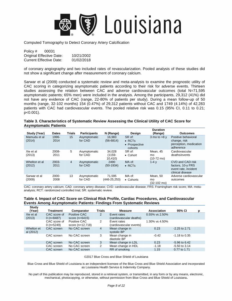
of coronary angiography and two included rates of revascularization. Pooled analysis of these studies did not show a significant change after measurement of coronary calcium. Sarwar et al (2009) conducted a systematic review and meta-analysis to examine the prognostic utility of CAC scoring in categorizing asymptomatic patients according to their risk for adverse events. Thirteen studies assessing the relation between CAC and adverse cardiovascular outcomes (total N=71,595 asymptomatic patients; 65% men) were included in the analysis. Among the participants, 29,312 (41%) did not have any evidence of CAC (range, 22-80% of patients per study). During a mean follow-up of 50 months (range, 32-102 months) 154 (0.47%) of 29,312 patients without CAC and 1749 (4.14%) of 42,283 patients with CAC had cardiovascular events. The pooled relative risk was 0.15 (95% CI, 0.11 to 0.21; p<0.001). Table 3. Characteristics of Systematic Review Assessing the Clinical Utility of CAC Score for Asymptomatic Patients
Study (Year) Dates Trials Participants
N (Range) Design Duration (Range) Outcomes
Mamudu et al (2014)
1996-2014
15 Asymptomatic for CAD
16,983 (56-6814)
SR of:
RCTs
Prospective cohorts
3 mo to >8 y Positive behavioral change, risk perception, medication adherence
Xie et al (2013)
2008-2011
5 Asymptomatic for CAD
34,028 (1159-10,410)
SR of:
Cohort
Mean, 45 mo (10-72 mo)
Cardiovascular deaths/events
Whelton et al (2012)
2003-2011
4 Asymptomatic for CAD
2490 (50-1934)
MA of:
RCTs
1-4 y CVD and CAD risk factors, 10-y FRS event rate, incident clinical disease
Sarwar et al (2009)
2000-2008
13 Asymptomatic for CAD
71,595 (446-25,253)
MA of:
Cohorts
Mean, 50 mo (32-102 mo)
Adverse cardiovascular outcomes
CAC: coronary artery calcium; CAD: coronary artery disease; CVD: cardiovascular disease; FRS: Framingham risk score; MA: meta-analysis; RCT: randomized controlled trial; SR: systematic review.
Table 4. Impact of CAC Score on Clinical Risk Profile, Cardiac Procedures, and Cardiovascular Events Among Asymptomatic Patients: Findings From Systematic Reviews
Study (Year) Treatment Comparator Trials Measure Association 95% CI p
Xie et al (2013)
CAC score of 0 (n=8487)
Positive CAC score (n=6415)
2 Event rates (cardiovascular deaths)
0.55% vs 2.50%
CAC score of 0 (n=5249)
Positive CAC score (n=12,718)
2 Event rates (cardiovascular events)
1.30% vs 4.50%
Whelton et al (2012)
CAC screen No CAC screen 4 Mean change in systolic BP
0.23 -2.25 to 2.71
CAC screen No CAC screen 3 Mean change in diastolic BP
-0.42 -1.18 to 0.35
CAC screen No CAC screen 3 Mean change in LDL 0.23 -5.96 to 6.42 CAC screen No CAC screen 2 Mean change in HDL -1.18 -5.50 to 3.14 CAC screen No CAC screen RR of smoking 1.15 0.77 to 1.71

Computed Tomography to Detect Coronary Artery Calcification Policy # 00031 Original Effective Date: 10/21/2002 Current Effective Date: 01/02/2018
©2017 Blue Cross and Blue Shield of Louisiana
Blue Cross and Blue Shield of Louisiana is an independent licensee of the Blue Cross and Blue Shield Association and incorporated
as Louisiana Health Service & Indemnity Company.
No part of this publication may be reproduced, stored in a retrieval system, or transmitted, in any form or by any means, electronic, mechanical, photocopying, or otherwise, without permission from Blue Cross and Blue Shield of Louisiana.
Page 9 of 22
cessation CAC screen No CAC screen RR of angiography 1.17 0.68 to 1.99 CAC screen No CAC screen RR of revascularization 1.35 0.69 to 2.63 Sarwar et al (2009)
CAC score of 0 (n=29,312)
Positive CAC score (n=42,283)
13 RR of adverse cardiovascular outcome
0.15 0.11 to 0.21 <0.001
BP: blood pressure; CAC: coronary artery calcium; CI: confidence interval; HDL: high-density lipoprotein; LDL: low-density lipoprotein; RR: relative risk.
Randomized Controlled Trials RCTs by O’Malley et al (2003) and Rozanski et al (2011), included in the 2012 Whelton systematic review captured the effect of incorporating CAC scoring in clinical practice on CAD risk factors and overall CAD risk. O’Malley et al (2003) conducted an RCT among a consecutive sample of 450 asymptomatic active-duty U.S. Army personnel ages 39 to 45 years to assess the effects of incorporating EBCT as a motivational factor into a cardiovascular screening program. The program offered intensive case management or usual
care and assessed treatment impact on 10-year FRS over 1 year. The authors used a 22 factorial design and patients were randomized to 1 of the 4 intervention arms: EBCT results provided in the setting of intensive case management (n=111) or usual care (n=119) or EBCT results withheld in the setting of intensive case management (n=124) or usual care (n=96). Mean absolute risk change in 10-year FRS between groups receiving and not receiving results was +0.30 and +0.36 (p=0.81), respectively. The trial was not powered for clinical end points. EBCT did not produce any benefits regarding a difference in FRS at 1 year. Rozanski et al (2011) conducted an RCT to evaluate the impact of CT scanning for CAC on cardiac risk factors. A total of 2137 healthy volunteers were randomized in a 2:1 ratio to CT scanning (n=1424) or no CT scanning (n=713) and followed for 4 years. At baseline, both groups received 1 session of risk factor counseling by a nurse practitioner. The primary end point was 4-year change in CAD risk factors and FRS. At the 4-year follow-up, there was differential dropout among the groups, with 88.2% (1256/1424) of follow-up in the scan group vs 81.9% (584/713) in the no-scan group. Compared with the no-scan group, the scan group showed a net favorable change in systolic blood pressure (p=0.02), low-density lipoprotein cholesterol (p=0.04), and waist circumference for those with increased abdominal girth (p=0.01), and a tendency to weight loss among overweight subjects (p=0.07). While there was a mean rise in FRS in the no-scan group (0.7, SD=5.1), FRS remained static in the scan group (0.002, SD=4.9; p=0.003). Downstream medical testing and costs in the scan group were comparable with those of the no-scan group, balanced by lower and higher resource utilization for subjects with normal CAC scans and CAC scores of 400 or higher, respectively. This trial highlights the potential benefit of CAC screening in modifying cardiac risk profile but is not definitive in demonstrating improved outcomes. Trial limitations included differing intensities of interventions between groups and differential dropout. It is possible that the small differences reported in the trial result from bias related to these methodologic limitations. Also, this trial did not compare the impact of other types of risk factor intervention, most notably more intensive risk factor counseling. Finally, the generalizability of

Computed Tomography to Detect Coronary Artery Calcification Policy # 00031 Original Effective Date: 10/21/2002 Current Effective Date: 01/02/2018
©2017 Blue Cross and Blue Shield of Louisiana
Blue Cross and Blue Shield of Louisiana is an independent licensee of the Blue Cross and Blue Shield Association and incorporated
as Louisiana Health Service & Indemnity Company.
No part of this publication may be reproduced, stored in a retrieval system, or transmitted, in any form or by any means, electronic, mechanical, photocopying, or otherwise, without permission from Blue Cross and Blue Shield of Louisiana.
Page 10 of 22
the findings is uncertain, because this was a volunteer population that might have been highly motivated for change. Observational Studies Gepner et al (2017) prospectively evaluated CVD, CHD, and stroke/TIA events using data from MESA to compare the abilities of CAC and carotid plaque scores to predict CVD events. After 11.3 years of follow-up among 4955 participants (mean age, 61.6 years), 709 CVD, 498 CHD, and 262 stroke/TIA events occurred. CAC scoring compared with carotid plaque scoring was a stronger predictor of CVD events (HR=1.78 [95% CI, 1.16 to 1.98; p<0.001] vs HR=1.27 [95% CI, 1.16 to 1.40; p<0.001]) and CHD events (HR=2.09 [95% CI, 1.84 to 2.38; p<0.001] vs HR, 1.35 [95% CI, 1.21 to 1.51; p<0.001]), respectively. Nakanishi et al (2016) conducted a study among 13,092 consecutive asymptomatic individuals without known CAD (mean age, 58 years) to examine the predictive ability of CAC scoring on 5- and 15-year mortality rates among men and women; the study included individuals clinically referred for a CAC scan between 1997 and 2011 at university medical center. During a median follow-up of 11.0 years, there were 522 (4.0%) deaths. Compared with a CAC score of 0, increasing CAC was associated with higher mortality rate for CAC scores ranging from: 1 to 99 (HR=1.5; 95% CI, 1.1 to 2.1); 100 to 399 (HR=1.8, 95% CI, 1.3 to 2.5); and 400 or higher (HR=2.6, 95% CI, 1.9 to 3.6). Kelkar et al (2016) conducted a prospective study to determine the long-term prognosis of asymptomatic women and men classified as low-intermediate risk undergoing screening with CAC scoring. A total of 2363 participants with a low-intermediate FRS (10-year predicted risk, 6%-9.9%) underwent CAC screening during 1996 to 1999 and were followed for a median of 14.6 years. Women (n=1072) were older than men (n=1291) participating in the study (mean, 55.6 years vs 46.7 years; p<0.001). For women, 15-year mortality rates ranged from 5.0% for a CAC score of 0 to 23.5% for a CAC score of 400 or higher (p<0.001). For men, 15-year mortality ranged from 3.5% for CAC score of 0 to 18.0% for a CAC score of 400 or higher (p<0.001). Adjusting for risk factors, relative hazards for death for women with CAC scores of 1 to 10, 11 to 99, 100 to 399, and 400 or higher during the 15-year follow-up were 1.92 (95% CI, 0.82 to 4.47), 2.37 (95% CI, 1.29 to 4.35), 2.99 (95% CI, 1.60 to 5.60), and 6.53 (95% CI, 3.50 to 12.21), respectively. For men with CAC scores of 1 to 10, 11 to 99, 100 to 399, and 400 or higher, adjusted relative hazards for the same period were 1.73 (95% CI, 0.74 to 4.02), 2.88 (95% CI, 1.59 to 5.23), 4.10 (95% CI, 2.17 to 7.74), and 2.71 (95% CI, 1.10 to 6.69), respectively. Jacobs et al (2012), one of the studies included in the 2013 Xie systematic review, conducted CAC scoring among 7557 lung cancer screening participants without symptoms of CAD and followed them for a median of 10 months (range, 1-21 months) for cardiovascular events. Compared with those who had a CAC score of 0 (n=1814), subjects with CAC scores ranging from 1 to 100 (n=2191), 101 to 1000 (n=2267), and greater than 1000 (n=1285) had an increased risk of cardiovascular event, with adjusted HRs of 1.8 (95% CI, 0.8 to 3.9), 1.9 (95% CI, 0.9 to 4.2), and 5.3 (95% CI, 2.5 to 11.6), respectively. Another study by Jacobs et al (2011) followed a routine clinical population (N=10,410) for 18 months after CAC scoring for cardiovascular events. Compared with subjects who had a visual score 0, subjects with visual score 1 to 2, 3 to 5, and 6 to 12 had a 2.2 (95% CI, 1.6 to 3.0), 2.5 (95% CI, 1.8 to 3.4), and 3.7 (95% CI, 2.7 to 5.2)

Computed Tomography to Detect Coronary Artery Calcification Policy # 00031 Original Effective Date: 10/21/2002 Current Effective Date: 01/02/2018
©2017 Blue Cross and Blue Shield of Louisiana
Blue Cross and Blue Shield of Louisiana is an independent licensee of the Blue Cross and Blue Shield Association and incorporated
as Louisiana Health Service & Indemnity Company.
No part of this publication may be reproduced, stored in a retrieval system, or transmitted, in any form or by any means, electronic, mechanical, photocopying, or otherwise, without permission from Blue Cross and Blue Shield of Louisiana.
Page 11 of 22
times higher adjusted hazard of cardiovascular events during the follow-up period, respectively. Overall, 47 (0.55%) cardiovascular deaths were reported in 8487 subjects with a CAC score of 0 whereas 72 cardiovascular events (1.3%) occurred in 5249 subjects with a CAC score of 0. One hundred sixty cardiovascular deaths (2.5%) were found in 6415 subjects with a positive calcium score, whereas 570 cardiovascular events (4.5%) occurred in 12,718 subjects with a positive calcium score. Budoff et al (2013) evaluated the association between coronary calcium scores and CHD events during 5-year follow-up of 2232 adults from MESA (discussed above), and 3119 subjects from the Heinz Nixdorf RECALL (Risk factors, Evaluation of Coronary Calcium and Lifestyle Factors) study. Increasing Agatston scores were associated with increased risk of CHD. In MESA, compared with a CAC score of 0, having a score greater than 400 was associated with a hazard for CHD of 3.31 (95% CI, 1.12 to 9.8) after adjusting for CHD risk factors; a score ranging from 100 to 399 was associated with a hazard of 3.27 (95% CI, 1.19 to 8.95). In the RECALL study, compared with a CAC score of 0, having a score greater than 400 was associated with a hazard for CHD of 2.96 (95% CI, 1.22 to 7.19). Lower CAC scores were not significantly associated with CHD after adjusting for other risk factors. Additional analysis of MESA data found that CAC scores are associated with CHD events among individuals at either high or low CHD risk based on traditional risk factors. Gibson et al (2014) also used MESA data to evaluate the relation between CAC and incidence of cerebrovascular events, including all strokes and TIAs. Over an average of 9.5 years of follow-up, 234 (3.5%) cerebrovascular events occurred. Having an elevated CAC score was independently predictive of both cerebrovascular events (HR=1.70; 95% CI, 1.24 to 2.35; p=0.001) and stroke (HR=1.59; 95% CI, 1.11 to 2.07; p=0.01). Chang et al (2015) prospectively evaluated whether CAC scoring added incremental predictive value to exercise treadmill testing and stress myocardial perfusion single-photon emission CT testing when used to assess risk of cardiac events (a composite of cardiac death, nonfatal myocardial infarction, and the need for coronary revascularization) in a cohort of 988 asymptomatic and symptomatic low-risk patients without known CHD. Over a median follow-up of 6.9 years, the cardiac event rate was 11.2% (1.6% per year). Annual event rates were higher in patients with CAC scores above 400 (3.7% per year) compared with those with CAC scores of 10 or less (0.6% per year; p<0.001). The addition of CAC score to risk stratification based on the FRS improved risk prediction. Johnson et al (2015) assessed the association between CAC score and subsequent health behavior change. The study included a convenience sample of 174 adults with CHD risk factors who underwent CAC scoring. The authors found no significant between-group change in risk perception measured by Perception of Risk of Heart Disease Scale scores (CAC score range, 0, 1-10, 11-100, 101-400, >400), with the exception of a small increase in the moderate-risk group (CAC score, 101-400) from 55.5 to 58.7 (p=0.004). All groups demonstrated increases in health-promoting behavior over time. Section Summary: Clinical Utility Multiple prospective studies have found that CAC scoring is associated with future risk of CHD events. CAC scores likely add to the predictive ability of clinical risk prediction models. However, relevant studies

Computed Tomography to Detect Coronary Artery Calcification Policy # 00031 Original Effective Date: 10/21/2002 Current Effective Date: 01/02/2018
©2017 Blue Cross and Blue Shield of Louisiana
Blue Cross and Blue Shield of Louisiana is an independent licensee of the Blue Cross and Blue Shield Association and incorporated
as Louisiana Health Service & Indemnity Company.
No part of this publication may be reproduced, stored in a retrieval system, or transmitted, in any form or by any means, electronic, mechanical, photocopying, or otherwise, without permission from Blue Cross and Blue Shield of Louisiana.
Page 12 of 22
enrolled different populations, assessed different traditional risk factors, and assessed different coronary disease outcomes. Different calcium score cutoffs were analyzed in these studies. Given the variation across studies, the magnitude of increased risk conferred by a given calcium score is still uncertain. Studies that evaluated use of CAC scoring in asymptomatic patients have reported mixed findings on whether the score led to improved cardiovascular risk profiles or improvements in other meaningful clinical outcomes. The meta-analysis of RCTs did not find significant improvements in cardiac risk profiles, smoking cessation, or incidence of subsequent cardiac procedures with the use of CAC scoring. CAC SCORING IN SYMPTOMATIC PATIENTS In certain clinical situations, such as patients presenting with chest pain, it is uncertain whether the symptoms are due to CAD. Coronary calcium measurement has been proposed as a method to rule out CAD in certain patients if their CAC score is 0. The presence of any coronary calcium can be a sensitive but not specific test for coronary disease because CAD rarely occurs in the absence of coronary calcium, False positives occur because the calcium may not be associated with an ischemic lesion. The absence of any coronary calcium can be a specific test for the absence of coronary disease and direct the diagnostic workup toward other causes of the patient’s symptoms. In this context, coronary calcium measurement is not used to make a positive diagnosis but as a diagnostic “filter” to rule out an atherosclerotic cause for the patient’s symptoms. Clinical Context and Test Purpose The use of CAC scoring with CT in symptomatic patients can rule out the atherosclerotic etiology of CAD. The question addressed in this evidence review is: In individuals with symptoms suggestive of CAD does CAC scoring rule out urgent or emergent CAD and improve net health outcomes? The following PICOTS were used to select literature to inform this review. Patients The population of interest includes individuals who have signs and/or symptoms suggestive of CAD.
Interventions The intervention of interest is CAC scoring using fast CT imaging, including EBCT and spiral CT. Comparators The comparator of interest is standard diagnostic testing (functional testing, exercise electrocardiograph [ECG]). Outcomes The outcomes of interest include overall survival, test accuracy, test validity, morbid events (e.g., MACEs, need for ICA and revascularization).

Computed Tomography to Detect Coronary Artery Calcification Policy # 00031 Original Effective Date: 10/21/2002 Current Effective Date: 01/02/2018
©2017 Blue Cross and Blue Shield of Louisiana
Blue Cross and Blue Shield of Louisiana is an independent licensee of the Blue Cross and Blue Shield Association and incorporated
as Louisiana Health Service & Indemnity Company.
No part of this publication may be reproduced, stored in a retrieval system, or transmitted, in any form or by any means, electronic, mechanical, photocopying, or otherwise, without permission from Blue Cross and Blue Shield of Louisiana.
Page 13 of 22
Timing The timing of use of CT CAC scoring is when individuals require evaluation for persistent stable angina or experience onset of acute chest pain. Setting The setting is a cardiology practice or emergent care setting for patients undergoing evaluation of chest pain. Technical Reliability The technical reliability of CAC scoring using fast CT imaging, including EBCT and spiral CT, was described in the previous section (Coronary Artery Calcium Scoring in Asymptomatic Individuals) and the 1998 TEC Assessment. Clinical Validity
Systematic Reviews Chaikriangkrai et al (2016) conducted a systematic review and meta-analysis to examine the prognostic value and accuracy of a CAC score of 0 for identifying patients presenting with acute chest pain at acceptable low risk for future cardiovascular events. The systematic review included only prospective cohort studies that used MDCT or EBCT to calculate CAC scores using the Agatston method and reported MACEs at 1 month and beyond the index emergency department visit. Eight studies evaluating 3556 patients with a median follow-up of 10.5 months were selected. Reviewers conducted a subgroup analysis of 6 studies at predominantly white patients (n=2432 patients) to estimate the prognostic accuracy indices of CAC scores (0, >0) for cardiovascular events (MACEs, all-cause deaths, nonfatal myocardial infarction). Pooled sensitivity, specificity, as well as positive and negative likelihood ratios were 96% (I
2=0%), 60% (I
2=15.1%),
2.36 (I2=0%), and 0.07 (I
2=0%), respectively (see Table 5).
Sarwar et al (2009) conducted a systematic review and meta-analysis to examine the clinical, diagnostic, and prognostic significance of a CAC score of 0. Eighteen studies from 1992 to 2007, in which 10,355 symptomatic patients with suspected CAD underwent CAC testing as well as ICA, were selected in the analysis to examine the diagnostic accuracy of CAC scoring for stenosis on ICA. A total of 5805 (56%) patients had significant coronary stenosis (defined as >50%) on ICA. Pooled data revealed that the presence of calcium had a sensitivity, a specificity, as well as a positive and a negative likelihood ratio of 98%, 40%, 1.63, and 0.06, respectively, for predicting coronary artery stenosis. The summary negative predictive value was 92% (95% CI, 88% to 95%; p<0.001). The summary positive predictive value was 68% (95% CI, 64% to 72%; p<0.001) (see Table 5). Table 5. Pooled Diagnostic Performance of CAC Score for CAD Among Symptomatic Individuals
Test Studies N Sensitivity (95% CI), %
Specificity (95% CI), % LR+ (95% CI) LR- (95% CI)
Chaikriangkrai et al (2016)
CAC score (0, >0) 6 2432 96 (93 to 98) 60 (58 to 62) 2.36 (2.22 to 2.51) 0.07 (0.04 to 0.14)

Computed Tomography to Detect Coronary Artery Calcification Policy # 00031 Original Effective Date: 10/21/2002 Current Effective Date: 01/02/2018
©2017 Blue Cross and Blue Shield of Louisiana
Blue Cross and Blue Shield of Louisiana is an independent licensee of the Blue Cross and Blue Shield Association and incorporated
as Louisiana Health Service & Indemnity Company.
No part of this publication may be reproduced, stored in a retrieval system, or transmitted, in any form or by any means, electronic, mechanical, photocopying, or otherwise, without permission from Blue Cross and Blue Shield of Louisiana.
Page 14 of 22
Sarwar et al (2009)
CAC score (0, >0) 18 10,355 98 (97 to 98) 40 (38 to 41) 1.63 (1.59-1.67) 0.06 (0.05-0.07)
CI: confidence interval; LR: likelihood ratio; CAC: coronary artery calcium; CAD: coronary artery disease.
Randomized Controlled Trials Lubbers et al (2016) conducted a multicenter RCT to compare the effectiveness and safety of a cardiac CT algorithm with functional testing in patients with symptoms (stable chest pain or angina equivalent symptoms) suggestive of CAD. A total of 350 patients with stable angina were prospectively randomized 2:1 to cardiac CT and functional testing, such as exercise ECG, myocardial perfusion imaging, or stress echocardiography. Patients in the cardiac CT arm (n=242) initially underwent calcium scanning followed by computed tomography angiography (CCTA) if the Agatston calcium score was between 1 and 400. CAD was ruled out if the patients had a CAC score of 0. The original primary end point of the trial was the proportion of patients undergoing catheter angiography followed by revascularization, but because of insufficient funding, authors could not assess that end point and chose clinical effectiveness as the alternative primary outcome, defined as the absence of chest pain complaints after 1 year. After 1 year, fewer patients randomized to CT reported angina symptoms that those in the functional testing group (39% vs 25%, p=0.012), although the proportion of patients with similar or worsened symptoms was comparable (26% vs 29%, p=0.595). The tiered protocol study design is a strength of this study, but the unplanned change in end points limits analysis and conclusions. Observational Studies In 2015, Pursnani et al published results from a subgroup analysis of the ROMICAT II trial. It evaluated the incremental diagnostic value of CAC scoring plus CTA in low- to intermediate-risk patients presenting to the emergency department with symptoms (chest pain or angina equivalent of ≥5 minutes duration within 24 hours) suggesting acute coronary syndrome (ACS). The ROMICAT II trial randomized patients with possible ACS to CTA as part of an initial evaluation or to the standard emergency department evaluation strategy, as directed by local caregivers. As part of the trial protocol, all patients undergoing CTA had a CAC scan; the present analysis included 473 patients who underwent both CTA and CAC scanning. Among these patients, the ACS rate (defined as unstable angina and myocardial infarction during the index hospitalization) was 8% (n=38). Patients with lower CAC scores were less likely to have a discharge diagnosis of ACS. Among 253 patients with a CAC score of 0, 2 (0.8%) patients were diagnosed with ACS (95% CI, 0.1% to 2.8%). Receiver operating characteristic curve analysis was used to predict the risk of ACS by CAC score greater than 0, continuous CAC score, CTA results, and combined CAC and CTA score. The optimal cut point of CAC for ACS detection was 22 (C statistic, 0.81), with 318 (67%) patients having a CAC score less than 22. All CTA strategies had high sensitivity for ACS detection, without significant differences in stenosis thresholds. CAC was inferior to CTA for predicting ACS (C range, 0.86 vs 0.92; p=0.03). The addition of CAC score to CTA (i.e., using selective CTA only for patients with CAC score >22 or >0) did not significantly improve the detection of ACS (CAC+CTA C=0.93 vs CTA C=0.92; p=0.88). Overall, this trial suggested that CAC scoring does not provide incremental value beyond CTA in predicting the likelihood of ACS in a low- to intermediate-risk population presenting to the emergency department.

Computed Tomography to Detect Coronary Artery Calcification Policy # 00031 Original Effective Date: 10/21/2002 Current Effective Date: 01/02/2018
©2017 Blue Cross and Blue Shield of Louisiana
Blue Cross and Blue Shield of Louisiana is an independent licensee of the Blue Cross and Blue Shield Association and incorporated
as Louisiana Health Service & Indemnity Company.
No part of this publication may be reproduced, stored in a retrieval system, or transmitted, in any form or by any means, electronic, mechanical, photocopying, or otherwise, without permission from Blue Cross and Blue Shield of Louisiana.
Page 15 of 22
In 2014, Hulten et al published results from a retrospective cohort study among symptomatic patients without a history of CAD to evaluate the accuracy of CAC scoring for excluding coronary stenosis, using CTA as the criterion standard. The study included 1145 patients who had symptoms possibly consistent with CAD who underwent noncontrast CAC scoring and contrast-enhanced CTA from 2004 to 2011. For detection of greater than 50% stenosis, CAC had a sensitivity, specificity, and negative predictive value of 98%, 55%, and 99%, respectively. For prediction of cardiovascular death or myocardial infarction, the addition of either or both CAC and CTA to a clinical prediction score did not significantly improve prognostic value. Chaikriangkrai et al (2015) retrospectively evaluated whether CAC added incremental value to CTA for predicting coronary artery stenosis in 805 symptomatic patients without known CHD. CAC score was significantly associated with the presence of coronary artery stenosis on CTA. Both CAC score and the presence of CTA stenosis were significantly associated with MACE rates, including cardiac death, nonfatal myocardial infarction, and late coronary revascularization. Patients with more than 50% stenosis on CTA had higher MACE rates, compared with those who had a normal CTA (4.5% vs 0.1%, p<0.001) and with those who had less than 50% stenosis (4.5% vs 1.4%, p=0.002). Those with a CAC score of more than 400 had higher MACE rates than those with scores between 1 and 100 (4.2% vs 1.4%, p=0.014) and those with scores of 0 (4.2% vs 0% p<0.001). The addition of CAC score to a risk prediction model for MACE, which included clinical risk factors and CTA stenosis, significantly improved the model’s predictive performance
(global 2 score, 108 vs 70, p=0.019).
Dharampal et al (2013) retrospectively evaluated a cohort of 1975 symptomatic patients (those with chest pain referred by their cardiologist for CTA) who underwent clinical evaluation and CAC scoring and CTA or ICA. The primary outcome was obstructive CAD (≥50% stenosis) on ICA or CTA (if ICA was not done). The authors evaluated the NRI with the addition of CAC score to a clinical prediction model for patients who had an intermediate probability of CHD (10%-90%) after clinical evaluation based on chest pain characteristic, age, sex, risk factors, and electrocardiogram. Discrimination of CAD was significantly improved by incorporating the CAC score into the clinical evaluation (AUC, 0.80 vs 0.89, p<0.001). Yoon et al (2012) conducted a prospective study among 136 Korean men (58% men; age, 56 years) who presented to the emergency department with acute chest pain and nondiagnostic ECG to examine the diagnostic usefulness of the “zero calcium score criteria” as a decision-making strategy to rule out significant CAD as the etiology of acute chest pain. All patients underwent 64-slice CT for calcium scoring and CTCA. Ninety-two (68%) of 136 patients did not show detectable CAC, and 14 (15%) of these 92 without CAC had 50% or more stenosis on CTA. Sensitivity, specificity, positive predictive value, and negative predictive value of a CAC score of 0 for the detection of 50% or more stenosis were 66% (95% CI, 50% to 80%), 83% (95% CI, 74% to 90%), 64% (95% CI, 48% to 77%), and 85% (95% CI, 75% to 91%), respectively. A calcium score of 0 did not necessarily guarantee the absence of significant CAD in an Asian population presenting to the emergency department with chest pain. Gottlieb et al (2010) conducted a prospective multicenter study to evaluate whether the absence of coronary calcium could be used to rule out 50% or more coronary stenosis or the need for

Computed Tomography to Detect Coronary Artery Calcification Policy # 00031 Original Effective Date: 10/21/2002 Current Effective Date: 01/02/2018
©2017 Blue Cross and Blue Shield of Louisiana
Blue Cross and Blue Shield of Louisiana is an independent licensee of the Blue Cross and Blue Shield Association and incorporated
as Louisiana Health Service & Indemnity Company.
No part of this publication may be reproduced, stored in a retrieval system, or transmitted, in any form or by any means, electronic, mechanical, photocopying, or otherwise, without permission from Blue Cross and Blue Shield of Louisiana.
Page 16 of 22
revascularization. The authors compared the diagnostic performance of 64-detector CT with that of ICA. Among 291 patients with suspected CAD included in the study, 214 (73%) were male, and the mean age was 59.3 years. Fifty-six percent of the patients had 50% or more stenosis. Among 72 patients with a CAC score of 0, 14 (19%) had at least 1 coronary artery with 50% or more stenosis. The overall sensitivity for a CAC score of 0 to predict the absence of 50% or more stenosis was 45%, specificity was 91%, negative predictive value was 68%, and positive predictive value was 81%. Additionally, 9 (12.5%) patients with a CAC score of 0 underwent revascularization within 30 days of calcium scoring. Section Summary: Clinical Validity Systematic reviews and meta-analyses have reported a very low negative likelihood ratio for CAC score in predicting MACEs and significant coronary stenosis, suggesting the potential value of calcium score of 0 in ruling out an atherosclerotic etiology of disease. However, multiple observational studies with angiographic (CTA or ICA) have suggested that a CAC score of 0 may not rule out the presence of significant atherosclerotic CAD among symptomatic patients. Clinical Utility
Systematic Reviews The 2016 systematic review by Chaikriangkrai et al (discussed above) assessed studies of relevance to our analysis of clinical utility. Specifically, in 8 studies (total N=3556 patients), those with a CAC score of 0 had a significantly lower risk of MACEs compared with patients with CAC scores greater than 0 (RR=0.06; 95% CI, 0.04 to 0.11; p<0.001; I
2=0%). The risk difference was 0.19 (95% CI, 0.11 to 0.27).
Subgroup analyses in the 5 studies evaluating death or nonfatal myocardial infarction showed that the patients with a CAC score of 0 had a significantly lower risk of death or nonfatal myocardial infarction compared with patients with CAC scores greater than 0 (RR=0.19; 95% CI, 0.08 to 0.47; I
2=0%). The risk
difference was 0.03 (95% CI, 0 to 0.05). The pooled event rate for death or nonfatal myocardial infarction with a CAC score of 0 (0.5%/year [0.04 death/myocardial infarction per 100 patient-months, or 6 deaths/myocardial infarction in 13,656 patient-months]) was significantly lower than with a CAC scores greater than 0 (3.5%/year [0.29 death/myocardial infarction per 100 patient-months, or 33 deaths/myocardial infarction in 11,350 patient-months]). In the 2009 systematic review by Sarwar et al (also discussed above), 7 studies assessing the prognostic value of CAC in the symptomatic population (n=3924) were selected. Overall, 921 (23%) patients did not have any evidence of CAC. During a mean follow-up of 42 months (range, 30-84 months) 17 (1.8%) of 921 patients without CAC had a cardiovascular event compared with 270 (8.99%) of 3003 patients with CAC. The cumulative relative risk was 0.09 (95% CI, 0.04 to 0.20; p<0.001). Observational Studies Yerramasu et al (2014) prospectively assessed an evaluation algorithm including CAC scoring for patients presenting to a rapid access chest pain clinic with stable chest pain possibly consistent with CHD. Three hundred patients presenting with acute chest pain to 1 of 3 chest pain clinics underwent CAC scoring. If the

Computed Tomography to Detect Coronary Artery Calcification Policy # 00031 Original Effective Date: 10/21/2002 Current Effective Date: 01/02/2018
©2017 Blue Cross and Blue Shield of Louisiana
Blue Cross and Blue Shield of Louisiana is an independent licensee of the Blue Cross and Blue Shield Association and incorporated
as Louisiana Health Service & Indemnity Company.
No part of this publication may be reproduced, stored in a retrieval system, or transmitted, in any form or by any means, electronic, mechanical, photocopying, or otherwise, without permission from Blue Cross and Blue Shield of Louisiana.
Page 17 of 22
CAC score was 1000 or more Agatston units, ICA was performed; if the CAC score was less than 1000, CTCA was performed. All patients with a CAC score of 0 and low pretest likelihood of CHD had no obstructive CHD on CTCA and were event-free during follow-up. Of the 18 patients with CAC scores from 400 to 1000, 17 (94%) had greater than 50% obstruction on subsequent CTCA and were referred for further evaluation, 14 (78%) of whom had obstructive CHD. Of 15 patients with CAC scores 1000 or more and who were referred for coronary angiography, obstructive CHD was present in 13 (87%). This study suggested that CAC scoring can be used in the acute chest pain setting to stratify decision making for further testing. Ten Kate et al (2013) prospectively evaluated the accuracy of cardiac CT, including CAC scoring with or without CTCA, in distinguishing heart failure due to CAD from heart failure due to non-CAD causes. Data on the predictive ability of a negative CAC score in ruling out CAD was also included. The study included 93 symptomatic patients with newly diagnosed heart failure of unknown etiology, all of whom underwent CAC scoring. Those with a CAC score greater than 0 underwent CTCA and, if the CTCA was positive for CAD (>20% luminal diameter narrowing), ICA was recommended. Forty-six percent of patients had a CAC score of 0. At a mean follow-up of 20 months, no patient with a CAC score of 0 had a myocardial infarction, underwent percutaneous coronary intervention, had a coronary artery bypass graft, or had signs of CAD. Section Summary: Clinical Utility Currently, evidence from nonrandomized observational studies suggests very low short or long term risk of cardiovascular events or death in patients having calcium scores of 0 compared with those having positive (more than 0) calcium scores. However, considering the inconsistency in evidence regarding the diagnostic accuracy of calcium scoring and lack of evidence from RCTs, further research is needed to examine the clinical utility of ruling out atherosclerotic CAD based on CAC score of 0. SUMMARY OF EVIDENCE For individuals who are asymptomatic with risk of CAD who receive CAC scoring, the evidence includes multiple systematic reviews, RCTs, and nonrandomized observational studies. Relevant outcomes are overall survival, test accuracy and validity, morbid events, and resource utilization. There is extensive evidence on the predictive value of CAC score screening for CVD among asymptomatic patients, and this evidence has demonstrated that scanning has incremental predictive accuracy above traditional risk factor measurement. However, high-quality evidence demonstrating that the use of CAC scores in clinical practice leads to changes in patient management or in individual risk behaviors that improve cardiac outcomes is lacking. A meta-analysis of RCTs reported no significant change in coronary risk profile, downstream testing, or revascularization following screening using CAC scoring compared with no CAC scoring. The evidence is insufficient to determine the effects of the technology on health outcomes For individuals with signs and/or symptoms suggestive of CAD who receive CAC scoring before other diagnostic testing, the evidence includes prospective and retrospective nonrandomized studies. Relevant outcomes are overall survival, test accuracy and validity, morbid events, and resource utilization. CAC scoring has potential as a diagnostic test to rule out CAD in patients presenting with symptoms or as a “gatekeeper” test before invasive imaging is performed. Evidence from observational studies has suggested that negative results on CAC scoring rule out CAD with good reliability. However, the evidence has been

Computed Tomography to Detect Coronary Artery Calcification Policy # 00031 Original Effective Date: 10/21/2002 Current Effective Date: 01/02/2018
©2017 Blue Cross and Blue Shield of Louisiana
Blue Cross and Blue Shield of Louisiana is an independent licensee of the Blue Cross and Blue Shield Association and incorporated
as Louisiana Health Service & Indemnity Company.
No part of this publication may be reproduced, stored in a retrieval system, or transmitted, in any form or by any means, electronic, mechanical, photocopying, or otherwise, without permission from Blue Cross and Blue Shield of Louisiana.
Page 18 of 22
inconsistent, with some studies reporting lack of value when using a zero calcium score to rule out CAD. Further prospective trials would be needed to demonstrate that such a strategy is effective in practice and is at least as effective as alternative strategies for ruling out CAD. To demonstrate that use of calcium scores improves the efficiency or accuracy of the diagnostic workup of symptomatic patients, rigorous studies defining exactly how CAC scores would be used in combination with other tests to triage patients would be necessary. The evidence is insufficient to determine the effects of the technology on health outcomes.
References 1. Blue Cross and Blue Shield Association, Medical Policy Reference Manual, “Computed Tomography to Detect Coronary Artery
Calcification”, 6.01.03, 9:2017. 2. Blue Cross and Blue Shield Association Technology Evaluation Center (TEC). Diagnosis and screening for coronary artery
disease with electron beam computed tomography. TEC Assessments. 1998;Volume 13:Tab 27. 3. Xie X, Zhao Y, de Bock GH, et al. Validation and prognosis of coronary artery calcium scoring in nontriggered thoracic computed
tomography: systematic review and meta-analysis. Circulation Cardiovascular imaging. 2013;6(4):514-521. 4. Choi AD, Leifer ES, Yu J, et al. Prospective evaluation of the influence of iterative reconstruction on the reproducibility of coronary
calcium quantification in reduced radiation dose 320 detector row CT. Journal of cardiovascular computed tomography. 2016;10(5):359-363.
5. Williams MC, Golay SK, Hunter A, et al. Observer variability in the assessment of CT coronary angiography and coronary artery calcium score: substudy of the Scottish COmputed Tomography of the HEART (SCOT-HEART) trial. Open Heart. 2015;2(1):e000234.
6. Sabour S, Rutten A, van der Schouw YT, et al. Inter-scan reproducibility of coronary calcium measurement using Multi Detector-Row Computed Tomography (MDCT). Eur J Epidemiol. 2007;22(4):235-243.
7. Nakanishi R, Li D, Blaha MJ, et al. All-cause mortality by age and gender based on coronary artery calcium scores. European heart journal cardiovascular Imaging. 2016;17(11):1305-1314.
8. Gepner AD, Young R, Delaney JA, et al. Comparison of carotid plaque score and coronary artery calcium score for predicting cardiovascular disease events: the multi-ethnic study of atherosclerosis. J Am Heart Assoc. 2017;6(2).
9. Blaha MJ, Cainzos-Achirica M, Greenland P, et al. Role of coronary artery calcium score of zero and other negative risk markers for cardiovascular disease: the Multi-Ethnic Study of Atherosclerosis (MESA). Circulation. 2016;133(9):849-858.
10. Polonsky TS, McClelland RL, Jorgensen NW, et al. Coronary artery calcium score and risk classification for coronary heart disease prediction. Jama. 2010;303(16):1610-1616.
11. Elias-Smale SE, Wieberdink RG, Odink AE, et al. Burden of atherosclerosis improves the prediction of coronary heart disease but not cerebrovascular events: the Rotterdam Study. Eur Heart J. 2011;32(16):2050-2058.
12. Won KB, Chang HJ, Niinuma H, et al. Evaluation of the predictive value of coronary artery calcium score for obstructive coronary artery disease in asymptomatic Korean patients with type 2 diabetes mellitus. Coron Artery Dis. 2015;26(2):150-156.
13. Mamudu HM, Paul TK, Veeranki SP, Budoff M. The effects of coronary artery calcium screening on behavioral modification, risk perception, and medication adherence among asymptomatic adults: a systematic review. Atherosclerosis. 2014;236(2):338-350.
14. Whelton SP, Nasir K, Blaha MJ, et al. Coronary artery calcium and primary prevention risk assessment: what is the evidence? An updated meta-analysis on patient and physician behavior. Circulation Cardiovascular quality and outcomes. 2012;5(4):601-607.
15. Sarwar A, Shaw LJ, Shapiro MD, et al. Diagnostic and prognostic value of absence of coronary artery calcification. JACC Cardiovascular imaging. 2009;2(6):675-688.
16. O'Malley PG, Feuerstein IM, Taylor AJ. Impact of electron beam tomography, with or without case management, on motivation, behavioral change, and cardiovascular risk profile: a randomized controlled trial. JAMA. 2003;289(17):2215-2223.
17. Rozanski A, Gransar H, Shaw LJ, et al. Impact of coronary artery calcium scanning on coronary risk factors and downstream testing the EISNER (Early Identification of Subclinical Atherosclerosis by Noninvasive Imaging Research) prospective randomized trial. J Am Coll Cardiol. 2011;57(15):1622-1632.
18. Kelkar AA, Schultz WM, Khosa F, et al. Long-term prognosis after coronary artery calcium scoring among low-intermediate risk women and men. Circulation Cardiovascular imaging. 2016;9(4):e003742.
19. Jacobs PC, Gondrie MJ, van der Graaf Y, et al. Coronary artery calcium can predict all-cause mortality and cardiovascular events on low-dose CT screening for lung cancer. AJR American journal of roentgenology. 2012;198(3):505-511.

Computed Tomography to Detect Coronary Artery Calcification Policy # 00031 Original Effective Date: 10/21/2002 Current Effective Date: 01/02/2018
©2017 Blue Cross and Blue Shield of Louisiana
Blue Cross and Blue Shield of Louisiana is an independent licensee of the Blue Cross and Blue Shield Association and incorporated
as Louisiana Health Service & Indemnity Company.
No part of this publication may be reproduced, stored in a retrieval system, or transmitted, in any form or by any means, electronic, mechanical, photocopying, or otherwise, without permission from Blue Cross and Blue Shield of Louisiana.
Page 19 of 22
20. Jacobs PC, Gondrie MJ, Mali WP, et al. Unrequested information from routine diagnostic chest CT predicts future cardiovascular events. European radiology. 2011;21(8):1577-1585.
21. Budoff MJ, Mohlenkamp S, McClelland R, et al. A comparison of outcomes with coronary artery calcium scanning in unselected populations: the Multi-Ethnic Study of Atherosclerosis (MESA) and Heinz Nixdorf RECALL study (HNR). Journal of cardiovascular computed tomography. 2013;7(3):182-191.
22. Silverman MG, Blaha MJ, Krumholz HM, et al. Impact of coronary artery calcium on coronary heart disease events in individuals at the extremes of traditional risk factor burden: the Multi-Ethnic Study of Atherosclerosis. Eur Heart J. 2014;35(33):2232-2241.
23. Gibson AO, Blaha MJ, Arnan MK, et al. Coronary artery calcium and incident cerebrovascular events in an asymptomatic cohort. The MESA Study. JACC Cardiovascular imaging. 2014;7(11):1108-1115.
24. Chang SM, Nabi F, Xu J, et al. Value of CACS compared with ETT and myocardial perfusion imaging for predicting long-term cardiac outcome in asymptomatic and symptomatic patients at low risk for coronary disease: clinical implications in a multimodality imaging world. JACC Cardiovascular imaging. 2015;8(2):134-144.
25. Johnson JE, Gulanick M, Penckofer S, Kouba J. Does knowledge of coronary artery calcium affect cardiovascular risk perception, likelihood of taking action, and health-promoting behavior change? The Journal of cardiovascular nursing. 2015;30(1):15-25.
26. Chaikriangkrai K, Palamaner Subash Shantha G, Jhun HY, et al. Prognostic value of coronary artery calcium score in acute chest pain patients without known coronary artery disease: systematic review and meta-analysis. Ann Emerg Med. 2016;68(6):659-670.
27. Chaikriangkrai K, Velankar P, Schutt R, et al. Additive prognostic value of coronary artery calcium score over coronary computed tomographic angiography stenosis assessment in symptomatic patients without known coronary artery disease. The American journal of cardiology. 2015;115(6):738-744.
28. Lubbers M, Dedic A, Coenen A, et al. Calcium imaging and selective computed tomography angiography in comparison to functional testing for suspected coronary artery disease: the multicentre, randomized CRESCENT trial. Eur Heart J. 2016;37(15):1232-1243.
29. Pursnani A, Chou ET, Zakroysky P, et al. Use of coronary artery calcium scanning beyond coronary computed tomographic angiography in the emergency department evaluation for acute chest pain: the ROMICAT II trial. Circulation Cardiovascular imaging. 2015;8(3).
30. Hulten E, Bittencourt MS, Ghoshhajra B, et al. Incremental prognostic value of coronary artery calcium score versus CT angiography among symptomatic patients without known coronary artery disease. Atherosclerosis. 2014;233(1):190-195.
31. Dharampal AS, Rossi A, Dedic A, et al. Restriction of the referral of patients with stable angina for CT coronary angiography by clinical evaluation and calcium score: impact on clinical decision making. European radiology. 2013;23(10):2676-2686.
32. Yoon YE, Chang SA, Choi SI, et al. The absence of coronary artery calcification does not rule out the presence of significant coronary artery disease in Asian patients with acute chest pain. The international journal of cardiovascular imaging. 2012;28(2):389-398.
33. Gottlieb I, Miller JM, Arbab-Zadeh A, et al. The absence of coronary calcification does not exclude obstructive coronary artery disease or the need for revascularization in patients referred for conventional coronary angiography. J Am Coll Cardiol. 2010;55(7):627-634.
34. Yerramasu A, Lahiri A, Venuraju S, et al. Diagnostic role of coronary calcium scoring in the rapid access chest pain clinic: prospective evaluation of NICE guidance. European heart journal cardiovascular Imaging. 2014;15(8):886-892.
35. ten Kate GJ, Caliskan K, Dedic A, et al. Computed tomography coronary imaging as a gatekeeper for invasive coronary angiography in patients with newly diagnosed heart failure of unknown aetiology. European journal of heart failure. 2013;15(9):1028-1034.
36. Budoff MJ, Achenbach S, Blumenthal RS, et al. Assessment of coronary artery disease by cardiac computed tomography: a scientific statement from the American Heart Association Committee on Cardiovascular Imaging and Intervention, Council on Cardiovascular Radiology and Intervention, and Committee on Cardiac Imaging, Council on Clinical Cardiology. Circulation. 2006;114(16):1761-1791.
37. Greenland P, Bonow RO, Brundage BH, et al. ACCF/AHA 2007 clinical expert consensus document on coronary artery calcium scoring by computed tomography in global cardiovascular risk assessment and in evaluation of patients with chest pain: a report of the American College of Cardiology Foundation Clinical Expert Consensus Task Force (ACCF/AHA Writing Committee to Update the 2000 Expert Consensus Document on Electron Beam Computed Tomography) developed in collaboration with the Society of Atherosclerosis Imaging and Prevention and the Society of Cardiovascular Computed Tomography. J Am Coll Cardiol. 2007;49(3):378-402.
38. Taylor AJ, Cerqueira M, Hodgson JM, et al. ACCF/SCCT/ACR/AHA/ASE/ASNC/NASCI/SCAI/SCMR 2010 appropriate use criteria for cardiac computed tomography. A report of the American College of Cardiology Foundation Appropriate Use Criteria

Computed Tomography to Detect Coronary Artery Calcification Policy # 00031 Original Effective Date: 10/21/2002 Current Effective Date: 01/02/2018
©2017 Blue Cross and Blue Shield of Louisiana
Blue Cross and Blue Shield of Louisiana is an independent licensee of the Blue Cross and Blue Shield Association and incorporated
as Louisiana Health Service & Indemnity Company.
No part of this publication may be reproduced, stored in a retrieval system, or transmitted, in any form or by any means, electronic, mechanical, photocopying, or otherwise, without permission from Blue Cross and Blue Shield of Louisiana.
Page 20 of 22
Task Force, the Society of Cardiovascular Computed Tomography, the American College of Radiology, the American Heart Association, the American Society of Echocardiography, the American Society of Nuclear Cardiology, the North American Society for Cardiovascular Imaging, the Society for Cardiovascular Angiography and Interventions, and the Society for Cardiovascular Magnetic Resonance. J Am Coll Cardiol. 2010;56(22):1864-1894.
39. Fihn SD, Gardin JM, Abrams J, et al. 2012 ACCF/AHA/ACP/AATS/PCNA/SCAI/STS Guideline for the diagnosis and management of patients with stable ischemic heart disease: a report of the American College of Cardiology Foundation/American Heart Association Task Force on Practice Guidelines, and the American College of Physicians, American Association for Thoracic Surgery, Preventive Cardiovascular Nurses Association, Society for Cardiovascular Angiography and Interventions, and Society of Thoracic Surgeons. J Am Coll Cardiol. 2012;60(24):e44-e164.
40. Fihn SD, Blankenship JC, Alexander KP, et al. 2014 ACC/AHA/AATS/PCNA/SCAI/STS focused update of the guideline for the diagnosis and management of patients with stable ischemic heart disease: a report of the American College of Cardiology/American Heart Association Task Force on Practice Guidelines, and the American Association for Thoracic Surgery, Preventive Cardiovascular Nurses Association, Society for Cardiovascular Angiography and Interventions, and Society of Thoracic Surgeons. J Am Coll Cardiol. 2014;64(18):1929-1949.
41. Excellence TNIfHaC. Chest pain of recent onset: assessment and diagnosis. 2016; https://www.nice.org.uk/guidance/cg95/chapter/Recommendations.
42. Smeeth L, Skinner JS, Ashcroft J, Hemingway H, Timmis A, Chest Pain Guideline Development G. NICE clinical guideline: chest pain of recent onset. Br J Gen Pract. 2010;60(577):607-610.
43. Helfand M, Buckley DI, Freeman M, et al. Emerging risk factors for coronary heart disease: a summary of systematic reviews conducted for the U.S. Preventive Services Task Force. Ann Intern Med. 2009;151(7):496-507.
44. U. S. Preventive Services Task Force. Using nontraditional risk factors in coronary heart disease risk assessment: U.S. Preventive Services Task Force recommendation statement. Ann Intern Med. 2009;151(7):474-482.
Policy History Original Effective Date: 10/21/2002 Current Effective Date: 01/02/2018 10/19/2002 Medical Policy Committee review 10/21/2002 Managed Care Advisory Council approval 10/05/2004 Medical Director review 11/16/2004 Medical Policy Committee review. Format revision. No substance change to policy 11/29/2004 Managed Care Advisory Council approval 07/07/2006 Format revision, including, addition of FDA and or other governmental regulatory approval and rationale/source. Coverage eligibility unchanged. 11/01/2006 Medical Director review 11/15/2006 Medical Policy Committee approval. Coverage eligibility unchanged. 11/05/2008 Medical Director review 11/18/2008 Medical Policy Committee approval. No change to coverage eligibility. 01/01/2010 Coding revision. 11/04/2010 Medical Policy Committee approval 11/16/2010 Medical Policy Implementation Committee approval. No change to coverage eligibility. 02/01/2011 Coding revision 11/03/2011 Medical Policy Committee approval 11/16/2011 Medical Policy Implementation Committee approval. No change to coverage eligibility. 11/01/2012 Medical Policy Committee approval 11/28/2012 Medical Policy Implementation Committee approval. No change to coverage eligibility. 11/07/2013 Medical Policy Committee review 11/20/2013 Medical Policy Implementation Committee approval. Title changed from “Electron Beam/Spiral
Computed Tomography to Detect Coronary Calcification” to “Computed Tomography to Detect Coronary Calcification”. Coverage eligibility unchanged.

Computed Tomography to Detect Coronary Artery Calcification Policy # 00031 Original Effective Date: 10/21/2002 Current Effective Date: 01/02/2018
©2017 Blue Cross and Blue Shield of Louisiana
Blue Cross and Blue Shield of Louisiana is an independent licensee of the Blue Cross and Blue Shield Association and incorporated
as Louisiana Health Service & Indemnity Company.
No part of this publication may be reproduced, stored in a retrieval system, or transmitted, in any form or by any means, electronic, mechanical, photocopying, or otherwise, without permission from Blue Cross and Blue Shield of Louisiana.
Page 21 of 22
11/06/2014 Medical Policy Committee review 11/21/2014 Medical Policy Implementation Committee approval. Coverage eligibility unchanged. 08/03/2015 Coding update: ICD10 Diagnosis code section added; ICD9 Procedure code section removed. 10/29/2015 Medical Policy Committee approval 11/16/2015 Medical Policy Implementation Committee approval. Title change. No change to coverage eligibility.
Title change. 11/03/2016 Medical Policy Committee approval 11/16/2016 Medical Policy Implementation Committee approval. Coverage eligibility unchanged. 01/01/2017 Coding update: Removing ICD-9 Diagnosis Codes 11/02/2017 Medical Policy Committee approval 11/15/2017 Medical Policy Implementation Committee approval. Coverage eligibility unchanged. Next Scheduled Review Date: 11/2018
Coding The five character codes included in the Blue Cross Blue Shield of Louisiana Medical Policy Coverage Guidelines are
obtained from Current Procedural Terminology (CPT®)‡, copyright 2016 by the American Medical Association (AMA).
CPT is developed by the AMA as a listing of descriptive terms and five character identifying codes and modifiers for reporting medical services and procedures performed by physician. The responsibility for the content of Blue Cross Blue Shield of Louisiana Medical Policy Coverage Guidelines is with Blue Cross and Blue Shield of Louisiana and no endorsement by the AMA is intended or should be implied. The AMA disclaims responsibility for any consequences or liability attributable or related to any use, nonuse or interpretation of information contained in Blue Cross Blue Shield of Louisiana Medical Policy Coverage Guidelines. Fee schedules, relative value units, conversion factors and/or related components are not assigned by the AMA, are not part of CPT, and the AMA is not recommending their use. The AMA does not directly or indirectly practice medicine or dispense medical services. The AMA assumes no liability for data contained or not contained herein. Any use of CPT outside of Blue Cross Blue Shield of Louisiana Medical Policy Coverage Guidelines should refer to the most current Current Procedural Terminology which contains the complete and most current listing of CPT codes and descriptive terms. Applicable FARS/DFARS apply. CPT is a registered trademark of the American Medical Association. Codes used to identify services associated with this policy may include (but may not be limited to) the following:
Code Type Code
CPT 75571, 75572, 75573, 75574
HCPCS S8092
ICD-10 Diagnosis All related diagnoses
*Investigational – A medical treatment, procedure, drug, device, or biological product is Investigational if the effectiveness has not been clearly tested and it has not been incorporated into standard medical practice. Any determination we make that a medical treatment, procedure, drug, device, or biological product is Investigational will be based on a consideration of the following:
A. Whether the medical treatment, procedure, drug, device, or biological product can be lawfully marketed without approval of the U.S. Food and Drug Administration (FDA) and whether such approval has been granted at the time the medical treatment, procedure, drug, device, or biological product is sought to be furnished; or
B. Whether the medical treatment, procedure, drug, device, or biological product requires further studies or clinical trials to determine its maximum tolerated dose, toxicity, safety, effectiveness, or effectiveness as compared with the standard means
Computed Tomography to Detect Coronary Artery Calcification Policy # 00031 Original Effective Date: 10/21/2002 Current Effective Date: 01/02/2018
©2017 Blue Cross and Blue Shield of Louisiana
Blue Cross and Blue Shield of Louisiana is an independent licensee of the Blue Cross and Blue Shield Association and incorporated
as Louisiana Health Service & Indemnity Company.
No part of this publication may be reproduced, stored in a retrieval system, or transmitted, in any form or by any means, electronic, mechanical, photocopying, or otherwise, without permission from Blue Cross and Blue Shield of Louisiana.
Page 22 of 22
of treatment or diagnosis, must improve health outcomes, according to the consensus of opinion among experts as shown by reliable evidence, including:
1. Consultation with the Blue Cross and Blue Shield Association technology assessment program (TEC) or other nonaffiliated technology evaluation center(s);
2. Credible scientific evidence published in peer-reviewed medical literature generally recognized by the relevant medical community; or
3. Reference to federal regulations. ‡ Indicated trademarks are the registered trademarks of their respective owners. NOTICE: Medical Policies are scientific based opinions, provided solely for coverage and informational purposes. Medical Policies should not be construed to suggest that the Company recommends, advocates, requires, encourages, or discourages any particular treatment, procedure, or service, or any particular course of treatment, procedure, or service.