Embed Size (px)
Citation preview
Conflicts of interest: none to declare
Monika Klimkowska MD PhDDepartment of Pathology and Cytology
Karolinska University HospitalStockholm, Sweden
Multilineage dysplasia in bone marrow- a common phenomenon
in untreated Gaucher disease type 1Monika Klimkowska MD PhD
Department of Pathology and Cytology
Karolinska University Hospital
Stockholm, Sweden
Email: [email protected]
Dysplasia
Abnormal differentiation and maturation pattern of cells
Disordered tissue growth
Response to pathological conditions (reversible)• inflammation, • deficiency states, • radiation-induced tissue damage
Hallmark of neoplasia
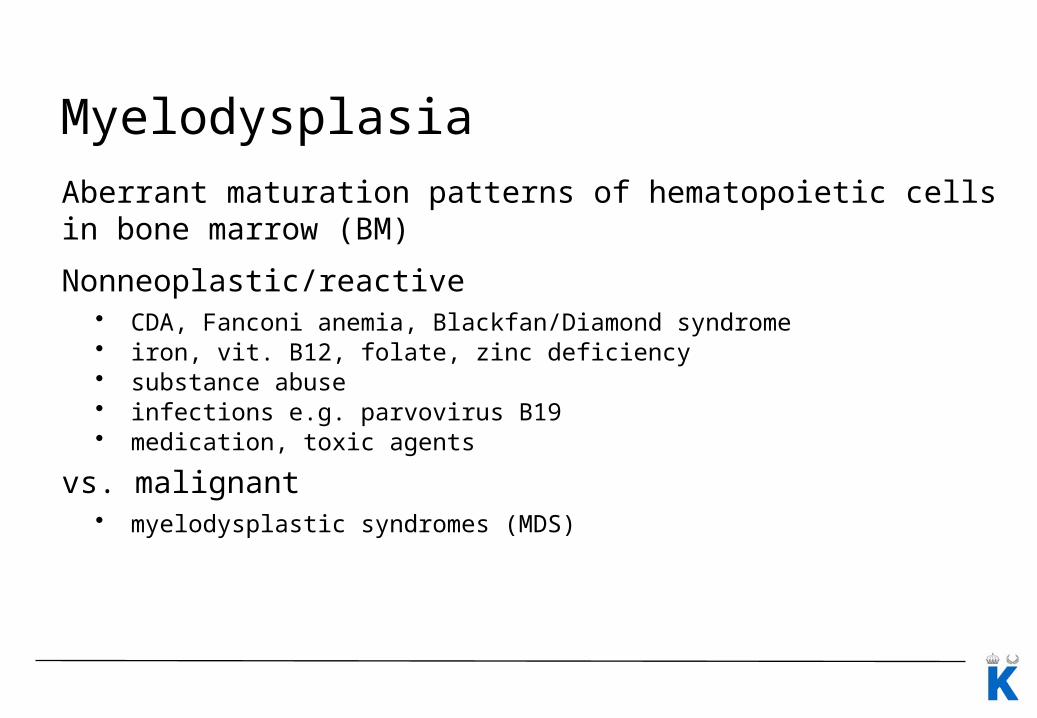
Myelodysplasia
Aberrant maturation patterns of hematopoietic cells in bone marrow (BM)
Nonneoplastic/reactive• CDA, Fanconi anemia, Blackfan/Diamond syndrome • iron, vit. B12, folate, zinc deficiency• substance abuse• infections e.g. parvovirus B19• medication, toxic agents
vs. malignant• myelodysplastic syndromes (MDS)
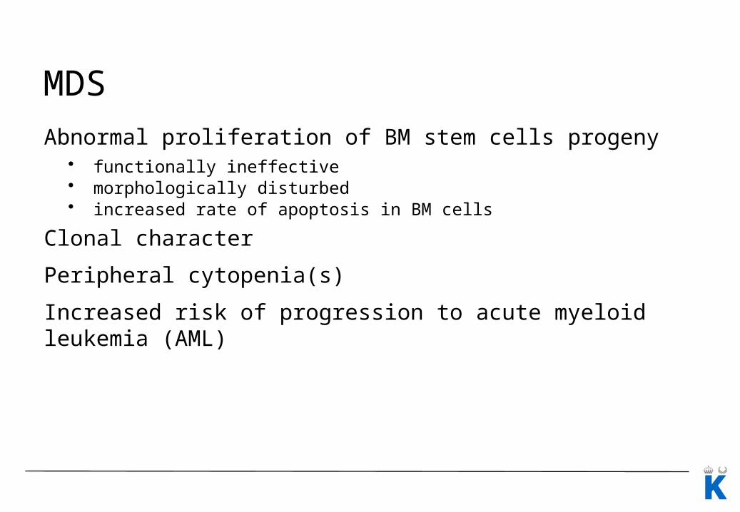
MDS
Abnormal proliferation of BM stem cells progeny• functionally ineffective • morphologically disturbed• increased rate of apoptosis in BM cells
Clonal character
Peripheral cytopenia(s)
Increased risk of progression to acute myeloid leukemia (AML)
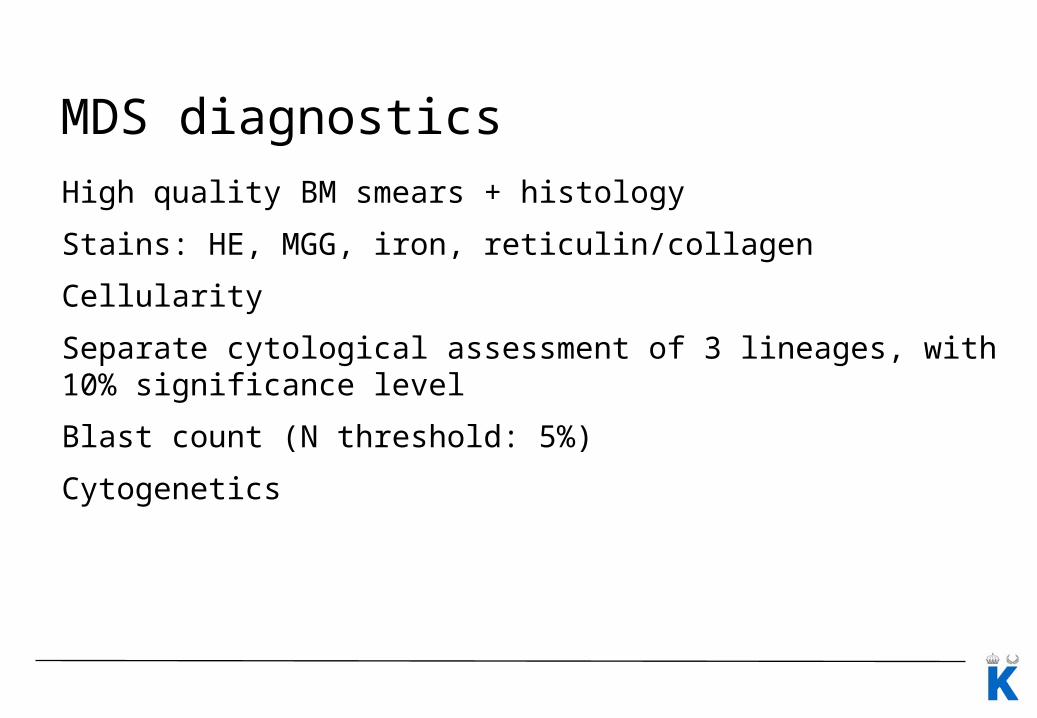
MDS diagnostics
High quality BM smears + histology
Stains: HE, MGG, iron, reticulin/collagen
Cellularity
Separate cytological assessment of 3 lineages, with 10% significance level
Blast count (N threshold: 5%)
Cytogenetics

MDS diagnosticsClassification: WHO 2008• Refractory cytopenia with unilineage dysplasia (RCUD)• Refractory anemia with ring sideroblasts (RARS)• Refreactory cytopenia with multilineage dysplasia (RCMD)• Refractory anemia with excess blasts (RAEB)• Myelodysplastic syndrome with isolated del(5q)• Myelodysplastic syndrome, unclassifiable• Childhood myelodysplastic syndrome
Prognostic and predictive factors: IPSS 1997• Percentage BM blasts• Cytogenetic findings• Number of cytopenias
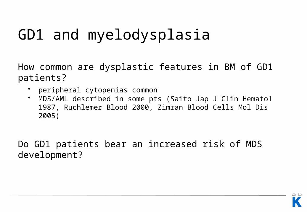
GD1 and myelodysplasia
How common are dysplastic features in BM of GD1 patients?• peripheral cytopenias common• MDS/AML described in some pts (Saito Jap J Clin Hematol 1987, Ruchlemer
Blood 2000, Zimran Blood Cells Mol Dis 2005)
Do GD1 patients bear an increased risk of MDS development?
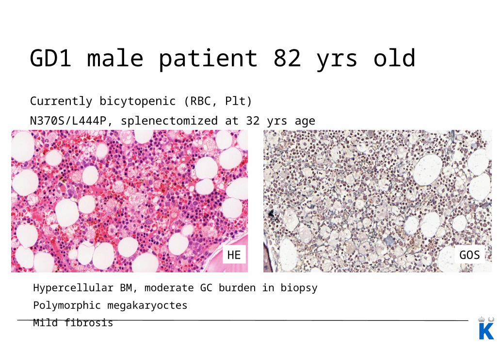
GD1 male patient 82 yrs old
Currently bicytopenic (RBC, Plt)
N370S/L444P, splenectomized at 32 yrs age
Hypercellular BM, moderate GC burden in biopsy
Polymorphic megakaryoctes
Mild fibrosis
HE GOS
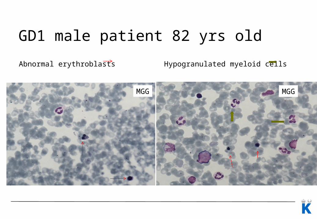
GD1 male patient 82 yrs old
Abnormal erythroblasts Hypogranulated myeloid cells
MGG MGG
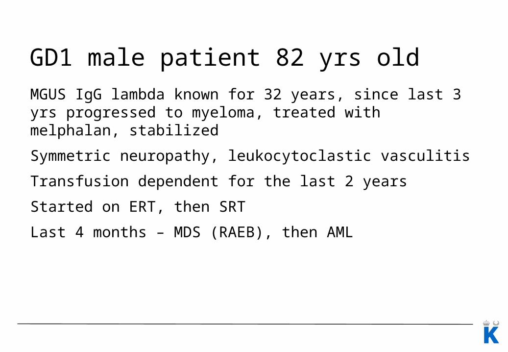
GD1 male patient 82 yrs old
MGUS IgG lambda known for 32 years, since last 3 yrs progressed to myeloma, treated with melphalan, stabilized
Symmetric neuropathy, leukocytoclastic vasculitis
Transfusion dependent for the last 2 years
Started on ERT, then SRT
Last 4 months – MDS (RAEB), then AML
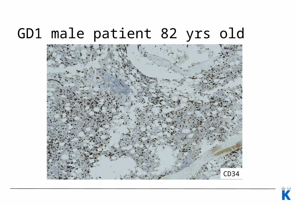
GD1 male patient 82 yrs old
CD34
Material and methodsDiagnostic BM specimens (aspirates, biopsies) from 17 untreated pts with GD1 from Sweden, Lithuania, and Poland
Central reassesment by a hematopathologist blinded to clinical data• BM specimens from 15 patients (7 women, 8 men) eligible for assessment; • BM smears from 2 pts were extremely diluted with PB. One of them had both
high cellularity and GCs burden but at the same time advanced fibrosis, and the other one had hypocellular BM.
Median pt age at evaluation: 56 years (range 21–86 years), four of them (27%) were splenectomized.
All but two pts carried at least one allele with a N370S (c.1226A>G) mutation in the GBA1 gene.
Only one patient did not have any cytopenia in PB, 7 pts had unilineage cytopenia, 5 pts had bicytopenia, and 2 pts had pancytopenia.
Two pts had vitamin B12 deficiency.
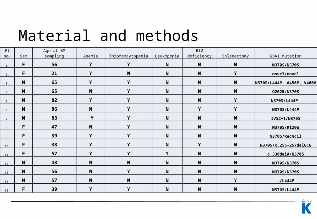
Material and methodsPt no. Sex Age at BM sampling Anemia Thrombocytopenia Leukopenia B12 deficiency Splenectomy GBA1 mutation
1. F 56 Y Y N N N N370S/N370S
2. F 21 Y N N N Y novel/novel
3. M 65 Y Y N N NN370S/L444P, A456P,
V460V
4. M 65 N Y N N N G202R/N370S
5. M 82 Y Y N N Y N370S/L444P
6. M 86 N Y N Y Y N370S/L444P
7. M 83 Y Y N N N IVS2+1/N370S
8. F 47 N Y N N N N370S/R120W
9. F 39 Y Y N N N N370S/RecNcil
10. F 38 Y Y N Y N N370S/c.255-257delGCG
11. F 57 Y Y Y N N c.330delA/N370S
12. M 48 N N N N N N370S/N370S
13. M 56 N Y N N N N370S/N370S
14. M 57 N N N N Y -/L444P
15. F 39 Y Y N N N N370S/L444P
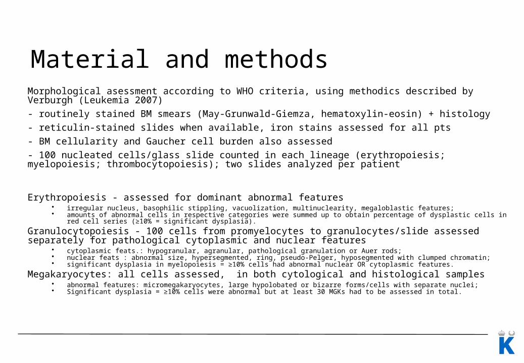
Material and methodsMorphological asessment according to WHO criteria, using methodics described by Verburgh (Leukemia 2007)
- routinely stained BM smears (May-Grunwald-Giemza, hematoxylin-eosin) + histology
- reticulin-stained slides when available, iron stains assessed for all pts
- BM cellularity and Gaucher cell burden also assessed
- 100 nucleated cells/glass slide counted in each lineage (erythropoiesis; myelopoiesis; thrombocytopoiesis); two slides analyzed per patient
Erythropoiesis - assessed for dominant abnormal features• irregular nucleus, basophilic stippling, vacuolization, multinuclearity, megaloblastic features; • amounts of abnormal cells in respective categories were summed up to obtain percentage of dysplastic cells in red cell series (≥10% = significant
dysplasia).
Granulocytopoiesis - 100 cells from promyelocytes to granulocytes/slide assessed separately for pathological cytoplasmic and nuclear features
• cytoplasmic feats.: hypogranular, agranular, pathological granulation or Auer rods; • nuclear feats : abnormal size, hypersegmented, ring, pseudo-Pelger, hyposegmented with clumped chromatin; • significant dysplasia in myelopoiesis = ≥10% cells had abnormal nuclear OR cytoplasmic features.
Megakaryocytes: all cells assessed, in both cytological and histological samples• abnormal features: micromegakaryocytes, large hypolobated or bizarre forms/cells with separate nuclei;• Significant dysplasia = ≥10% cells were abnormal but at least 30 MGKs had to be assessed in total.
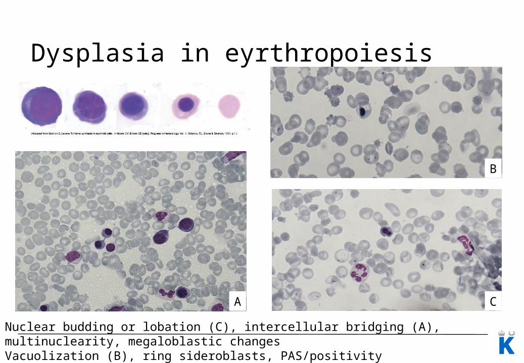
Dysplasia in eyrthropoiesis
Nuclear budding or lobation (C), intercellular bridging (A), multinuclearity, megaloblastic changesVacuolization (B), ring sideroblasts, PAS/positivity
A
B
C
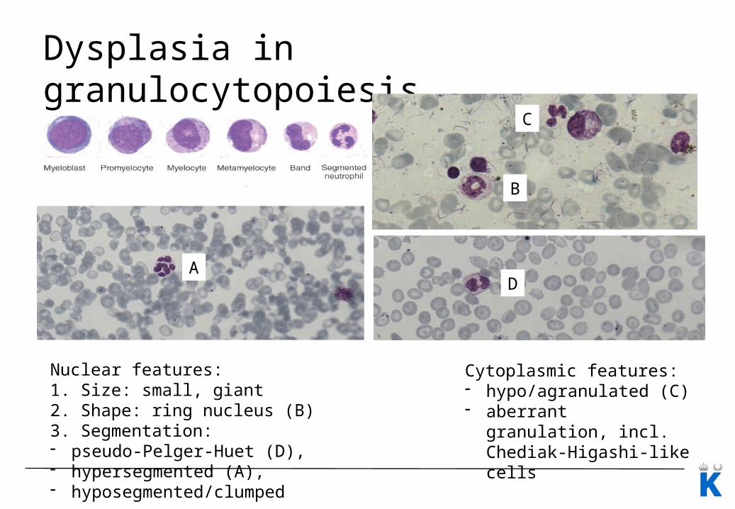
Dysplasia in granulocytopoiesis
Cytoplasmic features:- hypo/agranulated (C)- aberrant granulation, incl.
Chediak-Higashi-like cells
Nuclear features:1. Size: small, giant2. Shape: ring nucleus (B)3. Segmentation: - pseudo-Pelger-Huet (D), - hypersegmented (A), - hyposegmented/clumped
AD
B
C
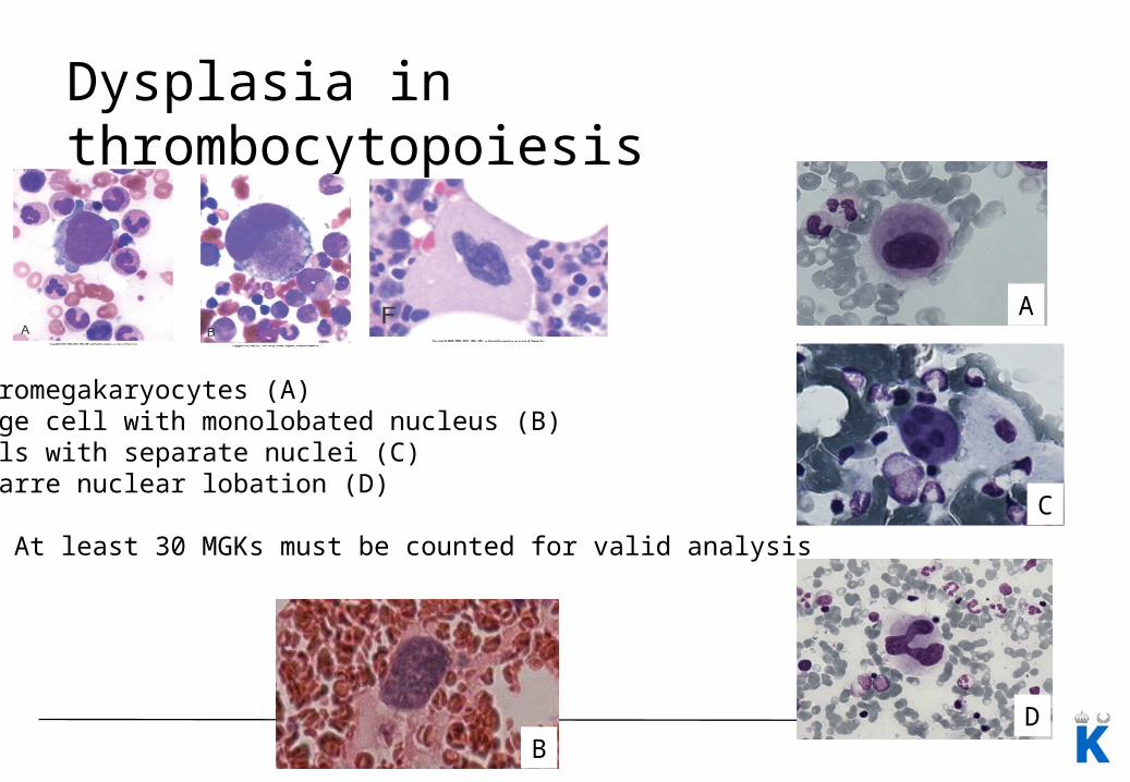
Dysplasia in thrombocytopoiesis
Micromegakaryocytes (A)Large cell with monolobated nucleus (B)Cells with separate nuclei (C)Bizarre nuclear lobation (D)
(!) At least 30 MGKs must be counted for valid analysis
A
C
DB
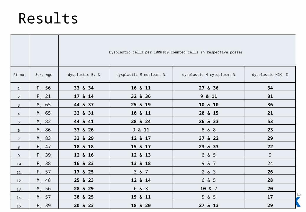
Results
Dysplastic cells per 100&100 counted cells in respective poeses
Pt no. Sex, Age dysplastic E, % dysplastic M nuclear, % dysplastic M cytoplasm, % dysplastic MGK, %
1. F, 56 33 & 34 16 & 11 27 & 36 34
2. F, 21 17 & 14 32 & 36 9 & 11 31
3. M, 65 44 & 37 25 & 19 10 & 10 36
4. M, 65 33 & 31 10 & 11 20 & 15 21
5. M, 82 44 & 41 28 & 24 26 & 33 53
6. M, 86 33 & 26 9 & 11 8 & 8 23
7. M, 83 33 & 29 12 & 17 37 & 22 29
8. F, 47 18 & 18 15 & 17 23 & 33 22
9. F, 39 12 & 16 12 & 13 6 & 5 9
10. F, 38 16 & 23 13 & 18 9 & 7 24
11. F, 57 17 & 25 3 & 7 2 & 3 26
12. M, 48 25 & 23 12 & 14 6 & 5 28
13. M, 56 28 & 29 6 & 3 10 & 7 20
14. M, 57 30 & 25 15 & 11 5 & 5 17
15. F, 39 20 & 23 18 & 20 27 & 13 29
Results
Cellularity: 35-85%
GD burden: 5-70%
Blast count: no pt above 5%
Ring sideroblasts: only single detected, no pt above 15%
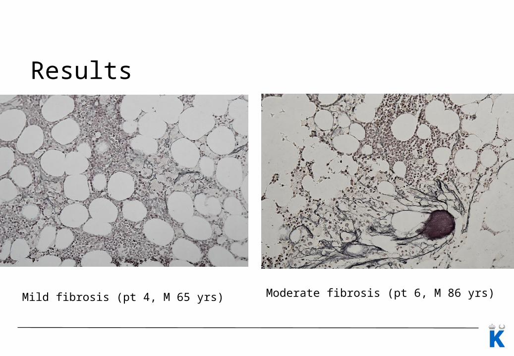
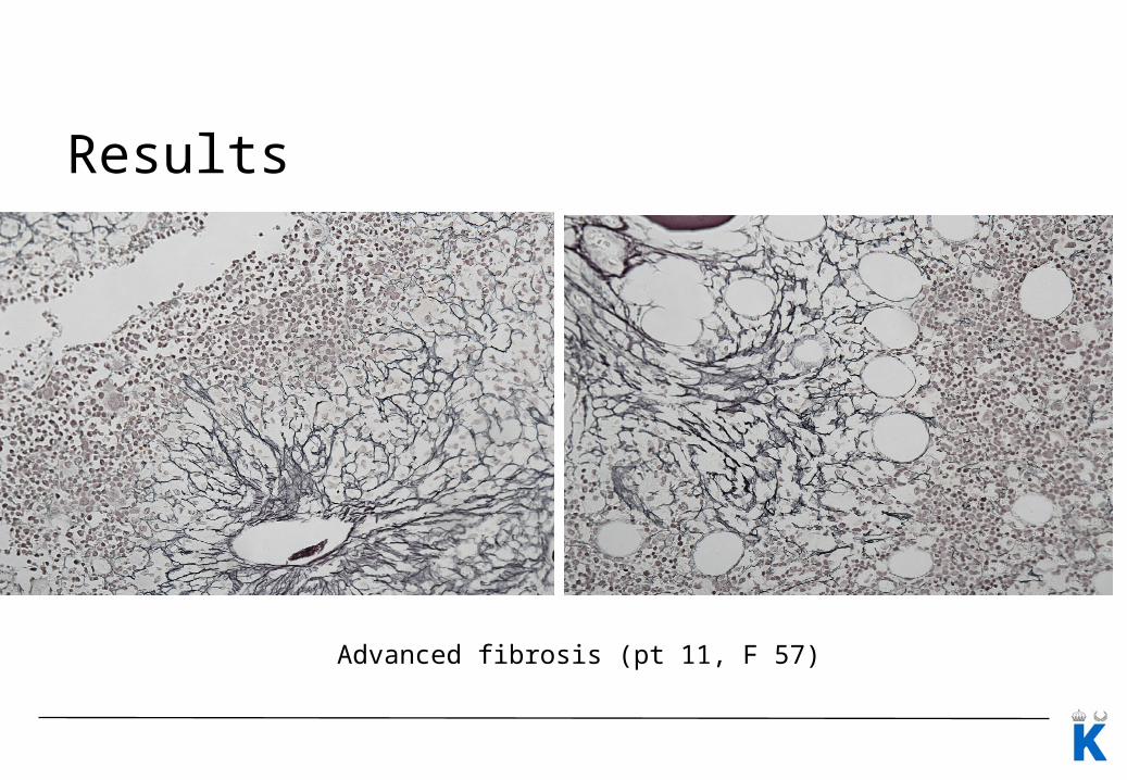
Fibrosis:
- variable, from mild to advanced,
- higher density in GC-rich areas compared to hematopoietic islands
Cytogenetics available only for single pts
Results
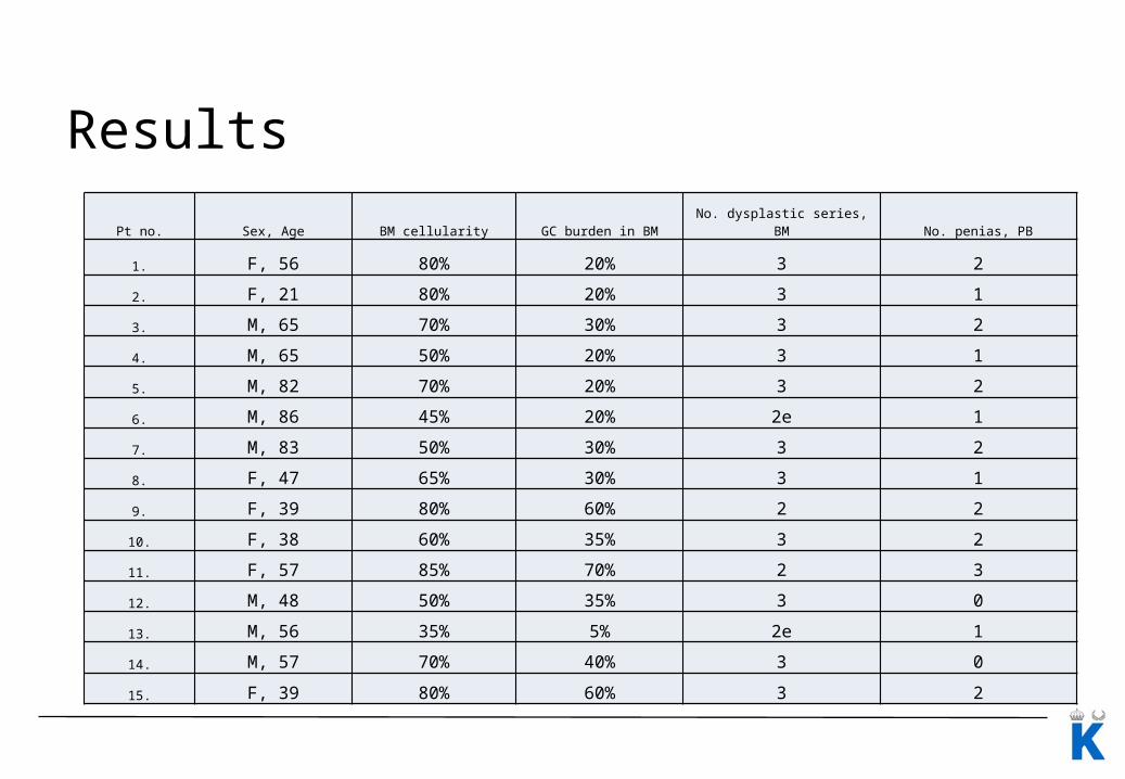
Pt no. Sex, Age BM cellularity GC burden in BM No. dysplastic series, BM No. penias, PB
1. F, 56 80% 20% 3 2
2. F, 21 80% 20% 3 1
3. M, 65 70% 30% 3 2
4. M, 65 50% 20% 3 1
5. M, 82 70% 20% 3 2
6. M, 86 45% 20% 2e 1
7. M, 83 50% 30% 3 2
8. F, 47 65% 30% 3 1
9. F, 39 80% 60% 2 2
10. F, 38 60% 35% 3 2
11. F, 57 85% 70% 2 3
12. M, 48 50% 35% 3 0
13. M, 56 35% 5% 2e 1
14. M, 57 70% 40% 3 0
15. F, 39 80% 60% 3 2
Results
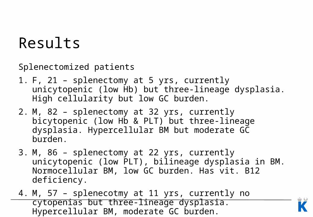
Splenectomized patients
1. F, 21 – splenectomy at 5 yrs, currently unicytopenic (low Hb) but three-lineage dysplasia. High cellularity but low GC burden.
2. M, 82 – splenectomy at 32 yrs, currently bicytopenic (low Hb & PLT) but three-lineage dysplasia. Hypercellular BM but moderate GC burden.
3. M, 86 – splenectomy at 22 yrs, currently unicytopenic (low PLT), bilineage dysplasia in BM. Normocellular BM, low GC burden. Has vit. B12 deficiency.
4. M, 57 – splenecotmy at 11 yrs, currently no cytopenias but three-lineage dysplasia. Hypercellular BM, moderate GC burden.
Results
Mild fibrosis (pt 4, M 65 yrs) Moderate fibrosis (pt 6, M 86 yrs)
Results
Advanced fibrosis (pt 11, F 57)
Conclusions
1. Significant dysplastic changes can be found in most patients with GD1, even in persons without overt cytopenias or after splenectomy.
2. Percentage aberrant cells in respective poeses can be low, however, morphological abnormalities are of the same type as in myelodysplastic syndromes.
Underlying mechanisms for abnormal hematopoietic cell maturation in GD pts are not known
• Can GCs infiltrates adversely affect neighboring hematopoietic islands by means of local cytokine interactions?
• Can GC infiltrates affect local blood & nutrient supply? Impact of advanced local fibrosis?
Further issues
• Fibrosis in BM may also strongly affect quality of the investigation. Aspirated material can be heavily diluted and virtually non-diagnostic, on the contrary to BM biopsies.
• Potential clonal nature of the observed abnormalities in hematopoietic cells in patients with GD remains to be investigated.
• Studies of patients who developed true MDS or acute myeloid leukemia in course of GD can be of value for assessment of potential increased risk in this patient population.
Department of Medicine at Huddinge, Karolinska Institute, Stockholm, Sweden
Maciej MachaczkaHans Hägglund
Children’s Memorial Health Institute, Department of Metabolic Diseases, Endocrinology and Diabetology, Warsaw, Poland
Anna Tylki-Szymańska
Department of Pathology, Collegium Medicum of the Jagiellonian University, Cracow, Poland
Krystyna Gałązka
Department of Hematology, Collegium Medicum of the Jagiellonian University, Cracow, Poland
Wojciech Jurczak
Department of Pathology and Cytology, Karolinska University Hospital, Stockholm, Sweden
Agnieszka Bulanda
Centre of Hematology and Oncology, Children’s Hospital, Affiliate of Vilnius University Hospital Santariskiu Clinics, Lithuania
Gražina Kleinotiene
Lina Rageliene
Department of Hematology, Oncology and Transfusion Medicine Center, Vilnius University Hospital Santariskiu Clinics, Lithuania
Regina Pileckyte
National Centre of Pathology, Vilnius University, Hospital Santariskiu Clinics affiliate, Lithuania
Ugnius Mickys