Embed Size (px)
Citation preview
Congenital Adrenal Hyperplasia and
Testicular Feminization Syndromes
Dr. Ahmed Hussain A. Mujamammi
Objectives
• Adrenal steroidogenesis
• Congenital adrenal hyperplasia syndrome
Types
Biochemical characteristics
Clinical manifestations
• Testicular feminization syndrome
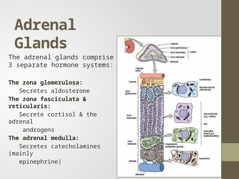
Adrenal GlandsThe adrenal glands comprise 3 separate hormone systems:
The zona glomerulosa:
Secretes aldosterone
The zona fasciculata & reticularis:
Secrete cortisol & the adrenal
androgens
The adrenal medulla:
Secretes catecholamines (mainly
epinephrine)

Hermaphroditism or Intersex
A person who has neither standard male or standard female anatomy.
Discrepancy between the type of gonads and the external genitalia
True hermaphrodite (ovary plus testis)
Female pseudohermaphrodite (FPH, only ovary)
Male pseudohermaphrodite (MPH, only testis)
Glucocorticoids & Mineralocorticoids
• Glucocorticoids:• Steroids with cortisol-like activity• Potent metabolic regulators & immunosuppressants
• Mineralocorticoids:• Steroids with aldosterone-like activity• Promote renal sodium reabsorption
Steroidogenesis and Congenital adrenal hyperplasia
syndrome
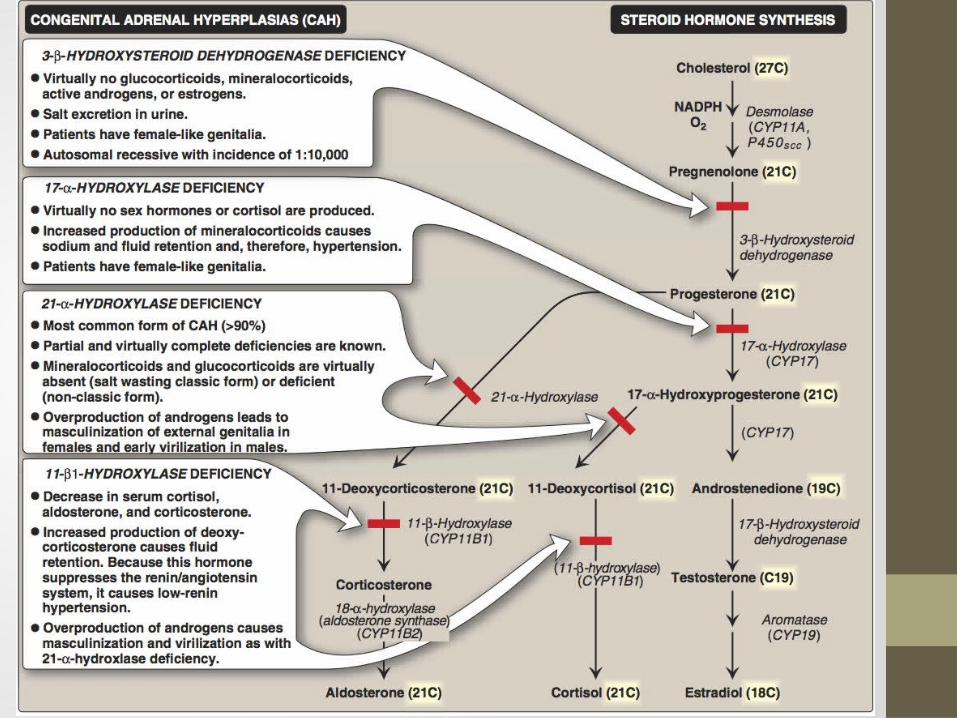
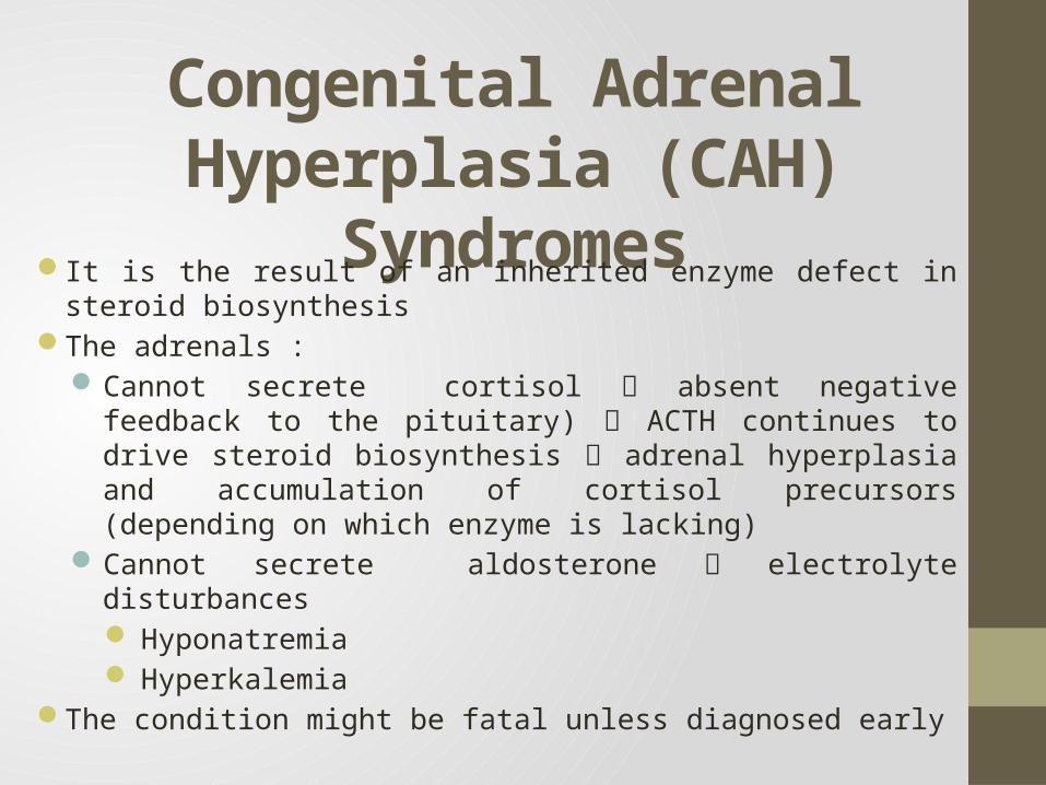
Congenital Adrenal Hyperplasia (CAH) Syndromes
It is the result of an inherited enzyme defect in steroid biosynthesisThe adrenals :
Cannot secrete cortisol absent negative feedback to the pituitary) ACTH continues to drive steroid biosynthesis adrenal hyperplasia and accumulation of cortisol precursors (depending on which enzyme is lacking)
Cannot secrete aldosterone electrolyte disturbancesHyponatremiaHyperkalemia
The condition might be fatal unless diagnosed early
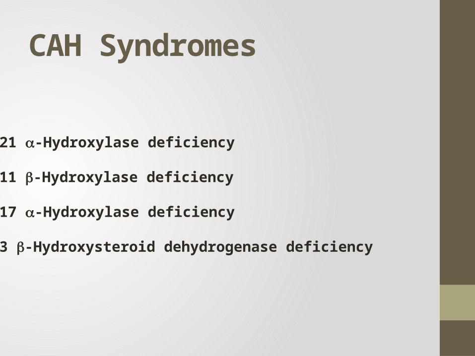
CAH Syndromes
21 -Hydroxylase deficiency
11 -Hydroxylase deficiency
17 -Hydroxylase deficiency
3 -Hydroxysteroid dehydrogenase deficiency
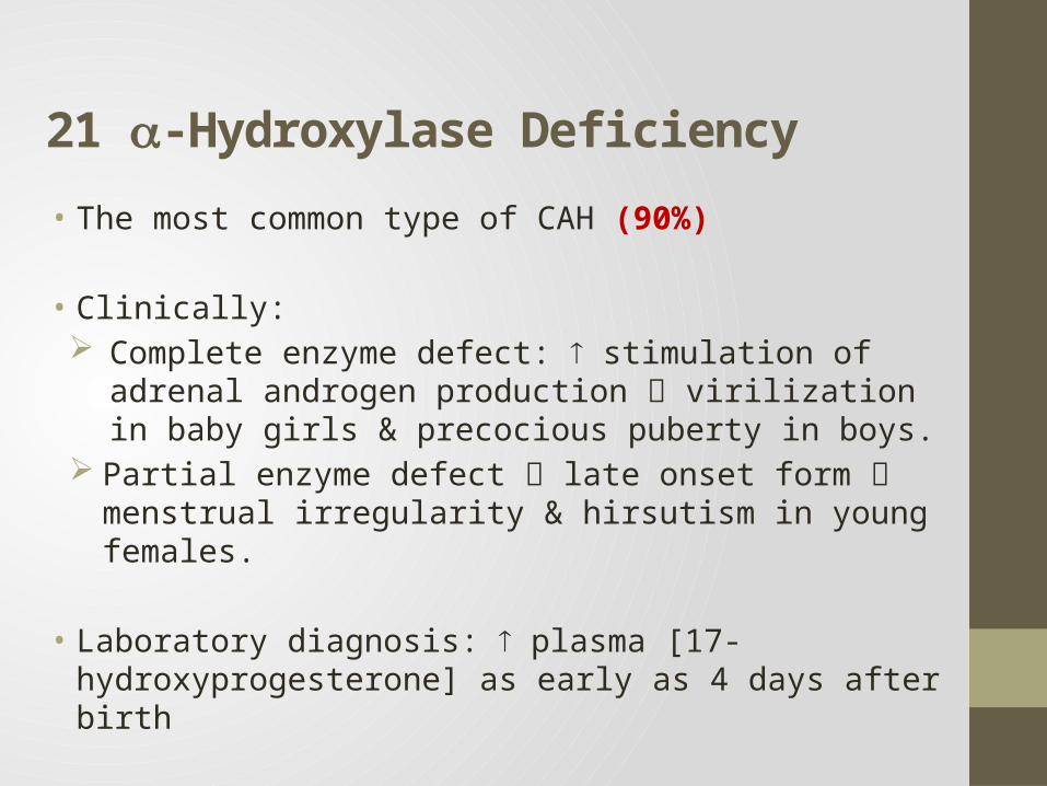
21 -Hydroxylase Deficiency • The most common type of CAH (90%)
• Clinically: Complete enzyme defect: stimulation of adrenal androgen
production virilization in baby girls & precocious puberty in boys.
Partial enzyme defect late onset form menstrual irregularity & hirsutism in young females.
• Laboratory diagnosis: plasma [17-hydroxyprogesterone] as early as 4 days after birth
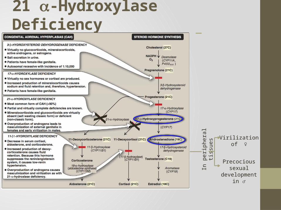
21 -Hydroxylase Deficiency
Virilization of ♀
Precocious sexual development in ♂
X X
In p
erip
hera
l tis
sues
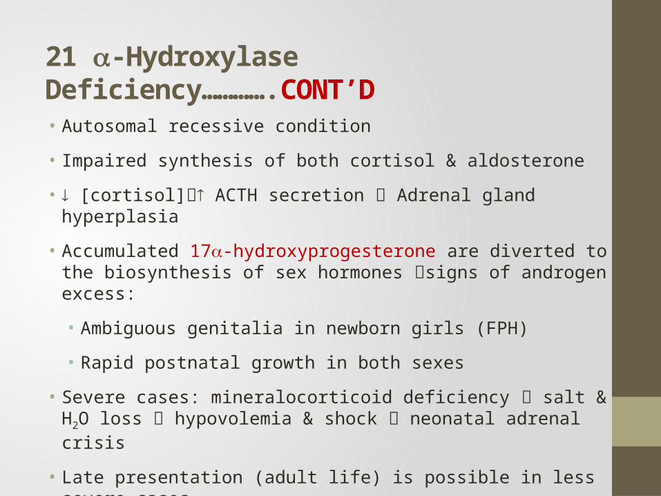
21 -Hydroxylase Deficiency………….CONT’D • Autosomal recessive condition
• Impaired synthesis of both cortisol & aldosterone
• [cortisol] ACTH secretion Adrenal gland hyperplasia
• Accumulated 17-hydroxyprogesterone are diverted to the biosynthesis of sex hormones signs of androgen excess:
• Ambiguous genitalia in newborn girls (FPH)
• Rapid postnatal growth in both sexes
• Severe cases: mineralocorticoid deficiency salt & H2O loss hypovolemia & shock neonatal adrenal crisis
• Late presentation (adult life) is possible in less severe cases
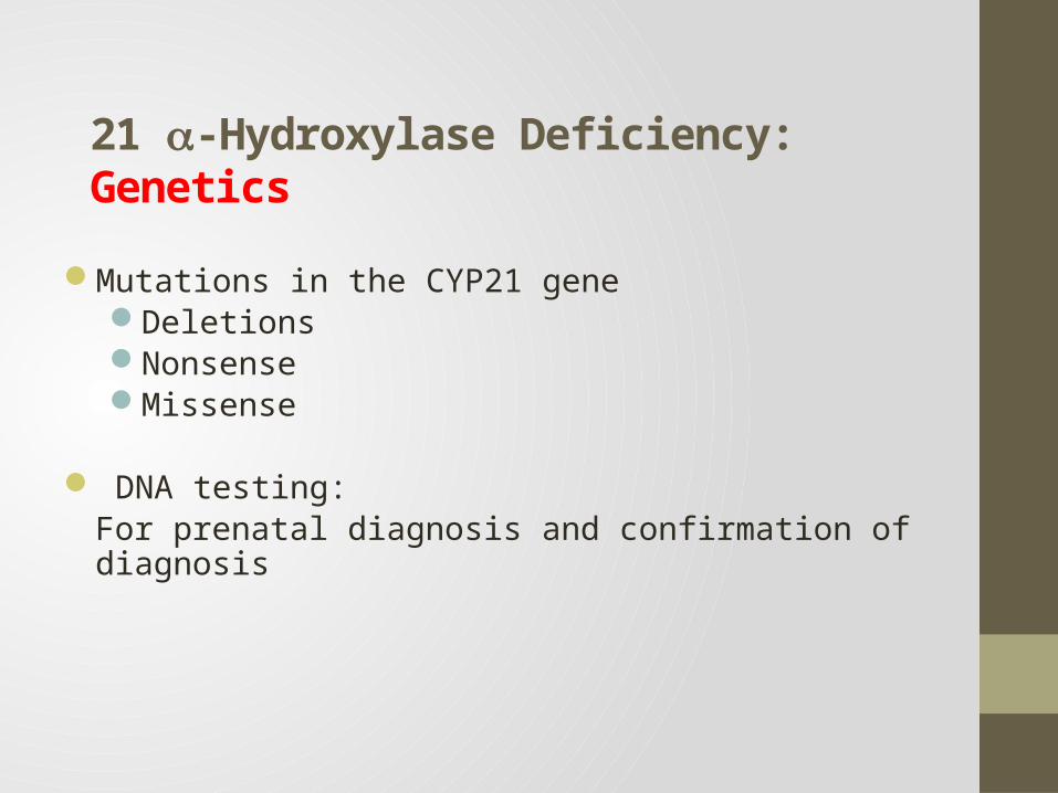
21 -Hydroxylase Deficiency: Genetics
Mutations in the CYP21 geneDeletionsNonsenseMissense
DNA testing: For prenatal diagnosis and confirmation of diagnosis
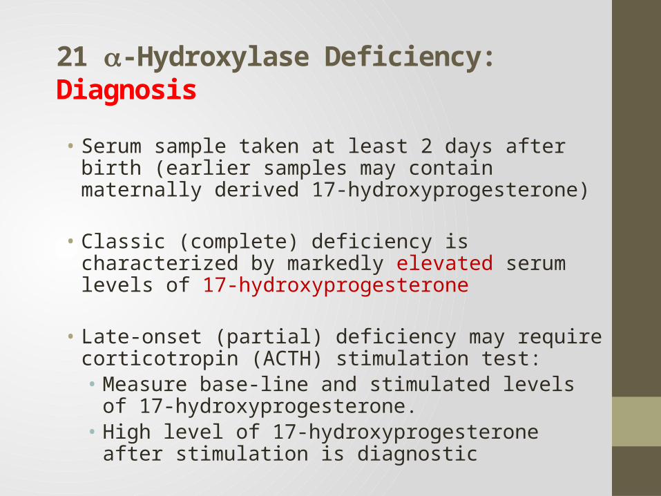
21 -Hydroxylase Deficiency: Diagnosis
• Serum sample taken at least 2 days after birth (earlier samples may contain maternally derived 17-hydroxyprogesterone)
• Classic (complete) deficiency is characterized by markedly elevated serum levels of 17-hydroxyprogesterone
• Late-onset (partial) deficiency may require corticotropin (ACTH) stimulation test:• Measure base-line and stimulated levels of 17-
hydroxyprogesterone. • High level of 17-hydroxyprogesterone after stimulation is
diagnostic
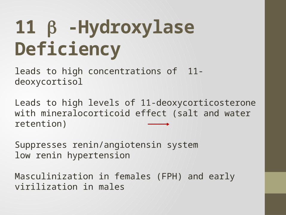
11 -Hydroxylase Deficiency
leads to high concentrations of 11-deoxycortisol
Leads to high levels of 11-deoxycorticosterone with mineralocorticoid effect (salt and water retention)
Suppresses renin/angiotensin system low renin hypertension
Masculinization in females (FPH) and early virilization in males
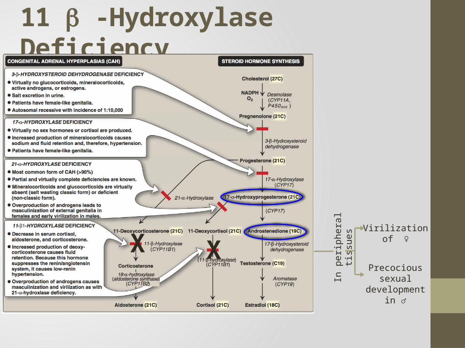
11 -Hydroxylase Deficiency
X XVirilization of ♀
Precocious sexual development in ♂In
per
iphe
ral t
issu
es
Testicular Feminization Syndrome (Androgen Insensitivity
Syndrome)
Disorders of Male Sexual Differentiation
• They are rare group of disorders• The defect may be in:• Androgen receptors (inactive androgen receptors target tissues
cannot respond to stimulation by circulating testosterone; e.g., Testicular feminization syndrome)
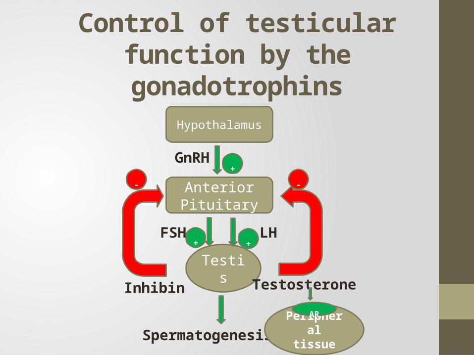
Control of testicular function by the gonadotrophins
Hypothalamus
Anterior Pituitary
Testis
+
+ +FSH LH
TestosteroneInhibin
- -
Spermatogenesis
GnRH
Peripheral tissue
AR
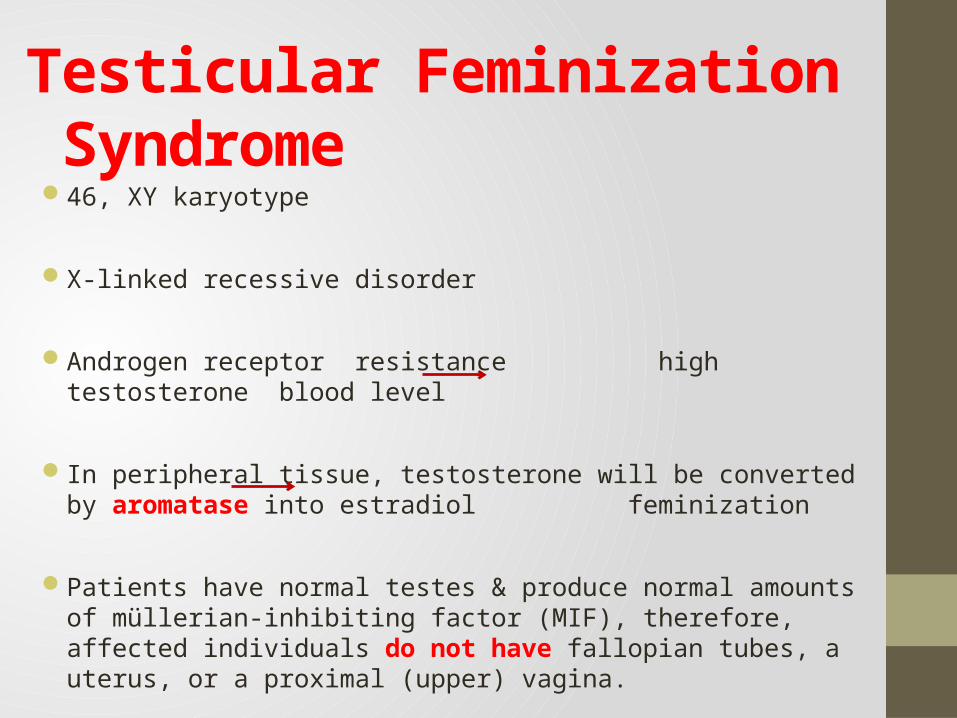
Testicular Feminization Syndrome46, XY karyotype
X-linked recessive disorder
Androgen receptor resistance high testosterone blood level
In peripheral tissue, testosterone will be converted by aromatase into estradiol feminization
Patients have normal testes & produce normal amounts of müllerian-inhibiting factor (MIF), therefore, affected individuals do not have fallopian tubes, a uterus, or a proximal (upper) vagina.
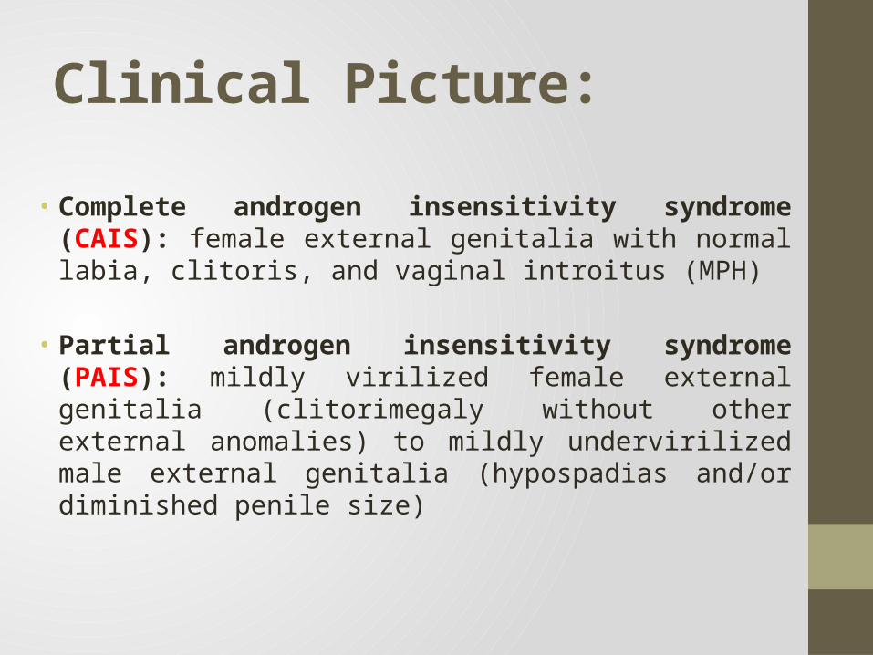
Clinical Picture:
• Complete androgen insensitivity syndrome (CAIS): female external genitalia with normal labia, clitoris, and vaginal introitus (MPH)
• Partial androgen insensitivity syndrome (PAIS): mildly virilized female external genitalia (clitorimegaly without other external anomalies) to mildly undervirilized male external genitalia (hypospadias and/or diminished penile size)
Karyotype: differentiate an undermasculinized male from a masculinized female.
Fluorescent in situ hybridization (FISH): Presence of a Y chromosome can be confirmed by probes for the SRY region of the Y chromosome. These offer a much quicker turnaround time than conventional karyotypes.
Increased (or normal) testosterone and dihydrotestosterone blood levels
DNA tests and mutation analysis for androgen receptor gene:
Complete or partial gene deletions, point mutations, or small insertions/deletions
Laboratory Diagnosis
Further Investigations
Imaging Studies “Pelvic ultrasound”:
Absence of fallopian tubes and uterus


![Endometrium presentation - Dr Wright[1] · Endometrial Hyperplasia Simple hyperplasia Complex hyperplasia (adenomatous) Simple atypical hyperplasia ... Progression of Hyperplasia](https://img.pdfslide.net/doc/110x75/5b8a421e7f8b9a50388bc13d/endometrium-presentation-dr-wright1-endometrial-hyperplasia-simple-hyperplasia.jpg)