Embed Size (px)
Citation preview
Congenital anomalies
Antonín Šípek, MD, PhD
Department of Medical GeneticsThomayer Faculty Hospital
Etiology, diagnosis and incidence
http://www.vrozene-vady.cz/
Index of Presentation
1.Definitions
2.Etiology, teratogens
3.Selected congenital anomalies
4.Prenatal diagnosis
5.Incidence of selected congenital anomalies
6.Summary
This presentation can be downloaded from our website.
So there is not necessary to rewrite all the text from slides
Just visit http://www.vrozene-vady.cz/ and download a PDF file.
Congenital anomalies
1.Definitions
2.Etiology, teratogens
3.Selected congenital anomalies
4.Prenatal diagnosis
5.Incidence of selected congenital anomalies
6.Summary
Definitions I
Congenital anomaly
Congenital Anomaly (CA) is an anomaly that affects a body
part or physiologic function and is present at birth.
It is caused by the abnormal ontogenetic development of the fetus.
The process is affected by genetic, environmental or both factors.
The disturbance of the regulation and development cascades take
place on the level of tissue, cell or molecule.
Definitions II
Other „synonyms“
Congenital Malformation is a congenital anomaly of the
structure of some body part.
Birth Defect or Congenital Disorder are nearly synonyms for
the term Congenital Anomaly.
Chromosomal Aberrations are the numerical or structural
abnormalities of the karyotype.
Genetic Disorders are the conditions caused by the mutation of
the gene(s).
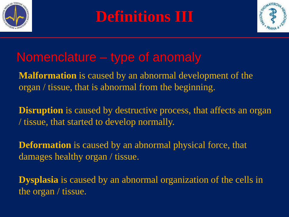
Definitions III
Nomenclature – type of anomaly
Malformation is caused by an abnormal development of the
organ / tissue, that is abnormal from the beginning.
Disruption is caused by destructive process, that affects an organ
/ tissue, that started to develop normally.
Deformation is caused by an abnormal physical force, that
damages healthy organ / tissue.
Dysplasia is caused by an abnormal organization of the cells in
the organ / tissue.
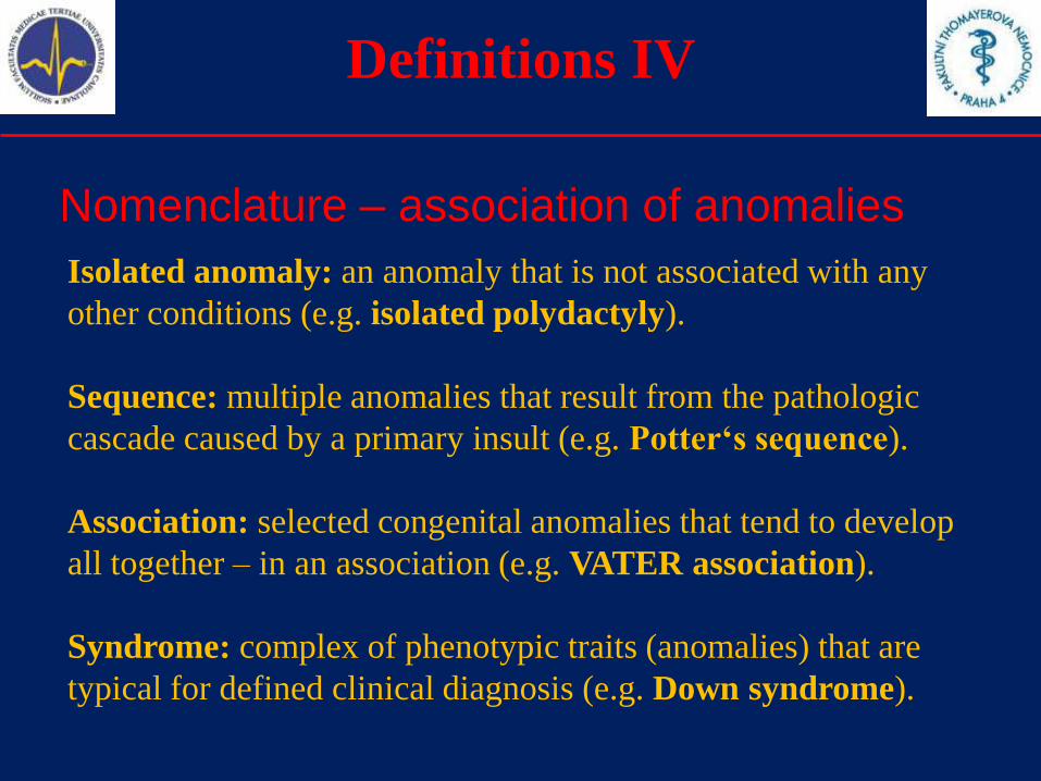
Definitions IV
Nomenclature – association of anomalies
Isolated anomaly: an anomaly that is not associated with any
other conditions (e.g. isolated polydactyly).
Sequence: multiple anomalies that result from the pathologic
cascade caused by a primary insult (e.g. Potter‘s sequence).
Association: selected congenital anomalies that tend to develop
all together – in an association (e.g. VATER association).
Syndrome: complex of phenotypic traits (anomalies) that are
typical for defined clinical diagnosis (e.g. Down syndrome).
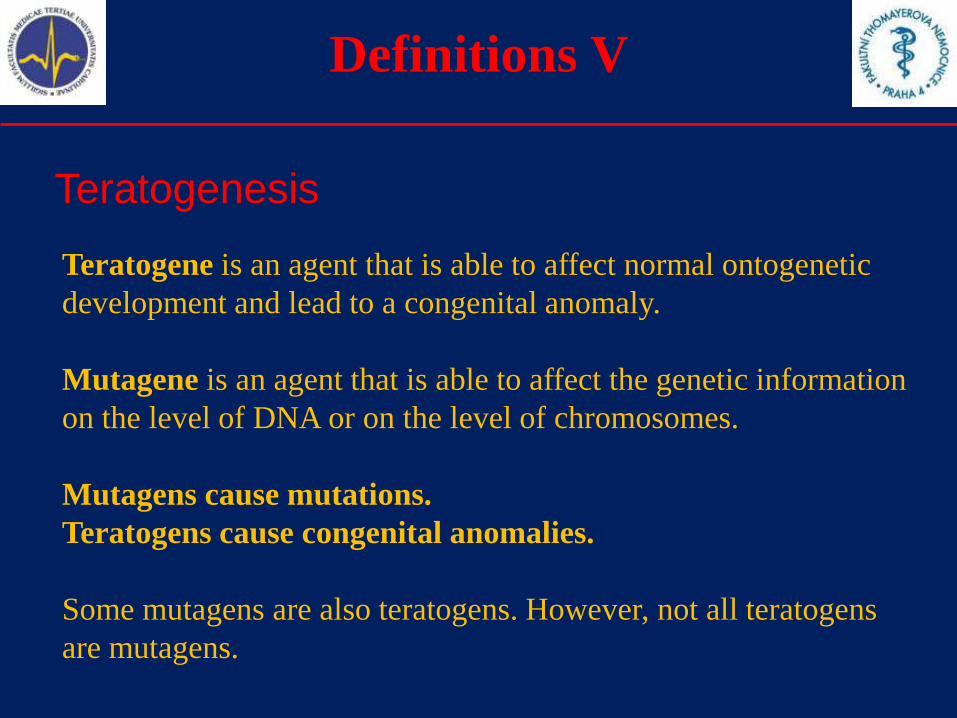
Definitions V
Teratogenesis
Teratogene is an agent that is able to affect normal ontogenetic
development and lead to a congenital anomaly.
Mutagene is an agent that is able to affect the genetic information
on the level of DNA or on the level of chromosomes.
Mutagens cause mutations.
Teratogens cause congenital anomalies.
Some mutagens are also teratogens. However, not all teratogens
are mutagens.
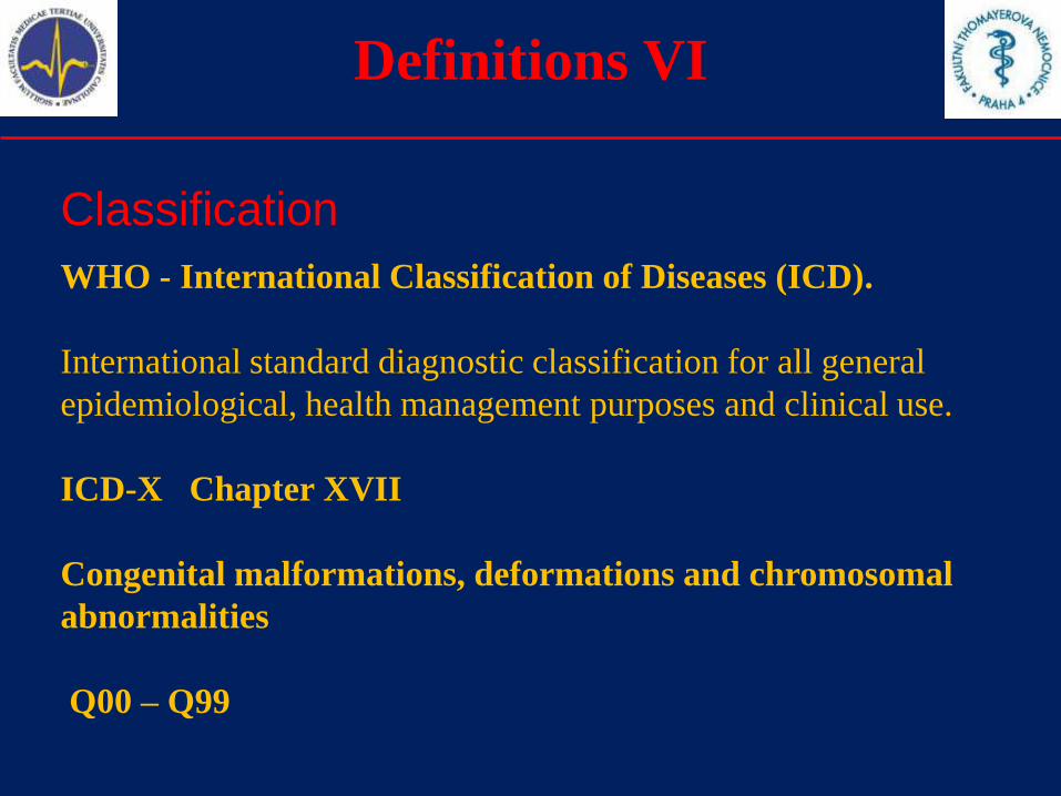
Definitions VI
Classification
WHO - International Classification of Diseases (ICD).
International standard diagnostic classification for all general
epidemiological, health management purposes and clinical use.
ICD-X Chapter XVII
Congenital malformations, deformations and chromosomal
abnormalities
Q00 – Q99
Definitions VII
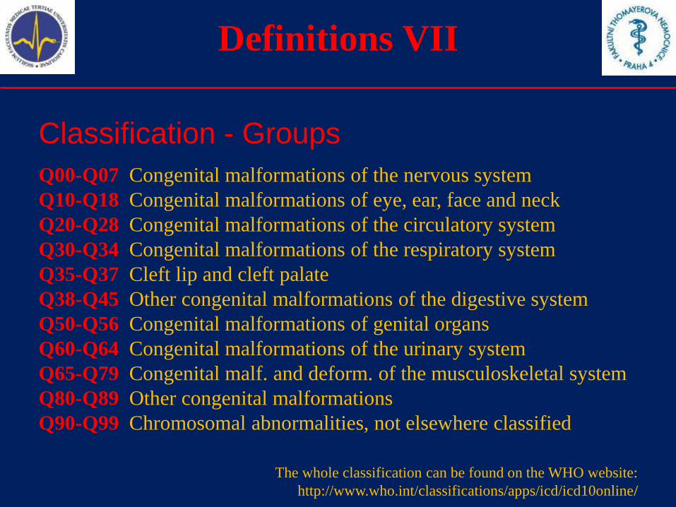
Classification - Groups
Q00-Q07 Congenital malformations of the nervous system
Q10-Q18 Congenital malformations of eye, ear, face and neck
Q20-Q28 Congenital malformations of the circulatory system
Q30-Q34 Congenital malformations of the respiratory system
Q35-Q37 Cleft lip and cleft palate
Q38-Q45 Other congenital malformations of the digestive system
Q50-Q56 Congenital malformations of genital organs
Q60-Q64 Congenital malformations of the urinary system
Q65-Q79 Congenital malf. and deform. of the musculoskeletal system
Q80-Q89 Other congenital malformations
Q90-Q99 Chromosomal abnormalities, not elsewhere classified
The whole classification can be found on the WHO website:
http://www.who.int/classifications/apps/icd/icd10online/
Congenital anomalies
1.Definitions
2.Etiology, teratogens
3.Selected congenital anomalies
4.Prenatal diagnosis
5.Incidence of selected congenital anomalies
6.Summary
Etiology I
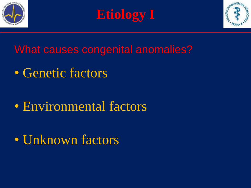
What causes congenital anomalies?
• Genetic factors
• Environmental factors
• Unknown factors
Etiology II
What causes congenital anomalies?
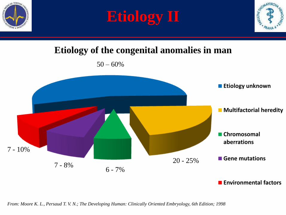
Etiology unknown
Multifactorial heredity
Chromosomal aberrations
Gene mutations
Environmental factors
From: Moore K. L., Persaud T. V. N.; The Developing Human: Clinically Oriented Embryology, 6th Edition; 1998
50 – 60%
20 - 25%
6 - 7%7 - 8%
7 - 10%
Etiology of the congenital anomalies in man
Genetics I
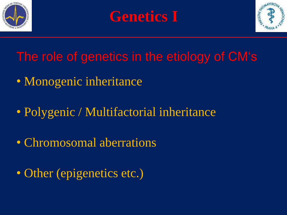
The role of genetics in the etiology of CM‘s
• Monogenic inheritance
• Polygenic / Multifactorial inheritance
• Chromosomal aberrations
• Other (epigenetics etc.)

Genetics II
Monogenic inheritance
Some congenital malformation are inherited as a monogenic trait.
There are several genes, whose mutations are associated with
selected congenital anomalies. The reference can be found in
Mendelian Inheritance in Man (MIM). Prenatal diagnosis and
management of the genetic counseling is less difficult.
• Marfan syndrome (Q87.4; MIM: 154700)
• Ehlers – Dahnlos syndrome (Q79.6; MIM: 13000)
• Osteogenesis imperfecta (Q78.0; MIM: 166200)
• Achondroplasia (Q77.4; MIM: 100800)
• Holoprosencephaly (Q04.2; MIM: 236100)
• Xeroderma pigmentosum (Q82.1; MIM: 278830)
Genetics III
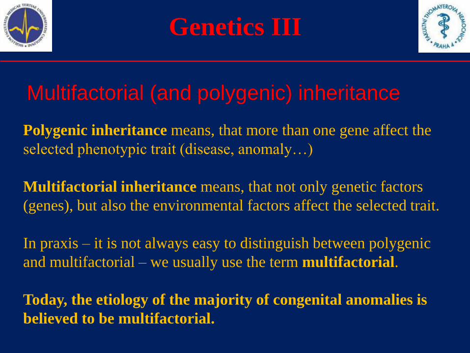
Multifactorial (and polygenic) inheritance
Polygenic inheritance means, that more than one gene affect the
selected phenotypic trait (disease, anomaly…)
Multifactorial inheritance means, that not only genetic factors
(genes), but also the environmental factors affect the selected trait.
In praxis – it is not always easy to distinguish between polygenic
and multifactorial – we usually use the term multifactorial.
Today, the etiology of the majority of congenital anomalies is
believed to be multifactorial.
Genetics IV
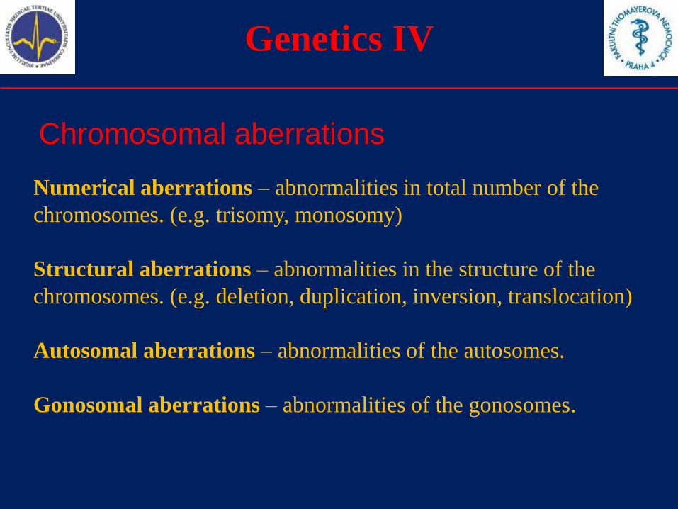
Chromosomal aberrations
Numerical aberrations – abnormalities in total number of the
chromosomes. (e.g. trisomy, monosomy)
Structural aberrations – abnormalities in the structure of the
chromosomes. (e.g. deletion, duplication, inversion, translocation)
Autosomal aberrations – abnormalities of the autosomes.
Gonosomal aberrations – abnormalities of the gonosomes.
Genetics V
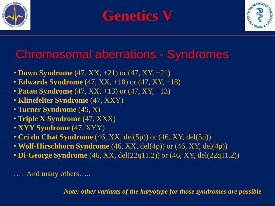
Chromosomal aberrations - Syndromes
• Down Syndrome (47, XX, +21) or (47, XY, +21)
• Edwards Syndrome (47, XX, +18) or (47, XY, +18)
• Patau Syndrome (47, XX, +13) or (47, XY, +13)
• Klinefelter Syndrome (47, XXY)
• Turner Syndrome (45, X)
• Triple X Syndrome (47, XXX)
• XYY Syndrome (47, XYY)
• Cri du Chat Syndrome (46, XX, del(5p)) or (46, XY, del(5p))
• Wolf-Hirschhorn Syndrome (46, XX, del(4p)) or (46, XY, del(4p))
• Di-George Syndrome (46, XX, del(22q11.2)) or (46, XY, del(22q11.2))
….. And many others…..
Note: other variants of the karyotype for those syndromes are possible
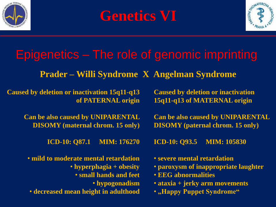
Genetics VI
Epigenetics – The role of genomic imprinting
Prader – Willi Syndrome X Angelman Syndrome
Caused by deletion or inactivation 15q11-q13
of PATERNAL origin
Can be also caused by UNIPARENTAL
DISOMY (maternal chrom. 15 only)
ICD-10: Q87.1 MIM: 176270
• mild to moderate mental retardation
• hyperphagia + obesity
• small hands and feet
• hypogonadism
• decreased mean height in adulthood
Caused by deletion or inactivation
15q11-q13 of MATERNAL origin
Can be also caused by UNIPARENTAL
DISOMY (paternal chrom. 15 only)
ICD-10: Q93.5 MIM: 105830
• severe mental retardation
• paroxysm of inappropriate laughter
• EEG abnormalities
• ataxia + jerky arm movements
• „Happy Puppet Syndrome“
Teratogens I
Environmental factors
There are many environmental factors that cause congenital
anomalies, or are able to cause them in specific situations.
Those factors are commonly known as teratogens.
However - the effect of teratogens is dependent on the genetics.
The genotype can modify the teratogenic effect.
There are three main groups of teratogens:
1. Physical
2. Chemical
3. Biological
Teratogens II
Physical teratogens
• X-rays (common diagnostic doses are not dangerous)
• Ionizing radiation (e.g. gamma radiation)
• High temperature (sauna, fever)
• Mechanical factors (amniotic bands, oligohydramnion)
Ultrasonography and electromagnetic field seem to be safe.
Teratogens III
Chemical teratogens
• chemical substances used in industry or agriculture
(organic solvents, paints, polychlorinated biphenyls,
heavy metals)
• alcohol (cause Fetal alcohol syndrome)
• products of cigarette smoking (teratogenic effect of
marihuana smoking was also proved)
• other drugs (e.g. cocaine), doping (steroids)
• cytostatics and some other groups of medicaments
(antiepileptics, antibiotics, warfarine, ACE-inhibitors)

Teratogens IV
Drugs and teratogenic effect
The intensive study of teratogenic effect of the drugs started after the
„thalidomide affair“ in the sixties of 20th century.
The current boom of pharmaceutical industry provides many new medicaments
each year. The safety of those substances must be tested.
Teratogenic effect is species-dependent. It is possible, that human embryo will
not be affected by the same substance, that affects rat embryos.
The same dose of substance could be teratogenic in human, but needn‘t to be
teratogenic at all in rats or other animals. The effect is dose-dependent.
The same substance can be teratogenic only in a specific week of pregnancy. It
can only affect the development of a specific organ / tissue. The effect is time-
dependent.
It is not easy to prove, that the congenital malformation was caused by the
usage of a specific drug during pregnancy. Usually, there is not enough data. It is
necessary to collect all data possible about such risk – pregnancies.

Teratogens V
Drugs and teratogenic effect
During the time of blastogenesis, the damage caused by the
teratogens cause no anomalies. The embryo is either able to repair
all damage taken, or it stops to develop and dies.
The time of organogenesis (3th-12th week of pregnancy) is the
critical period for most teratogens. The morphologic anomalies
are usually caused during this period.
The second and third trimester is not so critical, however the
toxic effect of some substances is pathologic as well.
The teratogenic effect of the drugs: 1) proved 2) presumable
3) possible 4) couldn‘t be excluded
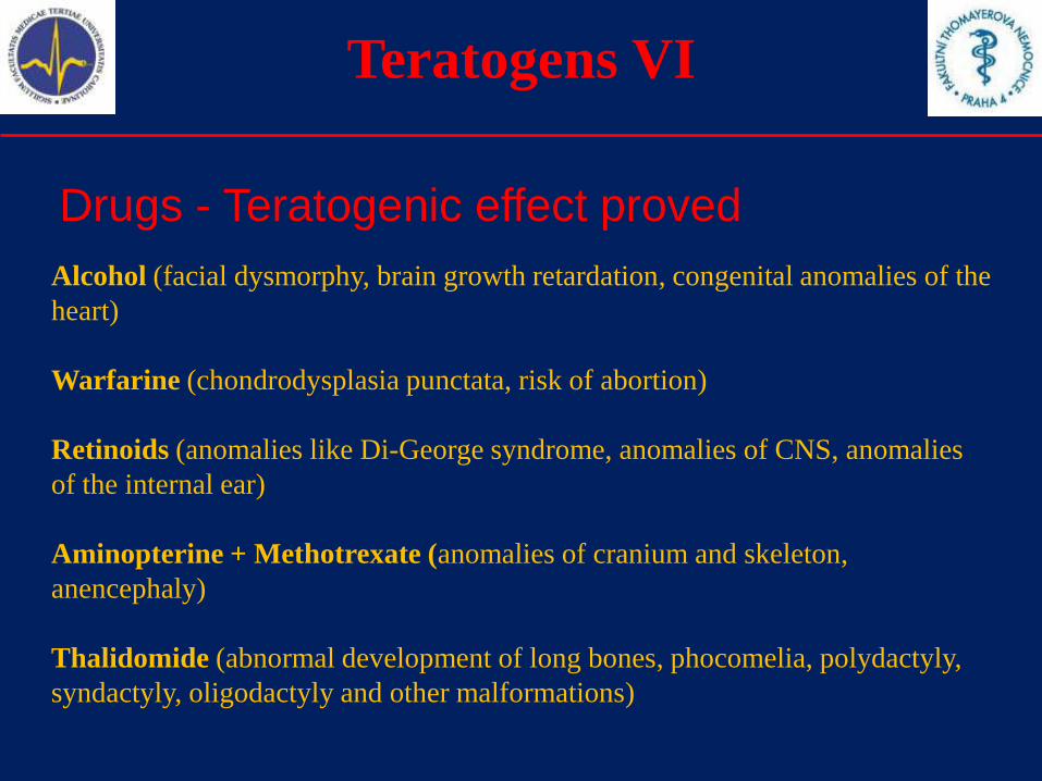
Teratogens VI
Drugs - Teratogenic effect proved
Alcohol (facial dysmorphy, brain growth retardation, congenital anomalies of the
heart)
Warfarine (chondrodysplasia punctata, risk of abortion)
Retinoids (anomalies like Di-George syndrome, anomalies of CNS, anomalies
of the internal ear)
Aminopterine + Methotrexate (anomalies of cranium and skeleton,
anencephaly)
Thalidomide (abnormal development of long bones, phocomelia, polydactyly,
syndactyly, oligodactyly and other malformations)
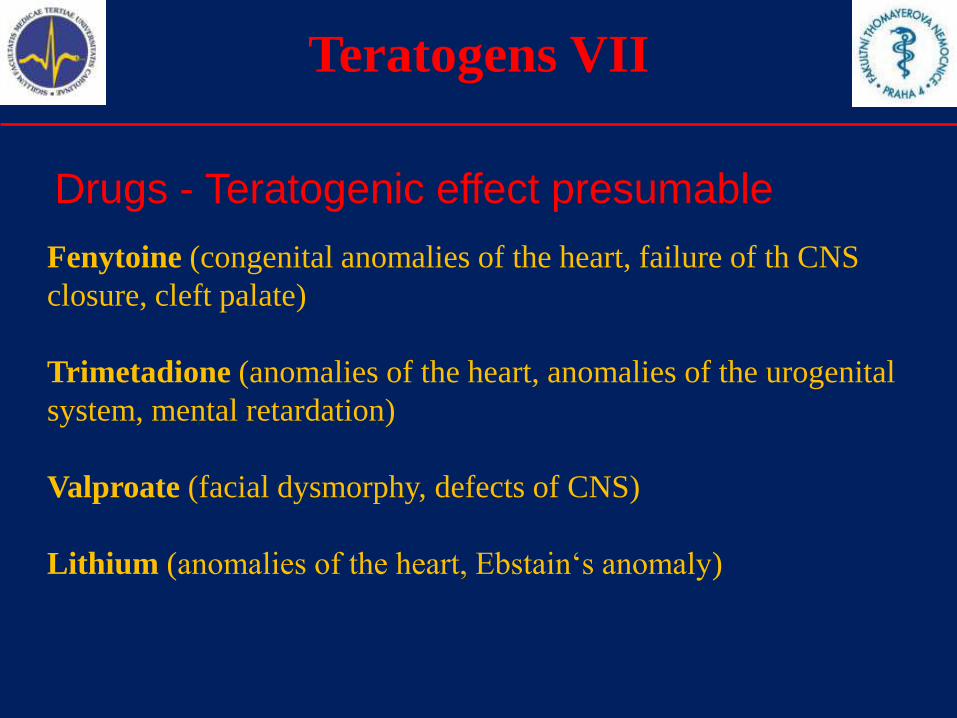
Teratogens VII
Drugs - Teratogenic effect presumable
Fenytoine (congenital anomalies of the heart, failure of th CNS
closure, cleft palate)
Trimetadione (anomalies of the heart, anomalies of the urogenital
system, mental retardation)
Valproate (facial dysmorphy, defects of CNS)
Lithium (anomalies of the heart, Ebstain‘s anomaly)
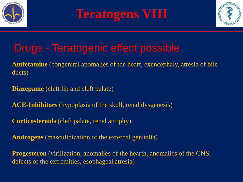
Teratogens VIII
Drugs - Teratogenic effect possible
Amfetamine (congenital anomalies of the heart, exencephaly, atresia of bile
ducts)
Diazepame (cleft lip and cleft palate)
ACE-Inhibitors (hypoplasia of the skull, renal dysgenesis)
Corticosteroids (cleft palate, renal atrophy)
Androgens (masculinization of the external genitalia)
Progesteron (virilization, anomalies of the hearth, anomalies of the CNS,
defects of the extremities, esophageal atresia)
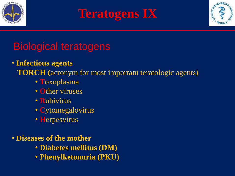
Teratogens IX
Biological teratogens
• Infectious agents
TORCH (acronym for most important teratologic agents)
• Toxoplasma
• Other viruses
• Rubivirus
• Cytomegalovirus
• Herpesvirus
• Diseases of the mother
• Diabetes mellitus (DM)
• Phenylketonuria (PKU)
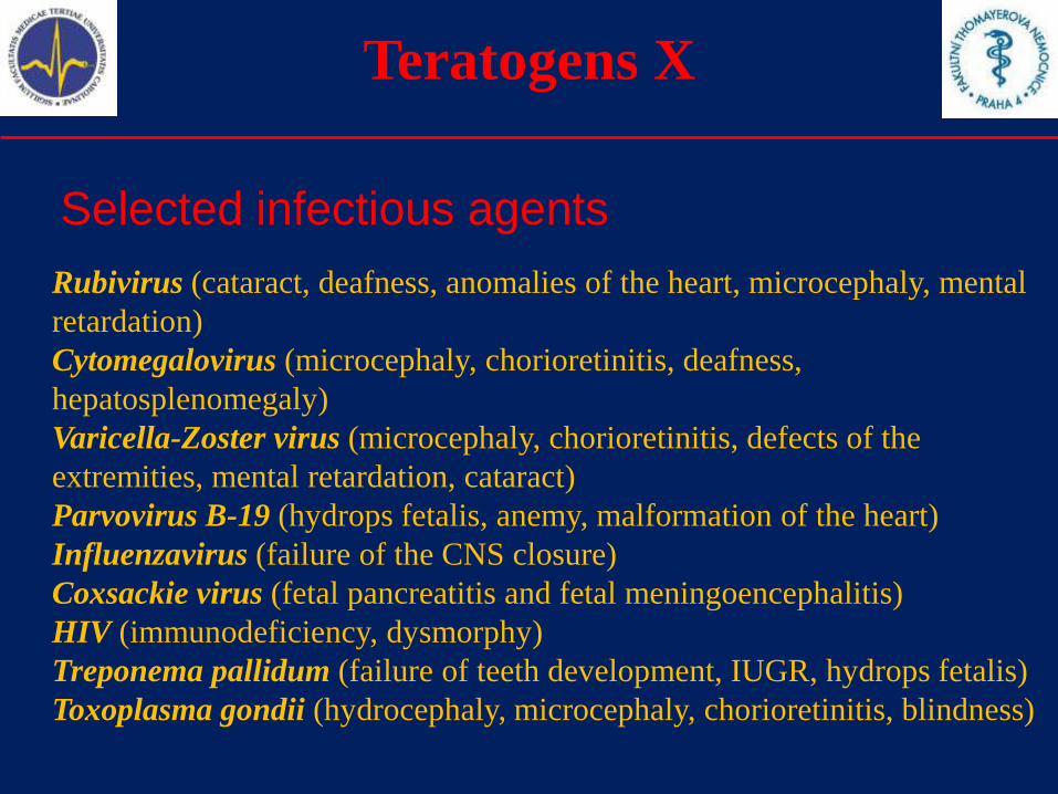
Teratogens X
Selected infectious agents
Rubivirus (cataract, deafness, anomalies of the heart, microcephaly, mental
retardation)
Cytomegalovirus (microcephaly, chorioretinitis, deafness,
hepatosplenomegaly)
Varicella-Zoster virus (microcephaly, chorioretinitis, defects of the
extremities, mental retardation, cataract)
Parvovirus B-19 (hydrops fetalis, anemy, malformation of the heart)
Influenzavirus (failure of the CNS closure)
Coxsackie virus (fetal pancreatitis and fetal meningoencephalitis)
HIV (immunodeficiency, dysmorphy)
Treponema pallidum (failure of teeth development, IUGR, hydrops fetalis)
Toxoplasma gondii (hydrocephaly, microcephaly, chorioretinitis, blindness)
Teratogens XI
Sources of information about drugs
State Institute of Drug Control of Czech Republic
http://www.sukl.cz/
European Medicines Agency
http://www.emea.europa.eu/
U S Food and Drug Administration
http://www.fda.gov/
Teratogens XII
Teratology Information Services / Societies
Czech Teratology Information Service (CZTIS)
http://old.lf3.cuni.cz/histologie/english/33.htm
European Network Teratology Information Services
http://www.entis-org.com/
Organization of Teratology Information Specialists
http://www.otispregnancy.org/
Congenital anomalies
1.Definitions
2.Etiology, teratogens
3.Selected congenital anomalies
4.Prenatal diagnosis
5.Incidence of selected congenital anomalies
6.Summary, credits
Congenital Anomalies I
The groups selected for this lecture
• Neural tube defects (NTD)
• Abdominal wall defects (AWD)
• Congenital anomalies of the kidneys
• Chromosomal aberrations
In descriptions we use the official definitions provided by ICBDSR
organization - http://www.icbdsr.org/
Congenital Anomalies II
From: Smith's Recognizable Patterns Of Human Malformation 6ed
From: Smith's Recognizable Patterns Of Human Malformation 6ed
From: Smith's Recognizable Patterns Of Human Malformation 6ed From: Smith's Recognizable Patterns Of Human Malformation 6edFrom: Smith's Recognizable Patterns Of Human Malformation 6ed
Neural tube defects
Defect in closure of the neural
groove. Normal neural tube is
not formed.
Anencephaly represents a defect
in closure at the anterior part of
the neural groove.
Defects at the mid- or caudal
neural groove cause
meningo(myelo)cele or spina
bifida.
Congenital Anomalies III
Anencephaly
Anencephaly is a congenital anomaly characterized by
the total or partial absence of the cranial vault , the
covering skin, and the brain missing or reduced to
small mass(e).
Include craniorachischisis. Include infants with
iniencephaly and other neural tube defects as
encephalocele or open spina bifida, when associated with
anencephaly.
Exclude acephaly, that is, absence of head observed in
amorphous acardiac twins.
Congenital Anomalies IV
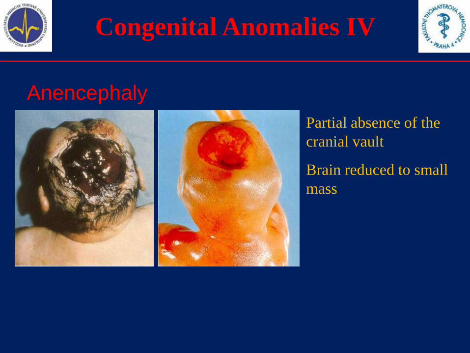
Anencephaly
Partial absence of the
cranial vault
Brain reduced to small
mass
Congenital Anomalies V
Spina Bifida
Spina bifida is a family of congenital anomalies defects
in the closure of the spinal column characterized by
herniation or exposure of the spinal cord and/or
meninges through an incompletely closed spine.
Include meningocele, meningomyelocele, myelocele,
myelomeningocele, rachischisis. Spina bifida is not
counted when present with anencephaly.
Exclude: spina bifida occulta, sacrococcygeal teratoma
without dysraphism.
Congenital Anomalies VI
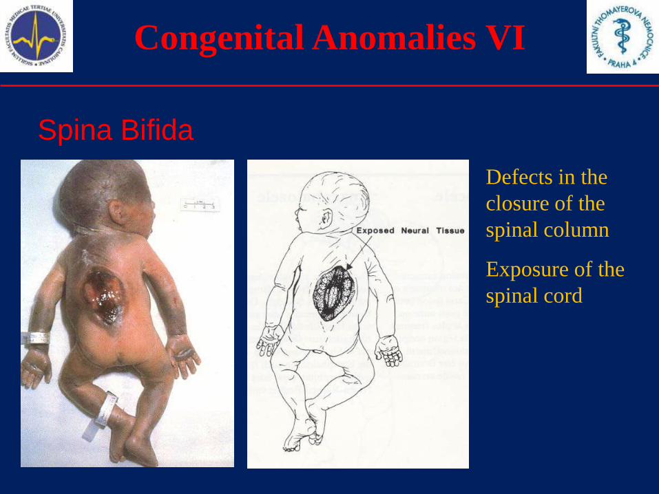
Spina Bifida
Defects in the
closure of the
spinal column
Exposure of the
spinal cord
Congenital Anomalies VII
Encephalocele
Encephalocele is a congenital anomaly characterized by
herniation of the brain and/or meninges through a
defect in the skull. Encephalocele is not counted when
present with spina bifida.
Congenital Anomalies VIII
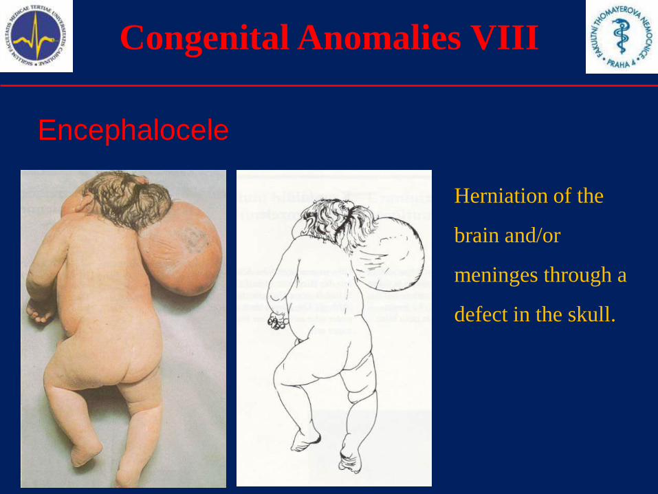
Encephalocele
Herniation of the
brain and/or
meninges through a
defect in the skull.
Congenital Anomalies IX
Omphalocele
Omphalocele is a congenital anomaly characterized by
herniation of abdominal contents through the
umbilical insertion and covered by a membrane which
may or may not be intact.
Exclude gastroschisis (para-umbilical hernia), aplasia or
hypoplasia of abdominal muscles, skin-covered umbilical
hernia.
Congenital Anomalies X
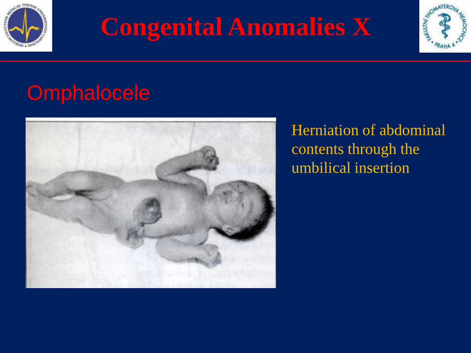
Omphalocele
Herniation of abdominal
contents through the
umbilical insertion
Congenital Anomalies XI
Gastroschisis
Gastroschisis is a congenital anomaly characterized by
visceral herniation through an abdominal wall defect
lateral to an intact umbilical cord and not covered by a
membrane.
Exclude a- or hypoplasia of abdominal muscles, skin-
covered umbilical hernia, omphalocele.
Congenital Anomalies XII
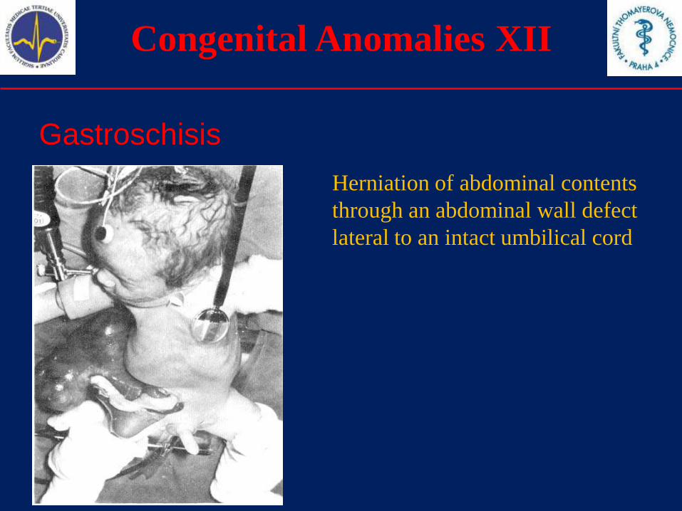
Gastroschisis
Herniation of abdominal contents
through an abdominal wall defect
lateral to an intact umbilical cord
Congenital Anomalies XIII
Diaphragmatic hernia
Diaphragmatic hernia is a congenital anomaly
characterized by herniation into thorax of abdominal
contents through a defect of the diaphragm. Include
total absence of the diaphragm.
Exclude hiatus hernia, eventration and phrenic palsy.
Congenital Anomalies XIV
Hydrocephaly
Hydrocephaly is a congenital anomaly characterized by
dilatation of the cerebral ventricles, not associated
with a primary brain atrophy, with or without
enlargement of the head, and diagnosed at birth.
Not counted when present with encephalocele or spina
bifida.
Exclude: macrocephaly without dilatation of ventricular
system, skull of macerated fetus, hydranencephaly,
holoprosencephaly, and postnatally acquired
hydrocephalus.
Congenital Anomalies XV
Congenital anomalies of the kidneys
Renal agenesis is a congenital anomaly characterized by
complete absence of kidneys bilaterally or severely
dysplastic kidneys.
Cystic kidney is a congenital anomaly characterized by
multiple cysts in the kidney.
Include infantile polycystic kidney, multicystic kidney,
other forms of cystic kidney and unspecified cystic kidney.
Exclude single kidney cyst.
Congenital Anomalies XVI
Polycystic kidney disease - Genetics
Autosomal dominant form (MIM: 173900)
• PKD1 gene mutation (16p13.3-p13.12, MIM: 601313)
• PKD2 gene mutation (4q21-q23, MIM: 173910)
• PKD3 gene mutation ?? (not yet localized, MIM: 600666)
Autosomal recessive form (MIM: 263200)
• PKHD1 gene mutation (6p21.1-p12, MIM: 606702)

Congenital Anomalies XVII
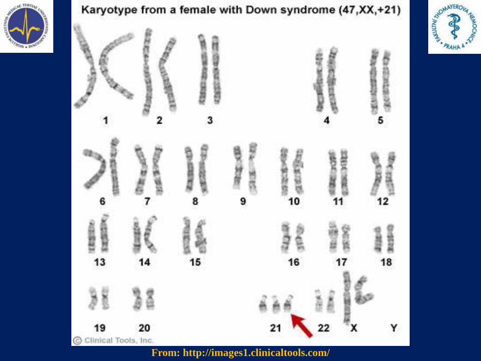
Down syndrome
Down syndrome is a congenital chromosomal anomaly
syndrome characterized by a well known pattern of minor
and major anomalies and associated with excess
chromosomal 21 material.
Include trisomy mosaicism and translocations of
chromosome 21.
Common karyotype – trisomy 21
47, XX, +21 (female) or 47, XY, +21 (male)
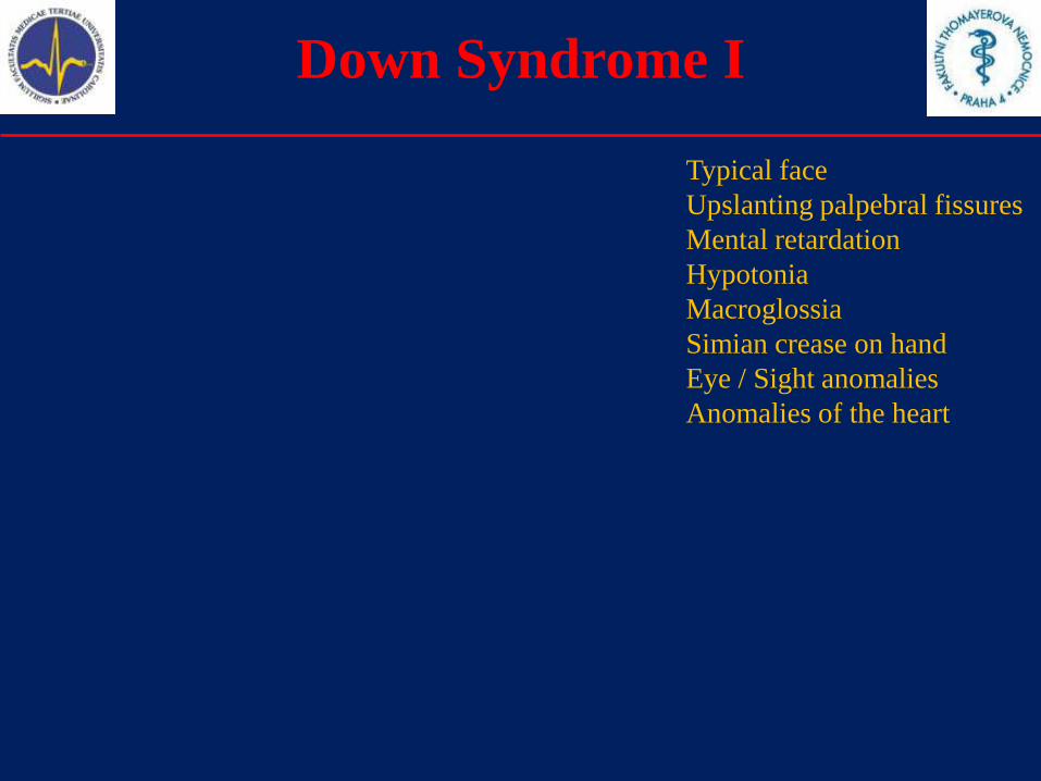
Down Syndrome I
From: Smith's Recognizable Patterns Of Human Malformation 6edTypical face
Upslanting palpebral fissures
Mental retardation
Hypotonia
Macroglossia
Simian crease on hand
Eye / Sight anomalies
Anomalies of the heart
From: http://images1.clinicaltools.com/
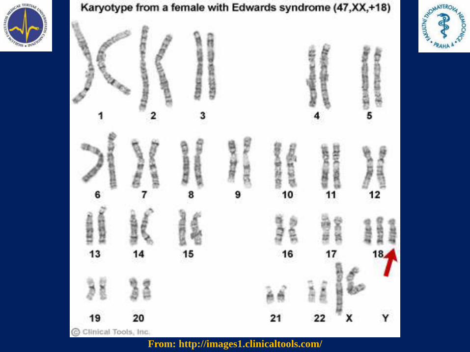
Congenital Anomalies XVIII
Edwards syndrome
Edwards syndrome is a congenital chromosomal anomaly
syndrome characterized by a well known pattern of minor
and major anomalies and associated with extra chromosome
18 material.
Include translocation and mosaic trisomy 18.
Common karyotype – trisomy 18
47, XX, +18 (female) or 47, XY, +18 (male)
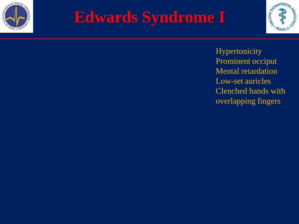
Edwards Syndrome I
From: Smith's Recognizable Patterns Of Human Malformation 6ed
From: Smith's Recognizable Patterns Of Human Malformation 6ed Hypertonicity
Prominent occiput
Mental retardation
Low-set auricles
Clenched hands with
overlapping fingers
From: http://images1.clinicaltools.com/
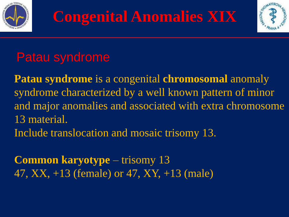
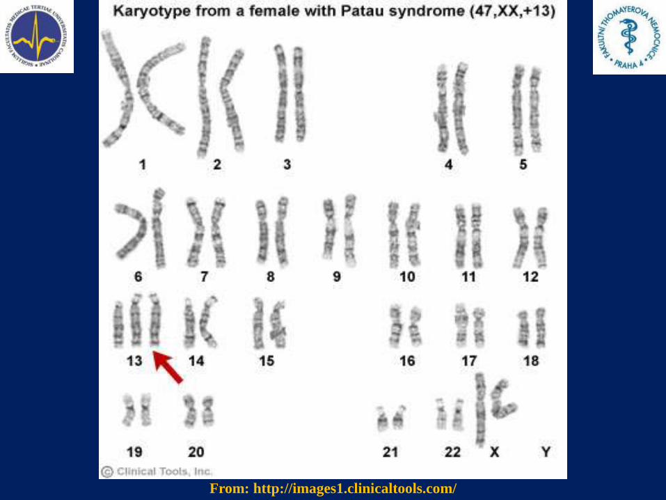
Congenital Anomalies XIX
Patau syndrome
Patau syndrome is a congenital chromosomal anomaly
syndrome characterized by a well known pattern of minor
and major anomalies and associated with extra chromosome
13 material.
Include translocation and mosaic trisomy 13.
Common karyotype – trisomy 13
47, XX, +13 (female) or 47, XY, +13 (male)
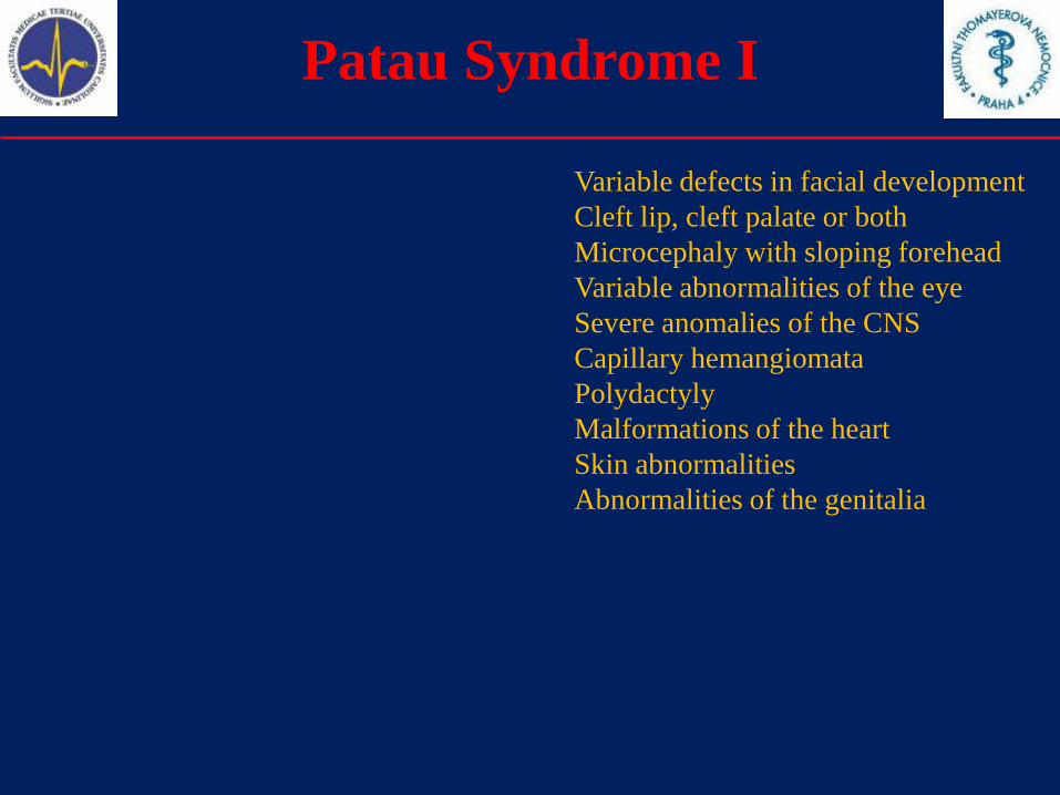
Patau Syndrome I
From: Smith's Recognizable Patterns Of Human Malformation 6ed
From: Smith's Recognizable Patterns Of Human Malformation 6ed
From: Smith's Recognizable Patterns Of Human Malformation 6ed
Variable defects in facial development
Cleft lip, cleft palate or both
Microcephaly with sloping forehead
Variable abnormalities of the eye
Severe anomalies of the CNS
Capillary hemangiomata
Polydactyly
Malformations of the heart
Skin abnormalities
Abnormalities of the genitalia
From: http://images1.clinicaltools.com/
Congenital Anomalies XX
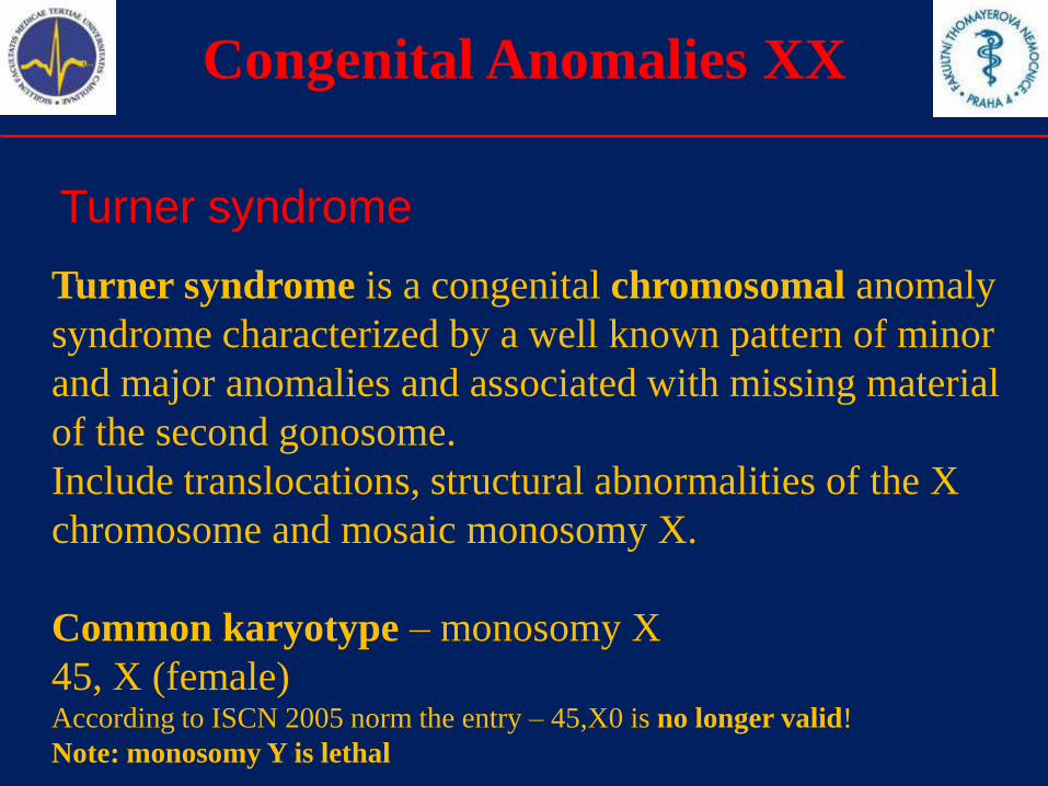
Turner syndrome
Turner syndrome is a congenital chromosomal anomaly
syndrome characterized by a well known pattern of minor
and major anomalies and associated with missing material
of the second gonosome.
Include translocations, structural abnormalities of the X
chromosome and mosaic monosomy X.
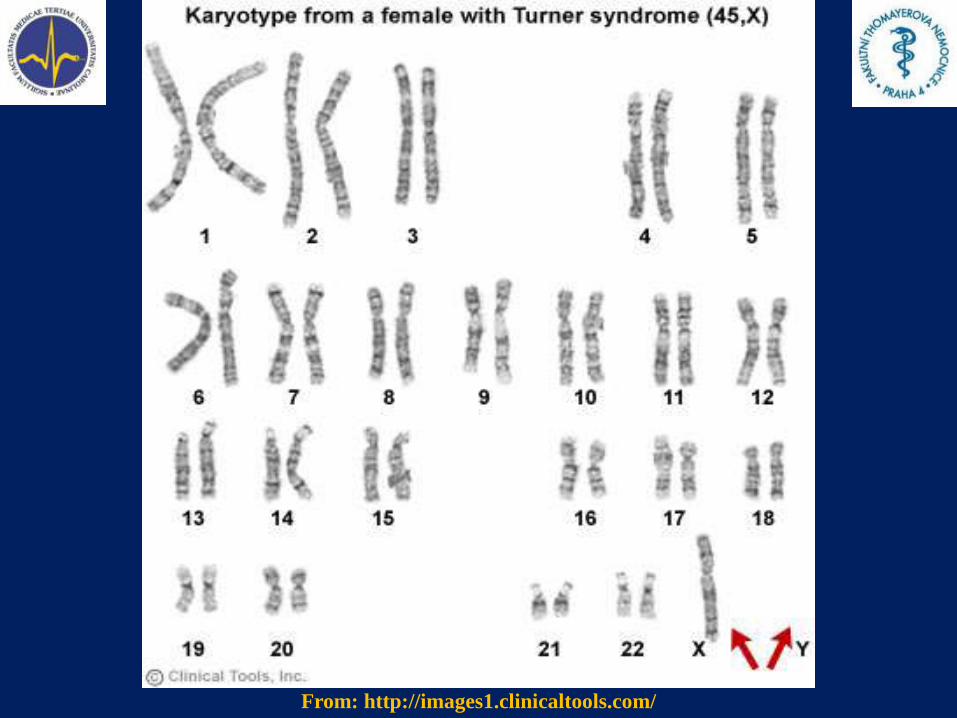
Common karyotype – monosomy X
45, X (female)According to ISCN 2005 norm the entry – 45,X0 is no longer valid!
Note: monosomy Y is lethal
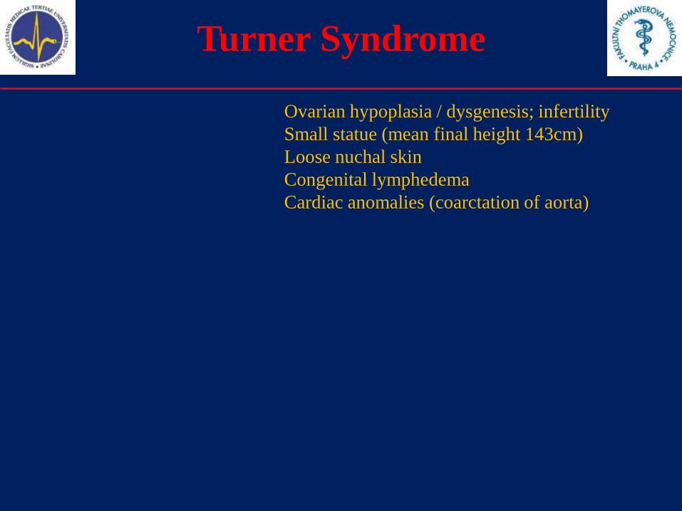
Turner Syndrome
From: Smith's Recognizable Patterns Of Human Malformation 6ed
From: Smith's Recognizable Patterns Of Human Malformation 6ed
From: Smith's Recognizable Patterns Of Human Malformation 6edFrom: Smith's Recognizable Patterns Of Human Malformation 6ed
Ovarian hypoplasia / dysgenesis; infertility
Small statue (mean final height 143cm)
Loose nuchal skin
Congenital lymphedema
Cardiac anomalies (coarctation of aorta)
From: http://images1.clinicaltools.com/
Congenital Anomalies XXI
Useful links
OMIM – Online Mendelian Inheritance in Man
http://www.ncbi.nlm.nih.gov/sites/entrez?db=omim
eMedicine - The original open access comprehensive medical textbook
http://www.emedicine.com/
Orphanet - The portal for rare diseases and orphan drugs
http://www.orpha.net/
Congenital anomalies
1.Definitions
2.Etiology, teratogens
3.Selected congenital anomalies
4.Prenatal diagnosis
5.Incidence of selected congenital anomalies
6.Summary
Prenatal diagnosis I
Primary prevention
The main goal is to prevent anomaly or malformation before they
develop (that means before conception or during pregnancy).
The women should avoid the pregnancy in very low or very high
age. The pregnancy should be planned.
The parents should avoid any contact with mutagens or teratogens.
No stress, smoking, drugs and alcohol during pregnancy.
Clinical geneticist should be consulted in advance – if necessary
(repeated abortions, congenital anomalies in family, genetic
diseases).
Good compensation of mother‘s diseases (DM, PKU etc.)
Supplementation with folic acid
Prenatal diagnosis II
Secondary prevention
The main goal is to prevent the birth of a child with a congenital
anomaly. However, the termination itself is not a prevention.
We can terminate the pregnancy in order to prevent such a birth.
However, the termination may not be legal in each country.
In the Czech Republic it is legal to terminate the pregnancy
because of severe genetic reasons till 24th week of pregnancy.
Prenatal diagnosis is therefore very important, because we need the
best information available about the condition of the fetus. If a
severe condition is diagnosed, we may offer termination of
pregnancy, prenatal therapy or special treatment in perinatologic
period.
Prenatal diagnosis III
Methods
There are two main groups of methods in prenatal diagnosis:
Invasive methodsAmniocentesis (AMC), Chorionic villus sampling (CVS),
Cordocentesis (CC), Fetoscopy, Fetal biopsy
Noninvasive methodsBiochemical screening (requires mother‘s blood sample),
Ultrasound, Magnetic resonance
Prenatal diagnosis IV
Amniocentesis
Amniocentesis is commonly used invasive method, that is able to
obtain a sample of the amniotic fluid, including amniocytes. Those
cells can be cultivated for cytogenetic analysis. Using QF-PCR we
can have preliminary results (for most common trisomies) in 2 days
(rather than in 2 weeks – we need for cultivation and karyotype).
Early amniocentesis can be made earlier then the standard
amniocentesis (14th week – rather than 16th week). However, we
cannot obtain so much amniotic fluid.
Prenatal diagnosis V
Other Invasive methods
CVS can be made very soon, after the 11th week of gestation. This
allows early diagnosis. However, there is a risk of confined placental
mosaicism (CPM), what can make the decision more difficult.
Cordocentesis is usually made after the 20th week. The sample of
blood allows us to make hematologic tests, as well as the cultivation.
Fetoscopy and fetal biopsy are very rare today. Fetoscopy can be
used in order to confirm some dermatologic anomalies (Ichtyosis
vulgaris). Biopsy can obtain a sample of fetal skin (or other tissue) for
histopathological analysis.
Prenatal diagnosis VI
Biochemical screenings
Combined screening of the first trimester
This test combines the ultrasound diagnostics (NT – Nuchal
Translucency measurement, NB – nasal bone presence / absence,
tricuspidal regurgitation) with biochemical test of maternal serum
(using PAPP-A and free beta-HCG subunit).
Some other biochemical markers can be examined in 1st trimester.
Biochemical screening of the second trimester
The blood sample from the mother is obtained usually after 16th
week. uE3 (unconjugated estriol), HCG (human chorionic
gonadotropine) and AFP (alfa-fetoprotein) are analyzed.
Prenatal diagnosis VII
Imaging methods
Ultrasound diagnostics
Ultrasound diagnosis is commonly used and provide detailed
morphological information. Therefore - the majority of structural
anomalies is diagnosed thanks to USG diagnostics. Special methods –
like fetal echocardiography – may provide additional information.
Magnetic resonance imaging
MRI is relatively new method in prenatal diagnosis and is still quite
rare. The image processing must be very quick, because the fetus is
moving. However - this method is excellent in detection of brain and
other soft-tissue anomalies.
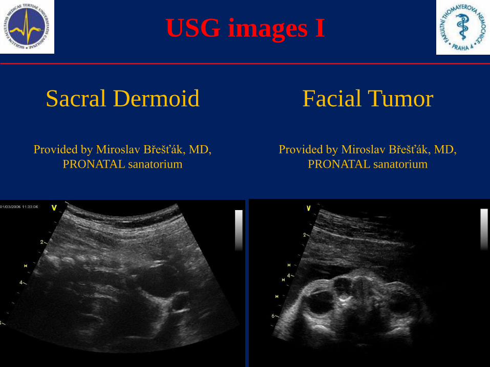
USG images I
Sacral Dermoid
Provided by Miroslav Břešťák, MD,
PRONATAL sanatorium
Facial Tumor
Provided by Miroslav Břešťák, MD,
PRONATAL sanatorium

USG images II
Nasal Bone absence
Provided by Miroslav Břešťák, MD,
PRONATAL sanatorium
Polycystic Kidney
Provided by Miroslav Břešťák, MD,
PRONATAL sanatorium
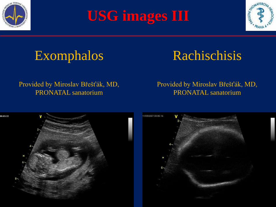
USG images III
Exomphalos
Provided by Miroslav Břešťák, MD,
PRONATAL sanatorium
Rachischisis
Provided by Miroslav Břešťák, MD,
PRONATAL sanatorium
Prenatal diagnosis VIII
Preimplantation genetic diagnostics
PGD is a special method that is used during In Vitro Fertilization
process. The material for diagnosis (either cytogenetic or molecular-
genetic) is obtained before the embryo is implanted (one blastomere
or a polar body may be used for this diagnosis).
This method may be extremely useful for parents with balanced
chromosomal translocations or for parents who are carriers of severe
genetic diseases (e.g. muscular dystrophies).
Only the embryos with normal karyotype / without genetic mutation
shall be implanted.
However, there can be problem with blastomeric mosaicism, that can
cause false-positive or false-negative results.
Prenatal diagnosis
History of prenatal diagnosis
Prenatal diagnosis in the Czech Republic
1970 – cultivation of the amniocytes
1971 – first prenatally diagnosed Down Syndrome
1977 – placentocentesis
1978 – fetoscopy
1980 – ultrasound diagnostics
1983 – CVS
1985 – prenatal molecular-genetic analysis – hemophilia
1987 – cordocentesis
1988 – early amniocentesis
1990 – prenatal biochemical screening
2000 – preimplantation genetic diagnostics (PGD)
Congenital anomalies
1.Definitions
2.Etiology, teratogens
3.Selected congenital anomalies
4.Prenatal diagnosis
5.Incidence of selected congenital anomalies
6.Summary
Population teratology
Definitions
Population teratology analyses the mean incidences of
congenital anomalies (CAs) in selected population. Long,
consensual monitoring is necessary in order to find any
trends that may appear in the incidence of some
congenital anomalies (either increase or decrease).
Other findings may be accumulation of CAs, clustering
of CAs, cyclic changes in the incidence of CAs, or so
called nesting.
Incidence I
Czech Republic
History of Congenital Anomalies Monitoring Program of Czech
Republic is very long. The regular monitoring started in 1964. The
registration itself undergo many changes during more than 40 years
of existence.
Currently we register any diagnosis from ICD-X Q00-Q99 group,
that was diagnosed prenatally or postnatally until (finished) 15th
year of live.
The data for the registry are collected in the Institute of Health
Information and Statistics of the Czech Republic (ÚZIS ČR).
The Czech Registry was one of founding members of ICBDSR
(International Clearinghouse for Birth Defects Surveillance and
Research) international organization.
Incidence II
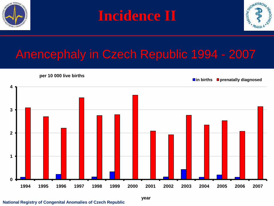
Anencephaly in Czech Republic 1994 - 2007
0
1
2
3
4
1994 1995 1996 1997 1998 1999 2000 2001 2002 2003 2004 2005 2006 2007
in births prenatally diagnosedper 10 000 live births
yearNational Registry of Congenital Anomalies of Czech Republic

Incidence III
Anencephaly in Czech Republic 1994 - 2007
0
10
20
30
40
50
60
70
80
90
100
1994 1995 1996 1997 1998 1999 2000 2001 2002 2003 2004 2005 2006 2007
year
efficiency of prenatal diagnosis - %
National Registry of Congenital Anomalies of Czech Republic
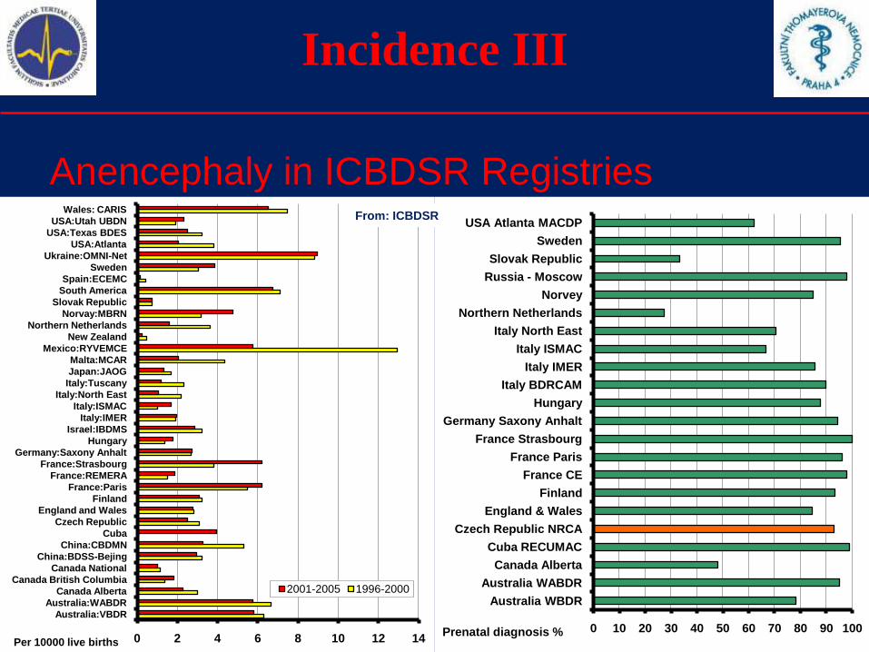
Incidence III
Anencephaly in ICBDSR Registries
0 10 20 30 40 50 60 70 80 90 100
Australia WBDR
Australia WABDR
Canada Alberta
Cuba RECUMAC
Czech Republic NRCA
England & Wales
Finland
France CE
France Paris
France Strasbourg
Germany Saxony Anhalt
Hungary
Italy BDRCAM
Italy IMER
Italy ISMAC
Italy North East
Northern Netherlands
Norvey
Russia - Moscow
Slovak Republic
Sweden
USA Atlanta MACDP
Prenatal diagnosis %0 2 4 6 8 10 12 14
Australia:VBDR
Australia:WABDR
Canada Alberta
Canada British Columbia
Canada National
China:BDSS-Bejing
China:CBDMN
Cuba
Czech Republic
England and Wales
Finland
France:Paris
France:REMERA
France:Strasbourg
Germany:Saxony Anhalt
Hungary
Israel:IBDMS
Italy:IMER
Italy:ISMAC
Italy:North East
Italy:Tuscany
Japan:JAOG
Malta:MCAR
Mexico:RYVEMCE
New Zealand
Northern Netherlands
Norvay:MBRN
Slovak Republic
South America
Spain:ECEMC
Sweden
Ukraine:OMNI-Net
USA:Atlanta
USA:Texas BDES
USA:Utah UBDN
Wales: CARIS
2001-2005 1996-2000
Per 10000 live births
From: ICBDSR
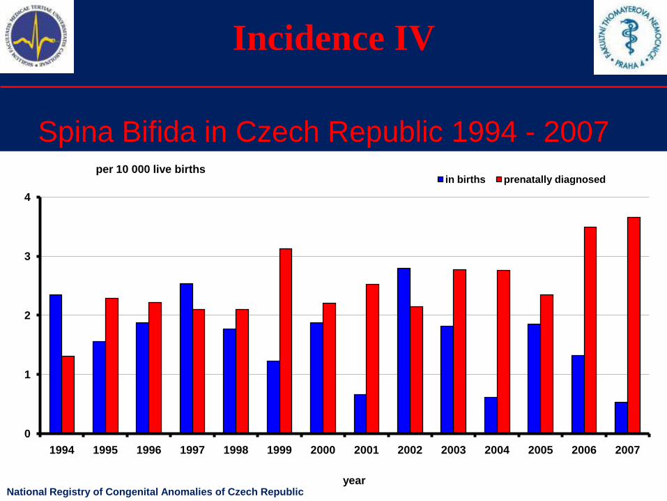
Incidence IV
Spina Bifida in Czech Republic 1994 - 2007
0
1
2
3
4
1994 1995 1996 1997 1998 1999 2000 2001 2002 2003 2004 2005 2006 2007
in births prenatally diagnosedper 10 000 live births
yearNational Registry of Congenital Anomalies of Czech Republic
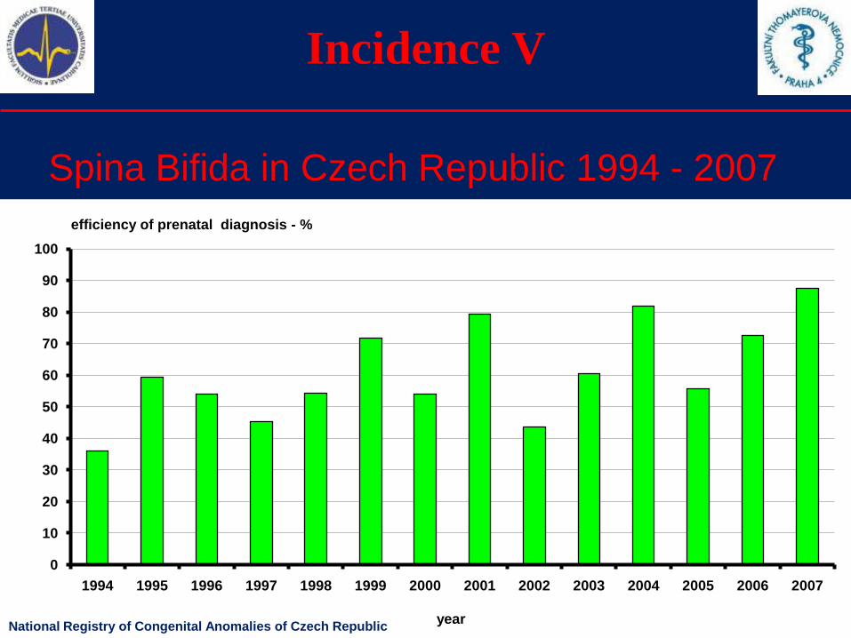
Incidence V
Spina Bifida in Czech Republic 1994 - 2007
0
10
20
30
40
50
60
70
80
90
100
1994 1995 1996 1997 1998 1999 2000 2001 2002 2003 2004 2005 2006 2007
year
efficiency of prenatal diagnosis - %
National Registry of Congenital Anomalies of Czech Republic
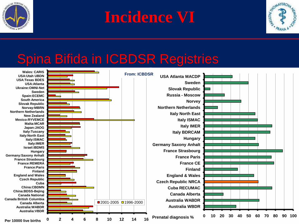
Incidence VI
Spina Bifida in ICBDSR Registries
0 2 4 6 8 10 12 14 16
Australia:VBDR
Australia:WABDR
Canada Alberta
Canada British Columbia
Canada National
China:BDSS-Bejing
China:CBDMN
Cuba
Czech Republic
England and Wales
Finland
France:Paris
France:REMERA
France:Strasbourg
Germany:Saxony Anhalt
Hungary
Israel:IBDMS
Italy:IMER
Italy:ISMAC
Italy:North East
Italy:Tuscany
Japan:JAOG
Malta:MCAR
Mexico:RYVEMCE
New Zealand
Northern Netherlands
Norvay:MBRN
Slovak Republic
South America
Spain:ECEMC
Sweden
Ukraine:OMNI-Net
USA:Atlanta
USA:Texas BDES
USA:Utah UBDN
Wales: CARIS
2001-2005 1996-2000
Per 10000 live births
0 10 20 30 40 50 60 70 80 90 100
Australia WBDR
Australia WABDR
Canada Alberta
Cuba RECUMAC
Czech Republic NRCA
England & Wales
Finland
France CE
France Paris
France Strasbourg
Germany Saxony Anhalt
Hungary
Italy BDRCAM
Italy IMER
Italy ISMAC
Italy North East
Northern Netherlands
Norvey
Russia - Moscow
Slovak Republic
Sweden
USA Atlanta MACDP
Prenatal diagnosis %
From: ICBDSR
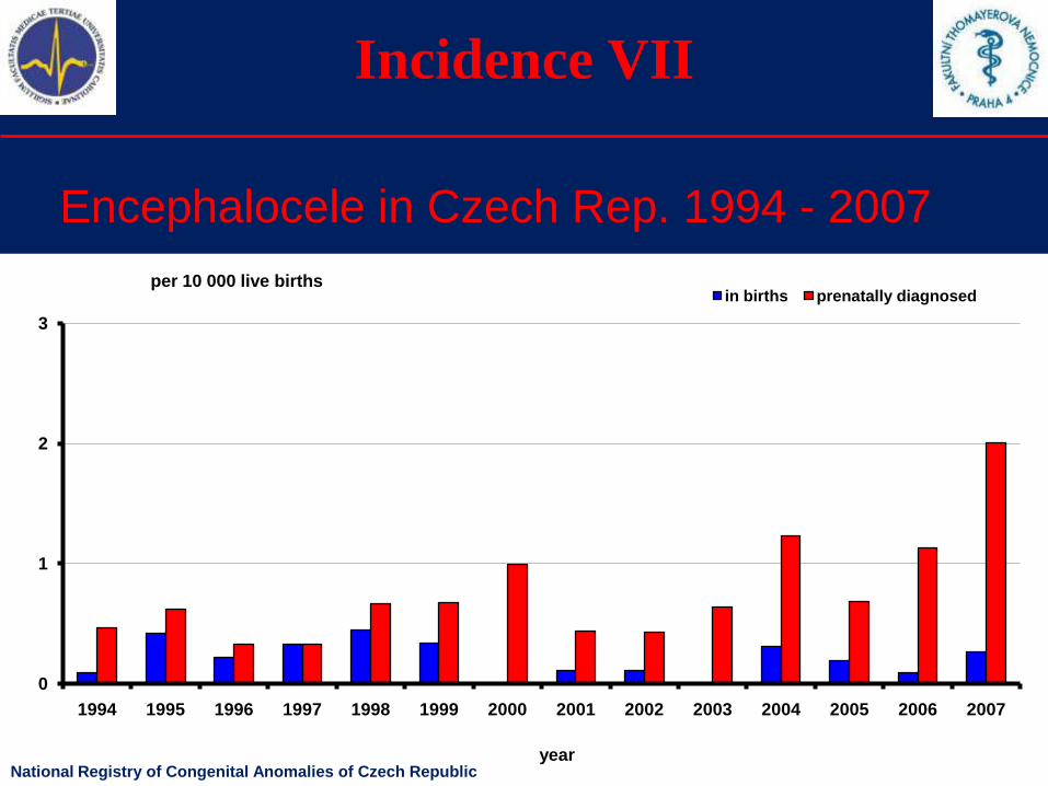
Incidence VII
Encephalocele in Czech Rep. 1994 - 2007
0
1
2
3
1994 1995 1996 1997 1998 1999 2000 2001 2002 2003 2004 2005 2006 2007
in births prenatally diagnosedper 10 000 live births
yearNational Registry of Congenital Anomalies of Czech Republic
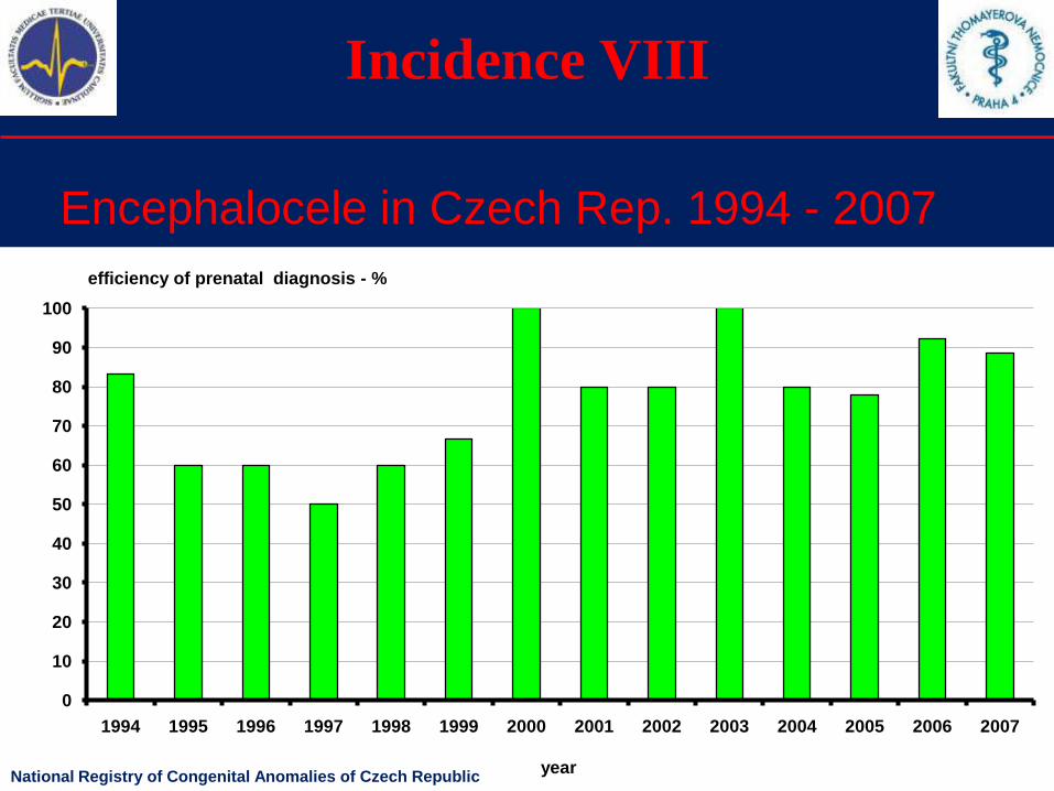
Incidence VIII
Encephalocele in Czech Rep. 1994 - 2007
0
10
20
30
40
50
60
70
80
90
100
1994 1995 1996 1997 1998 1999 2000 2001 2002 2003 2004 2005 2006 2007
year
efficiency of prenatal diagnosis - %
National Registry of Congenital Anomalies of Czech Republic
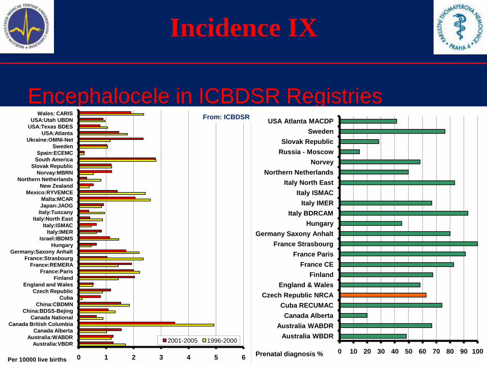
Incidence IX
Encephalocele in ICBDSR Registries
0 1 2 3 4 5 6
Australia:VBDR
Australia:WABDR
Canada Alberta
Canada British Columbia
Canada National
China:BDSS-Bejing
China:CBDMN
Cuba
Czech Republic
England and Wales
Finland
France:Paris
France:REMERA
France:Strasbourg
Germany:Saxony Anhalt
Hungary
Israel:IBDMS
Italy:IMER
Italy:ISMAC
Italy:North East
Italy:Tuscany
Japan:JAOG
Malta:MCAR
Mexico:RYVEMCE
New Zealand
Northern Netherlands
Norvay:MBRN
Slovak Republic
South America
Spain:ECEMC
Sweden
Ukraine:OMNI-Net
USA:Atlanta
USA:Texas BDES
USA:Utah UBDN
Wales: CARIS
2001-2005 1996-2000
Per 10000 live births
0 10 20 30 40 50 60 70 80 90 100
Australia WBDR
Australia WABDR
Canada Alberta
Cuba RECUMAC
Czech Republic NRCA
England & Wales
Finland
France CE
France Paris
France Strasbourg
Germany Saxony Anhalt
Hungary
Italy BDRCAM
Italy IMER
Italy ISMAC
Italy North East
Northern Netherlands
Norvey
Russia - Moscow
Slovak Republic
Sweden
USA Atlanta MACDP
Prenatal diagnosis %
From: ICBDSR
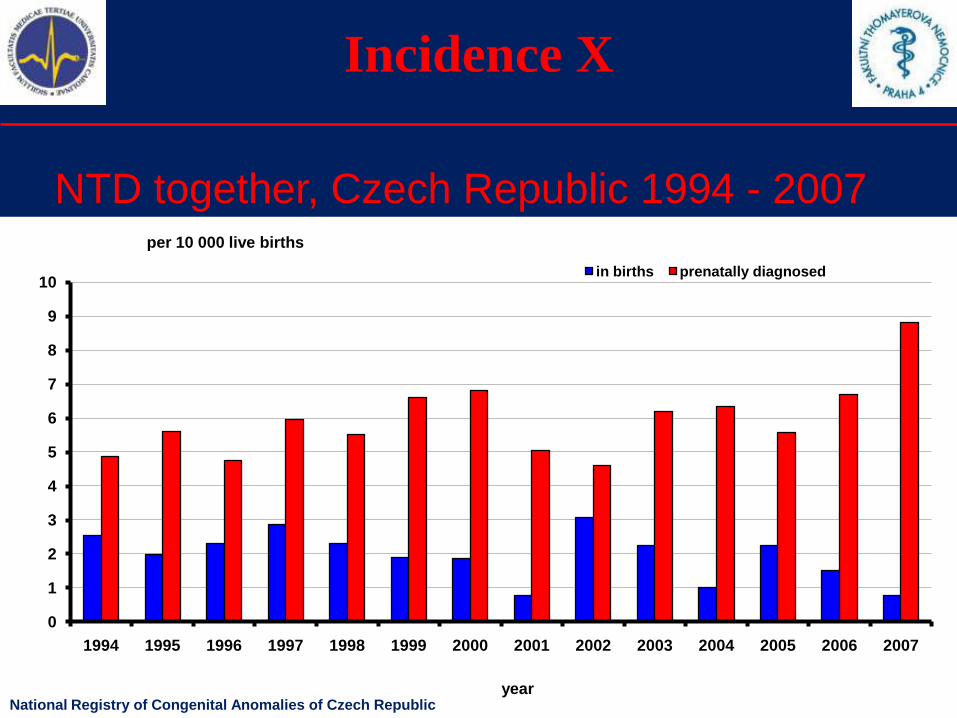
Incidence X
NTD together, Czech Republic 1994 - 2007
0
1
2
3
4
5
6
7
8
9
10
1994 1995 1996 1997 1998 1999 2000 2001 2002 2003 2004 2005 2006 2007
in births prenatally diagnosed
per 10 000 live births
yearNational Registry of Congenital Anomalies of Czech Republic
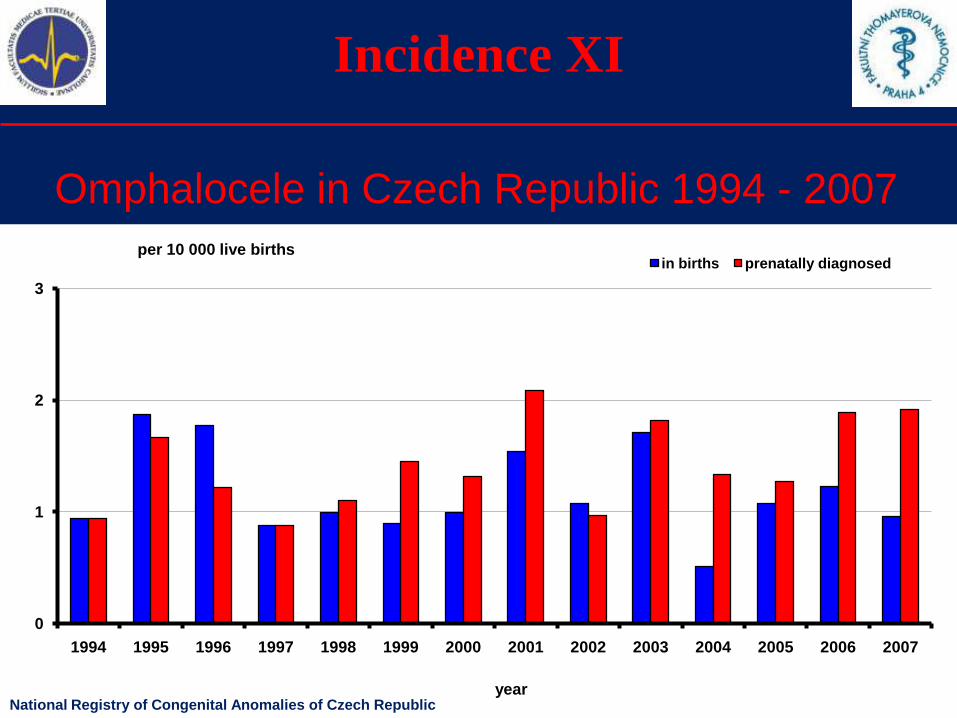
Incidence XI
Omphalocele in Czech Republic 1994 - 2007
0
1
2
3
1994 1995 1996 1997 1998 1999 2000 2001 2002 2003 2004 2005 2006 2007
in births prenatally diagnosedper 10 000 live births
yearNational Registry of Congenital Anomalies of Czech Republic
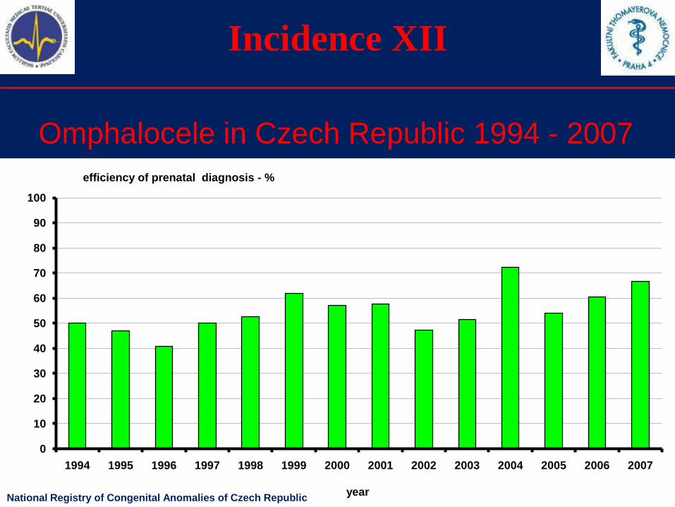
Incidence XII
Omphalocele in Czech Republic 1994 - 2007
0
10
20
30
40
50
60
70
80
90
100
1994 1995 1996 1997 1998 1999 2000 2001 2002 2003 2004 2005 2006 2007
year
efficiency of prenatal diagnosis - %
National Registry of Congenital Anomalies of Czech Republic
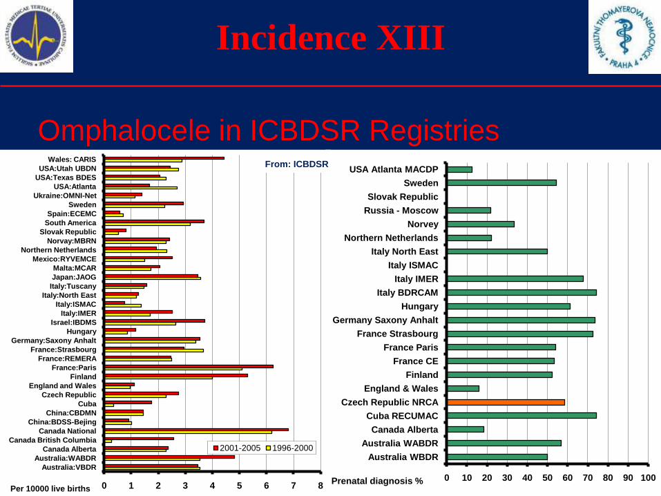
Incidence XIII
Omphalocele in ICBDSR Registries
0 10 20 30 40 50 60 70 80 90 100
Australia WBDR
Australia WABDR
Canada Alberta
Cuba RECUMAC
Czech Republic NRCA
England & Wales
Finland
France CE
France Paris
France Strasbourg
Germany Saxony Anhalt
Hungary
Italy BDRCAM
Italy IMER
Italy ISMAC
Italy North East
Northern Netherlands
Norvey
Russia - Moscow
Slovak Republic
Sweden
USA Atlanta MACDP
Prenatal diagnosis %0 1 2 3 4 5 6 7 8
Australia:VBDR
Australia:WABDR
Canada Alberta
Canada British Columbia
Canada National
China:BDSS-Bejing
China:CBDMN
Cuba
Czech Republic
England and Wales
Finland
France:Paris
France:REMERA
France:Strasbourg
Germany:Saxony Anhalt
Hungary
Israel:IBDMS
Italy:IMER
Italy:ISMAC
Italy:North East
Italy:Tuscany
Japan:JAOG
Malta:MCAR
Mexico:RYVEMCE
Northern Netherlands
Norvay:MBRN
Slovak Republic
South America
Spain:ECEMC
Sweden
Ukraine:OMNI-Net
USA:Atlanta
USA:Texas BDES
USA:Utah UBDN
Wales: CARIS
2001-2005 1996-2000
Per 10000 live births
From: ICBDSR
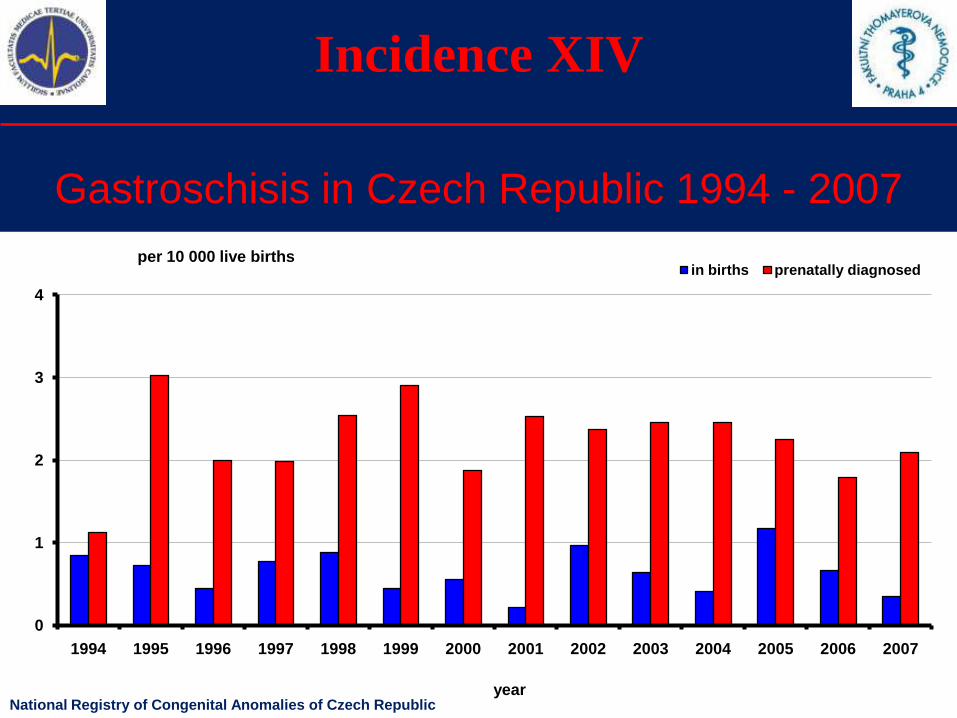
Incidence XIV
Gastroschisis in Czech Republic 1994 - 2007
0
1
2
3
4
1994 1995 1996 1997 1998 1999 2000 2001 2002 2003 2004 2005 2006 2007
in births prenatally diagnosedper 10 000 live births
yearNational Registry of Congenital Anomalies of Czech Republic
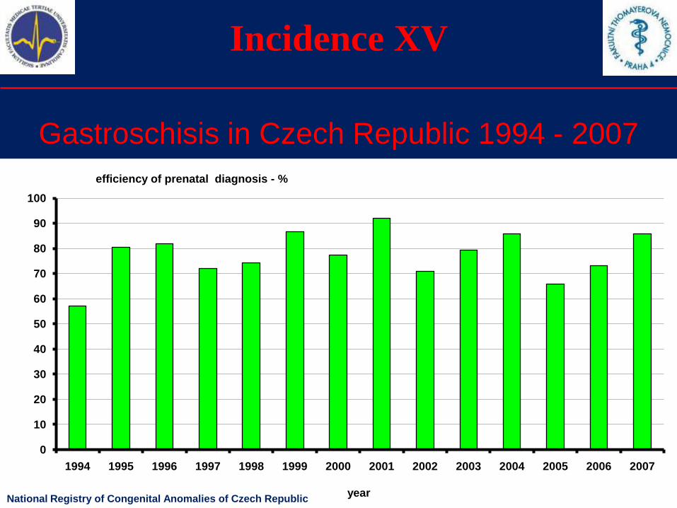
Incidence XV
Gastroschisis in Czech Republic 1994 - 2007
0
10
20
30
40
50
60
70
80
90
100
1994 1995 1996 1997 1998 1999 2000 2001 2002 2003 2004 2005 2006 2007
year
efficiency of prenatal diagnosis - %
National Registry of Congenital Anomalies of Czech Republic
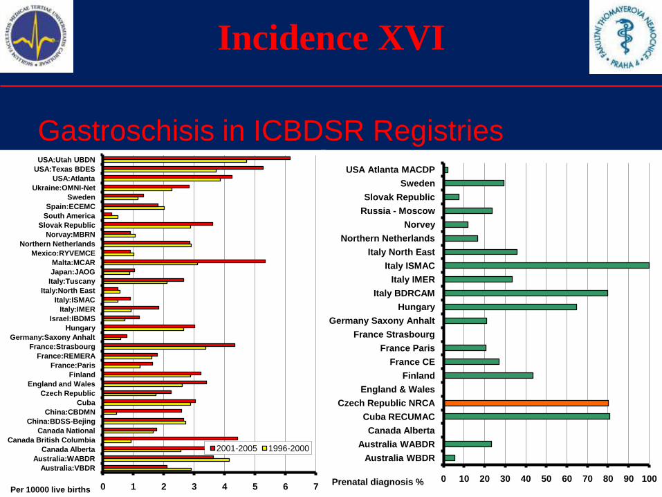
Incidence XVI
Gastroschisis in ICBDSR Registries
0 10 20 30 40 50 60 70 80 90 100
Australia WBDR
Australia WABDR
Canada Alberta
Cuba RECUMAC
Czech Republic NRCA
England & Wales
Finland
France CE
France Paris
France Strasbourg
Germany Saxony Anhalt
Hungary
Italy BDRCAM
Italy IMER
Italy ISMAC
Italy North East
Northern Netherlands
Norvey
Russia - Moscow
Slovak Republic
Sweden
USA Atlanta MACDP
Prenatal diagnosis %0 1 2 3 4 5 6 7
Australia:VBDR
Australia:WABDR
Canada Alberta
Canada British Columbia
Canada National
China:BDSS-Bejing
China:CBDMN
Cuba
Czech Republic
England and Wales
Finland
France:Paris
France:REMERA
France:Strasbourg
Germany:Saxony Anhalt
Hungary
Israel:IBDMS
Italy:IMER
Italy:ISMAC
Italy:North East
Italy:Tuscany
Japan:JAOG
Malta:MCAR
Mexico:RYVEMCE
Northern Netherlands
Norvay:MBRN
Slovak Republic
South America
Spain:ECEMC
Sweden
Ukraine:OMNI-Net
USA:Atlanta
USA:Texas BDES
USA:Utah UBDN
2001-2005 1996-2000
Per 10000 live births
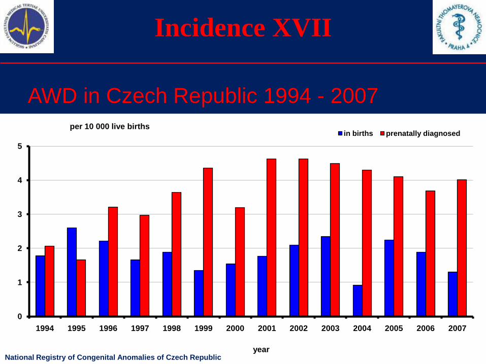
Incidence XVII
AWD in Czech Republic 1994 - 2007
0
1
2
3
4
5
1994 1995 1996 1997 1998 1999 2000 2001 2002 2003 2004 2005 2006 2007
in births prenatally diagnosedper 10 000 live births
yearNational Registry of Congenital Anomalies of Czech Republic
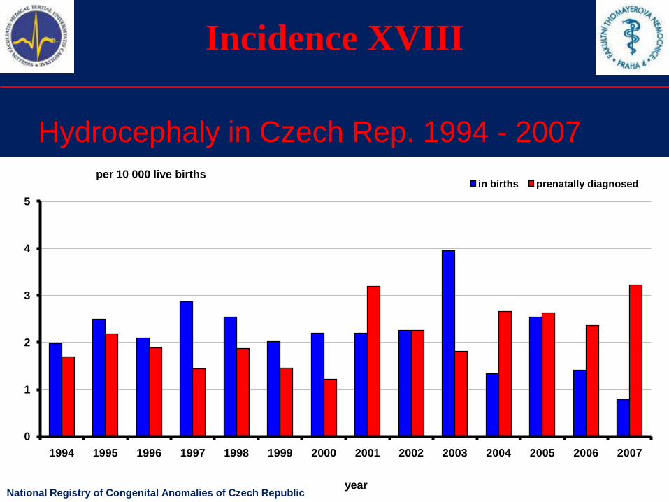
Incidence XVIII
Hydrocephaly in Czech Rep. 1994 - 2007
0
1
2
3
4
5
1994 1995 1996 1997 1998 1999 2000 2001 2002 2003 2004 2005 2006 2007
in births prenatally diagnosedper 10 000 live births
yearNational Registry of Congenital Anomalies of Czech Republic
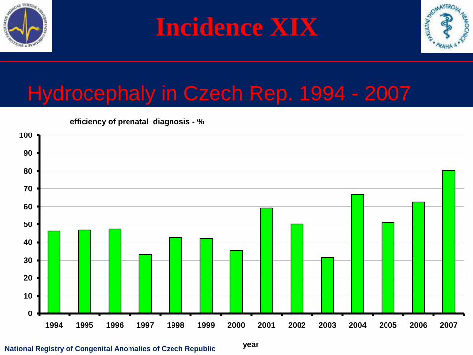
Incidence XIX
Hydrocephaly in Czech Rep. 1994 - 2007
0
10
20
30
40
50
60
70
80
90
100
1994 1995 1996 1997 1998 1999 2000 2001 2002 2003 2004 2005 2006 2007
year
efficiency of prenatal diagnosis - %
National Registry of Congenital Anomalies of Czech Republic
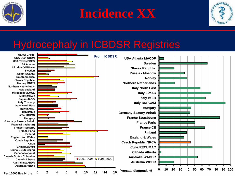
Incidence XX
Hydrocephaly in ICBDSR Registries
0 10 20 30 40 50 60 70 80 90 100
Australia WBDR
Australia WABDR
Canada Alberta
Cuba RECUMAC
Czech Republic NRCA
England & Wales
Finland
France CE
France Paris
France Strasbourg
Germany Saxony Anhalt
Hungary
Italy BDRCAM
Italy IMER
Italy ISMAC
Italy North East
Northern Netherlands
Norvey
Russia - Moscow
Slovak Republic
Sweden
USA Atlanta MACDP
Prenatal diagnosis %0 2 4 6 8 10 12 14 16
Australia:VBDR
Australia:WABDR
Canada Alberta
Canada British Columbia
Canada National
China:BDSS-Bejing
China:CBDMN
Cuba
Czech Republic
England and Wales
Finland
France:Paris
France:REMERA
France:Strasbourg
Germany:Saxony Anhalt
Hungary
Israel:IBDMS
Italy:IMER
Italy:ISMAC
Italy:North East
Italy:Tuscany
Japan:JAOG
Malta:MCAR
Mexico:RYVEMCE
New Zealand
Northern Netherlands
Norvay:MBRN
Slovak Republic
South America
Spain:ECEMC
Sweden
Ukraine:OMNI-Net
USA:Atlanta
USA:Texas BDES
USA:Utah UBDN
Wales: CARIS
2001-2005 1996-2000
Per 10000 live births
From: ICBDSR
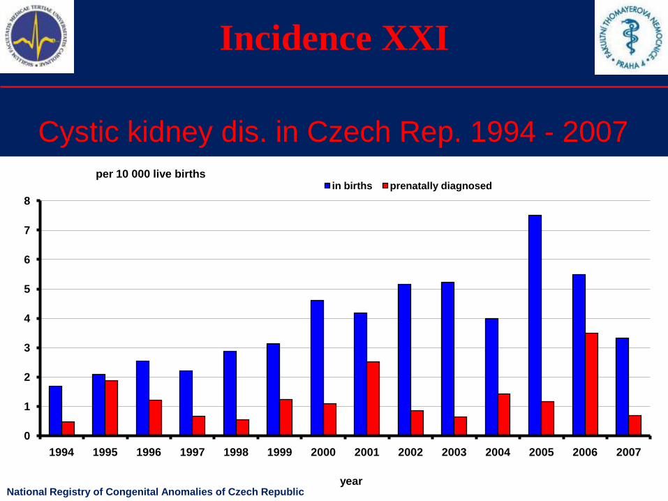
Incidence XXI
Cystic kidney dis. in Czech Rep. 1994 - 2007
0
1
2
3
4
5
6
7
8
1994 1995 1996 1997 1998 1999 2000 2001 2002 2003 2004 2005 2006 2007
in births prenatally diagnosedper 10 000 live births
yearNational Registry of Congenital Anomalies of Czech Republic
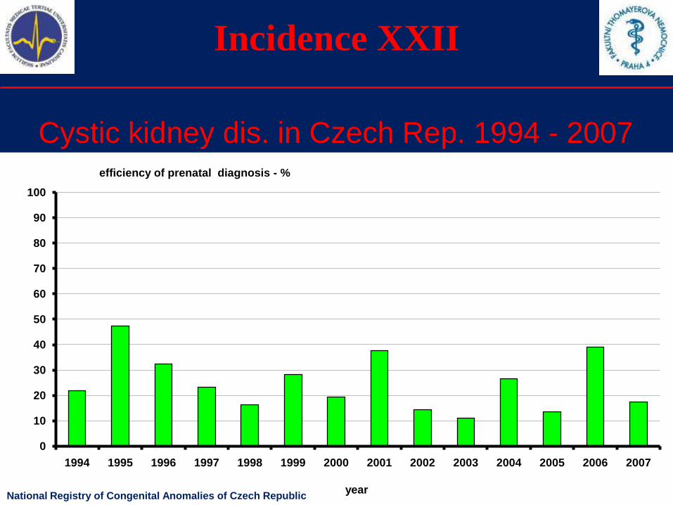
Incidence XXII
Cystic kidney dis. in Czech Rep. 1994 - 2007
0
10
20
30
40
50
60
70
80
90
100
1994 1995 1996 1997 1998 1999 2000 2001 2002 2003 2004 2005 2006 2007
year
efficiency of prenatal diagnosis - %
National Registry of Congenital Anomalies of Czech Republic
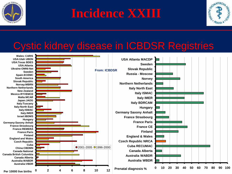
Incidence XXIII
Cystic kidney disease in ICBDSR Registries
0 10 20 30 40 50 60 70 80 90 100
Australia WBDR
Australia WABDR
Canada Alberta
Cuba RECUMAC
Czech Republic NRCA
England & Wales
Finland
France CE
France Paris
France Strasbourg
Germany Saxony Anhalt
Hungary
Italy BDRCAM
Italy IMER
Italy ISMAC
Italy North East
Northern Netherlands
Norvey
Russia - Moscow
Slovak Republic
Sweden
USA Atlanta MACDP
Prenatal diagnosis %0 2 4 6 8 10 12
Australia:VBDR
Australia:WABDR
Canada Alberta
Canada British Columbia
Canada National
China:CBDMN
Cuba
Czech Republic
England and Wales
Finland
France:Paris
France:REMERA
France:Strasbourg
Germany:Saxony Anhalt
Hungary
Israel:IBDMS
Italy:IMER
Italy:ISMAC
Italy:North East
Italy:Tuscany
Japan:JAOG
Malta:MCAR
Mexico:RYVEMCE
New Zealand
Northern Netherlands
Norvay:MBRN
Slovak Republic
South America
Spain:ECEMC
Sweden
Ukraine:OMNI-Net
USA:Atlanta
USA:Texas BDES
USA:Utah UBDN
Wales: CARIS
2001-2005 1996-2000
Per 10000 live births
From: ICBDSR
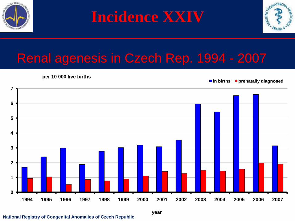
Incidence XXIV
Renal agenesis in Czech Rep. 1994 - 2007
0
1
2
3
4
5
6
7
1994 1995 1996 1997 1998 1999 2000 2001 2002 2003 2004 2005 2006 2007
in births prenatally diagnosedper 10 000 live births
yearNational Registry of Congenital Anomalies of Czech Republic
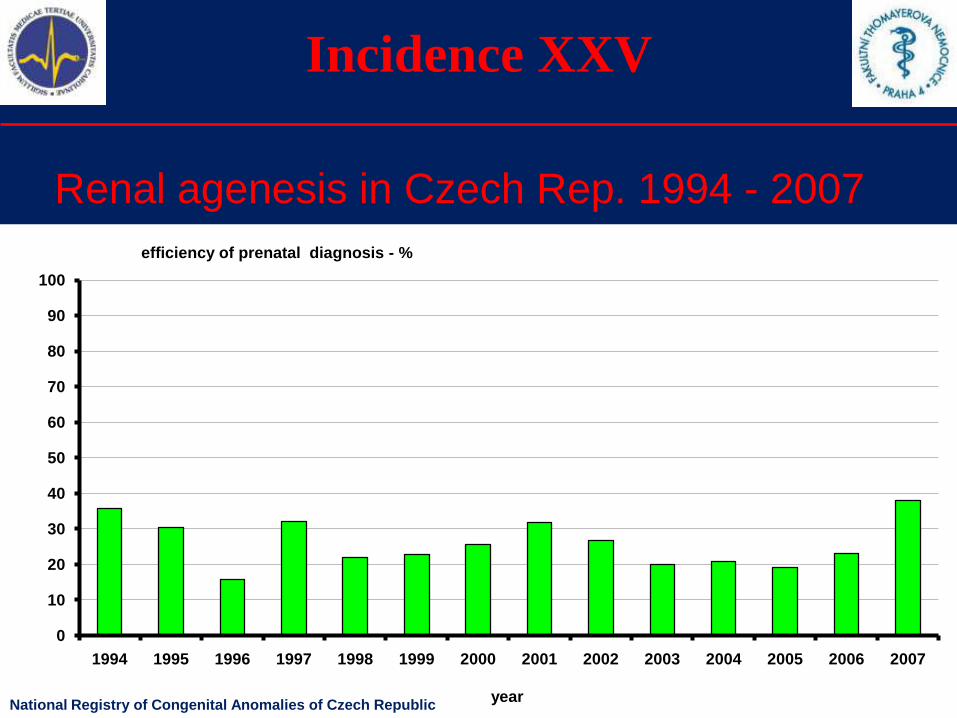
Incidence XXV
Renal agenesis in Czech Rep. 1994 - 2007
0
10
20
30
40
50
60
70
80
90
100
1994 1995 1996 1997 1998 1999 2000 2001 2002 2003 2004 2005 2006 2007
year
efficiency of prenatal diagnosis - %
National Registry of Congenital Anomalies of Czech Republic
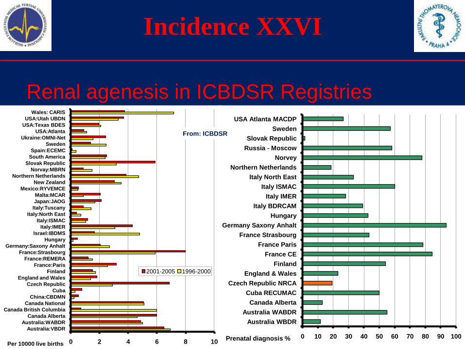
Incidence XXVI
Renal agenesis in ICBDSR Registries
0 10 20 30 40 50 60 70 80 90 100
Australia WBDR
Australia WABDR
Canada Alberta
Cuba RECUMAC
Czech Republic NRCA
England & Wales
Finland
France CE
France Paris
France Strasbourg
Germany Saxony Anhalt
Hungary
Italy BDRCAM
Italy IMER
Italy ISMAC
Italy North East
Northern Netherlands
Norvey
Russia - Moscow
Slovak Republic
Sweden
USA Atlanta MACDP
Prenatal diagnosis %0 2 4 6 8 10
Australia:VBDR
Australia:WABDR
Canada Alberta
Canada British Columbia
Canada National
China:CBDMN
Cuba
Czech Republic
England and Wales
Finland
France:Paris
France:REMERA
France:Strasbourg
Germany:Saxony Anhalt
Hungary
Israel:IBDMS
Italy:IMER
Italy:ISMAC
Italy:North East
Italy:Tuscany
Japan:JAOG
Malta:MCAR
Mexico:RYVEMCE
New Zealand
Northern Netherlands
Norvay:MBRN
Slovak Republic
South America
Spain:ECEMC
Sweden
Ukraine:OMNI-Net
USA:Atlanta
USA:Texas BDES
USA:Utah UBDN
Wales: CARIS
2001-2005 1996-2000
Per 10000 live births
From: ICBDSR
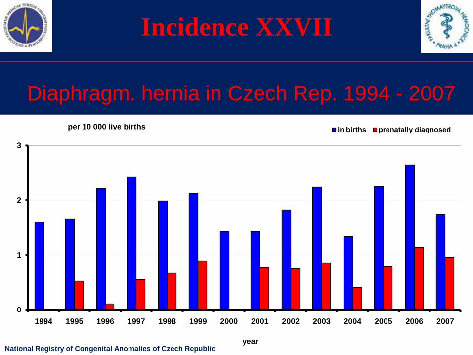
Incidence XXVII
Diaphragm. hernia in Czech Rep. 1994 - 2007
0
1
2
3
1994 1995 1996 1997 1998 1999 2000 2001 2002 2003 2004 2005 2006 2007
in births prenatally diagnosedper 10 000 live births
yearNational Registry of Congenital Anomalies of Czech Republic

Incidence XXVIII
Diaphragm. hernia in Czech Rep. 1994 - 2007
0
10
20
30
40
50
60
70
80
90
100
1994 1995 1996 1997 1998 1999 2000 2001 2002 2003 2004 2005 2006 2007
year
efficiency of prenatal diagnosis - %
National Registry of Congenital Anomalies of Czech Republic
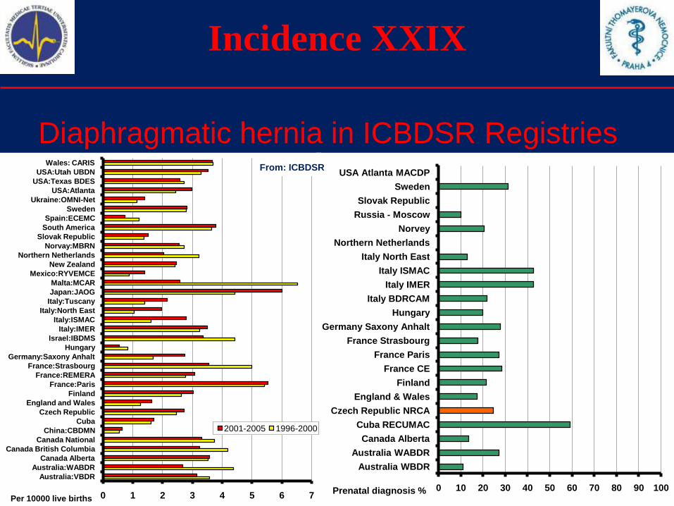
Incidence XXIX
Diaphragmatic hernia in ICBDSR Registries
0 10 20 30 40 50 60 70 80 90 100
Australia WBDR
Australia WABDR
Canada Alberta
Cuba RECUMAC
Czech Republic NRCA
England & Wales
Finland
France CE
France Paris
France Strasbourg
Germany Saxony Anhalt
Hungary
Italy BDRCAM
Italy IMER
Italy ISMAC
Italy North East
Northern Netherlands
Norvey
Russia - Moscow
Slovak Republic
Sweden
USA Atlanta MACDP
0 1 2 3 4 5 6 7
Australia:VBDR
Australia:WABDR
Canada Alberta
Canada British Columbia
Canada National
China:CBDMN
Cuba
Czech Republic
England and Wales
Finland
France:Paris
France:REMERA
France:Strasbourg
Germany:Saxony Anhalt
Hungary
Israel:IBDMS
Italy:IMER
Italy:ISMAC
Italy:North East
Italy:Tuscany
Japan:JAOG
Malta:MCAR
Mexico:RYVEMCE
New Zealand
Northern Netherlands
Norvay:MBRN
Slovak Republic
South America
Spain:ECEMC
Sweden
Ukraine:OMNI-Net
USA:Atlanta
USA:Texas BDES
USA:Utah UBDN
Wales: CARIS
2001-2005 1996-2000
Per 10000 live birthsPrenatal diagnosis %
From: ICBDSR
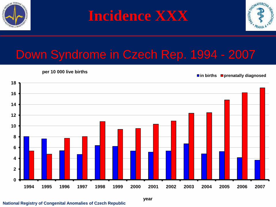
Incidence XXX
Down Syndrome in Czech Rep. 1994 - 2007
0
2
4
6
8
10
12
14
16
18
1994 1995 1996 1997 1998 1999 2000 2001 2002 2003 2004 2005 2006 2007
in births prenatally diagnosedper 10 000 live births
yearNational Registry of Congenital Anomalies of Czech Republic

Incidence XXXI
Down Syndrome in Czech Rep. 1994 - 2007
0
10
20
30
40
50
60
70
80
90
100
1994 1995 1996 1997 1998 1999 2000 2001 2002 2003 2004 2005 2006 2007
year
efficiency of prenatal diagnosis - %
National Registry of Congenital Anomalies of Czech Republic
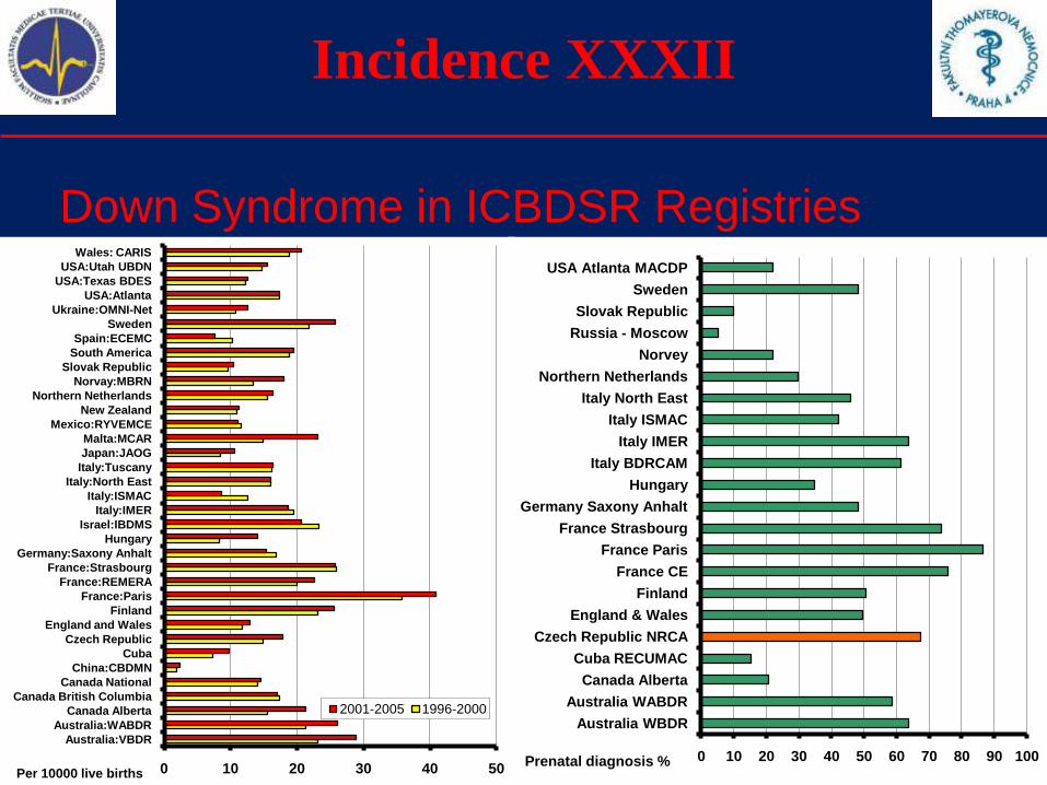
Incidence XXXII
Down Syndrome in ICBDSR Registries
0 10 20 30 40 50 60 70 80 90 100
Australia WBDR
Australia WABDR
Canada Alberta
Cuba RECUMAC
Czech Republic NRCA
England & Wales
Finland
France CE
France Paris
France Strasbourg
Germany Saxony Anhalt
Hungary
Italy BDRCAM
Italy IMER
Italy ISMAC
Italy North East
Northern Netherlands
Norvey
Russia - Moscow
Slovak Republic
Sweden
USA Atlanta MACDP
Prenatal diagnosis %0 10 20 30 40 50
Australia:VBDR
Australia:WABDR
Canada Alberta
Canada British Columbia
Canada National
China:CBDMN
Cuba
Czech Republic
England and Wales
Finland
France:Paris
France:REMERA
France:Strasbourg
Germany:Saxony Anhalt
Hungary
Israel:IBDMS
Italy:IMER
Italy:ISMAC
Italy:North East
Italy:Tuscany
Japan:JAOG
Malta:MCAR
Mexico:RYVEMCE
New Zealand
Northern Netherlands
Norvay:MBRN
Slovak Republic
South America
Spain:ECEMC
Sweden
Ukraine:OMNI-Net
USA:Atlanta
USA:Texas BDES
USA:Utah UBDN
Wales: CARIS
2001-2005 1996-2000
Per 10000 live births
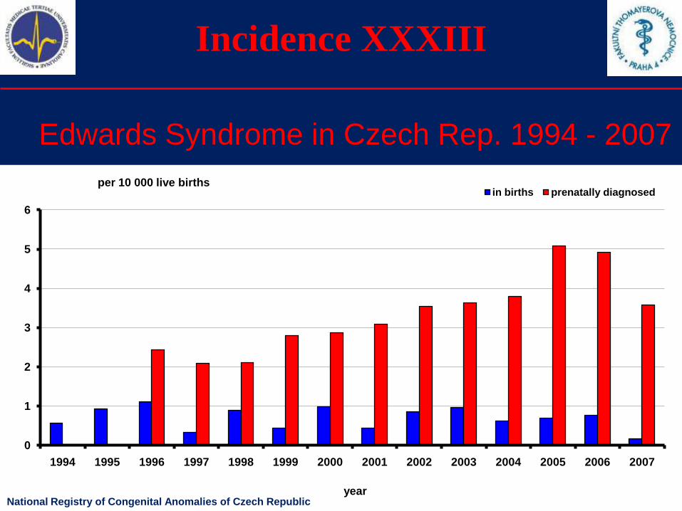
Incidence XXXIII
Edwards Syndrome in Czech Rep. 1994 - 2007
0
1
2
3
4
5
6
1994 1995 1996 1997 1998 1999 2000 2001 2002 2003 2004 2005 2006 2007
in births prenatally diagnosedper 10 000 live births
yearNational Registry of Congenital Anomalies of Czech Republic
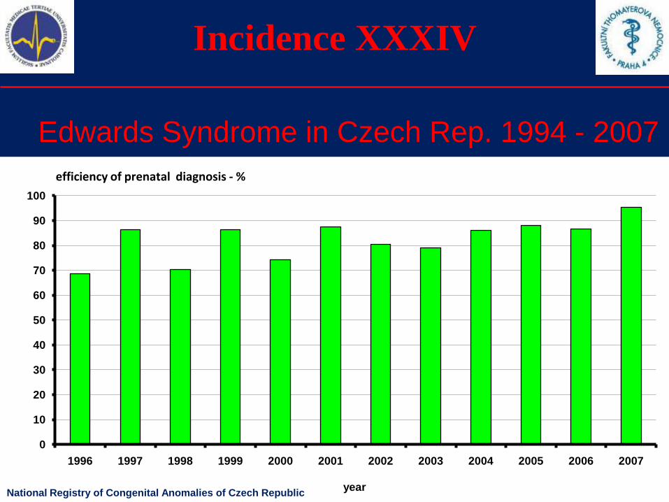
Incidence XXXIV
Edwards Syndrome in Czech Rep. 1994 - 2007
0
10
20
30
40
50
60
70
80
90
100
1996 1997 1998 1999 2000 2001 2002 2003 2004 2005 2006 2007
year
efficiency of prenatal diagnosis - %
National Registry of Congenital Anomalies of Czech Republic
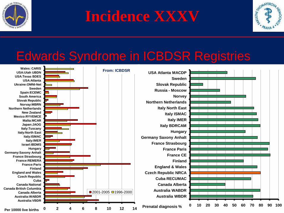
Incidence XXXV
Edwards Syndrome in ICBDSR Registries
0 10 20 30 40 50 60 70 80 90 100
Australia WBDR
Australia WABDR
Canada Alberta
Cuba RECUMAC
Czech Republic NRCA
England & Wales
Finland
France CE
France Paris
France Strasbourg
Germany Saxony Anhalt
Hungary
Italy BDRCAM
Italy IMER
Italy ISMAC
Italy North East
Northern Netherlands
Norvey
Russia - Moscow
Slovak Republic
Sweden
USA Atlanta MACDP
Prenatal diagnosis %0 2 4 6 8 10 12 14
Australia:VBDR
Australia:WABDR
Canada Alberta
Canada British Columbia
Canada National
Cuba
Czech Republic
England and Wales
Finland
France:Paris
France:REMERA
France:Strasbourg
Germany:Saxony Anhalt
Hungary
Israel:IBDMS
Italy:IMER
Italy:ISMAC
Italy:North East
Italy:Tuscany
Japan:JAOG
Malta:MCAR
Mexico:RYVEMCE
New Zealand
Northern Netherlands
Norvay:MBRN
Slovak Republic
South America
Spain:ECEMC
Sweden
Ukraine:OMNI-Net
USA:Atlanta
USA:Texas BDES
USA:Utah UBDN
Wales: CARIS
2001-2005 1996-2000
Per 10000 live births
From: ICBDSR
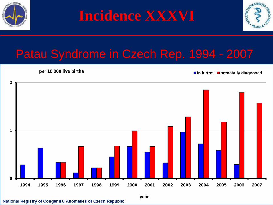
Incidence XXXVI
Patau Syndrome in Czech Rep. 1994 - 2007
0
1
2
1994 1995 1996 1997 1998 1999 2000 2001 2002 2003 2004 2005 2006 2007
in births prenatally diagnosedper 10 000 live births
yearNational Registry of Congenital Anomalies of Czech Republic
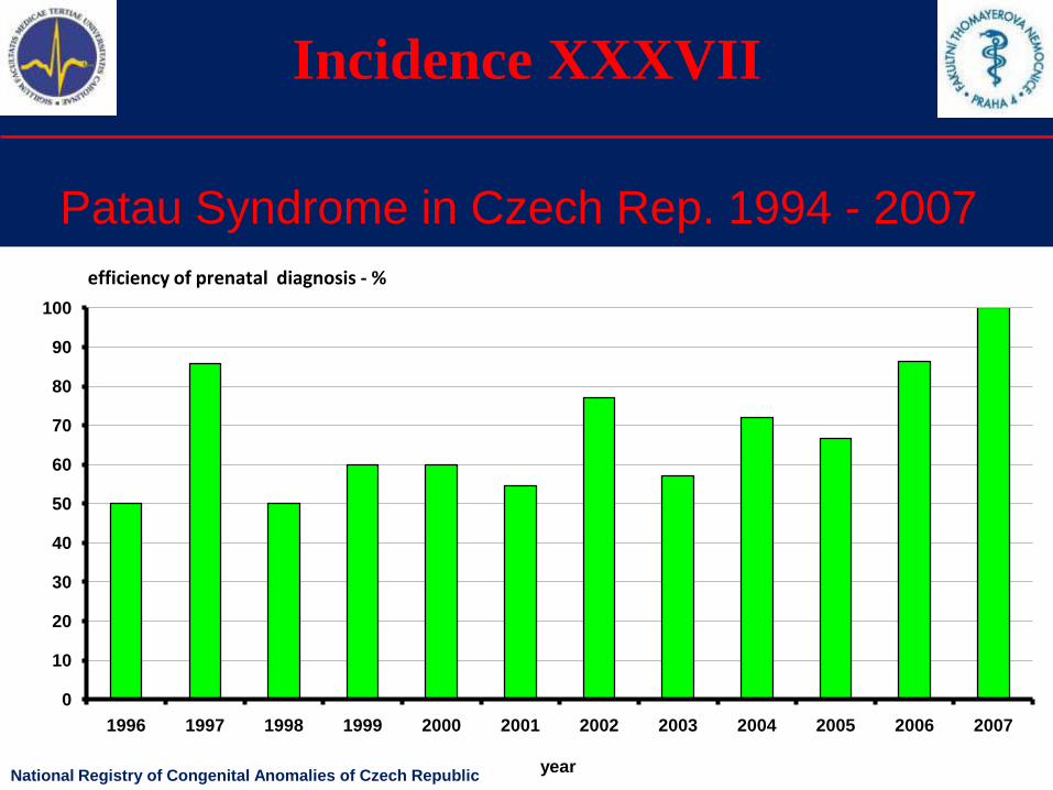
Incidence XXXVII
Patau Syndrome in Czech Rep. 1994 - 2007
0
10
20
30
40
50
60
70
80
90
100
1996 1997 1998 1999 2000 2001 2002 2003 2004 2005 2006 2007
year
efficiency of prenatal diagnosis - %
National Registry of Congenital Anomalies of Czech Republic
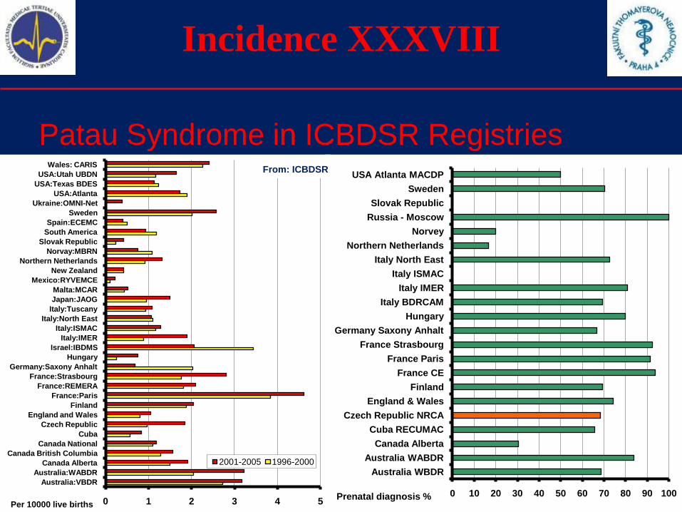
Incidence XXXVIII
Patau Syndrome in ICBDSR Registries
0 10 20 30 40 50 60 70 80 90 100
Australia WBDR
Australia WABDR
Canada Alberta
Cuba RECUMAC
Czech Republic NRCA
England & Wales
Finland
France CE
France Paris
France Strasbourg
Germany Saxony Anhalt
Hungary
Italy BDRCAM
Italy IMER
Italy ISMAC
Italy North East
Northern Netherlands
Norvey
Russia - Moscow
Slovak Republic
Sweden
USA Atlanta MACDP
Prenatal diagnosis %0 1 2 3 4 5
Australia:VBDR
Australia:WABDR
Canada Alberta
Canada British Columbia
Canada National
Cuba
Czech Republic
England and Wales
Finland
France:Paris
France:REMERA
France:Strasbourg
Germany:Saxony Anhalt
Hungary
Israel:IBDMS
Italy:IMER
Italy:ISMAC
Italy:North East
Italy:Tuscany
Japan:JAOG
Malta:MCAR
Mexico:RYVEMCE
New Zealand
Northern Netherlands
Norvay:MBRN
Slovak Republic
South America
Spain:ECEMC
Sweden
Ukraine:OMNI-Net
USA:Atlanta
USA:Texas BDES
USA:Utah UBDN
Wales: CARIS
2001-2005 1996-2000
Per 10000 live births
From: ICBDSR
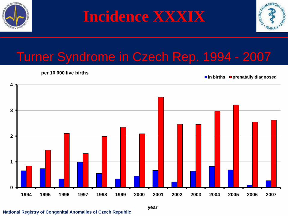
Incidence XXXIX
Turner Syndrome in Czech Rep. 1994 - 2007
0
1
2
3
4
1994 1995 1996 1997 1998 1999 2000 2001 2002 2003 2004 2005 2006 2007
in births prenatally diagnosedper 10 000 live births
yearNational Registry of Congenital Anomalies of Czech Republic
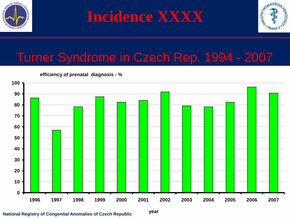
Incidence XXXX
Turner Syndrome in Czech Rep. 1994 - 2007
0
10
20
30
40
50
60
70
80
90
100
1996 1997 1998 1999 2000 2001 2002 2003 2004 2005 2006 2007
year
efficiency of prenatal diagnosis - %
National Registry of Congenital Anomalies of Czech Republic
Mortality and Morbidity
The role of congenital anomalies
Congenital anomalies are important causes of perinatal
mortality and morbidity.
Perinatal mortality covers both Stillbirths and Early
neonatal mortality.
The role of congenital anomalies in Stillbirths and in
Early neonatal mortality is commonly analyzed.
We provide the results from our Registry.
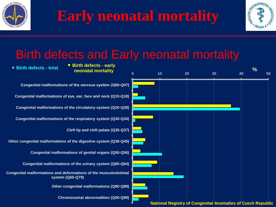
Early neonatal mortality
Birth defects and Early neonatal mortalityBirth defects - early neonatal mortality
Birth defects - total
0 10 20 30 40 50
Congenital malformations of the nervous systém (Q00-Q07)
Congenital malformations of eye, ear, face and neck (Q10-Q18)
Congenital malformations of the circulatory system (Q20-Q28)
Congenital malformations of the respiratory system (Q30-Q34)
Cleft lip and cleft palate (Q35-Q37)
Other congenital malformations of the digestive system (Q38-Q45)
Congenital malformations of genital organs (Q50-Q56)
Congenital malformations of the urinary system (Q60-Q64)
Congenital malformations and deformations of the musculoskeletal system (Q65-Q79)
Other congenital malformations (Q80-Q89)
Chromosomal abnormalities (Q90-Q99)
%
National Registry of Congenital Anomalies of Czech Republic
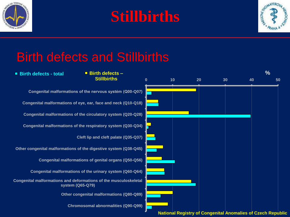
Stillbirths
Birth defects and Stillbirths%
National Registry of Congenital Anomalies of Czech Republic
Birth defects –Stillbirths
Birth defects - total
0 10 20 30 40 50
Congenital malformations of the nervous systém (Q00-Q07)
Congenital malformations of eye, ear, face and neck (Q10-Q18)
Congenital malformations of the circulatory system (Q20-Q28)
Congenital malformations of the respiratory system (Q30-Q34)
Cleft lip and cleft palate (Q35-Q37)
Other congenital malformations of the digestive system (Q38-Q45)
Congenital malformations of genital organs (Q50-Q56)
Congenital malformations of the urinary system (Q60-Q64)
Congenital malformations and deformations of the musculoskeletal system (Q65-Q79)
Other congenital malformations (Q80-Q89)
Chromosomal abnormalities (Q90-Q99)
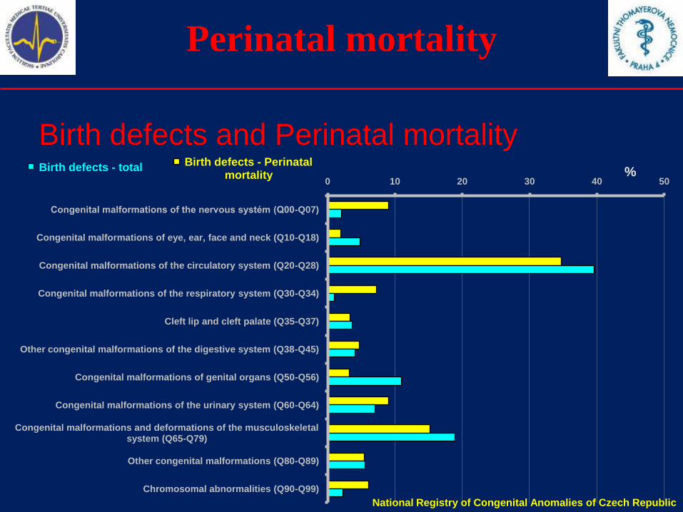
Perinatal mortality
Birth defects and Perinatal mortality%
National Registry of Congenital Anomalies of Czech Republic
Birth defects - Perinatal mortality
Birth defects - total
0 10 20 30 40 50
Congenital malformations of the nervous systém (Q00-Q07)
Congenital malformations of eye, ear, face and neck (Q10-Q18)
Congenital malformations of the circulatory system (Q20-Q28)
Congenital malformations of the respiratory system (Q30-Q34)
Cleft lip and cleft palate (Q35-Q37)
Other congenital malformations of the digestive system (Q38-Q45)
Congenital malformations of genital organs (Q50-Q56)
Congenital malformations of the urinary system (Q60-Q64)
Congenital malformations and deformations of the musculoskeletal system (Q65-Q79)
Other congenital malformations (Q80-Q89)
Chromosomal abnormalities (Q90-Q99)

Useful links
Registries and organizations
National Registry of Congenital Anomalies of the Czech Republic
http://www.uzis.cz/ http://www.vrozene-vady.cz/
International Clearinghouse for Birth Defects Surveillance and Research
http://www.icbdsr.org/
European Surveillance of Congenital Anomalies
http://www.eurocat.ulster.ac.uk/
Congenital anomalies
1.Definitions
2.Etiology, teratogens
3.Selected congenital anomalies
4.Prenatal diagnosis
5.Incidence of selected congenital anomalies
6.Summary
Summary I
Congenital anomalies can be caused by genetic, environmental or
both factors.
The effects of teratogens is species-dependent, dose-dependent and
time-dependent.
Be very careful with medicament therapy during pregnancy.
Prenatal diagnosis is complex and interdisciplinary domain, that
requires the cooperation of medical geneticist, gynecologist,
obstetrician, ultrasound diagnostician, clinical biochemist and
(sometimes) other experts.
Summary II
The absolute and relative number of congenital anomalies in the
Czech Republic is increasing. We believe, it is caused:
1) by the improvement of the prenatal diagnosis (especially by the
combined screening of 1st trimester implementation), which is able to
diagnose selected anomalies during the pregnancy earlier (some of
those cases would formerly end as spontaneous abortions without
diagnose)
2) by the increase of mean age of delivering women (what is a
common trend today).
Congenital anomalies are important causes of perinatal mortality and
morbidity. Prenatal diagnosis may bring useful information before
delivery, so there is time to ensure essential perinatologic therapy.

Recommended literature
For those, who are further interested…
Jones, K.L.
Smith's Recognizable Patterns Of Human
Malformation
Sixth edition
Saunders; 6th edition (August 17, 2005)
Contacts
Email: [email protected]
Website: http://www.vrozene-vady.cz/This presentation (and other materials) can be downloaded from our website.
Correspondence address:Antonin Sipek, MD, PhD
National Registry of Congenital Anomalies of the Czech Republic
Department of Medical Genetics
Thomayer University Hospital
Videnska 800
140 59, Prague 4

Thank you
Thank you for your attention.