Embed Size (px)
Citation preview
AGRICULTURAL RESEARCH COMMUNICATION CENTREwww.arccjournals.com/www.ijaronline.in
*Corresponding author’s e-mail: [email protected]
Indian J. Anim. Res., 50 (5) 2016 : 816-819Print ISSN:0367-6722 / Online ISSN:0976-0555
Congenital omphalocele in four calves, their surgical management and outcomeMujeeb ur Rehman Fazili*, Shahid Hussain Dar, H.K. Bhattacharyya and Manzoor ur Rehman
Teaching Veterinary Clinical Service Complex, Faculty of Veterinary Sciences & AH,SKUAST-K, Shuhama-190 006, Srinagar, Kashmir, India.Received: 19-07-2014 Accepted: 15-05-2015 DOI:10.18805/ijar.11168
ABSTRACTFour newborn dairy calves with omphalocele including one additionally with atresia ani and anury were presented fortreatment. The mass (diameter: 12.0 and 23.0cm respectively) in two calves was covered by continuation of the umbilicalcord. In the remaining two calves the covering had disrupted and the intestines were hanging from the umbilicus.Herniorrhaphy was performed under sedation using diazepam and local infiltration analgesia with lignocaine hydrochloride(2%). In the calf with atresia ani, anal opening was created under caudal epidural block. Three of the calves recoveredcompletely and the remaining one died. It was concluded that the surgical treatment of calves with omphalocele have goodprognosis provided they are presented promptly without mutilation of the mass.
Key words: Anury, Atresia Ani, Calves, Congenital, Herniorrhaphy, Omphalocele.
INTRODUCTIONOmphalocele is a rare type of congenital abdominal
wall defect that allows intestines (and sometimes a portionof liver) covered by a paper thin membrane (amnion) toprotrude from the body. The condition occurs when one ofthe four body folds fails to migrate normally in embryologicdevelopment (Baird, 2008). The true prevalence in animalsis difficult to determine owing to the unreported deaths(Baird, 1993, Smeak, 1993). However, in one study, theincidence of omphalocele among full-term calves was 0.1per cent (Mee, 1994). Other associated congenitalabnormalities have not been described in veterinary reports(Baird, 2008). In this paper, the authors put on record thedetails of omphalocele in four neonatal dairy calves(including one additionally with atresia ani and absence oftail/anury), their surgical management and the outcome.MATERIALS AND METHODS
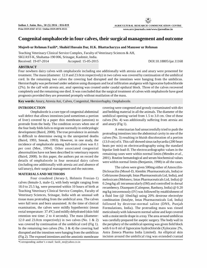
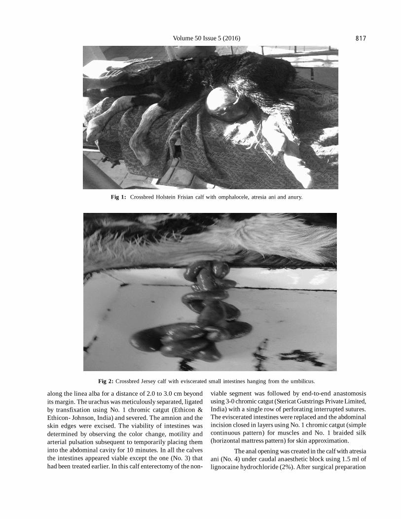
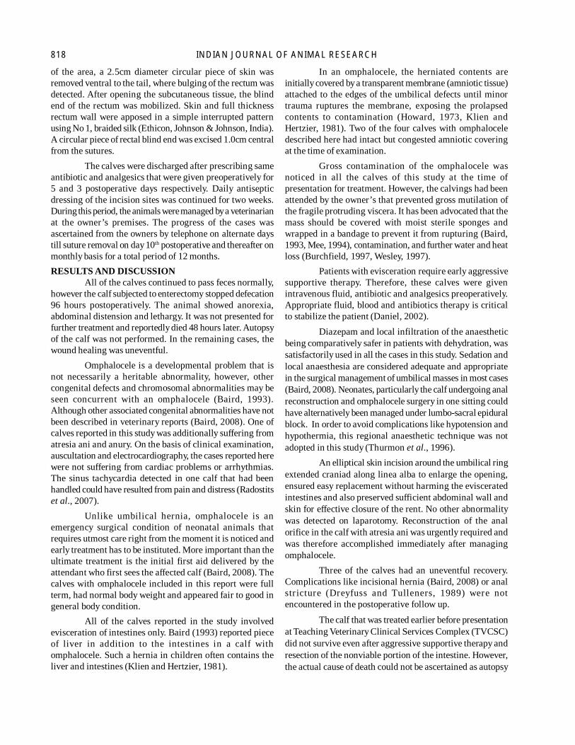
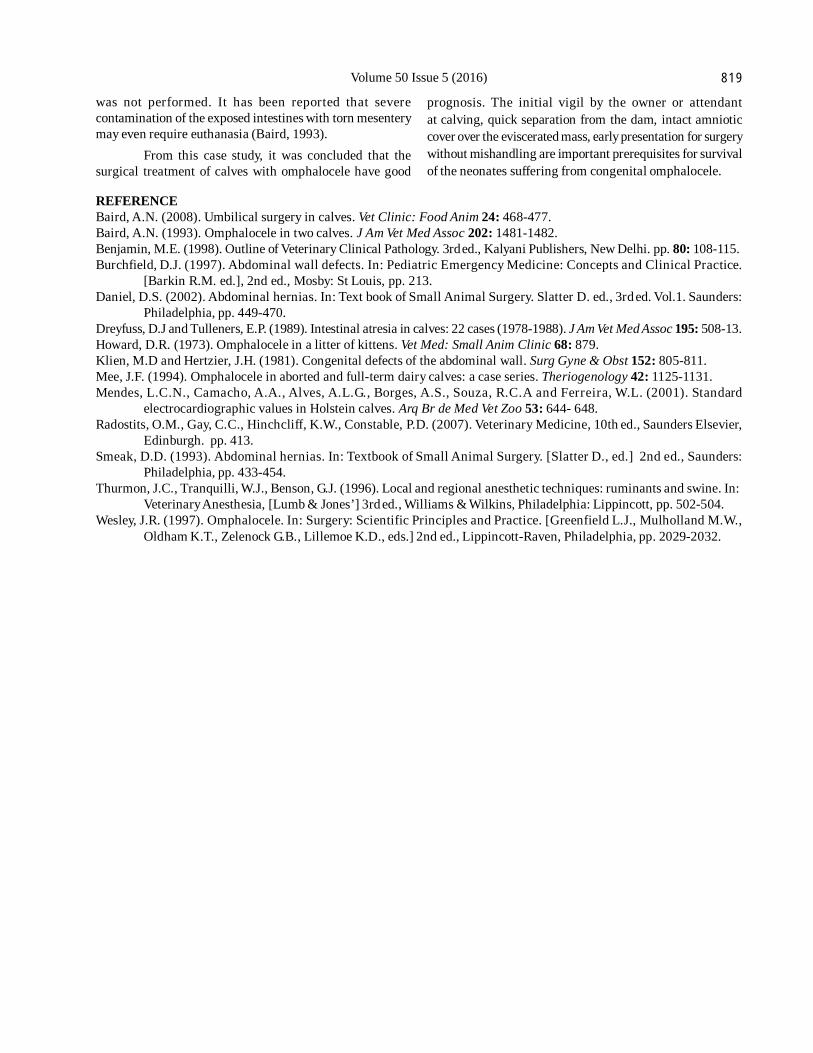
Four crossbred (Jersey-3, Holstein Fresian-1)calves (female-3, male-1), with body weight ranging from18.0 to 25.5 kg, were presented within 10 hours of birth atTeaching Veterinary Clinical Service Complex, Faculty ofVeterinary Sciences, Srinagar, India with an unusual softtissue mass protruding from the umbilical area. The calveswere full term and born unassisted. At the time of clinicalexamination, the calves were mildly hypothermic (meanrectal temperature: 37.40C) and mildly dehydrated (skin foldretention test time: 2 to 4 seconds). The mass (diameter:12.0 and 23.0cm respectively) in two calves (No. 1 & 2)was covered by continuation of the umbilical cord (Fig 1).In the remaining two calves (No. 3 & 4) the covering haddisrupted and the intestines were hanging from the umbilicus(Fig 2). The exposed intestines and the amniotic membrane
covering were congested and grossly contaminated with dirtand bedding material in all the animals. The diameter of theumbilical opening varied from 1.5 to 3.0 cm. One of thesecalves (No. 4) was additionally suffering from atresia aniand anury (Fig 1).
A veterinarian had unsuccessfully tried to push theprotruding intestines into the abdominal cavity in one of thecalves (No. 3) resulting in bluish discoloration of a portion(13.0 cm) of it. This calf showed sinus tachycardia (200 heartbeats per min) on electrocardiography using the standardbipolar limb lead-II. The electrocardiographic values in theremaining cases were within normal limits (Mendes et al.,2001). Routine hematological and serum biochemical valueswere within normal limits (Benjamin, 1998) in all the cases.
The calves were given 500mg either of Amoxcilin-Dicloxacilin (Moxel-D, Alembic Pharmaceuticals, India) orCeftriaxone (Intaceph; Intas Pharmaceuticals Ltd, India), andmeloxicam (Melonex; Intas Pharmaceuticals Ltd, India) @0.2mg/kg all intramuscularly (IM) and controlled in dorsalrecumbency. Diazepam (Calmpose, Ranbaxy, India) @ 0.20mg/kg intravenously (IV) was followed by establishment ofa fluid line (@ 10ml/kg) using 20% dextrose electrolytecombination (Intalyte, Intas Pharmaceuticals Ltd, India)followed by dextrose-normal saline (DNS, PunjabFormulations, India). The protruding mass was rinsedmeticulously with lukewarm normal saline and kept coveredwith a moist sterile drape in a tray. The area around umbilicuswas carefully prepared for aseptic surgery. The body wall inthe periphery of the umbilical opening was given field blockwith 6 to 8 ml of lignocaine hydrochloride (Xylocaine 2%,Astra Zeneca Pharma India Limited). An elliptical skinincision around the umbilical ring was extended craniad
Volume 50 Issue 5 (2016) 817
Fig 1: Crossbred Holstein Frisian calf with omphalocele, atresia ani and anury.
Fig 2: Crossbred Jersey calf with eviscerated small intestines hanging from the umbilicus.
along the linea alba for a distance of 2.0 to 3.0 cm beyondits margin. The urachus was meticulously separated, ligatedby transfixation using No. 1 chromic catgut (Ethicon &Ethicon- Johnson, India) and severed. The amnion and theskin edges were excised. The viability of intestines wasdetermined by observing the color change, motility andarterial pulsation subsequent to temporarily placing theminto the abdominal cavity for 10 minutes. In all the calvesthe intestines appeared viable except the one (No. 3) thathad been treated earlier. In this calf enterectomy of the non-
viable segment was followed by end-to-end anastomosisusing 3-0 chromic catgut (Stericat Gutstrings Private Limited,India) with a single row of perforating interrupted sutures.The eviscerated intestines were replaced and the abdominalincision closed in layers using No. 1 chromic catgut (simplecontinuous pattern) for muscles and No. 1 braided silk(horizontal mattress pattern) for skin approximation.
The anal opening was created in the calf with atresiaani (No. 4) under caudal anaesthetic block using 1.5 ml oflignocaine hydrochloride (2%). After surgical preparation
818 INDIAN JOURNAL OF ANIMAL RESEARCH
of the area, a 2.5cm diameter circular piece of skin wasremoved ventral to the tail, where bulging of the rectum wasdetected. After opening the subcutaneous tissue, the blindend of the rectum was mobilized. Skin and full thicknessrectum wall were apposed in a simple interrupted patternusing No 1, braided silk (Ethicon, Johnson & Johnson, India).A circular piece of rectal blind end was excised 1.0cm centralfrom the sutures.
The calves were discharged after prescribing sameantibiotic and analgesics that were given preoperatively for5 and 3 postoperative days respectively. Daily antisepticdressing of the incision sites was continued for two weeks.During this period, the animals were managed by a veterinarianat the owner’s premises. The progress of the cases wasascertained from the owners by telephone on alternate daystill suture removal on day 10th postoperative and thereafter onmonthly basis for a total period of 12 months.RESULTS AND DISCUSSION
All of the calves continued to pass feces normally,however the calf subjected to enterectomy stopped defecation96 hours postoperatively. The animal showed anorexia,abdominal distension and lethargy. It was not presented forfurther treatment and reportedly died 48 hours later. Autopsyof the calf was not performed. In the remaining cases, thewound healing was uneventful.
Omphalocele is a developmental problem that isnot necessarily a heritable abnormality, however, othercongenital defects and chromosomal abnormalities may beseen concurrent with an omphalocele (Baird, 1993).Although other associated congenital abnormalities have notbeen described in veterinary reports (Baird, 2008). One ofcalves reported in this study was additionally suffering fromatresia ani and anury. On the basis of clinical examination,auscultation and electrocardiography, the cases reported herewere not suffering from cardiac problems or arrhythmias.The sinus tachycardia detected in one calf that had beenhandled could have resulted from pain and distress (Radostitset al., 2007).
Unlike umbilical hernia, omphalocele is anemergency surgical condition of neonatal animals thatrequires utmost care right from the moment it is noticed andearly treatment has to be instituted. More important than theultimate treatment is the initial first aid delivered by theattendant who first sees the affected calf (Baird, 2008). Thecalves with omphalocele included in this report were fullterm, had normal body weight and appeared fair to good ingeneral body condition.
All of the calves reported in the study involvedevisceration of intestines only. Baird (1993) reported pieceof liver in addition to the intestines in a calf withomphalocele. Such a hernia in children often contains theliver and intestines (Klien and Hertzier, 1981).
In an omphalocele, the herniated contents areinitially covered by a transparent membrane (amniotic tissue)attached to the edges of the umbilical defects until minortrauma ruptures the membrane, exposing the prolapsedcontents to contamination (Howard, 1973, Klien andHertzier, 1981). Two of the four calves with omphaloceledescribed here had intact but congested amniotic coveringat the time of examination.
Gross contamination of the omphalocele wasnoticed in all the calves of this study at the time ofpresentation for treatment. However, the calvings had beenattended by the owner’s that prevented gross mutilation ofthe fragile protruding viscera. It has been advocated that themass should be covered with moist sterile sponges andwrapped in a bandage to prevent it from rupturing (Baird,1993, Mee, 1994), contamination, and further water and heatloss (Burchfield, 1997, Wesley, 1997).
Patients with evisceration require early aggressivesupportive therapy. Therefore, these calves were givenintravenous fluid, antibiotic and analgesics preoperatively.Appropriate fluid, blood and antibiotics therapy is criticalto stabilize the patient (Daniel, 2002).
Diazepam and local infiltration of the anaestheticbeing comparatively safer in patients with dehydration, wassatisfactorily used in all the cases in this study. Sedation andlocal anaesthesia are considered adequate and appropriatein the surgical management of umbilical masses in most cases(Baird, 2008). Neonates, particularly the calf undergoing analreconstruction and omphalocele surgery in one sitting couldhave alternatively been managed under lumbo-sacral epiduralblock. In order to avoid complications like hypotension andhypothermia, this regional anaesthetic technique was notadopted in this study (Thurmon et al., 1996).
An elliptical skin incision around the umbilical ringextended craniad along linea alba to enlarge the opening,ensured easy replacement without harming the evisceratedintestines and also preserved sufficient abdominal wall andskin for effective closure of the rent. No other abnormalitywas detected on laparotomy. Reconstruction of the analorifice in the calf with atresia ani was urgently required andwas therefore accomplished immediately after managingomphalocele.
Three of the calves had an uneventful recovery.Complications like incisional hernia (Baird, 2008) or analstr icture (Dreyfuss and Tulleners, 1989) were notencountered in the postoperative follow up.
The calf that was treated earlier before presentationat Teaching Veterinary Clinical Services Complex (TVCSC)did not survive even after aggressive supportive therapy andresection of the nonviable portion of the intestine. However,the actual cause of death could not be ascertained as autopsy
Volume 50 Issue 5 (2016) 819
was not performed. It has been reported that severecontamination of the exposed intestines with torn mesenterymay even require euthanasia (Baird, 1993).
From this case study, it was concluded that thesurgical treatment of calves with omphalocele have good
prognosis. The initial vigil by the owner or attendantat calving, quick separation from the dam, intact amnioticcover over the eviscerated mass, early presentation for surgerywithout mishandling are important prerequisites for survivalof the neonates suffering from congenital omphalocele.
REFERENCEBaird, A.N. (2008). Umbilical surgery in calves. Vet Clinic: Food Anim 24: 468-477.Baird, A.N. (1993). Omphalocele in two calves. J Am Vet Med Assoc 202: 1481-1482.Benjamin, M.E. (1998). Outline of Veterinary Clinical Pathology. 3rd ed., Kalyani Publishers, New Delhi. pp. 80: 108-115.Burchfield, D.J. (1997). Abdominal wall defects. In: Pediatric Emergency Medicine: Concepts and Clinical Practice.
[Barkin R.M. ed.], 2nd ed., Mosby: St Louis, pp. 213.Daniel, D.S. (2002). Abdominal hernias. In: Text book of Small Animal Surgery. Slatter D. ed., 3rd ed. Vol.1. Saunders:
Philadelphia, pp. 449-470.Dreyfuss, D.J and Tulleners, E.P. (1989). Intestinal atresia in calves: 22 cases (1978-1988). J Am Vet Med Assoc 195: 508-13.Howard, D.R. (1973). Omphalocele in a litter of kittens. Vet Med: Small Anim Clinic 68: 879.Klien, M.D and Hertzier, J.H. (1981). Congenital defects of the abdominal wall. Surg Gyne & Obst 152: 805-811.Mee, J.F. (1994). Omphalocele in aborted and full-term dairy calves: a case series. Theriogenology 42: 1125-1131.Mendes, L.C.N., Camacho, A.A., Alves, A.L.G., Borges, A.S., Souza, R.C.A and Ferreira, W.L. (2001). Standard
electrocardiographic values in Holstein calves. Arq Br de Med Vet Zoo 53: 644- 648.Radostits, O.M., Gay, C.C., Hinchcliff, K.W., Constable, P.D. (2007). Veterinary Medicine, 10th ed., Saunders Elsevier,
Edinburgh. pp. 413.Smeak, D.D. (1993). Abdominal hernias. In: Textbook of Small Animal Surgery. [Slatter D., ed.] 2nd ed., Saunders:
Philadelphia, pp. 433-454.Thurmon, J.C., Tranquilli, W.J., Benson, G.J. (1996). Local and regional anesthetic techniques: ruminants and swine. In:
Veterinary Anesthesia, [Lumb & Jones’] 3rd ed., Williams & Wilkins, Philadelphia: Lippincott, pp. 502-504.Wesley, J.R. (1997). Omphalocele. In: Surgery: Scientific Principles and Practice. [Greenfield L.J., Mulholland M.W.,
Oldham K.T., Zelenock G.B., Lillemoe K.D., eds.] 2nd ed., Lippincott-Raven, Philadelphia, pp. 2029-2032.