Embed Size (px)
Citation preview
Journal of Medical Genetics (1975). 12, 280.
Congenital vertebral anomalies: aetiology andrelationship to spina bifida cystica*
RUTH WYNNE-DAVIESFrom the Department of Orthopaedic Surgery (Clinical Genetics Research), University of Edinburgh
Summary. A family survey of337 patients with congenital vertebral anomalieshas been carried out from the Scoliosis Clinics of Edinburgh and the Royal NationalOrthopaedic Hospital, London.From genetic and epidemiological evidence it is clear that multiple vertebral an-
omalies (without apparent spina bifida) are aetiologically related to anencephaly andspina bifida cystica, carrying a 5-10% risk to subsequent sibs for any one of thesedefects. The implications for prenatal diagnosis are discussed.
Solitary hemivertebrae and localized anterior defects ofthe vertebral bodies caus-ing kyphoscoliosis are sporadic (non-familial) in nature, carrying no risk to subse-quent sibs.
Developmental errors of vertebrae are extremelyvariable, involving anything from one level only tomultiple defects throughout the length of the spineand affecting any part of the vertebrae. Clinicaldata relating to these defects and to congenitalscoliosis and kyphoscoliosis have been reported byJames (1955; 1967), MacEwen (1967), Winter et al(1968; 1973), and MacEwen et al (1972). Groupsof anomalies based on anatomical types can be de-scribed and the following is taken mainly fromJames (1967).
1. Neural arch defects. Posterior arch de-fects may be partial or total, occur with or without ameningocele or myelomeningocele, and may beaccompanied by vertebral body defects at any levelof the spine (Fig. 1). Diastematomyelia may bepresent and lead to paraplegia as the child grows(Fig. 2). Scoliosis does not usually develop withthe simple neural arch defects.
2. Segmental defects ofthe vertebral bodies.These vary from a single hemivertebrae (Fig. 3) tomultiple defects over the whole length of the spine.Scoliosis is frequent, due to asymmetry of affectedvertebrae and to their continued unequal growth.Spinal curvature (Figs. 2 and 4) may be apparent at
Received 24 July 1974.* This paper is the subject of part of a PhD Thesis (Wynne-Davies,
1973), University of Edinburgh.280
birth, develop during growth, or perhaps never de-velop at all if the segmental defects on each side ofthe vertebral column 'balance out'. Sprengel'sshoulder and absence or fusion of ribs are common.Anomalous vertebrae tend to fuse during growthand a rigid bar of bone may develop on the concaveside of a curve, giving rise to a particularly intract-able form of scoliosis (Fig. 5).These defects are not only of cosmetic signifi-
cance: spinal curvature may be severe and rapidlydeveloping, leading to cor pulmonale and perhapsparaplegia-although this latter is more commonwhen diastematomyelia is present, and in the nextgroup of anterior vetebral body defects.
3. Anterior vertebral body defects. The de-formity resulting from anterior defects of the verte-bral bodies is kyphoscoliosis-the term being usedprecisely to mean a true kyphosis as well as a lateralcurvature of the spine (James, 1955; Winter et al,1973). There may be partial or complete absenceof one vertebral body (Fig. 6), fusion of two or three(Fig. 7), or a combination of these. It is character-istically a localized deformity extending only overone, two, or three vertebrae and the neural arch isusually unaffected. It is a potentially serious an-omaly because the increasing angulation ofthe verte-brae as the child grows causes pressure on the spinalcord and paraplegia may develop.
copyright. on 1 January 2019 by guest. P
rotected byhttp://jm
g.bmj.com
/J M
ed Genet: first published as 10.1136/jm
g.12.3.280 on 1 Septem
ber 1975. Dow
nloaded from

Congenital vertebral anomalies: aetiology and relatio,mship to spina bifida cystica 281
FIG. 1. A wide neural arch defect in the lumbar re-gion and multiple segmental defects in the upper tho-racic spine.
FIG. 2. The neural arch defect in the lumbar regionis accompanied by diastemstomyelia at the thoraco-lumbar junction, and anomialous (now fused) ver-tebrae immediately above.
FIG. 3. A simple hemivertebra at the second lumbarlevel.
FIG. 4. Multiple segmental defects in the mid-thorscic region of this infant, including fused ribs.Scoliosis is already developing. The type and extentof the anomalies is very clear in this early radiograph.
FIG. 5. In contrastwith Figs. 1 and 4, this radiographshows a 74-year-old child with a fused mass of boneand a rigid bar on the concave side of the curve.Rib anomalies are present, but no very detailedradiographic diagnosis can be made at this stage.
FIG. 6. A lateral radiograph of the spine of a one-year-old girl showing an absent vertebral body in thelower thoracic region. Already there is an acutekyphosis and paraplegia is likely to develop as shegrows. The antero-posterior radiograph at the sameage is shown on the right. There is very littleassociated scoliosis.
FIG. 7. A iateral radiograph of the spine showsfailure of development and fusion of three thoracic-vertebrae, withassociated kyphosis.
copyright. on 1 January 2019 by guest. P
rotected byhttp://jm
g.bmj.com
/J M
ed Genet: first published as 10.1136/jm
g.12.3.280 on 1 Septem
ber 1975. Dow
nloaded from

Ruth Wynne-Davies
4. Minor fusion anomalies. These may affecttwo or more vertebral bodies, laminae, or spines andmay occur with other segnental defects or as iso-lated features in an almost normal vertebral column.
Difficulties of an exact radiologicaldiagnosis
The extreme variability of segmental vertebraldefects defies classification and sometimes descrip-tion. The problem is further increased by de-formity of the spine (scoliosis and kyphosis) obscur-ing radiological detail. Lateral curvature will beaccompanied by rotation of vertebrae, and so whentaking an apparently antero-posterior view, the re-sult may be, for example, a true antero-posteriorview of the thoracic region with an oblique or lateralview of the lumbar spine.As the child grows, anomalous vertebrae tend to
fuse into a solid mass, and this, together with thespinal curvature may make exact diagnosis im-possible in the older child or adult. The most help-ful radiographs are those taken in infancy or earlychildhood, before significant deformity has de-veloped or fusion occurred.
Previous surveysThere have been no family studies in congenital
scoliosis, although there have been occasional reportsof more than one affected member of a family. Afamily survey of cervical vertebral defects ('KlippelFeil syndrome') was carried out by Gunderson et al(1967). They investigated 11 index patients andcarried out radiography of the cervical region in 121relatives. They concluded there were a number ofdifferent genetic patterns of inheritance (includingdominant and recessive types), varying with thenature of the lesion in the index patient. Some oftheir cases had vertebral anomalies other than cervi-cal.There have been many genetic and epidemio-
logical studies of the neural tube defects fromdifferent parts of the world (Record and McKeown,1950; Edwards, 1958; Williamson, 1965; Carter andFraser Roberts, 1967; Naggan and MacMahon,1967; Carter et al, 1968; Elwood, 1970; Fedrick,1970; Richards et al, 1972; Carter and Evans,1973a; 1973b). Some index patients must have hadcongenital vertebral defects in addition to spinabifida but how many is not known.The main findings of these surveys may be sum-
marized as follows:(1) Anencephaly and spina bifida/meningocele,
with or without hydrocephalus occur within the
same kindreds and are therefore aetiologically re-lated.
(2) Concordance in monozygous twins is low,perhaps only about 20% of co-twins of patients areaffected.
(3) The risk of a second sib being affected is ap-proximately 5% (higher in areas of high incidence);if two sibs are already affected, the risk to a third isapproximately 10%. The risk to children of adultsurvivors on the three small series available is ap-proximately 3%.
(4) The disorders are more common in the lowersocial classes.
(5) Some surveys report increased frequency inthe winter months.
(6) There is an excess of first born children, andof mothers who are older than average.
(7) Within the British Isles there are regionaldifferences in the frequency of the neural tube de-fects, being highest in Northern Ireland and lowestin south-east England.The genetic liability to neural tube defects is
thought to be polygepaic acting together with a strongenvironmental element.
The aim of the current survey was to try and es-tablish whether there was any aetiological relation-ship between spina bifida cystica, spina bifidaocculta, and other congenital vertebral defects.Evidence was obtained from a family survey, notingthe proportions of affected relatives with the same orother spinal anomalies, and from epidemiologicaldata.
Population incidence and frequency ofspma bifida occulta
The frequency of the neural tube defects has beenwell established from many surveys and in Britainranges from approximately 3 to 8 per 1000 births.These defects, however, are immediately apparentat birth. There is more difficulty in establishing apopulation frequency for other congenital vertebralanomalies which may not be discovered for manyyears, or in some cases not at all.
In a study of 15 000 minifilms of the chest fromthe State of Delaware, USA, Shands and Eisberg(1955) noted seven cases of congenital scoliosis withdefinite vertebral anomalies, giving an incidence of0.5 per 1000. This figure would exclude anomaliesin the lower lumbar region, since they studied onlychest x-rays. In the Edinburgh Scoliosis Clinic be-tween 1958 and 1968 (the period of this survey)there were 72 patients with congenital scoliosis whocame from Edinburgh City, the Lothians, and Fife.Over the same period, and attending the same clinic
282
copyright. on 1 January 2019 by guest. P
rotected byhttp://jm
g.bmj.com
/J M
ed Genet: first published as 10.1136/jm
g.12.3.280 on 1 Septem
ber 1975. Dow
nloaded from

Congenital vertebral anomalies: aetiology and relationship to spina bifida cystica
from the same area, there were 118 patients withidiopathic scoliosis. That is, idiopathic scoliosiswas 1.6 times more common. The external de-formity is similar and ascertainment for the two islikely to be comparable. The general populationincidence for idiopathic scoliosis has been estab-lished in Edinburgh as approximately 2 per 1000(Wynne-Davies, 1968) and so a figure of 1 per 1000for congenital vertebral anomalies is a reasonableguess; it is not too dissimilar from Shands' andEisberg's figure (1955) which is clearly too low anestimate.The frequency of very minor vertebral anomalies
is impossible to assess since there will be no reasonfor individuals to attend hospital for treatment, andeven if radiographs were taken of a large population,only some of such defects would be apparent.An investigation was carried out in Edinburgh to
determine the frequency of spina bifida occulta atthe 5th lumbar/lst sacral level. It was thought tobe very common in the general population and it washoped to assess the significance of relatives whomight be so affected. Lorber and Levick (1967)noted 20% of parents with spina bifida occulta andonly 5%0 of controls-though some anatomy textbooks refer to 5-10%. In view of the uncertaintyof seeing this minor anomaly on any one radiograph,a series was examined of 162 patients with idio-pathic scoliosis and 59 with paralytic scoliosis due toprevious poliomyelitis. These two groups werechosen because each individual had dozens of verte-bral column radiographs taken at intervals through-out the growing period-perhaps from 3 months to16 years of age. There was, therefore, a greaterchance of observing this minor vertebral anomaly.Results indicated that at least half this populationup to the age of 16 years (109 of 221 patients) hadspina bifida occulta at the 5th lumbar/lst sacrallevel, which must still be a minimum figure sincethis was only a radiographic survey.The reason for failure to observe a minor neural
arch defect on any one x-ray is that the two sidesof the arch instead of meeting and fusing, over-rideone another (Fig. 8) and it will not necessarily beapparent on any one radiograph that a defect is pre-sent. In view of these figures, spina bifida occultaat the 5th lumbar/lst sacral level has been ignoredboth in index patients and their relatives.
Material and methodsThree hundred and thirty-seven patients with verte-
bral anomalies were traced from two scoliosis clinics; 127from the Edinburgh Clinic (1958-68) and 210 from theRoyal National Orthopaedic Hospital, London (1948-68). No patient was included without adequate radio-
FIG. 8. Spina bifida occulta is apparent in the lower lumbar andsacral spine: the two halves of the neural arch of the fourth lumbarvertebra are tending to over-ride one another.
graphs, usually taken in early childhood, allowing asaccurate a diagnosis as possible.
Congenital vertebral defects excluded from the surveywere spina bifida cystica as a solitary defect (ie, unaccom-panied by other vertebral anomalies); the Klippel Feilsyndrome (although a number of patients were includedwith vertebral anomalies extending from the upperthoracic region into the neck); and anomalies of segmen-tation and spina bifida occulta at the lumbo-sacral junc-tion.The method of research was by family survey, noting
particularly the proportions of anencephaly, spina bifidacystica, and other vertebral anomalies amongst relativesof index patients, and comparing this figure with thegeneral population frequency. The homes of all 337index patients were visited, a total of 924 first-degreerelatives seen, and a history taken for relatives of second-and third-degree. Living relatives with clinical evi-dence of a vertebral anomaly where possible were broughtto hospital for further examination and radiography. Inall cases medical records, including necropsy reports,were sought; hearsay evidence of abnormality was notaccepted.The parents of the first 24 Edinburgh patients indexed
(48 individuals) had routine radiographic examination ofthe vertebral column, but results being entirely negativeit was thought unjustifiable to proceed with this investi-gation further. It is possible there are more relativeswith minor defects than are reported here, but radio-graphic examination would not be a complete answersince minor vertebral anomalies might not be visibleeven if the quality of the radiographs were good. Theonly certain method of diagnosis is by direct vision,either during an operative procedure, or at necropsy.Major vertebral anomalies are nearly always clinicallyapparent, anencephaly and spina bifida cystica, of course,always so.
283
copyright. on 1 January 2019 by guest. P
rotected byhttp://jm
g.bmj.com
/J M
ed Genet: first published as 10.1136/jm
g.12.3.280 on 1 Septem
ber 1975. Dow
nloaded from
Ruth Wynne-Davies
Epidemiological data was collected relating to thepregnancy and birth history, parental age, parity, seasonof birth, social class, and regional distribution of indexpatients. Figures were compared with those obtainedfrom the Annual Reports of the Registrar General andthe Edinburgh Register of the Newborn, 1964-68* (acity register of all births with congenital anomalies, anda control series over the same period of692 normal births,this latter data is used here).
ResultsThe 337 index patients were divided into four
groups.1. Spina bifida cystica group (39 patients). All
had vertebral body anomalies in addition to theneural arch defect.
2. Spina bifida occulta group (79 patients). Allhad vertebral body anomalies in addition to a neuralarch defect (39 had wide, extensive spina bifida butno meningocele and 40 had minor spina bifidaocculta other than at the 5th lumbar/ist sacrallevel).
3. Multiple vertebral body anomalies group (118patients) with no apparent neural arch defect.
4. Solitary vertebral body anomalies group (101patients; 45 with isolated hemivertebrae, 40 with ananterior body defect and kyphoscoliosis, and 16 withminor anomalies such as narrowing of disc spaces orfused vertebrae only).Group 3 is the least satisfactory in that it is pos-
sible a few patients in reality belong to group 2.Anomalous vertebrae frequently fuse together as thechild grows, and also unequal vertebral growthleads to their rotation-thus a minor defect of theneural arch may never be seen on a radiograph.However, the group as a whole is dissimilar to group2, and is quite clearly delineated both clinically andradiographically from group 4.
Results were noted separately for the Edinburghand London clinics in view of the known regionaldifferences in frequency.
Sex ratio. Females were more common in allgroups. The male/female sex ratio was: for spinabifida (groups 1 and 2) 0.37; for multiple vertebralanomalies (group 3) 0.31; and for solitary vertebralanomalies (group 4) 0.68.
Associated anomalies. Developmental anom-alies unrelated to the axial skeleton and spinal corddefect were numerous, but showed no particularpattern. Appproximately 8%of the index patients
in the spina bifida groups had associated anomaliesand 16% of the index patients in the other twogroups; this is far higher than would be expectedfrom a random survey of the population (2-3%).
Genetic datatTwins. All twins in all three groups were dis-
cordant (five dizygous and two monozygous pairs,the type oftwinning being decided on clinical criteriaalone).
Parents, sibs, and children. Only propor-tions of affected sibs are considered since no parentcan be anencephalic and very few spina bifida cys-tica patients have as yet had families. However,two female index patients in the multiple vertebralanomalies group each had a parent affected with thesame type of defect (one mother and one father).
It should be noted that the affected relatives can-not be grouped in so sophisticated a way as the indexpatients, because the latter had attended for treat-ment over many years and many excellent radio-graphs were available. The diagnosis in sibs isnecessarily less complete since many of them weredead and in others radiography was either incom-plete or of only limited value. Thus, while indexpatients are in four groups (see above), sibs can onlybe divided into three (those with anencephaly, spinabifida cystica, or other vertebral anomalies).The proportions of affected sibs are given in
Tables I-IV and expressed as percentages in TableV.
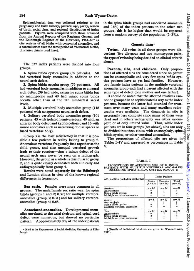
TABLE IPROPORTIONS OF AFFECTED SIBS OF 39 INDEX
PATIENTS WITH MULTIPLE VERTEBRAL ANOMALIESINCLUDING SPINA BIFIDA CYSTICA (GROUP 1)
Index PatientsAffected Sibs (including stillbirths) Males Females Total
(n = 13) (n = 26) (n = 39)
Brothers n=15 n=24 n = 39Anencephaly - 1 1Spina bifida cystica 2 _ 2Multiple vertebral anomalies - -
Sisters n=6 n=27 n 33Anencephaly _ 1 1Spina bifida cystica _ 1 1Multiple vertebral anomalies _ _
Total n=21 n=51 n= 72Anencephaly - 2 2Spina bifida cystica 2 1 3Multiple vertebral anomalies - - -
* Held at the Department of Social Medicine, University of Edin-burgh.
t (Details of individual kindreds are given in Wynne-Davies,1973).
284
copyright. on 1 January 2019 by guest. P
rotected byhttp://jm
g.bmj.com
/J M
ed Genet: first published as 10.1136/jm
g.12.3.280 on 1 Septem
ber 1975. Dow
nloaded from
Congenital vertebral anomalies: aetiology and relationship to spina bifida cystica
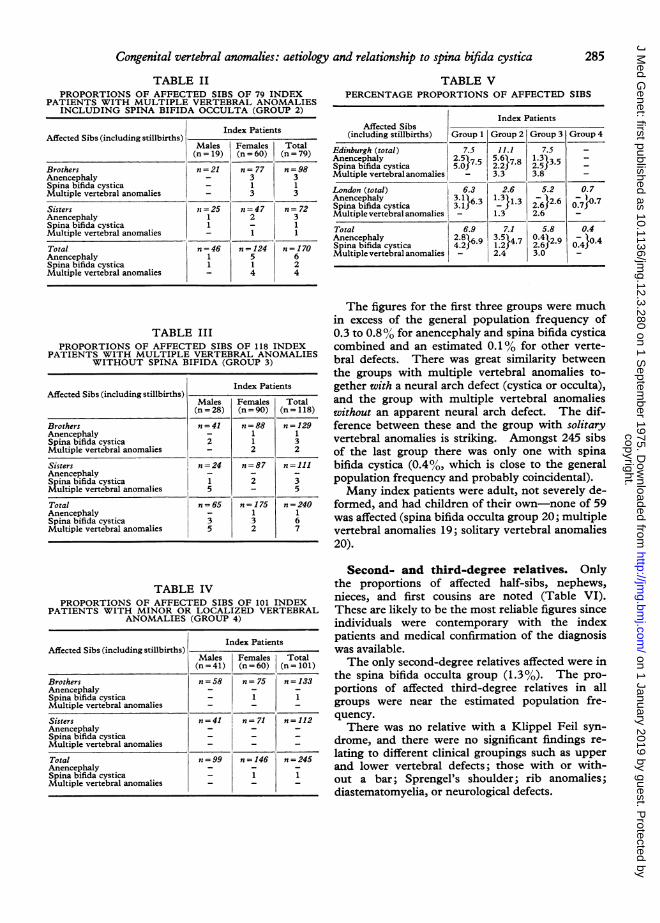
TABLE IIPROPORTIONS OF AFFECTED SIBS OF 79 INDEX
PATIENTS WITH MULTIPLE VERTEBRAL ANOMALIESINCLUDING SPINA BIFIDA OCCULTA (GROUP 2)
Index PatientsAffected Sibs (including stillbirths)
Males Females Total(n = 19) (n = 60) (n = 79)
Brothers n=21 n= 77 n=98Anencephaly _ 3 3Spina bifida cystica _ 1 1Multiple vertebral anomalies 3 3
Sisters n =25 n=47 n= 72Anencephaly 1 2 3Spina bifida cystica 1 _ 1Multiple vertebral anomalies 1
Total n=46 n=124 n=170Anencephaly 1 5 6Spina bifida cystica 1 1 2Multiple vertebral anomalies _ 4 4
TABLE IIIPROPORTIONS OF AFFECTED SIBS OF 118 INDEX
PATIENTS WITH MULTIPLE VERTEBRAL ANOMALIESWITHOUT SPINA BIFIDA (GROUP 3)
Index PatientsAffected SisS (sncluiung stilliSrtns)
Males Females Total(n = 28) (n = 90) (n= 118)
Brothers n=41 n = 88 n = 129Anencephaly - 1 1Spina bifida cystica 2 1 3Multiple vertebral anomalies - 2 2
Sisters n=24 n=87 n=111Anencephaly - - -
Spina bifida cystica 1 2 3Multiple vertebral anomalies 5 - 5
Total n =65 n = 175 n = 240Anencephaly _ 1 1Spina bifida cystica 3 3 6Multiple vertebral anomalies 5 2 7
TABLE IVPROPORTIONS OF AFFECTED SIBS OF 101 INDEX
PATIENTS WITH MINOR OR LOCALIZED VERTEBRALANOMALIES (GROUP 4)
Index PatientsAffected Sibs (includlng stillbirths)
Males Females Total(n = 41) (n = 60) (n = 101)
Brothers n = 58 n= 75 n = 133Anencephaly _ _Spina bifida cystica _ 1 1Multiple vertebral anomalies _ _
Sisters n=41 n=71 n=112Anencephaly _ _Spina bifida cystica _ _Multiple vertebral anomalies _ _
Total n =99 n = 146 n =245Anencephaly _ _Spina bifida cystica _ 1 1Multiple vertebral anomalies - -
TABLE VPERCENTAGE PROPORTIONS OF AFFECTED SIBS
Index PatientsAffected Sibs -
(including stillbirths) Group 1 Group 2 Group 3 Group 4
Edinburgh (total) 7.5 11.1 7.5 -
Anencephaly 2.557.5 5.6178 1.3 35Spina bifida cystica 5.0J 2.2 7 2.553Multiple vertebral anomalies - 3.3 3.8 -
London (total) 6.3 2.6 5.2 0.7Anencephaly 3.1}6.3 1.3?13 -)2.6 _ 7Spina bifida cystica 3.1 - 2.6 0.7fMultiple vertebral anomalies - 1.3 2.6
Total 6.9 7.1 5.8 0.4Anencephaly 2.8 6.9 3.5 4.7 0-412.9 °104Spina bifida cystica 4.29 1.27* }2.6 0.4}4Multiple vertebral anomalies - 2.4 3.0 -
The figures for the first three groups were muchin excess of the general population frequency of0.3 to 0.8% for anencephaly and spina bifida cysticacombined and an estimated 0.1% for other verte-bral defects. There was great similarity betweenthe groups with multiple vertebral anomalies to-gether with a neural arch defect (cystica or occulta),and the group with multiple vertebral anomalieswithout an apparent neural arch defect. The dif-ference between these and the group with solitaryvertebral anomalies is striking. Amongst 245 sibsof the last group there was only one with spinabifida cystica (0.40o, which is close to the generalpopulation frequency and probably coincidental).Many index patients were adult, not severely de-
formed, and had children of their own-none of 59was affected (spina bifida occulta group 20; multiplevertebral anomalies 19; solitary vertebral anomalies20).
Second- and third-degree relatives. Onlythe proportions of affected half-sibs, nephews,nieces, and first cousins are noted (Table VI).These are likely to be the most reliable figures sinceindividuals were contemporary with the indexpatients and medical confirmation of the diagnosiswas available.The only second-degree relatives affected were in
the spina bifida occulta group (1.3%). The pro-portions of affected third-degree relatives in allgroups were near the estimated population fre-quency.There was no relative with a Klippel Feil syn-
drome, and there were no significant findings re-
lating to different clinical groupings such as upperand lower vertebral defects; those with or with-out a bar; Sprengel's shoulder; rib anomalies;diastematomyelia, or neurological defects.
285
copyright. on 1 January 2019 by guest. P
rotected byhttp://jm
g.bmj.com
/J M
ed Genet: first published as 10.1136/jm
g.12.3.280 on 1 Septem
ber 1975. Dow
nloaded from

Ruth Wynne-Davies
TABLE VIPROPORTIONS OF AFFECTED SECOND- AND
THIRD-DEGREE RELATIVES
Index PatientsAffected Relatives
Group I Group 2 Group 3 Group 4
Second degree* n=14 n=80 n = 146 n = 142Anencephaly _ 1(1.3%) _Spina bifida cystica _ 1 (1.3%) _Multiple vertebral
anomalies -
Third degreet n = 405 n = 783 n = 1461 n = 944Anencephaly 1 (0.2%) 1(0.1%) 1 (0.1%)Spinabifidacystica 1 (0.2%) 2 (0.3%) 3 (0.2%) 1(0.1%)Multiple vertebral
anomalies
* Half sibs, nephews and nieces only.t First cousins only.
Consanguinous parents. There were two in-stances of related parents (first cousins), both indexpatients were in the solitary vertebral anomaliesgroup, one with kyphoscoliosis and the other with a
simple vertebral fusion. A total of seven sibs, twosons and three daughters, were all normal as werethe parents.
Epidemiology-environmental factorsMaternal obstetric and gynaecological his-
tory. Irregularities of the menstrual cycle are
very common, but it was noted that nearly half themothers of index patients in the spina bifida cysticaand occulta groups had a history of irregular men-struation, long periods of infertility, menorrhagia,severe dysmenorrhoea and so on, whereas amongstthe other two groups of index patients and amongstcontrols, rather less than one quarter of mothers hadsimilar complaints.The total number of previous abortions did not
differ significantly from the expected figure of 15%.However, when reviewed case by case, it became
apparent that a large number of abortions came
from very few families. In the spina bifida cysticaand occulta groups, seven mothers between themhad 27 abortions-over half of the total 50. Thesesame seven mothers between them also had twoanencephalic stillbirths, four children with spinabifida and meningocele, and three other (apparentlynormal) stillbirths. This type of history was not so
striking in the other two groups but only six of the219 mothers had one-third of the 71 abortions re-
corded.
Maternal illness. The only possible significantfinding under this heading related to maternalhypertension and toxaemia of pregnancy amongstmothers of both the spina bifida and the multiplevertebral anomalies groups. The diagnosis is a
clinical one and it is difficult to be precise in a retro-spective survey but findings were confirmed bymedical evidence wherever possible. Thirteen percent of mothers were affected compared with 7%of controls and of the localized vertebral anomaliesgroup.
Parental age and parity. The mean ages offathers and mothers and the mean of the paternal-maternal age difference were significantly raised forall groups, but not very markedly so. There was a
significant excess of first born children amongst thespina bifida occulta and multiple vertebral anomaliesgroups, but not amongst those with localized verte-bral anomalies (Table VII). (The figures did notreach a significant level in the spina bifida cysticagroup either, but numbers are small.)
Birthweight. The mean birthweight of indexpatients in the spina bifida and multiple vertebralanomalies groups was significantly low comparedwith their sibs (3040 ± 590 g, compared with 3356545 g).
TABLE VIIPARITY OF INDEX PATIENTS
Index Patients
Parity Group 1 Group 2 Group 3 Group 4
Obs Exp Obs Exp Obs Exp Obs Exp
First born 18 14.24 38 28.84 58 43.08 40 36.87Second born 11 12.19 13 24.68 31 36.87 29 31.56Third born or later 10 12.57 28 25.48 29 38.05 32 32.57
Total 39 79 118 101
Significance Not significant Highly significant Significant Not significant0.01 >p>0.001 0.02>p>0.01
286
copyright. on 1 January 2019 by guest. P
rotected byhttp://jm
g.bmj.com
/J M
ed Genet: first published as 10.1136/jm
g.12.3.280 on 1 Septem
ber 1975. Dow
nloaded from
Congenital vertebral anomalies: aetiology and relationship to spina bifida cystica
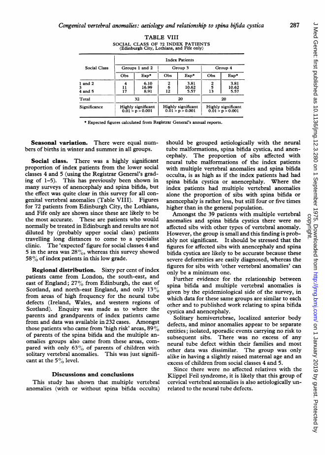
TABLE VIIISOCIAL CLASS OF 72 INDEX PATIENTS
(Edinburgh City, Lothians, and Fife only)
Index Patients
Social Class Groups 1 and 2 Group 3 Group 4
Obs Exp* Obs Exp* Obs Exp*
1 and 2 4 6.10 2 3.81 2 3.813 11 16.99 6 10.62 5 10.624 and 5 17 8.91 12 5.57 13 5.57
Total 32 20 20
Significance Highly significant Highly significant Highly significant0.01 >p>0.OOl 0.01 >p>0.001 0.01 >p>0.001
* Expected figures calculated from Registrar General's annual reports.
Seasonal variation. There were equal num-
bers of births in winter and summer in all groups.
Social class. There was a highly significantproportion of index patients from the lower socialclasses 4 and 5 (using the Registrar General's grad-ing of 1-5). This has previously been shown inmany surveys of anencephaly and spina bifida, butthe effect was quite clear in this survey for all con-
genital vertebral anomalies (Table VIII). Figuresfor 72 patients from Edinburgh City, the Lothians,and Fife only are shown since these are likely to bethe most accurate. These are patients who wouldnormally be treated in Edinburgh and results are notdiluted by (probably upper social class) patientstravelling long distances to come to a specialistclinic. The 'expected' figure for social classes 4 and5 in the area was 28%, whereas this survey showed580% of index patients in this low grade.
Regional distribution. Sixty per cent of indexpatients came from London, the south-east, andeast of England; 27% from Edinburgh, the east ofScotland, and north-east England, and only 13%from areas of high frequency for the neural tubedefects (Ireland, Wales, and western regions ofScotland). Enquiry was made as to where theparents and grandparents of index patients camefrom and data was available in 232 cases. Amongstthose patients who came from 'high risk' areas, 89%,'Oof parents of the spina bifida and the multiple an-
omalies groups also came from these areas, com-
pared with only 63% of parents of children withsolitary vertebral anomalies. This was just signifi-cant at the 5 0/ level.
Discussions and conclusionsThis study has shown that multiple vertebral
anomalies (with or without spina bifida occulta)
should be grouped aetiologically with the neuraltube malformations, spina bifida cystica, and anen-
cephaly. The proportion of sibs affected withneural tube malformations of the index patientswith multiple vertebral anomalies and spina bifidaocculta, is as high as if the index patients had hadspina bifida cystica or anencephaly. Where theindex patients had multiple vertebral anomaliesalone the proportion of sibs with spina bifida or
anencephaly is rather less, but still four or five timeshigher than in the general population.Amongst the 39 patients with multiple vertebral
anomalies and spina bifida cystica there were no
affected sibs with other types of vertebral anomaly.However, the group is small and this finding is prob-ably not significant. It should be stressed that thefigures for affected sibs with anencephaly and spinabifida cystica are likely to be accurate because thesesevere deformities are easily diagnosed, whereas thefigures for sibs with 'other vertebral anomalies' can
only be a minimum one.Further evidence for the relationship between
spina bifida and multiple vertebral anomalies isgiven by the epidemiological side of the survey, inwhich data for these same groups are similar to eachother and to published work relating to spina bifidacystica and anencephaly.
Solitary hemivertebrae, localized anterior bodydefects, and minor anomalies appear to be separateentities; isolated, sporadic events carrying no risk tosubsequent sibs. There was no excess of anyneural tube defect within their families and mostother data was dissimilar. The group was onlyalike in having a slightly raised maternal age and an
excess of children from social classes 4 and 5.Since there were no affected relatives with the
Klippel Feil syndrome, it is likely that this group ofcervical vertebral anomalies is also aetiologically un-related to the neural tube defects.
287
copyright. on 1 January 2019 by guest. P
rotected byhttp://jm
g.bmj.com
/J M
ed Genet: first published as 10.1136/jm
g.12.3.280 on 1 Septem
ber 1975. Dow
nloaded from
Ruth Wynne-Davies
Genetic advice and prenatal diagnosis. Therisk to sibs of cases with multiple vertebral ano-
malies is approximately 3 to 7% for neural tubemalformations and 2 to 3% for multiple vertebralanomalies alone; being higher in the Edinburgharea than in London. Aetiology is likely to bemultifactorial, with both genetic and environ-mental factors playing a major part in the aetiology.Now that the prenatal diagnosis of anencephaly
and spina bifida cystica is becoming possible by de-tecting alpha-fetoproteins in the amniotic fluid(Brock and Sutcliffe, 1972), the implications of add-ing other congenital defects to the same aetiologicalgroup are obvious. The alpha-fetoproteins are
thought to arise from free cerebrospinal fluid andwould therefore be unlikely to be present in a fetuswith only multiple vertebral anomalies and com-
plete skin cover. However, these families are
clearly at risk of a subsequent sib developing anen-
cephaly or spina bifida cystica and these risks shouldbe made known to them, since prenatal diagnosis ispossible for the 'open' defects.
Similarly, in families in whom anencephaly or
spina bifida cystica has already occurred, a subse-quent child may present with multiple congenitalvertebral defects. These anomalies are frequentlynot clinically apparent at birth, but there is a
potential risk of serious scoliosis, kyphosis, andparaplegia developing throughout the growingperiod. Whole-spine radiographs taken in theneonatal period would allow early diagnosis to bemade and preventive measures taken early.
Patients with multiple vertebral anomalies are
usually less severely handicapped than those withspina bifida cystica and more frequently have chil-dren of their own. In this survey, no child (of a
total of 59) was affected, but it is likely fromCarter's and Evans's figures (1973b) for the childrenof patients with spina bifida cystica that there wouldbe an increased risk of a neural tube defect pre-
senting in the next generation, with similar implica-tions for genetic counselling.
The index patients in this survey were all treated byProfessor J. I. P. James (London 1948-58; Edinburgh1958-68) and Mr C. W. Manning (Royal NationalOrthopaedic Hospital, London 1958-68) and I am mostgrateful to them both for access to case records and theirinterest in the project. A large number of people haveassisted with various aspects of the survey and detailed
acknowledgements are made at the end of the Thesis(Wynne-Davies, 1973).
REFmENCESBrock, D. J. H. and Sutcliffe, R. G. (1972). Alpha-fetoprotein in the
antenatal diagnosis of anencephaly and spina bifida. Lancet, 2,197-199.
Carter, C. O., David, P. A., and Laurence, K. M. (1968). A familystudy of major central nervous system malformations in SouthWales. Journal of Medical Genetics, 5, 81-106.
Carter, C. 0. and Evans, K. (1973a). Spina bifida and anencephalusin Greater London. Journal of Medical Genetics, 10, 209-234.
Carter, C. 0. and Evans, K. (1973b). Children of adult survivorswith spina bifida cystica. Lancet, 2, 924-926.
Carter, C. 0. and Fraser Roberts, J. A. (1967). The risk of re-
currence after two children with central-nervous-system malforma-tions. Lancet, 1, 306-308.
Edwards, J. H. (1958). Congenital malformations of the centralnervous system in Scotland. British Journal of Preventive andSocial Medicine, 12, 115-130.
Elwood, J. H. (1970). Anencephalus in Belfast, incidence andsecular and seasonal variations (1950-66). British Journal ofPreventive and Social Medicine, 24, 78-88.
Fedrick, J. (1970). Anencephalus: variation with maternal age,
parity, social class and region in England, Scotland, and Wales.Annals ofHuman Genetics, 34, 31-38.
Gunderson, C. E., Greenspan, R. H., Glaser, G. H., and Lubs, H. A.(1967). The Klippel-Feil syndrome, genetic and clinical re-
evaluation of cervical fusion. Medicine, 46, 491-512.James, J. I. P. (1955). Kyphoscoliosis. Journal of Bone and Joint
Surgery, 37B, 414-426.James, J. I. P. (1967). Scoliosis, ch. 8, pp. 96-136. Livingstone,Edinburgh and London.
Lorber, J. and Levick, K. (1967). Spina bifida cystica. Incidenceof spina bifida occulta in parents and in controls. Archives ofDisease in Childhood, 42, 171-173.
MacEwen, G. D. (1967). Congenital scoliosis with a unilateral bar.Journal of Bone and Joint Surgery, 49A, 1014.
MacEwen, G. D., Winter, R. B., and Hardy, J. H. (1972). Evalu-ation of kidney anomalies in congenital scoliosis. Journal ofBone and Joint Surgery, 54A, 1451-1454.
Naggan, L. and MacMahon, B. (1967). Ethnic differences in theprevalence of anencephaly and spina bifida in Boston, Massachu-setts. New England Journal of Medicine, 277, 1119-1123.
Record, R. G. and McKeown, T. (1950). Congenital malformationsof the central nervous system. British Journal of Social Medicine4, 26-50.
Registrar General for Scotland (1961-68). Anntal Reports 1939-1967. HMSO, Edinburgh.
Richards, I. D. G., McIntosh, H. T., and Sweenie, S. (1972). Agenetic study of anencephaly and spina bifida in Glasgow. De-velopmental Medicine and Child Neurology, 14, 626-639.
Shands, A. R. and Eisberg, H. B. (1955). The incidence of scoliosisin the State of Delaware. Journal of Bone and Joint Surgery, 37A,1243-1249.
Williamson, E. M. (1965). Incidence and family aggregation ofmajor congenital malformations of the central nervous system.Journal of Medical Genetics, 2, 161-172.
Winter, R. B., Moe, J. H., and Eilers, V. E. (1968). Congenitalscoliosis, a study of 234 patients treated and untreated. Journal ofBone and Joint Surgery, 50A, 15-47.
Winter, R. B., Moe, J. H., and Wang, J. F. (1973). Congenitalkyphosis. Journal of Bone and Joint Surgery, 55A, 223-256.
Wynne-Davies, R. (1968). Familial (idiopathic) scoliosis, a familysurvey. Journal of Bone and Joint Surgery, 50B, 24-30.
Wynne-Davies, R. (1973). Genetic and other Factors in the Aetio-logy of Scoliosis, pp. 62-120. PhD Thesis, University of Edin-burgh.
288
copyright. on 1 January 2019 by guest. P
rotected byhttp://jm
g.bmj.com
/J M
ed Genet: first published as 10.1136/jm
g.12.3.280 on 1 Septem
ber 1975. Dow
nloaded from