Embed Size (px)
Citation preview
301.150421
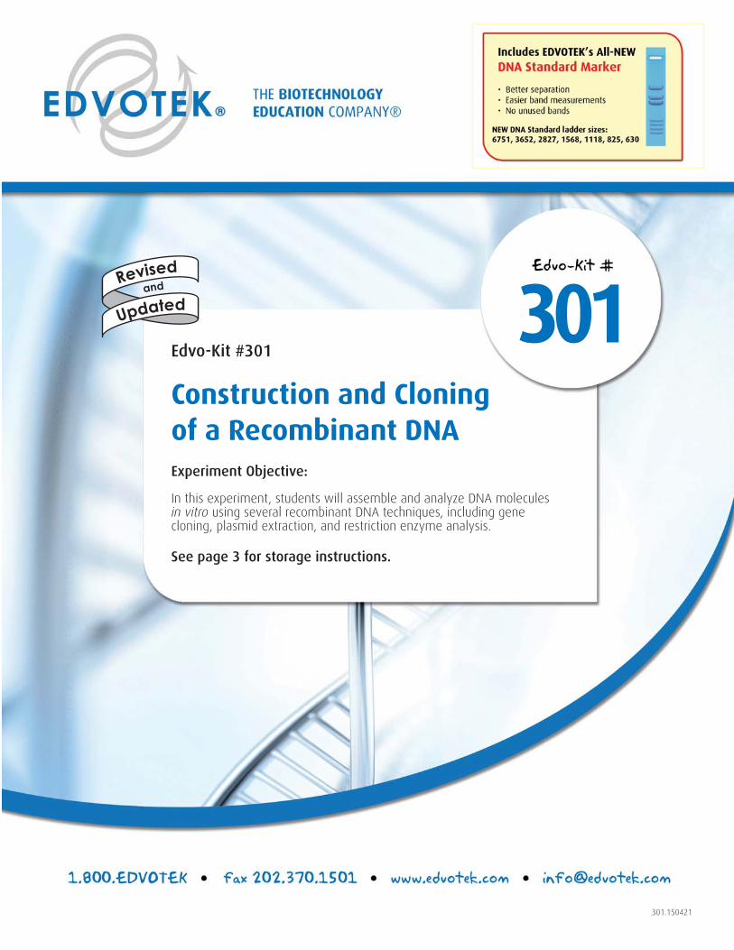
301Edvo-Kit #301
Construction and Cloning of a Recombinant DNAExperiment Objective:
In this experiment, students will assemble and analyze DNA molecules in vitro using several recombinant DNA techniques, including gene cloning, plasmid extraction, and restriction enzyme analysis.
See page 3 for storage instructions.
Updated
Revised
and
Table of Contents
Page
Experiment Components 3
Experiment Requirements 4
Background Information 5
Experiment Procedures
Experiment Overview and Laboratory Safety 11
Module I: Ligation of the Plasmid Vector to the kanR Gene Fragment 13
Module II: Transformation of the Recombinant DNA into E.coli 18
Module III: Culturing of kanR Transformants 22
Module IV: Extraction of Recombinant Plasmid DNA 23
Module V: Restriction Enzyme Analysis 25
Study Questions 27
Instructor's Guidelines
Organizing the Experiment 28
Overview of Pre-lab Preparations 29
Pre-Lab Preparations
Module I Preparations 30
Module II Preparations 32
Module III Preparations 35
Module IV Preparations 36
Module V Preparations 37
Experiment Results and Analysis 38
Study Questions and Answers 40
Appendices 41
A EDVOTEK® Troubleshooting Guide 42
B Bulk Preparation of Agarose Gels 46
C Data Analysis Using a Standard Curve 47
D LyphoCells™ For Transformation 50
Material Safety Data Sheets can be found on our website: www.edvotek.com
CLONING AND CONSTRUCTION OF A RECOMBINANT DNA EDVO-Kit 301
1.800.EDVOTEK • Fax 202.370.1501 • [email protected] • www.edvotek.com
2
Duplication of any part of this document is permitted for non-profi t educational purposes only. Copyright © 2015 EDVOTEK, Inc.,
all rights reserved. 301.150421
CLONING AND CONSTRUCTION OF A RECOMBINANT DNA EDVO-Kit 301
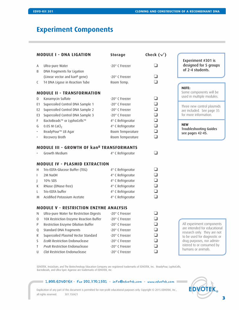
Experiment Components
EDVOTEK, InstaStain, and The Biotechnology Education Company are registered trademarks of EDVOTEK, Inc. ReadyPour, LyphoCells, BactoBeads, and Ultra-Spec Agarose are trademarks of EDVOTEK, Inc.
MODULE I - DNA LIGATION Storage Check (√ )
A Ultra-pure Water -20° C Freezer ❑B DNA Fragments for Ligation
(Linear vector and kanR gene) -20° C Freezer ❑C T4 DNA Ligase in Reaction Tube Room Temp. ❑
MODULE II - TRANSFORMATIOND Kanamycin Sulfate -20° C Freezer ❑E1 Supercoiled Control DNA Sample 1 -20° C Freezer ❑E2 Supercoiled Control DNA Sample 2 -20° C Freezer ❑E3 Supercoiled Control DNA Sample 3 -20° C Freezer ❑F BactoBeads™ or LyphoCells™ 4° C Refrigerator ❑G 0.05 M CaCl2 4° C Refrigerator ❑ • ReadyPour™ LB Agar Room Temperature ❑• Recovery Broth Room Temperature ❑
MODULE III - GROWTH OF kanR TRANSFORMANTS• Growth Medium 4° C Refrigerator ❑
MODULE IV - PLASMID EXTRACTIONH Tris-EDTA-Glucose Buffer (TEG) 4° C Refrigerator ❑I 2M NaOH 4° C Refrigerator ❑J 10% SDS 4° C Refrigerator ❑K RNase (DNase-free) 4° C Refrigerator ❑L Tris-EDTA buffer 4° C Refrigerator ❑M Acidifi ed Potassium Acetate 4° C Refrigerator ❑
MODULE V - RESTRICTION ENZYME ANALYSISN Ultra-pure Water for Restriction Digests -20° C Freezer ❑ O 10X Restriction Enzyme Reaction Buffer -20° C Freezer ❑P Restriction Enzyme Dilution Buffer -20° C Freezer ❑Q Standard DNA Fragments -20° C Freezer ❑R Supercoiled Plasmid Vector Standard -20° C Freezer ❑S EcoRI Restriction Endonuclease -20° C Freezer ❑T PvuII Restriction Endonuclease -20° C Freezer ❑U ClaI Restriction Endonuclease -20° C Freezer ❑
Experiment #301 is designed for 5 groups of 2-4 students.
All experiment components are intended for educational research only. They are not to be used for diagnostic or drug purposes, nor admin-istered to or consumed by humans or animals.
NOTE:Some components will be used in multiple modules.
Three new control plasmids are included. See page 35 for more information.
NEW Troubleshooting Guidessee pages 42-45.
CLONING AND CONSTRUCTION OF A RECOMBINANT DNAEDVO-Kit 301
3
1.800.EDVOTEK • Fax 202.370.1501 • [email protected] • www.edvotek.com
Duplication of any part of this document is permitted for non-profi t educational purposes only. Copyright © 2015 EDVOTEK, Inc.,
all rights reserved. 301.150421
CLONING AND CONSTRUCTION OF A RECOMBINANT DNAEDVO-Kit 301
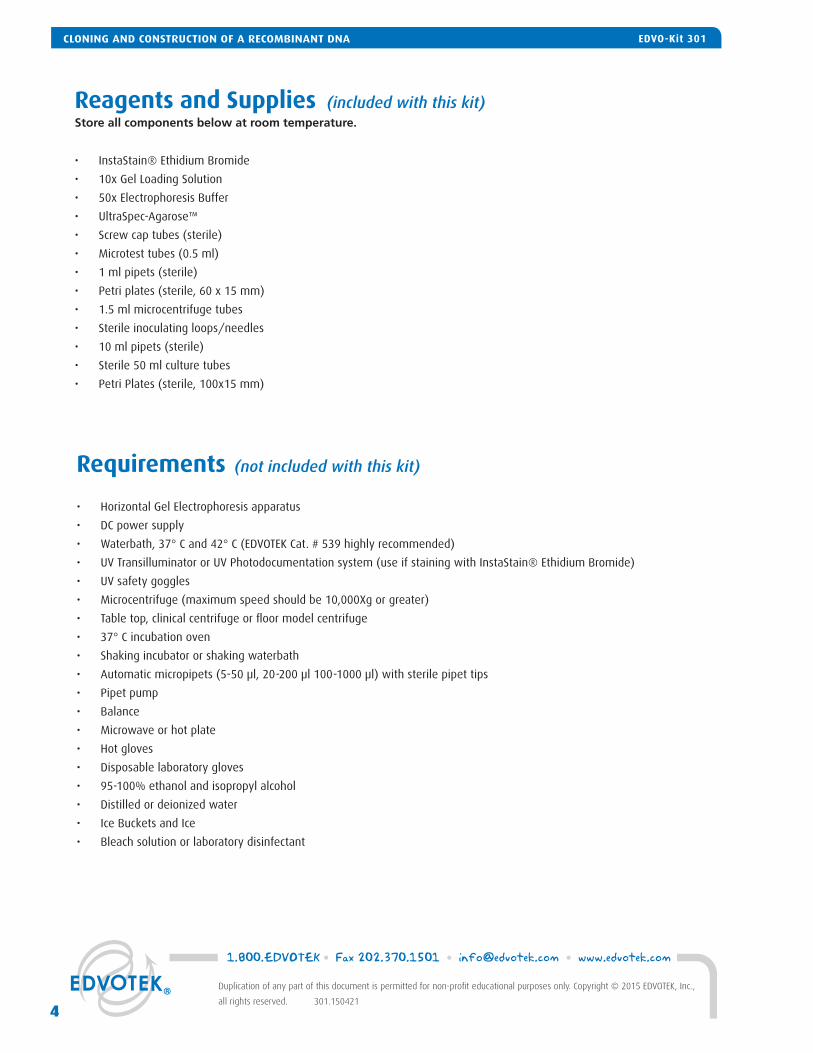
Requirements (not included with this kit)
• Horizontal Gel Electrophoresis apparatus
• DC power supply
• Waterbath, 37° C and 42° C (EDVOTEK Cat. # 539 highly recommended)
• UV Transilluminator or UV Photodocumentation system (use if staining with InstaStain® Ethidium Bromide)
• UV safety goggles
• Microcentrifuge (maximum speed should be 10,000Xg or greater)
• Table top, clinical centrifuge or fl oor model centrifuge
• 37° C incubation oven
• Shaking incubator or shaking waterbath
• Automatic micropipets (5-50 μl, 20-200 μl 100-1000 μl) with sterile pipet tips
• Pipet pump
• Balance
• Microwave or hot plate
• Hot gloves
• Disposable laboratory gloves
• 95-100% ethanol and isopropyl alcohol
• Distilled or deionized water
• Ice Buckets and Ice
• Bleach solution or laboratory disinfectant
Reagents and Supplies (included with this kit)Store all components below at room temperature.
• InstaStain® Ethidium Bromide
• 10x Gel Loading Solution
• 50x Electrophoresis Buffer
• UltraSpec-Agarose™
• Screw cap tubes (sterile)
• Microtest tubes (0.5 ml)
• 1 ml pipets (sterile)
• Petri plates (sterile, 60 x 15 mm)
• 1.5 ml microcentrifuge tubes
• Sterile inoculating loops/needles
• 10 ml pipets (sterile)
• Sterile 50 ml culture tubes
• Petri Plates (sterile, 100x15 mm)
1.800.EDVOTEK • Fax 202.370.1501 • [email protected] • www.edvotek.com
4
Duplication of any part of this document is permitted for non-profi t educational purposes only. Copyright © 2015 EDVOTEK, Inc.,
all rights reserved. 301.150421
CLONING AND CONSTRUCTION OF A RECOMBINANT DNA EDVO-Kit 301
CLONING AND CONSTRUCTION OF RECOMBINANT DNA
After James Watson and Francis Crick published the structure of the double helix, scientists around the world raced to unlock the secrets encoded in our DNA. However, they found it diffi cult to study the properties of an individual gene because a single chromosome can contain hundreds of genes. As such, gene cloning techniques were developed to isolate, combine, and repro-duce specifi c DNA sequences. First, using an enzyme called a restriction endonuclease, the DNA could be cut into smaller pieces. Then, a second enzyme called DNA ligase was used to ligate, or link, the DNA fragment with a bacterial plasmid. Finally, the hybrid DNA molecules were forced into bacteria, where they can be reproduced to create millions of copies of the starting DNA sequence. The development of recombinant DNA technology launched the era of biotechnology by making DNA mapping, sequencing and various genome-wide studies possible.
In this exploration, students will assemble and analyze recombinant DNA molecules using the same gene cloning techniques used in research laboratories. First, students will ligate the Kanamycin resistance (kanR) gene into a plasmid vector. The modi-fi ed plasmid is transformed into E.coli, which is then plated on media containing Kanamycin, and allowed to grow overnight. Because the newly-constructed plasmid contains the kanR gene, only successfully transformed cells should grow into colonies. However, the plasmid’s presence in the cells does not tell us anything about the orientation or the number of Kanamycin inserts present within the vector. To do this, the plasmid within a single transformant is grown, purifi ed, and analyzed using restriction digestion and agarose gel electrophoresis.
CREATING RECOMBINANT DNA
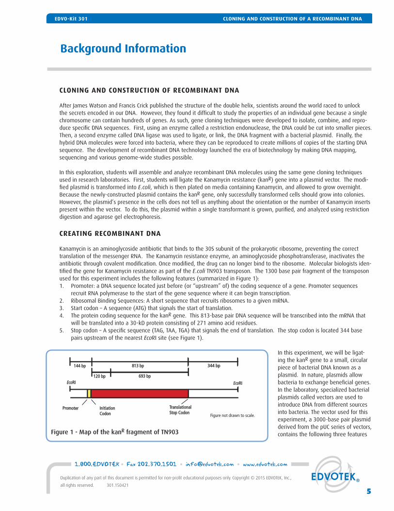
Kanamycin is an aminoglycoside antibiotic that binds to the 30S subunit of the prokaryotic ribosome, preventing the correct translation of the messenger RNA. The Kanamycin resistance enzyme, an aminoglycoside phosphotransferase, inactivates the antibiotic through covalent modifi cation. Once modifi ed, the drug can no longer bind to the ribosome. Molecular biologists iden-tifi ed the gene for Kanamycin resistance as part of the E.coli TN903 transposon. The 1300 base pair fragment of the transposon used for this experiment includes the following features (summarized in Figure 1):1. Promoter: a DNA sequence located just before (or “upstream” of) the coding sequence of a gene. Promoter sequences
recruit RNA polymerase to the start of the gene sequence where it can begin transcription.2. Ribosomal Binding Sequences: A short sequence that recruits ribosomes to a given mRNA.3. Start codon – A sequence (ATG) that signals the start of translation. 4. The protein coding sequence for the kanR gene. This 813-base pair DNA sequence will be transcribed into the mRNA that
will be translated into a 30-kD protein consisting of 271 amino acid residues.5. Stop codon – A specifi c sequence (TAG, TAA, TGA) that signals the end of translation. The stop codon is located 344 base
pairs upstream of the nearest EcoRI site (see Figure 1).
In this experiment, we will be ligat-ing the kanR gene to a small, circular piece of bacterial DNA known as a plasmid. In nature, plasmids allow bacteria to exchange benefi cial genes. In the laboratory, specialized bacterial plasmids called vectors are used to introduce DNA from different sources into bacteria. The vector used for this experiment, a 3000-base pair plasmid derived from the pUC series of vectors, contains the following three features
Background Information
Figure 1 - Map of the kanR fragment of TN903
144 bp144 bp
Figure not drawn to scale.
813 bp813 bp 344 bp344 bp
120 bp120 bp 693 bp693 bpEcoEcoRIRI EcoEcoRIRI
PromoterPromoter InitiationInitiationCodonCodon
TranslationalTranslationalStop CodonStop Codon
CLONING AND CONSTRUCTION OF A RECOMBINANT DNAEDVO-Kit 301
5
1.800.EDVOTEK • Fax 202.370.1501 • [email protected] • www.edvotek.com
Duplication of any part of this document is permitted for non-profi t educational purposes only. Copyright © 2015 EDVOTEK, Inc.,
all rights reserved. 301.150421
CLONING AND CONSTRUCTION OF A RECOMBINANT DNAEDVO-Kit 301
that make it useful for our experiment (Figure 2):1. Origin of Replication: a DNA sequence that allows bacteria to copy the plasmid.2. Multiple Cloning Site (or MCS): a short DNA sequence that contains many
unique restriction enzyme sites and allows scientists to insert specifi c genes into the plasmid.
3. Selectable marker: a gene that codes for resistance to a specifi c antibi-otic (in pUC plasmids, ampicillin). When using selective media, only cells containing the marker should grow into colonies. This allows researchers to identify transformed cells.
To link the kanR gene into this vector, both pieces of DNA are cut with an enzyme known as a restriction endonuclease. These endonucleases (also known as restriction enzymes) act like molecular scissors, cutting double-stranded DNA at specifi c sequences. Many restriction enzymes recognize and cut palindromic stretches of DNA, generally 4-8 base pairs in length. Diges-tion by a restriction enzyme generates DNA fragments with one of two types of DNA ends -- “blunt” or “sticky” (Figure 3). To illustrate this, fi rst consider the recognition site and cleavage pattern of HaeIII. This enzyme cuts both DNA strands at the same position, between the G and the neighboring C, which generates fragments without an overhang. These so-called “blunt” ends can be joined with any other blunt end.
In contrast to HaeIII, EcoRI cleaves between the G and neighboring A, as indicated by the arrows in the left side of the fi gure. It is important to note that the positions of the cleavage are staggered, so the resulting fragments project short overhangs of single-stranded DNA with complementary sequences. Such overhangs are referred to as “sticky” ends because the single-strands can interact with—or stick to—other overhangs with a complementary sequence. Digestion of the same piece of DNA using different enzymes can produce sticky ends of different lengths and strand orientation (5’ vs. 3’).
To create compatible sticky ends for our ligation, both pieces of DNA (the pUC vector and the kanR insert) are cut with the same restriction enzyme – EcoRI. This en-zyme cuts once within the multiple cloning site of our vector, creating a linear piece of DNA with sticky EcoRI overhangs at each end (Figure 4A). EcoRI overhangs are also created at each end of the kanR fragment when digested with the enzyme.
After the DNA fragments are cut, they are linked to-gether in vitro (Figure 4B). First, the unpaired nucleo-tides at the ends of the DNA fragments base-pair to one another, creating a circular DNA molecule. Then, DNA ligase catalyzes the formation of a phosphodiester bond between the 5' phosphate at the end of one frag-ment and 3' hydroxyl group at the end of the other fragment (Figure 4C). ATP is added to the reaction to supply the required energy for bond formation (Figure 4D). Although DNA ligase is most active at 37° C, ligation of DNA fragments with cohesive termini is usually performed between 4° C and 22° C. Lower temperatures allow for a more stable interaction between the sticky ends.
Once the two pieces of DNA are ligated together, we have created a recombinant DNA molecule. Ideally, a single kanR insert is ligated to the vector in the correct orientation, creating a 4300-base pair plasmid. In practice, the ligation is not limited to one insert per vector -- many combinations and orientations between vector and insert can be created using the two pieces of DNA (summarized in Figure 5). Because the vector is cut with one enzyme, the ends of the plasmid can self-ligate. Furthermore, a vector molecule can link to another vector instead of the insert, forming a long linear chain of plasmid DNA called a concate-
Figure 2: Plasmid Features
Plasmid Map
SelectableMarker
Origin ofReplication
Multiple cloning site
G A A T T C
C T T A A G
EcoRI
G G C C
C C G G
HaeIII
C T C G A G
Blunt EndSticky Ends
SacI
HaeIIISacI
G A G C T C
EcoRI
Figure 3: Different types of DNA ends produced byRestriction Enzymes.
1.800.EDVOTEK • Fax 202.370.1501 • [email protected] • www.edvotek.com
6
Duplication of any part of this document is permitted for non-profi t educational purposes only. Copyright © 2015 EDVOTEK, Inc.,
all rights reserved. 301.150421
CLONING AND CONSTRUCTION OF A RECOMBINANT DNA EDVO-Kit 301
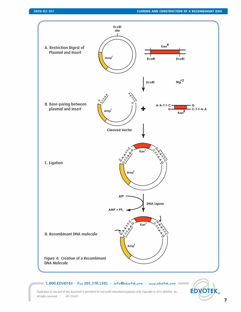
Figure 4: Creation of a Recombinant DNA Molecule
ATP
AMP + PP1
DNA Ligase
EcoRI Mg+2
KanR
A-A-T-T-C G
G C-T-T-A-A
Cleaved Vector
A. Restriction Digest of Plasmid and Insert
B. Base-paring between plasmid and insert
C. Ligation
D. Recombinant DNA molecule
G-A
-A-T-T-C
C-T-T-A
-A-GG
-A-A
-T-T
-C
C-T
-T-A
-A-G
Amp r
Kanr
G-A
-A-T-T-C
C-T-T-A
-A-GG
-A-A
-T-T
-C
C-T
-T-A
-A-G
Amp r
Amp r
Kanr
A-A
-T-T-C
GG
C-T
-T-A
-A
EcoRI
site
KanR
EcoRI EcoRI
+
Amp r
7
1.800.EDVOTEK • Fax 202.370.1501 • [email protected] • www.edvotek.com
Duplication of any part of this document is permitted for non-profi t educational purposes only. Copyright © 2015 EDVOTEK, Inc.,
all rights reserved. 301.150421
CLONING AND CONSTRUCTION OF A RECOMBINANT DNAEDVO-Kit 301
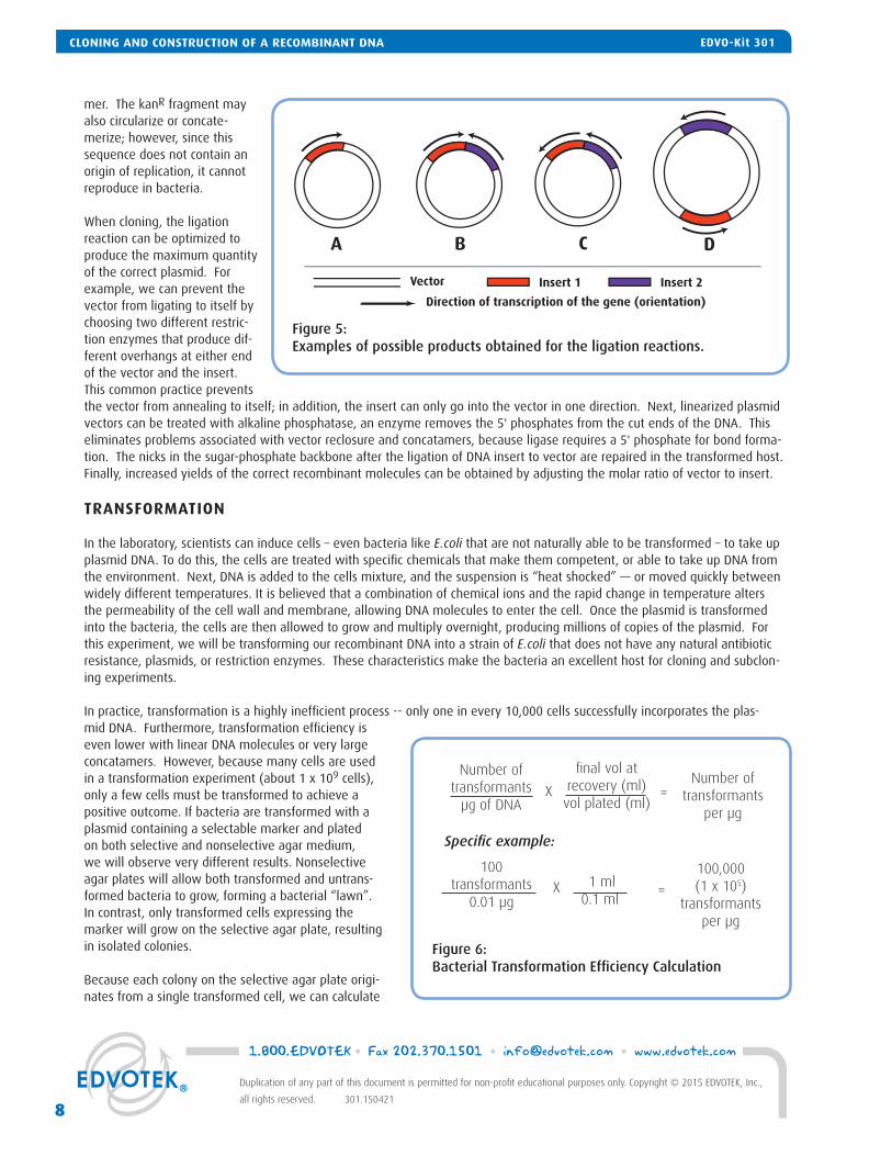
mer. The kanR fragment may also circularize or concate-merize; however, since this sequence does not contain an origin of replication, it cannot reproduce in bacteria.
When cloning, the ligation reaction can be optimized to produce the maximum quantity of the correct plasmid. For example, we can prevent the vector from ligating to itself by choosing two different restric-tion enzymes that produce dif-ferent overhangs at either end of the vector and the insert. This common practice prevents the vector from annealing to itself; in addition, the insert can only go into the vector in one direction. Next, linearized plasmid vectors can be treated with alkaline phosphatase, an enzyme removes the 5' phosphates from the cut ends of the DNA. This eliminates problems associated with vector reclosure and concatamers, because ligase requires a 5' phosphate for bond forma-tion. The nicks in the sugar-phosphate backbone after the ligation of DNA insert to vector are repaired in the transformed host. Finally, increased yields of the correct recombinant molecules can be obtained by adjusting the molar ratio of vector to insert.
TRANSFORMATION
In the laboratory, scientists can induce cells – even bacteria like E.coli that are not naturally able to be transformed – to take up plasmid DNA. To do this, the cells are treated with specifi c chemicals that make them competent, or able to take up DNA from the environment. Next, DNA is added to the cells mixture, and the suspension is “heat shocked” — or moved quickly between widely different temperatures. It is believed that a combination of chemical ions and the rapid change in temperature alters the permeability of the cell wall and membrane, allowing DNA molecules to enter the cell. Once the plasmid is transformed into the bacteria, the cells are then allowed to grow and multiply overnight, producing millions of copies of the plasmid. For this experiment, we will be transforming our recombinant DNA into a strain of E.coli that does not have any natural antibiotic resistance, plasmids, or restriction enzymes. These characteristics make the bacteria an excellent host for cloning and subclon-ing experiments.
In practice, transformation is a highly ineffi cient process -- only one in every 10,000 cells successfully incorporates the plas-mid DNA. Furthermore, transformation effi ciency is even lower with linear DNA molecules or very large concatamers. However, because many cells are used in a transformation experiment (about 1 x 10⁹ cells), only a few cells must be transformed to achieve a positive outcome. If bacteria are transformed with a plasmid containing a selectable marker and plated on both selective and nonselective agar medium, we will observe very different results. Nonselective agar plates will allow both transformed and untrans-formed bacteria to grow, forming a bacterial “lawn”. In contrast, only transformed cells expressing the marker will grow on the selective agar plate, resulting in isolated colonies.
Because each colony on the selective agar plate origi-nates from a single transformed cell, we can calculate
Number oftransformants
per μg
=
100 transformants
0.01 μg
Specifi c example:
X
fi nal vol at recovery (ml)vol plated (ml)
X
1 ml0.1 ml
=
100,000 (1 x 10⁵)
transformants per μg
Number of transformants
μg of DNA
Figure 6:Bacterial Transformation Effi ciency Calculation
Vector
A
Insert 1 Insert 2
Direction of transcription of the gene (orientation)
B C D
Figure 5: Examples of possible products obtained for the ligation reactions.
1.800.EDVOTEK • Fax 202.370.1501 • [email protected] • www.edvotek.com
8
Duplication of any part of this document is permitted for non-profi t educational purposes only. Copyright © 2015 EDVOTEK, Inc.,
all rights reserved. 301.150421
CLONING AND CONSTRUCTION OF A RECOMBINANT DNA EDVO-Kit 301
the transformation effi ciency, or the number of cells transformed per microgram (μg) of plasmid DNA (outlined in Figure 6). For example, if 10 nanograms (0.01 μg) of plasmid were used to transform one milliliter (ml) of cells, and plating 100 microliters (μl) of this mixture gives rise to 100 colonies, then there must have been 1,000 transformed bacteria in the one ml mixture. Dividing 1,000 transformants by 0.01 μg DNA means that the transformation effi ciency would be 1 x 10⁵ cells transformed per μg plasmid DNA. In general, transformation effi ciency ranges from 1 x 10⁵ to 1 x 10⁸ cells transformed per μg plasmid.
SELECTION AND PURIFICATION OF RECOMBINANT PLASMIDS
In most cases, the vector’s antibiotic resistance gene is used to select for bacteria that contain the recombinant DNA. The plasmid DNA is then screened to determine whether the insert is present. In this experiment, we can perform a simple and rapid selection for the insert by plating the transformants on nutrient agar plates containing Kanamycin. Only cells that contain the insert will grow. However, we must still analyze our recombinant DNA to verify the results of the ligation reaction. For example, the insert may be present in the reverse orientation, or multiple inserts may be present (Figure 5). To determine the nature of the recombinant plasmid, the DNA is digested with three restriction enzymes, used alone or in combination. The vari-ous digestions will create different patterns depending on the presence and the orientation of the insert.
Before we analyze the recombinant DNA, we must isolate plasmid from the cells. First, a single colony from the transfor-mation plate is used to inoculate liquid media. Because bacteria will often eliminate their plasmids, the liquid media contains Kanamycin to maintain this selective pressure. The bacteria are then allowed to grow and multiply in the media overnight, producing millions of copies of the plasmid. Finally, the plasmid is purifi ed from the bacteria using the alkaline-lysis method and analyzed.
The alkaline-lysis plasmid extraction technique is a simple and reliable method to purify the recombinant DNA from the bacte-rial cells. First, the cells are harvested from the liquid culture by centrifugation, creating a pellet of cells at the bottom of the tube. The cell pellet is then suspended in a solution that contains the detergent sodium dodecyl sulfate (or SDS). SDS disrupts the cell membrane, releasing the DNA from the bacteria in a process called lysis. The high pH of the solution irreversibly dena-tures the bacteria’s chromosomal DNA and aids in protein denaturation and RNA degradation. In addition, the nuclease RNase is added to the cell lysate to degrade any residual RNA in the sample. In contrast, supercoiled plasmid is not affected because its phosphate backbone remains unbroken.
Once the effects of lysis are complete, an acidic potassium acetate solution is added to the lysate to neutralize the pH and precipitate SDS and membrane/protein complexes. Furthermore, most of the chromosomal DNA will also precipitate because it is associated with the cell membrane. This leaves a solution that primarily contains the supercoiled plasmid. If high-quality plasmid is required for sequencing, the residual protein can be further removed from the DNA by organic solvents such as phe-nol and chloroform.
In this experiment, isopropyl alcohol is used to purify and concentrate DNA from cellular extracts. When the alcohol is added to the solution, electrostatic interactions between the water molecules and the sugar-phosphate backbone of the plasmid are disrupted, forcing the DNA out of solution as a sticky white precipitate. The precipitated plasmid is concentrated at the bot-tom of a micro-test tube by high-speed centrifugation. Once the supernatant is removed, the DNA pellet is dried before being resuspended in Tris-ETDA buffer for restriction digestion analysis.
RESTRICTION ENZYME ANALYSIS OF RECOMBINANT DNA
To verify the presence of the kanR insert, the recombinant plasmid is digested with EcoRI. Remember, this is the site that was used to clone the insert into the plasmid. Digestion of the plasmid with this enzyme will generate two fragments of 3000 and 1300-base pairs that represent the vector and the insert, respectively. This digestion does not confi rm the orientation of the insert nor does it identify the presence of multiple inserts because each insert will produce the same 1300-base pair product (Figure 3, A-C).
The presence of multiple inserts can be confi rmed by cleaving the plasmid with PvuII endonuclease. The only PvuII recognition site in the recombinant plasmid is found in the plasmid vector, about 180 base pairs downstream (in the 3' direction) from the
9
1.800.EDVOTEK • Fax 202.370.1501 • [email protected] • www.edvotek.com
Duplication of any part of this document is permitted for non-profi t educational purposes only. Copyright © 2015 EDVOTEK, Inc.,
all rights reserved. 301.150421
CLONING AND CONSTRUCTION OF A RECOMBINANT DNAEDVO-Kit 301
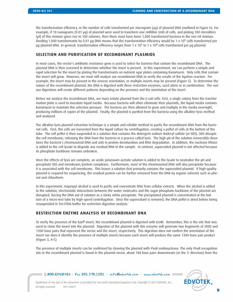
vector’s EcoRI site in the poly-linker. Consequently, when the DNA is digested with this enzyme, the plasmid will be linearized. The size of the linearized plasmid can be used to estimate how many inserts are present in the recombinant DNA molecule. For example, if two adjacent in-serts were ligated to the vector, the linearized plasmid would be 5600 base pairs in length (1300+1300+3000).
The single PvuII site in the vector can be used as a fi xed reference point to determine the orientation of the kanR insert. The insert possesses a single ClaI recognition site located ~264 base pairs from the 5’ end of the DNA (~144 base pairs upstream of start codon and ~120 base pairs from the gene). Because the relationship between the ClaI site and the PvuII site changes depending on the orientation of the kanR gene within the plasmid, a “double digest” of the recombinant DNA with these enzymes will produce a fragment with a distinguishing length, which can be used to determine the orientation of the insert (Figure 7). For example, a digest of the plasmid in Figure 7A will yield 1217 and 3083-base pair fragments after the double digest. When the insert is in the opposite orientation, a double digest will yield 444 and 3856-base pair fragments.
Statistically, one would expect to fi nd a 50:50 occurrence of the two insert orientations if many colonies were analyzed. It should be noted that the orientation of the kanR insert does not have any large effects on its expression because the fragment has its own promoter. However, when the vector supplies the promoter required for expression, one of the insert orientations can abolish expression of the subcloned gene.
If multiple inserts are present in a single circular vector, the determination of orientation becomes more complex (Figure 5B). Assume the PvuII site in the vector is 180 base pairs to the right (3') of RI restriction site (going in the clockwise direction). A PvuII - ClaI double digest of the recombinant in Figure 5A would yield three fragments having lengths of 1217, 1301 and 3084 base pairs.
ANALYSIS OF RESTRICTION DIGESTS
In order to view the results of a restriction digest, students will utilize agarose gel electrophoresis. At fi rst glance, an agarose gel appears to be a solid at room temperature. On the molecular level, the gel contains small channels through which the DNA can pass. A mixture of DNA molecules is added into depressions (or “wells”) within a gel, and then an electrical current is passed through the gel. Because the sugar-phosphate backbone of DNA has a strong negative charge, the current drives the DNA through the gel towards the positive electrode. Small DNA fragments move through these holes easily, but large DNA fragments have a more diffi cult time squeezing through channels. Therefore, molecules with dissimilar sizes and/or shapes travel at different speeds, become separated, and form discrete “bands” within the gel. After the current is stopped, the bands can be visualized using a stain that sticks to DNA.
Each student group will observe different banding patterns after electrophoresis of ligation reaction products due to the differ-ent plasmids that are built by the experiment. However, because plasmids can exist in different conformations, several unex-pected bands may be present in the undigested plasmid DNA samples. First, the supercoiled plasmid DNA is tightly wound in a compact secondary structure. This allows the DNA to be effi ciently packaged within the cell. This DNA will appear smaller than its molecular weight when analyzed by electrophoresis. In contrast, if the DNA backbone is nicked or cut during purifi cation, the plasmid will lose its compact structure and run at the appropriate size. This will create two distinct bands when analyzed by electrophoresis. Large chains of interlocked plasmids called catenanes are also present in the sample. The catenanes, being the largest of the DNA isoforms, will appear much larger than the linear DNA. All of these isoforms are digested by restriction enzymes, so they will produce the same patterns. After restriction digest, all of these isoforms will produce the same series of DNA fragments when analyzed by electrophoresis.
Figure 7 - Using double digests to determine insert orientation.
5’5’ClaClaI
5’5’3’3’
3’3’5’5’
5’5’3’3’
3’3’
ClaClaI
ATGATG
ATGATG
PvuPvuIIII PvuPvuIIII
A B
EcoEcoRIRI
EcoEcoRIRI
1.800.EDVOTEK • Fax 202.370.1501 • [email protected] • www.edvotek.com
10
Duplication of any part of this document is permitted for non-profi t educational purposes only. Copyright © 2015 EDVOTEK, Inc.,
all rights reserved. 301.150421
CLONING AND CONSTRUCTION OF A RECOMBINANT DNA EDVO-Kit 301
EXPERIMENT OBJECTIVE:
In this experiment, students will assemble and analyze DNA molecules in vitro using several recombinant DNA techniques, including gene cloning, plasmid extraction, and restriction enzyme analysis.
Experiment Overview
Wear gloves and safety goggles
LABORATORY SAFETY:
Transformation experiments contain antibiotics to select for transformed bacteria. Students who have allergies to anti-biotics such as Penicillin, Ampicillin, Kanamycin or Tetracycine should not participate in this experiment.
1. Wear gloves and goggles while working in the laboratory.
2. Exercise extreme caution when working in the laboratory - you will be heating and melting agar, which could be dangerous if performed incorrectly.
3. DO NOT MOUTH PIPET REAGENTS - USE PIPET PUMPS OR BULBS.
4. The E.coli bacteria used in this experiment is not considered pathogenic. Regardless, it is good practice to follow simple safety guidelines in handling and disposal of materials contaminated with bacteria.
A. Wipe down the lab bench with a 10% bleach solution or a laboratory disinfectant.
B. All materials, including petri plates, pipets, transfer pipets, loops and tubes, that come in contact with bacteria should be disinfected before disposal in the garbage. Disinfect materials as soon as possible after use in one of the following ways:
• Autoclave at 121° C for 20 minutes. Tape several petri plates together and close tube caps before disposal. Collect all contaminated materials in an auto-
clavable, disposable bag. Seal the bag and place it in a metal tray to prevent any possibility of liquid medium or agar from spilling into the sterilizer chamber.
• Soak in 10% bleach solution. Immerse petri plates, open tubes and other contaminated materials into a tub containing a 10% bleach solution. Soak
the materials overnight and then discard. Wear gloves and goggles when working with bleach.
5. Always wash hands thoroughly with soap and water after working in the laboratory.
6. If you are unsure of something, ASK YOUR INSTRUCTOR!
CLONING AND CONSTRUCTION OF A RECOMBINANT DNAEDVO-Kit 301
11
1.800.EDVOTEK • Fax 202.370.1501 • [email protected] • www.edvotek.com
Duplication of any part of this document is permitted for non-profi t educational purposes only. Copyright © 2015 EDVOTEK, Inc.,
all rights reserved. 301.150421
CLONING AND CONSTRUCTION OF A RECOMBINANT DNAEDVO-Kit 301
MODULE I-A OVERVIEW
LigationReaction60
µl
LigationControl
To Gel Analysis To Transformation(Module II)
Stock ligation reaction mixture:
Water, Vector and kanR fragments
Add 20 µl
Incubate 5 minat Room temp
Mix, then incubate30 min. at 16°C or1 hr. at room temp.
T4 DNA Ligase tube
T4 DNA Ligase tube
Add 5 µl Gel Loading Solution
C1 T4
C1
C1 R1
40µl
20µl
20µl
25µl
25µl
Add 20 µl
T4 DNA Ligase tube
Add 40 µl
T440µl
T420µl
1.800.EDVOTEK • Fax 202.370.1501 • [email protected] • www.edvotek.com
12
Duplication of any part of this document is permitted for non-profi t educational purposes only. Copyright © 2015 EDVOTEK, Inc.,
all rights reserved. 301.150421
CLONING AND CONSTRUCTION OF A RECOMBINANT DNA EDVO-Kit 301
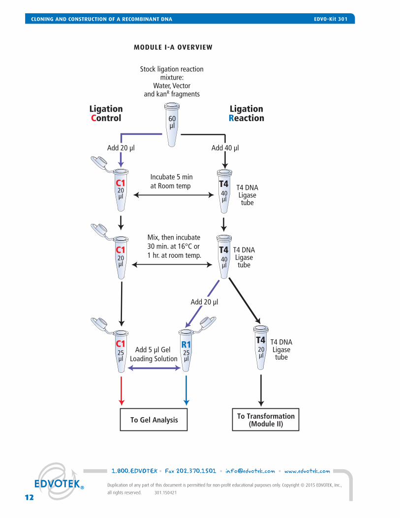
Module I-A: Ligation of the Plasmid Vector to the kanR Gene Fragment
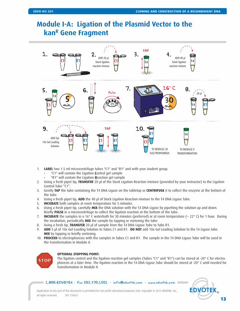
1. LABEL two 1.5 ml microcentrifuge tubes "C1" and "R1" and with your student group. • "C1" will contain the Ligation Control gel sample • "R1" will contain the Ligation Reaction gel sample2. Using a fresh pipet tip, TRANSFER 20 μl of the Stock Ligation Reaction mixture (provided by your instructor) to the Ligation
Control Tube “C1”. 3. Gently TAP the tube containing the T4 DNA Ligase on the tabletop or CENTRIFUGE it to collect the enzyme at the bottom of
the tube.4. Using a fresh pipet tip, ADD the 40 μl of Stock Ligation Reaction mixture to the T4 DNA Ligase Tube. 5. INCUBATE both samples at room temperature for 5 minutes. 6. Using a fresh pipet tip, carefully MIX the DNA solution with the T4 DNA Ligase by pipetting the solution up and down.
Briefl y PULSE in a microcentrifuge to collect the ligation reaction at the bottom of the tube. 7. INCUBATE the samples in a 16° C waterbath for 30 minutes (preferred) or at room temperature (~ 22° C) for 1 hour. During
the incubation, periodically MIX the sample by tapping or vortexing the tube. 8. Using a fresh tip, TRANSFER 20 μl of sample from the T4 DNA Ligase Tube to Tube R1. 9. ADD 5 μl of 10x Gel Loading Solution to Tubes C1 and R1. DO NOT add 10x Gel Loading Solution to the T4 Ligase tube. MIX by tapping or briefl y vortexing. 10. PROCEED to electrophoresis with the samples in Tubes C1 and R1. The sample in the T4 DNA Ligase Tube will be used in
the transformation in Module II.
OPTIONAL STOPPING POINT:The ligation control and the ligation reaction gel samples (Tubes "C1" and "R1") can be stored at -20° C for electro-phoresis at a later time. The ligation reaction in the T4 DNA Ligase Tube should be stored at -20° C until needed for Transformation in Module II.
1. 2. 3.
6.5.
99
16° C
4.
5min.
7. 8.
9.
30min.
R1ADD 20 µl
Stock ligationreaction mixture
C1 C1ADD 40 µl
Stock ligationreaction mixture
T4
Pulse 20 µl
ADD 5µl10x Gel Loading
Solution
TAP
R1C1 T4
T4
R1C1
10.
TO MODULE I-BELECTROPHORESIS
TO MODULE IITRANSFORMATION
C1
T4R1
C1 R1
T4
TAP
T4
CLONING AND CONSTRUCTION OF A RECOMBINANT DNAEDVO-Kit 301
13
1.800.EDVOTEK • Fax 202.370.1501 • [email protected] • www.edvotek.com
Duplication of any part of this document is permitted for non-profi t educational purposes only. Copyright © 2015 EDVOTEK, Inc.,
all rights reserved. 301.150421
CLONING AND CONSTRUCTION OF A RECOMBINANT DNAEDVO-Kit 301
60°C
1:001. 3.
4. 5.
7.
Caution! Flask will be HOT!
Concentratedbuffer
Distilledwater
Agarose
2.50x
Flask
60°C20min.
WAIT6.
Pour
Module I-B: Agarose Gel Electrophoresis
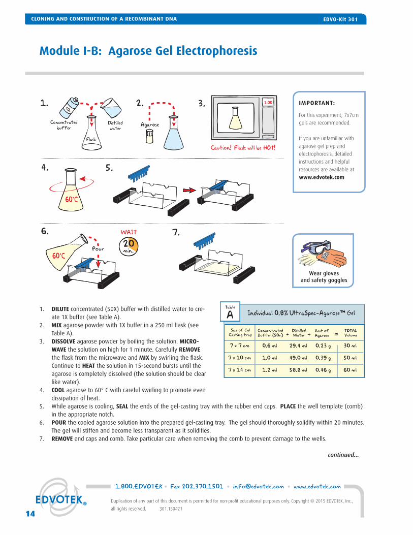
1. DILUTE concentrated (50X) buffer with distilled water to cre-ate 1X buffer (see Table A).
2. MIX agarose powder with 1X buffer in a 250 ml fl ask (see Table A).
3. DISSOLVE agarose powder by boiling the solution. MICRO-WAVE the solution on high for 1 minute. Carefully REMOVE the fl ask from the microwave and MIX by swirling the fl ask. Continue to HEAT the solution in 15-second bursts until the agarose is completely dissolved (the solution should be clear like water).
4. COOL agarose to 60° C with careful swirling to promote even dissipation of heat.
5. While agarose is cooling, SEAL the ends of the gel-casting tray with the rubber end caps. PLACE the well template (comb) in the appropriate notch.
6. POUR the cooled agarose solution into the prepared gel-casting tray. The gel should thoroughly solidify within 20 minutes. The gel will stiffen and become less transparent as it solidifi es.
7. REMOVE end caps and comb. Take particular care when removing the comb to prevent damage to the wells.
IMPORTANT:
For this experiment, 7x7cm
gels are recommended.
If you are unfamiliar with
agarose gel prep and
electrophoresis, detailed
instructions and helpful
resources are available at
www.edvotek.com
ConcentratedBuffer (50x)
Size of GelCasting tray
7 x 7 cm
7 x 10 cm
7 x 14 cm
0.6 ml
1.0 ml
1.2 ml
+DistilledWater
29.4 ml
49.0 ml
58.8 ml
+TOTALVolume
30 ml
50 ml
60 ml
=
Individual 0.8% UltraSpec-Agarose™ GelTable
AAmt ofAgarose
0.23 g
0.39 g
0.46 g
Wear gloves and safety goggles
continued...
CLONING AND CONSTRUCTION OF A RECOMBINANT DNA EDVO-Kit 301
1.800.EDVOTEK • Fax 202.370.1501 • [email protected] • www.edvotek.com
14
Duplication of any part of this document is permitted for non-profi t educational purposes only. Copyright © 2015 EDVOTEK, Inc.,
all rights reserved. 301.150421
CLONING AND CONSTRUCTION OF A RECOMBINANT DNA EDVO-Kit 301
Module I-B: Agarose Gel Electrophoresis, continued
1X DilutedBuffer
8. 9.
10. 11.
Pour
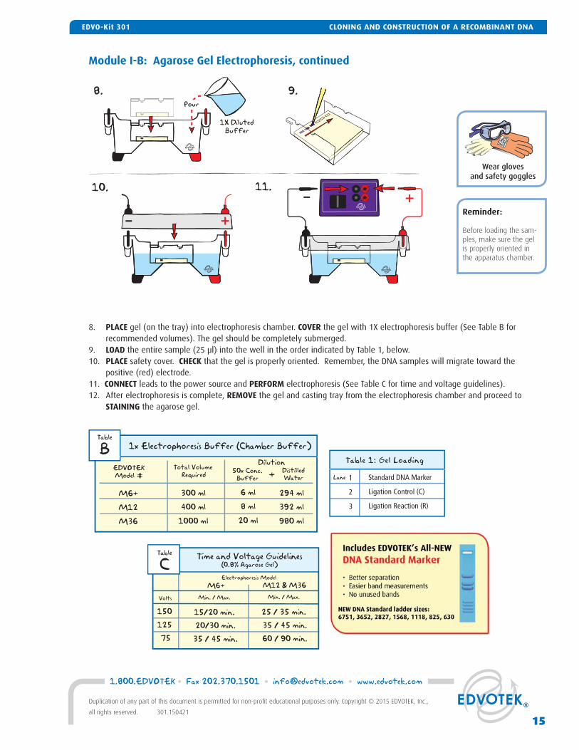
8. PLACE gel (on the tray) into electrophoresis chamber. COVER the gel with 1X electrophoresis buffer (See Table B for recommended volumes). The gel should be completely submerged.
9. LOAD the entire sample (25 μl) into the well in the order indicated by Table 1, below.10. PLACE safety cover. CHECK that the gel is properly oriented. Remember, the DNA samples will migrate toward the
positive (red) electrode.11. CONNECT leads to the power source and PERFORM electrophoresis (See Table C for time and voltage guidelines).12. After electrophoresis is complete, REMOVE the gel and casting tray from the electrophoresis chamber and proceed to
STAINING the agarose gel.
Reminder:
Before loading the sam-ples, make sure the gel is properly oriented in the apparatus chamber.
Time and Voltage Guidelines(0.8% Agarose Gel)
Min. / Max.Volts
150 125 75
15/20 min. 20/30 min. 35 / 45 min.
Table
CElectrophoresis Model
M6+ M12 & M36Min. / Max.
25 / 35 min. 35 / 45 min. 60 / 90 min.
50x Conc.Buffer
DistilledWater+
EDVOTEKModel #
Total Volume Required
1x Electrophoresis Buffer (Chamber Buffer)
M6+
M12
M36
300 ml
400 ml
1000 ml
Dilution
Table
B
6 ml
8 ml
20 ml
294 ml
392 ml
980 ml
Wear gloves and safety goggles
Lane 1
2
3
Standard DNA Marker
Ligation Control (C)
Ligation Reaction (R)
Table 1: Gel Loading
15
1.800.EDVOTEK • Fax 202.370.1501 • [email protected] • www.edvotek.com
Duplication of any part of this document is permitted for non-profi t educational purposes only. Copyright © 2015 EDVOTEK, Inc.,
all rights reserved. 301.150421
CLONING AND CONSTRUCTION OF A RECOMBINANT DNAEDVO-Kit 301
Module I-C: Staining with InstaStain® Ethidium Bromide
Wear gloves and UV safety goggles
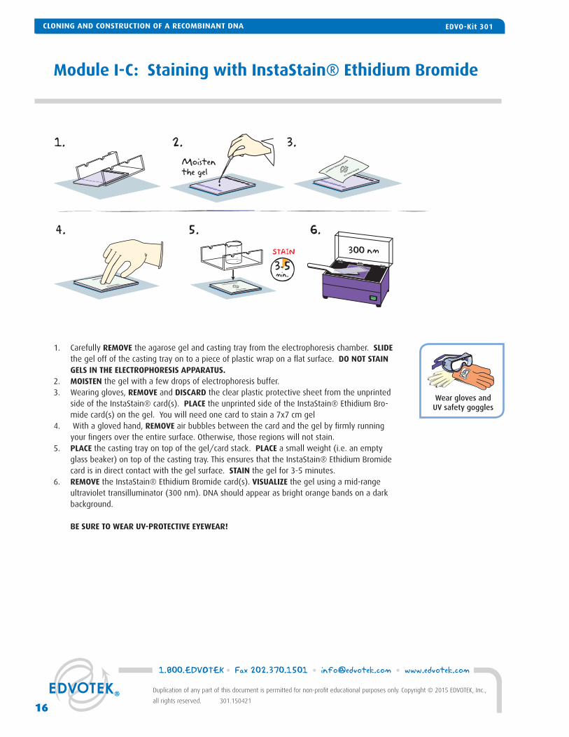
1. Carefully REMOVE the agarose gel and casting tray from the electrophoresis chamber. SLIDE the gel off of the casting tray on to a piece of plastic wrap on a fl at surface. DO NOT STAIN GELS IN THE ELECTROPHORESIS APPARATUS.
2. MOISTEN the gel with a few drops of electrophoresis buffer.3. Wearing gloves, REMOVE and DISCARD the clear plastic protective sheet from the unprinted
side of the InstaStain® card(s). PLACE the unprinted side of the InstaStain® Ethidium Bro-mide card(s) on the gel. You will need one card to stain a 7x7 cm gel
4. With a gloved hand, REMOVE air bubbles between the card and the gel by fi rmly running your fi ngers over the entire surface. Otherwise, those regions will not stain.
5. PLACE the casting tray on top of the gel/card stack. PLACE a small weight (i.e. an empty glass beaker) on top of the casting tray. This ensures that the InstaStain® Ethidium Bromide card is in direct contact with the gel surface. STAIN the gel for 3-5 minutes.
6. REMOVE the InstaStain® Ethidium Bromide card(s). VISUALIZE the gel using a mid-range ultraviolet transilluminator (300 nm). DNA should appear as bright orange bands on a dark background.
BE SURE TO WEAR UV-PROTECTIVE EYEWEAR!
Moistenthe gel
300 nm
1. 2.
4. 5. 6.
3.
3-5min.
STAIN
InstaStain® Ethidium Bromide
U.S. Patent Pending
InstaStain® Ethid
U.S. Patent Pending InstaStain® Ethidium Bromide
U.S. Patent Pending
-----
CLONING AND CONSTRUCTION OF A RECOMBINANT DNA EDVO-Kit 301
1.800.EDVOTEK • Fax 202.370.1501 • [email protected] • www.edvotek.com
16
Duplication of any part of this document is permitted for non-profi t educational purposes only. Copyright © 2015 EDVOTEK, Inc.,
all rights reserved. 301.150421
CLONING AND CONSTRUCTION OF A RECOMBINANT DNA EDVO-Kit 301
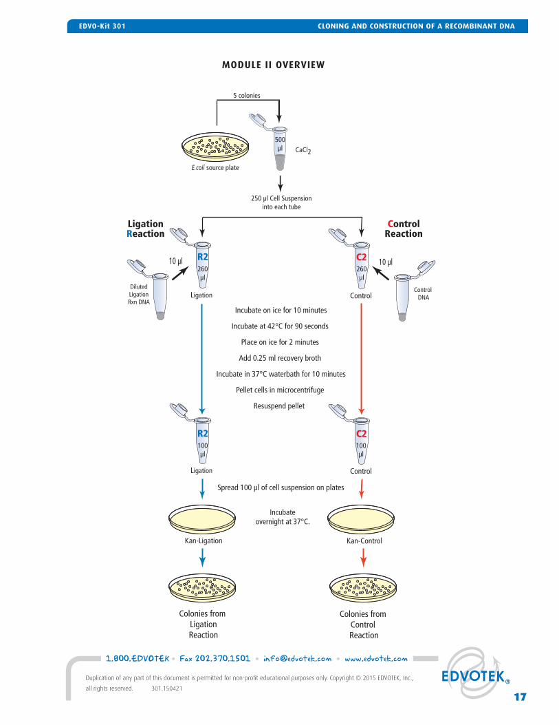
MODULE II OVERVIEW
Incubate on ice for 10 minutes
Incubate at 42°C for 90 seconds
Place on ice for 2 minutes
Add 0.25 ml recovery broth
Incubate in 37°C waterbath for 10 minutes
Pellet cells in microcentrifuge
Resuspend pellet
Spread 100 µl of cell suspension on plates
Colonies from Control Reaction
10 µl
Kan-Ligation Kan-Control
CaCl2
5 colonies
Control DNA
DilutedLigationRxn DNA
10 µl
Incubateovernight at 37°C.
Colonies from LigationReaction
250 µl Cell Suspensioninto each tube
Ligation Control
500µl
260µl
260µl
Ligation Control
100µl
100µl
LigationReaction
ControlReaction
C2R2
C2R2
E.coli source plate
17
1.800.EDVOTEK • Fax 202.370.1501 • [email protected] • www.edvotek.com
Duplication of any part of this document is permitted for non-profi t educational purposes only. Copyright © 2015 EDVOTEK, Inc.,
all rights reserved. 301.150421
CLONING AND CONSTRUCTION OF A RECOMBINANT DNAEDVO-Kit 301
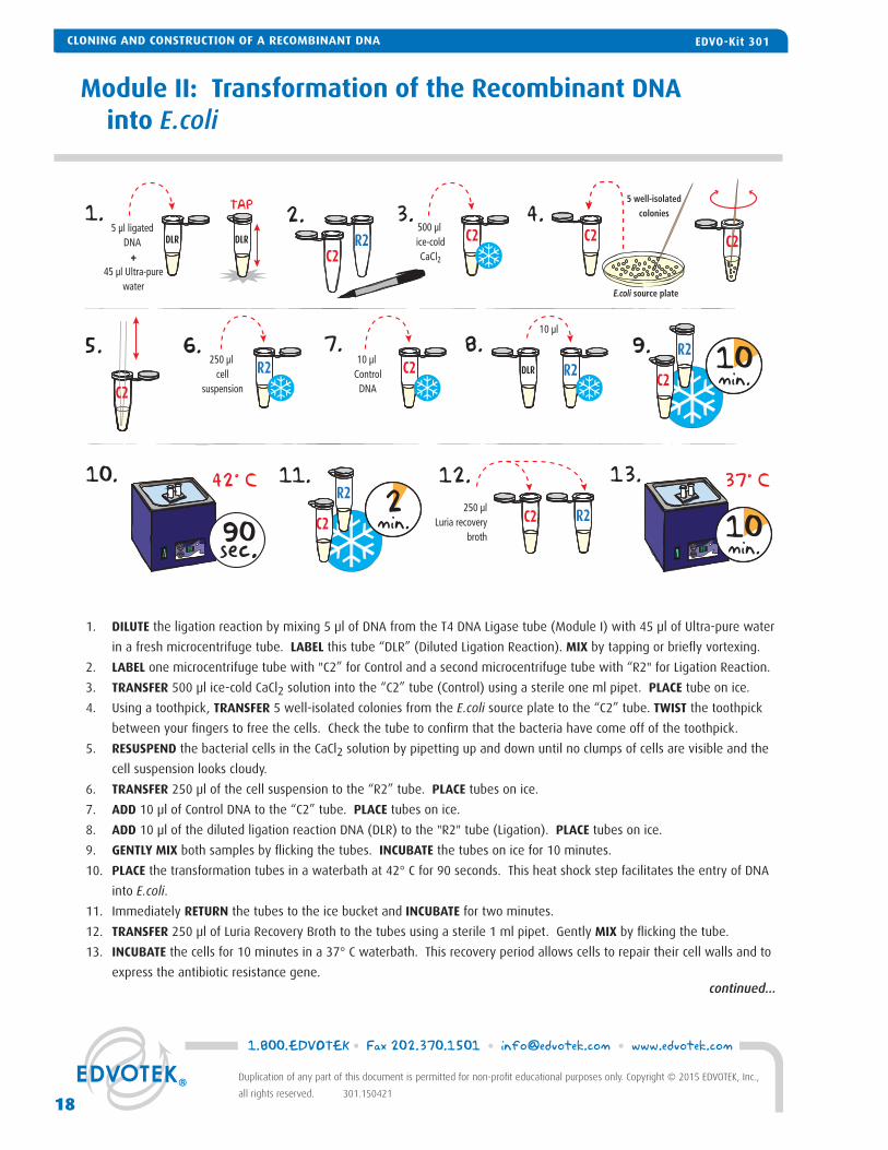
1. DILUTE the ligation reaction by mixing 5 μl of DNA from the T4 DNA Ligase tube (Module I) with 45 μl of Ultra-pure water
in a fresh microcentrifuge tube. LABEL this tube “DLR” (Diluted Ligation Reaction). MIX by tapping or briefl y vortexing.
2. LABEL one microcentrifuge tube with "C2” for Control and a second microcentrifuge tube with “R2" for Ligation Reaction.
3. TRANSFER 500 μl ice-cold CaCl2 solution into the “C2” tube (Control) using a sterile one ml pipet. PLACE tube on ice.
4. Using a toothpick, TRANSFER 5 well-isolated colonies from the E.coli source plate to the “C2” tube. TWIST the toothpick
between your fi ngers to free the cells. Check the tube to confi rm that the bacteria have come off of the toothpick.
5. RESUSPEND the bacterial cells in the CaCl2 solution by pipetting up and down until no clumps of cells are visible and the
cell suspension looks cloudy.
6. TRANSFER 250 μl of the cell suspension to the “R2” tube. PLACE tubes on ice.
7. ADD 10 μl of Control DNA to the “C2” tube. PLACE tubes on ice.
8. ADD 10 μl of the diluted ligation reaction DNA (DLR) to the "R2" tube (Ligation). PLACE tubes on ice.
9. GENTLY MIX both samples by fl icking the tubes. INCUBATE the tubes on ice for 10 minutes.
10. PLACE the transformation tubes in a waterbath at 42° C for 90 seconds. This heat shock step facilitates the entry of DNA
into E.coli.
11. Immediately RETURN the tubes to the ice bucket and INCUBATE for two minutes.
12. TRANSFER 250 μl of Luria Recovery Broth to the tubes using a sterile 1 ml pipet. Gently MIX by fl icking the tube.
13. INCUBATE the cells for 10 minutes in a 37° C waterbath. This recovery period allows cells to repair their cell walls and to
express the antibiotic resistance gene.
Module II: Transformation of the Recombinant DNA into E.coli
1. 2. 3.
6.5.
99
42° C
4.
7. 8.
10.
90sec. 99
37° C13.
R25 µl ligated
DNA +
45 µl Ultra-purewater
C2
500 µl ice-coldCaCl2
10 µl
250 µlLuria recovery
broth
TAP
R2
R2C2
DLR DLR
DLR
C2 C2
E.coli source plate
5 well-isolated
colonies
C2
250 µl cell
suspension
R210 µl
ControlDNA
C2 10min.
10min.
9.C2
R2
2min.
11. 12.
C2
R2
C2
continued...
CLONING AND CONSTRUCTION OF A RECOMBINANT DNA EDVO-Kit 301
1.800.EDVOTEK • Fax 202.370.1501 • [email protected] • www.edvotek.com
18
Duplication of any part of this document is permitted for non-profi t educational purposes only. Copyright © 2015 EDVOTEK, Inc.,
all rights reserved. 301.150421
CLONING AND CONSTRUCTION OF A RECOMBINANT DNA EDVO-Kit 301
Module II: Transformation of the Recombinant DNA into E.coli, continued
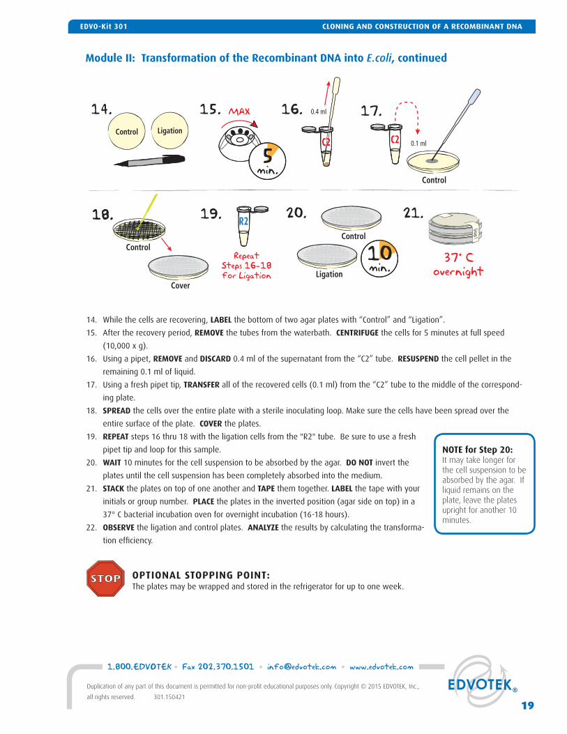
14. While the cells are recovering, LABEL the bottom of two agar plates with “Control” and “Ligation”.
15. After the recovery period, REMOVE the tubes from the waterbath. CENTRIFUGE the cells for 5 minutes at full speed
(10,000 x g).
16. Using a pipet, REMOVE and DISCARD 0.4 ml of the supernatant from the “C2” tube. RESUSPEND the cell pellet in the
remaining 0.1 ml of liquid.
17. Using a fresh pipet tip, TRANSFER all of the recovered cells (0.1 ml) from the “C2” tube to the middle of the correspond-
ing plate.
18. SPREAD the cells over the entire plate with a sterile inoculating loop. Make sure the cells have been spread over the
entire surface of the plate. COVER the plates.
19. REPEAT steps 16 thru 18 with the ligation cells from the "R2" tube. Be sure to use a fresh
pipet tip and loop for this sample.
20. WAIT 10 minutes for the cell suspension to be absorbed by the agar. DO NOT invert the
plates until the cell suspension has been completely absorbed into the medium.
21. STACK the plates on top of one another and TAPE them together. LABEL the tape with your
initials or group number. PLACE the plates in the inverted position (agar side on top) in a
37° C bacterial incubation oven for overnight incubation (16-18 hours).
22. OBSERVE the ligation and control plates. ANALYZE the results by calculating the transforma-
tion effi ciency.
14. 15. 16. 17.
0.1 ml
R2
Control Ligation
MAX
5min.
Control
C2
0.4 ml
18.18. 19. 20. 21.
Cover
C2
ControlRepeat
Steps 16-18for Ligation
Control
Ligation
10min.
37° Covernight
Group 1
OPTIONAL STOPPING POINT: The plates may be wrapped and stored in the refrigerator for up to one week.
NOTE for Step 20:It may take longer for the cell suspension to be absorbed by the agar. If liquid remains on the plate, leave the plates upright for another 10 minutes.
19
1.800.EDVOTEK • Fax 202.370.1501 • [email protected] • www.edvotek.com
Duplication of any part of this document is permitted for non-profi t educational purposes only. Copyright © 2015 EDVOTEK, Inc.,
all rights reserved. 301.150421
CLONING AND CONSTRUCTION OF A RECOMBINANT DNAEDVO-Kit 301
Module II: Transformation of the Recombinant DNA into E.coli
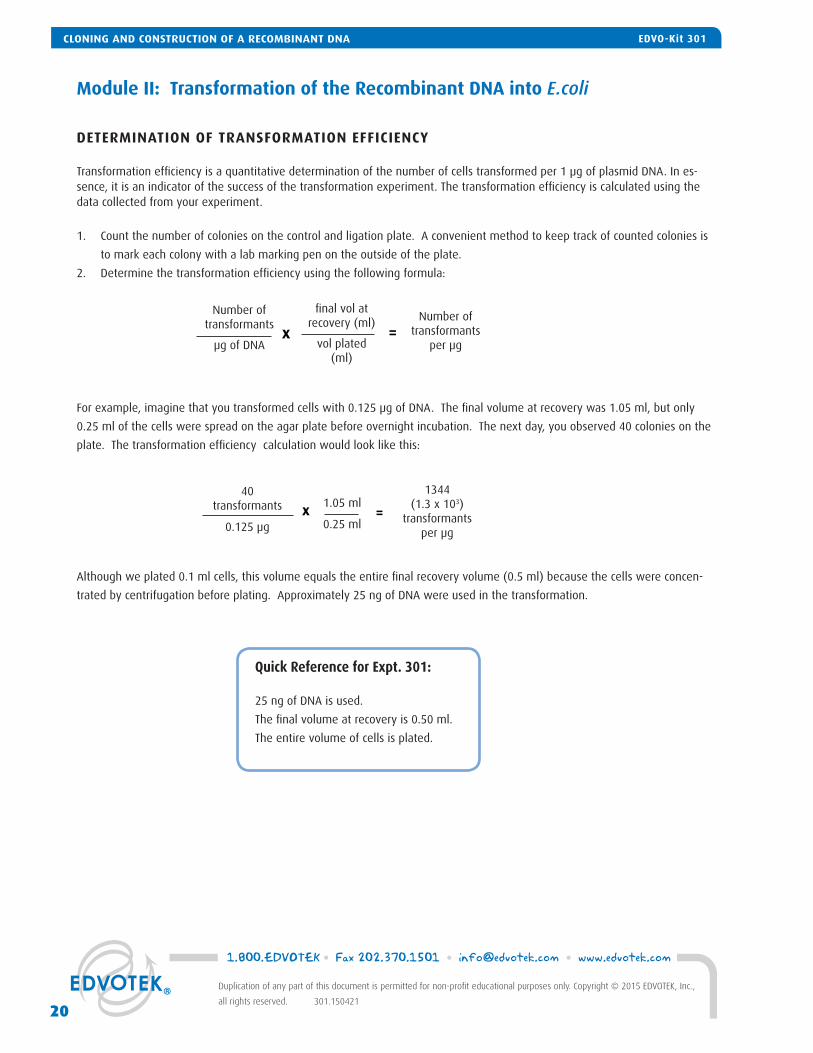
DETERMINATION OF TRANSFORMATION EFFICIENCY
Transformation effi ciency is a quantitative determination of the number of cells transformed per 1 μg of plasmid DNA. In es-sence, it is an indicator of the success of the transformation experiment. The transformation effi ciency is calculated using the data collected from your experiment.
1. Count the number of colonies on the control and ligation plate. A convenient method to keep track of counted colonies is
to mark each colony with a lab marking pen on the outside of the plate.
2. Determine the transformation effi ciency using the following formula:
Although we plated 0.1 ml cells, this volume equals the entire fi nal recovery volume (0.5 ml) because the cells were concen-
trated by centrifugation before plating. Approximately 25 ng of DNA were used in the transformation.
x =
fi nal vol at recovery (ml)
vol plated (ml)
Number of transformants
per μg
Number of transformants
μg of DNA
40transformants
0.125 μg
1344(1.3 x 10³)
transformants per μg
x 1.05 ml
0.25 ml=
Quick Reference for Expt. 301:
25 ng of DNA is used.
The fi nal volume at recovery is 0.50 ml.
The entire volume of cells is plated.
For example, imagine that you transformed cells with 0.125 μg of DNA. The fi nal volume at recovery was 1.05 ml, but only
0.25 ml of the cells were spread on the agar plate before overnight incubation. The next day, you observed 40 colonies on the
plate. The transformation effi ciency calculation would look like this:
1.800.EDVOTEK • Fax 202.370.1501 • [email protected] • www.edvotek.com
20
Duplication of any part of this document is permitted for non-profi t educational purposes only. Copyright © 2015 EDVOTEK, Inc.,
all rights reserved. 301.150421
CLONING AND CONSTRUCTION OF A RECOMBINANT DNA EDVO-Kit 301
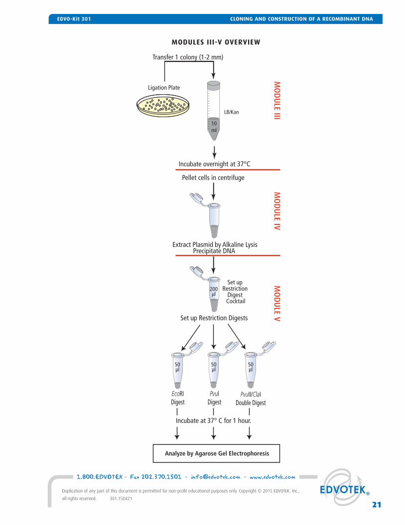
MODULES III-V OVERVIEW
Extract Plasmid by Alkaline LysisPrecipitate DNA
Set up Restriction Digests
Ligation Plate
LB/Kan
Transfer 1 colony (1-2 mm)
EcoRIDigest
PvuIDigest
PvuII/ClaIDouble Digest
Incubate overnight at 37°C
Pellet cells in centrifuge
Set up Restriction
Digest Cocktail
50µl
50µl
50µl
200µl
Analyze by Agarose Gel Electrophoresis
Incubate at 37° C for 1 hour.
MO
DU
LE IIIM
OD
ULE IV
MO
DU
LE V10ml
21
1.800.EDVOTEK • Fax 202.370.1501 • [email protected] • www.edvotek.com
Duplication of any part of this document is permitted for non-profi t educational purposes only. Copyright © 2015 EDVOTEK, Inc.,
all rights reserved. 301.150421
CLONING AND CONSTRUCTION OF A RECOMBINANT DNAEDVO-Kit 301
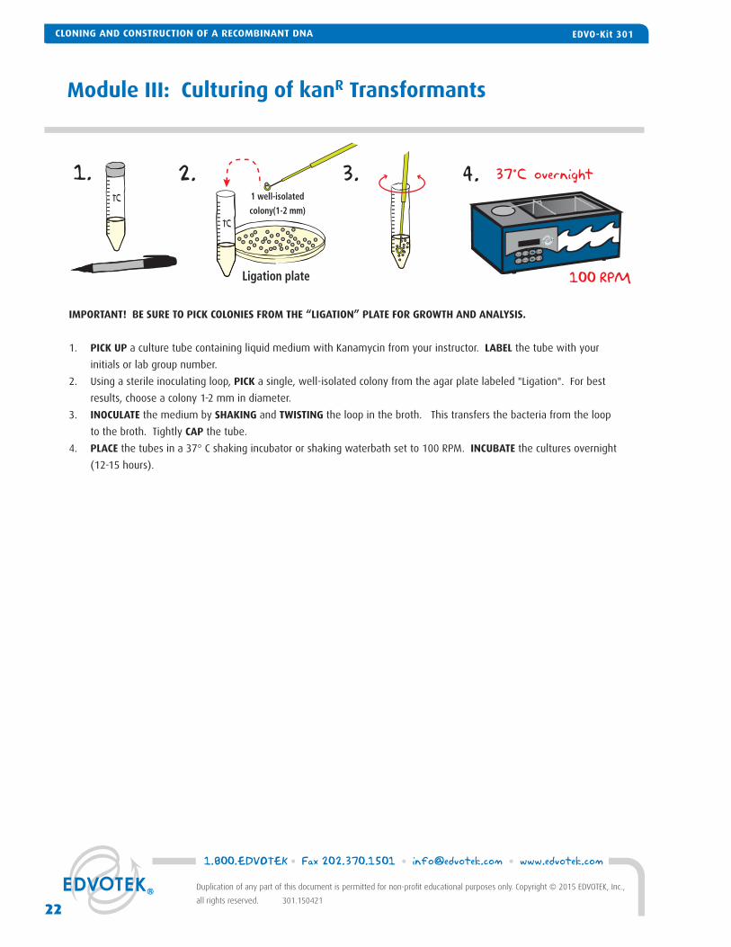
Module III: Culturing of kanR Transformants
IMPORTANT! BE SURE TO PICK COLONIES FROM THE “LIGATION” PLATE FOR GROWTH AND ANALYSIS.
1. PICK UP a culture tube containing liquid medium with Kanamycin from your instructor. LABEL the tube with your
initials or lab group number.
2. Using a sterile inoculating loop, PICK a single, well-isolated colony from the agar plate labeled "Ligation". For best
results, choose a colony 1-2 mm in diameter.
3. INOCULATE the medium by SHAKING and TWISTING the loop in the broth. This transfers the bacteria from the loop
to the broth. Tightly CAP the tube.
4. PLACE the tubes in a 37° C shaking incubator or shaking waterbath set to 100 RPM. INCUBATE the cultures overnight
(12-15 hours).
1. 2. 3. 4.
Ligation plate
1 well-isolated
colony(1-2 mm)
37°C overnight
100 RPM
TC
TC
CLONING AND CONSTRUCTION OF A RECOMBINANT DNA EDVO-Kit 301
1.800.EDVOTEK • Fax 202.370.1501 • [email protected] • www.edvotek.com
22
Duplication of any part of this document is permitted for non-profi t educational purposes only. Copyright © 2015 EDVOTEK, Inc.,
all rights reserved. 301.150421
CLONING AND CONSTRUCTION OF A RECOMBINANT DNA EDVO-Kit 301
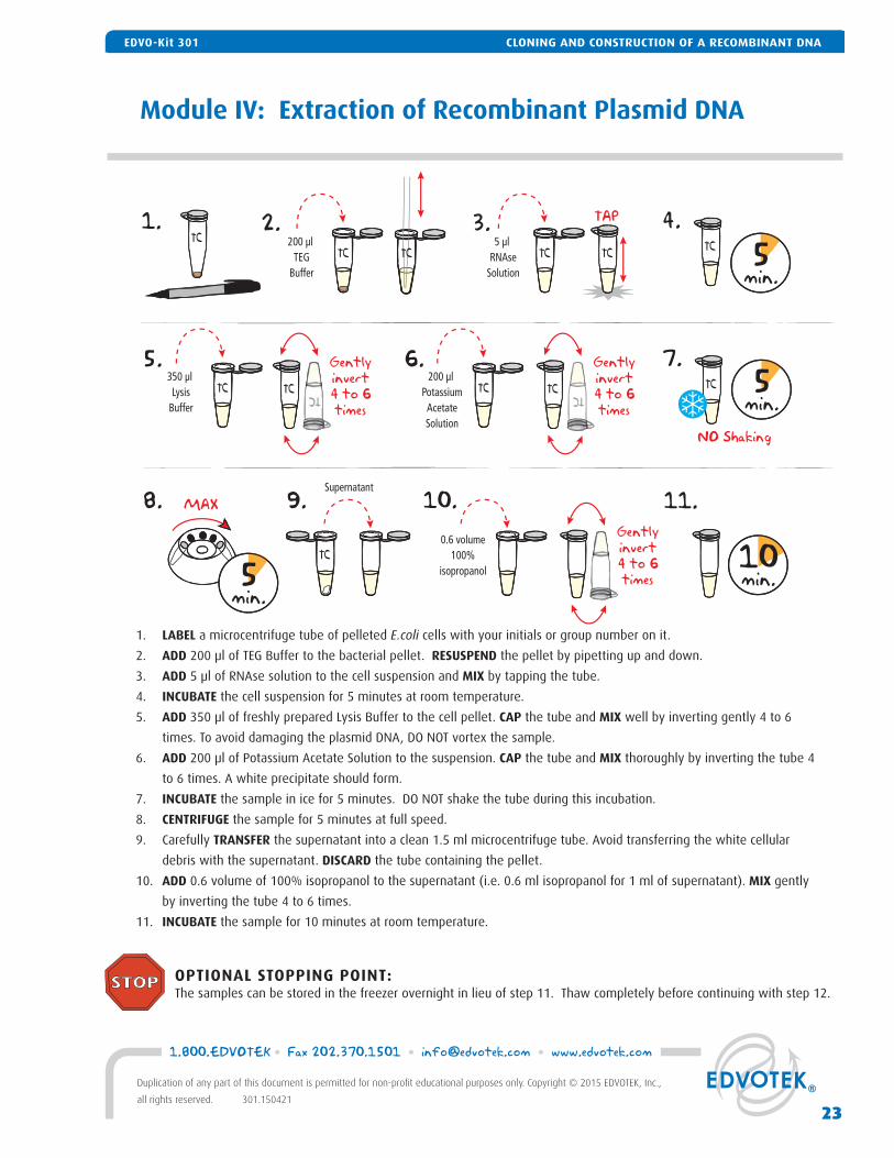
Module IV: Extraction of Recombinant Plasmid DNA
1. LABEL a microcentrifuge tube of pelleted E.coli cells with your initials or group number on it.
2. ADD 200 μl of TEG Buffer to the bacterial pellet. RESUSPEND the pellet by pipetting up and down.
3. ADD 5 μl of RNAse solution to the cell suspension and MIX by tapping the tube.
4. INCUBATE the cell suspension for 5 minutes at room temperature.
5. ADD 350 μl of freshly prepared Lysis Buffer to the cell pellet. CAP the tube and MIX well by inverting gently 4 to 6
times. To avoid damaging the plasmid DNA, DO NOT vortex the sample.
6. ADD 200 μl of Potassium Acetate Solution to the suspension. CAP the tube and MIX thoroughly by inverting the tube 4
to 6 times. A white precipitate should form.
7. INCUBATE the sample in ice for 5 minutes. DO NOT shake the tube during this incubation.
8. CENTRIFUGE the sample for 5 minutes at full speed.
9. Carefully TRANSFER the supernatant into a clean 1.5 ml microcentrifuge tube. Avoid transferring the white cellular
debris with the supernatant. DISCARD the tube containing the pellet.
10. ADD 0.6 volume of 100% isopropanol to the supernatant (i.e. 0.6 ml isopropanol for 1 ml of supernatant). MIX gently
by inverting the tube 4 to 6 times.
11. INCUBATE the sample for 10 minutes at room temperature.
1. 2.
5.
8. 9.
200 µl TEG
Buffer
TAP
10min.
TCTC
5 µl RNAse
Solution
TC TCTC
4.3.5
min.
TC
7.5
min.TC350 µl
LysisBuffer
TC
6.200 µl
PotassiumAcetateSolution
TCTC
Gentlyinvert4 to 6times
TC TC
Gentlyinvert4 to 6times
MAX
5min.
NO Shaking
10.Supernatant
TC0.6 volume
100%isopropanol
Gentlyinvert4 to 6times
11.
OPTIONAL STOPPING POINT: The samples can be stored in the freezer overnight in lieu of step 11. Thaw completely before continuing with step 12.
CLONING AND CONSTRUCTION OF A RECOMBINANT DNAEDVO-Kit 301
23
1.800.EDVOTEK • Fax 202.370.1501 • [email protected] • www.edvotek.com
Duplication of any part of this document is permitted for non-profi t educational purposes only. Copyright © 2015 EDVOTEK, Inc.,
all rights reserved. 301.150421
CLONING AND CONSTRUCTION OF A RECOMBINANT DNAEDVO-Kit 301
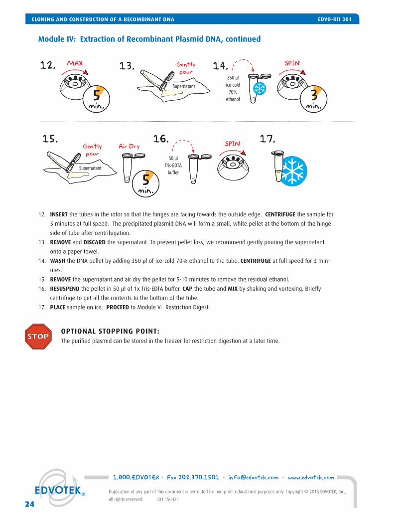
Module IV: Extraction of Recombinant Plasmid DNA, continued
12. INSERT the tubes in the rotor so that the hinges are facing towards the outside edge. CENTRIFUGE the sample for
5 minutes at full speed. The precipitated plasmid DNA will form a small, white pellet at the bottom of the hinge
side of tube after centrifugation.
13. REMOVE and DISCARD the supernatant. To prevent pellet loss, we recommend gently pouring the supernatant
onto a paper towel.
14. WASH the DNA pellet by adding 350 μl of ice-cold 70% ethanol to the tube. CENTRIFUGE at full speed for 3 min-
utes.
15. REMOVE the supernatant and air dry the pellet for 5-10 minutes to remove the residual ethanol.
16. RESUSPEND the pellet in 50 μl of 1x Tris-EDTA buffer. CAP the tube and MIX by shaking and vortexing. Briefl y
centrifuge to get all the contents to the bottom of the tube.
17. PLACE sample on ice. PROCEED to Module V: Restriction Digest.
12. MAX
5min.
SPIN
3min.
13. 14.
Supernatant
Gentlypour
350 µlice-cold
70%ethanol
SPIN15. 16. 17.
Supernatant
Gentlypour
50 µlTris-EDTA
buffer
5min.
Air Dry
OPTIONAL STOPPING POINT: The purifi ed plasmid can be stored in the freezer for restriction digestion at a later time.
1.800.EDVOTEK • Fax 202.370.1501 • [email protected] • www.edvotek.com
24
Duplication of any part of this document is permitted for non-profi t educational purposes only. Copyright © 2015 EDVOTEK, Inc.,
all rights reserved. 301.150421
CLONING AND CONSTRUCTION OF A RECOMBINANT DNA EDVO-Kit 301
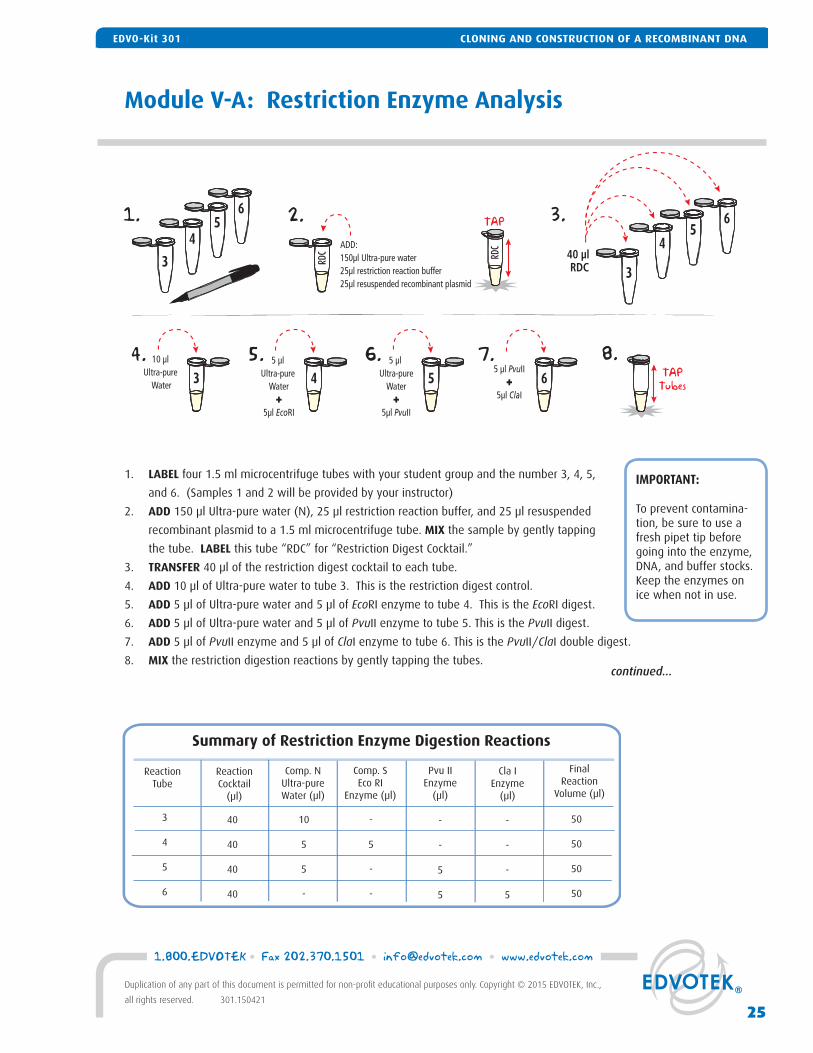
Module V-A: Restriction Enzyme Analysis
1. LABEL four 1.5 ml microcentrifuge tubes with your student group and the number 3, 4, 5,
and 6. (Samples 1 and 2 will be provided by your instructor)
2. ADD 150 μl Ultra-pure water (N), 25 μl restriction reaction buffer, and 25 μl resuspended
recombinant plasmid to a 1.5 ml microcentrifuge tube. MIX the sample by gently tapping
the tube. LABEL this tube “RDC” for “Restriction Digest Cocktail.”
3. TRANSFER 40 μl of the restriction digest cocktail to each tube.
4. ADD 10 μl of Ultra-pure water to tube 3. This is the restriction digest control.
5. ADD 5 μl of Ultra-pure water and 5 μl of EcoRI enzyme to tube 4. This is the EcoRI digest.
6. ADD 5 μl of Ultra-pure water and 5 μl of PvuII enzyme to tube 5. This is the PvuII digest.
7. ADD 5 μl of PvuII enzyme and 5 μl of ClaI enzyme to tube 6. This is the PvuII/ClaI double digest.
8. MIX the restriction digestion reactions by gently tapping the tubes.
40
40
40
40
-
-
-
5
ReactionTube
Summary of Restriction Enzyme Digestion Reactions
10
5
5
-
3
4
5
6
ReactionCocktail
(μl)
Comp. NUltra-pureWater (μl)
Comp. SEco RI
Enzyme (μl)
FinalReaction
Volume (μl)
Pvu IIEnzyme
(μl)
Cla IEnzyme
(μl)
50
50
50
50
-
5
-
-
-
-
5
5
IMPORTANT:
To prevent contamina-tion, be sure to use a fresh pipet tip before going into the enzyme, DNA, and buffer stocks. Keep the enzymes on ice when not in use.
1. 2.
8.
ADD:150µl Ultra-pure water25µl restriction reaction buffer25µl resuspended recombinant plasmid
TAP
40 µl RDCRD
C RDC
4. 10 µl Ultra-pure
Water
3
45
6 3.
3
45
6
3
5. 5 µl Ultra-pure
Water+
5µl EcoRI
4
6. 5 µl Ultra-pure
Water+
5µl PvuII
5
7.5 µl PvuII
+5µl ClaI
6 TAPTubes
99
continued...
CLONING AND CONSTRUCTION OF A RECOMBINANT DNAEDVO-Kit 301
25
1.800.EDVOTEK • Fax 202.370.1501 • [email protected] • www.edvotek.com
Duplication of any part of this document is permitted for non-profi t educational purposes only. Copyright © 2015 EDVOTEK, Inc.,
all rights reserved. 301.150421
CLONING AND CONSTRUCTION OF A RECOMBINANT DNAEDVO-Kit 301
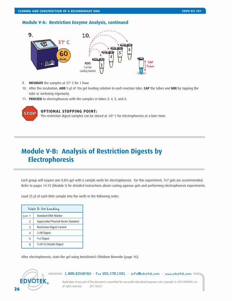
Module V-B: Analysis of Restriction Digests by Electrophoresis
Each group will require one 0.8% gel with 6 sample wells for electrophoresis. For this experiment, 7x7 gels are recommended.
Refer to pages 14-15 (Module I) for detailed instructions about casting agarose gels and performing electrophoresis experiments.
Load 25 μl of each DNA sample into the wells in the following order:
Lane 1
2
3
4
5
6
Standard DNA Marker
Supercoiled Plasmid Vector Standard
Restriction Digest Control
EcoRI Digest
PvuI Digest
PvuII/ClaI Double Digest
Table 2: Gel Loading
After electrophoresis, stain the gel using InstaStain® Ethidium Bromide (page 16).
9. INCUBATE the samples at 37° C for 1 hour.
10. After the incubation, ADD 5 μl of 10x gel loading solution to each reaction tube. CAP the tubes and MIX by tapping the
tube or vortexing vigorously.
11. PROCEED to electrophoresis with the samples in tubes 3, 4, 5, and 6.
9. 10.
99
37° C
60min.
ADD:5 µl Gel
Loading Solution
3
45
6
TAPTubes
Module V-A: Restriction Enzyme Analysis, continued
OPTIONAL STOPPING POINT: The restriction digest samples can be stored at -20° C for electrophoresis at a later time.
1.800.EDVOTEK • Fax 202.370.1501 • [email protected] • www.edvotek.com
26
Duplication of any part of this document is permitted for non-profi t educational purposes only. Copyright © 2015 EDVOTEK, Inc.,
all rights reserved. 301.150421
CLONING AND CONSTRUCTION OF A RECOMBINANT DNA EDVO-Kit 301
Study Questions
1. Did you observe a discreet band of DNA after the electrophoresis of the ligation reaction products? Explain. Did you observe any bands that migrated faster than the 1300 base pair kanR fragment? If so, how could these DNA forms have been generated? (Hint: DNA does not always circularize as a relaxed molecule).
2. Which of the following pairs could be ligated together? (All termini are cohesive and complementary.)
a. 5'-dephosphorylated linear insert DNA + linear vector b. Supercoiled vector + linear insert DNA c. 5'-dephosphorylated linear vector + linear 5'-dephosphorylated insert DNA d. Linear 5'-dephosphorylated vector + linear insert DNA e. Nicked vector + linear insert DNA
In general, which of the above possibilities would be the best approach in a subcloning experiment like the one you have done? Why?
3. Assume the transformants produced with the ligated DNA were also plated on ampicillin medium. Would you expect to see a signifi cant difference in the number of colonies compared to the Kanamycin plates? Why? (Hint: the linear vector was not dephosphorylated before the ligation). Why would it be unwise to pick a transformant from an ampicillin plate if you were trying to isolate the recombinant DNA? If you had, is there a step in this series of experiments that would have prevented the propagation of the incorrect plasmid?
4. Did the electrophoretic pattern of your uncut recombinant plasmid contain many forms of DNA like your ligation reaction? Explain.
5. Did your recombinant plasmid have more than one insert? What was the orientation of the insert(s)? Make a rough map of your recombinant plasmid.
6. Can the size of a supercoiled plasmid be calculated by comparison to linear DNA fragments of known size that have been run in parallel?
7. A kanR transformant was found to contain the supercoiled pUC vector without an insert in addition to the expected super-coiled recombinant plasmid. How can this be explained?
CLONING AND CONSTRUCTION OF A RECOMBINANT DNAEDVO-Kit 301
27
1.800.EDVOTEK • Fax 202.370.1501 • [email protected] • www.edvotek.com
Duplication of any part of this document is permitted for non-profi t educational purposes only. Copyright © 2015 EDVOTEK, Inc.,
all rights reserved. 301.150421
CLONING AND CONSTRUCTION OF A RECOMBINANT DNAEDVO-Kit 301
Instructor's Guide
ORGANIZING AND IMPLEMENTING THE EXPERIMENT
Prior to starting this experiment, carefully check the list of Components and Requirements on pages 3 and 4 to ensure that you have all the necessary components and equipment.
Some modules of this experiment may require multiple laboratory periods. The experiment can be tempo-rarily stopped after the completion of each module or at designated stopping points. Student results will not be compromised if the experiment is paused at these times. Please consider this in the planning and the implementation of this experiment.
Module IIIIIIIVV
Pre-Lab
1 hour2 hrs
30 min1.5 hrs1.5 hrs
Experiment
3.5 hrs1 hr
15 min1.5-2 hrs2.5 hrs
Approximate Time Requirements
1.800.EDVOTEK • Fax 202.370.1501 • [email protected] • www.edvotek.com
28
Duplication of any part of this document is permitted for non-profi t educational purposes only. Copyright © 2015 EDVOTEK, Inc.,
all rights reserved. 301.150421
INSTRUCTOR'S GUIDE CLONING AND CONSTRUCTION OF A RECOMBINANT DNA EDVO-Kit 301
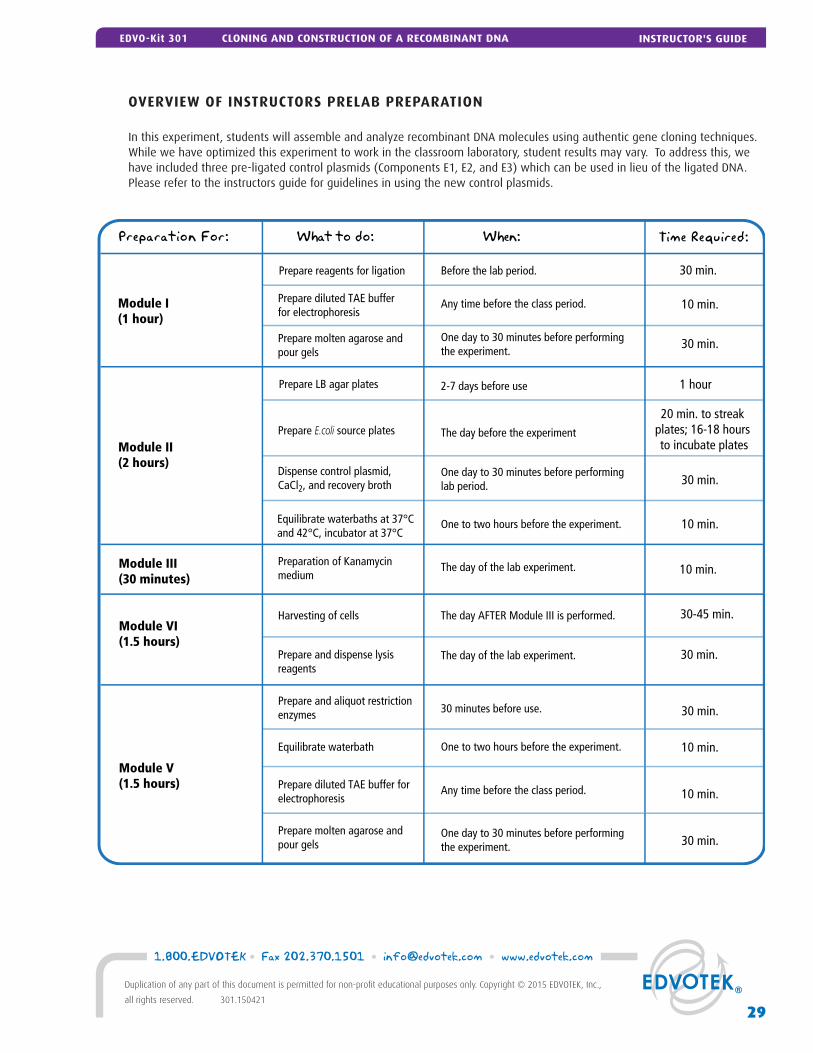
Preparation For: What to do: When: Time Required:
Module III (30 minutes)
Module VI(1.5 hours)
Prepare and dispense lysisreagents
30 min.
Preparation of Kanamycinmedium
The day of the lab experiment.
The day of the lab experiment.
30 minutes before use.
10 min.
30-45 min.
Module V(1.5 hours)
30 min.
Any time before the class period.Prepare diluted TAE bufferfor electrophoresis
10 min.
One day to 30 minutes before performingthe experiment.
30 min.
Module I (1 hour)
Prepare reagents for ligation Before the lab period. 30 min.
Module II (2 hours)
Prepare molten agarose andpour gels
One to two hours before the experiment. 10 min.Equilibrate waterbaths at 37°Cand 42°C, incubator at 37°C
The day before the experimentPrepare E.coli source plates20 min. to streak
plates; 16-18 hours to incubate plates
One day to 30 minutes before performinglab period. 30 min.
Prepare LB agar plates 2-7 days before use 1 hour
Dispense control plasmid, CaCl2, and recovery broth
The day AFTER Module III is performed.Harvesting of cells
Prepare and aliquot restrictionenzymes
One to two hours before the experiment. 10 min.Equilibrate waterbath
Any time before the class period. 10 min.Prepare diluted TAE buffer forelectrophoresis
One day to 30 minutes before performingthe experiment. 30 min.
Prepare molten agarose and pour gels
OVERVIEW OF INSTRUCTORS PRELAB PREPARATION
In this experiment, students will assemble and analyze recombinant DNA molecules using authentic gene cloning techniques. While we have optimized this experiment to work in the classroom laboratory, student results may vary. To address this, we have included three pre-ligated control plasmids (Components E1, E2, and E3) which can be used in lieu of the ligated DNA. Please refer to the instructors guide for guidelines in using the new control plasmids.
29
1.800.EDVOTEK • Fax 202.370.1501 • [email protected] • www.edvotek.com
Duplication of any part of this document is permitted for non-profi t educational purposes only. Copyright © 2015 EDVOTEK, Inc.,
all rights reserved. 301.150421
INSTRUCTOR'S GUIDEEDVO-Kit 301 CLONING AND CONSTRUCTION OF A RECOMBINANT DNA
MODULE I: Pre-Lab Preparations
MODULE I-A: LIGATION OF THE PLASMID VECTOR TO THE kanR GENE FRAGMENT
This kit provides enough reagents to perform 5 ligation reactions. We recommend fi nishing this module in one class period. If necessary, the samples can be stored in the freezer directly following the ligation (step 7). The samples should be completely thawed before proceeding to step 8.
We recommend the aliquoting the reagents into sets for each lab group as described below. Alternatively, the students can share the stock tubes placed in a central location. Note that sharing the tubes increases the risk of a spill or contamination.
1. Shortly before the lab begins, thaw the Ultra-pure Water (A) and DNA fragments for ligation (B) and place on ice.2. Prepare DNA for ligation: a. Label fi ve 0.5 ml microcentrifuge tubes with LRM for Ligation Reaction Mixture. Place the tubes on ice. b. Add 300 μl of Ultra-pure Water (A) to the tube containing the DNA fragments for ligation. Mix well by pipetting up
and down. c. Aliquot 65 μl for each student. Keep the tubes on ice until use.3. Each group requires one T4 DNA Ligase Reaction Tube (C). Label this tube "T4" before distributing.4. Equilibrate waterbath to 16°C if ligating at this temperature. This can be accomplished by setting the waterbath to 16°C
and placing it in a refrigerator or cold room.5. Each group requires 10 μl of 10x Gel Loading Solution.
MODULE I-B: AGAROSE GEL ELECTROPHORESIS
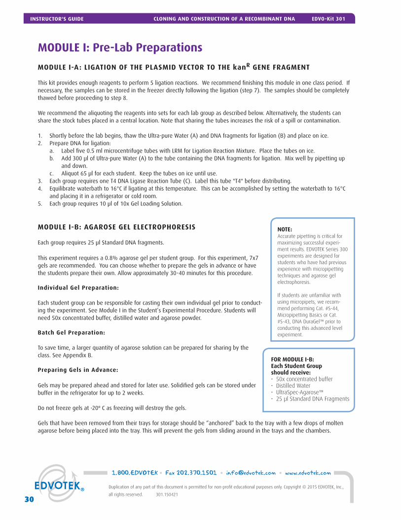
Each group requires 25 μl Standard DNA fragments.
This experiment requires a 0.8% agarose gel per student group. For this experiment, 7x7 gels are recommended. You can choose whether to prepare the gels in advance or have the students prepare their own. Allow approximately 30-40 minutes for this procedure.
Individual Gel Preparation:
Each student group can be responsible for casting their own individual gel prior to conduct-ing the experiment. See Module I in the Student’s Experimental Procedure. Students will need 50x concentrated buffer, distilled water and agarose powder.
Batch Gel Preparation:
To save time, a larger quantity of agarose solution can be prepared for sharing by the class. See Appendix B.
Preparing Gels in Advance:
Gels may be prepared ahead and stored for later use. Solidifi ed gels can be stored under buffer in the refrigerator for up to 2 weeks.
Do not freeze gels at -20º C as freezing will destroy the gels.
Gels that have been removed from their trays for storage should be “anchored” back to the tray with a few drops of molten agarose before being placed into the tray. This will prevent the gels from sliding around in the trays and the chambers.
NOTE:Accurate pipetting is critical for maximizing successful experi-ment results. EDVOTEK Series 300 experiments are designed for students who have had previous experience with micropipetting techniques and agarose gel electrophoresis.
If students are unfamiliar with using micropipets, we recom-mend performing Cat. #S-44, Micropipetting Basics or Cat. #S-43, DNA DuraGel™ prior to conducting this advanced level experiment.
FOR MODULE I-B:Each Student Groupshould receive:• 50x concentrated buffer• Distilled Water • UltraSpec-Agarose™• 25 μl Standard DNA Fragments
1.800.EDVOTEK • Fax 202.370.1501 • [email protected] • www.edvotek.com
30
Duplication of any part of this document is permitted for non-profi t educational purposes only. Copyright © 2015 EDVOTEK, Inc.,
all rights reserved. 301.150421
INSTRUCTOR'S GUIDE CLONING AND CONSTRUCTION OF A RECOMBINANT DNA EDVO-Kit 301
MODULE I-C: STAINING WITH INSTASTAIN® ETHIDIUM BROMIDE
InstaStain® Ethidium Bromide provides the sensitivity of ethidium bromide while minimizing the volume of liquid waste generated by staining and destaining a gel. An agarose gel stained with InstaStain® Ethidium Bromide is ready for visualization in as little as 3 minutes! Each In-staStain® card will stain 49 cm2 of gel (7 x 7 cm). You will need 1 card to stain a 7 x 7 cm gel.
Use a mid-range ultraviolet transilluminator (Cat. #558) to visualize gels stained with In-staStain® Ethidium Bromide. BE SURE TO WEAR UV-PROTECTIVE EYEWEAR!
• Standard DNA markers should be visible after staining even if other DNA samples are faint or absent. If bands appear faint, repeat staining with a fresh InstaStain card for an ad-ditional 3-5 min. If markers are not visible, troubleshoot for problems with electrophoretic separation.
• Ethidium bromide is a listed mutagen. Wear gloves and protective eyewear when using this product. UV protective eyewear is required for visualization with a UV transilluminator.
• InstaStain® Ethidium Bromide cards and stained gels should be discarded using institu-tional guidelines for solid chemical waste.
Photodocumentation (Optional)
Once gels are stained, you may wish to photograph your results. There are many different photodocumentation systems available, including digital systems that are interfaced directly with computers. Specifi c instructions will vary depending upon the type of photodocumentation system you are using.
MODULE I: Pre-Lab Preparations
FOR MODULE I-CEach Group should receive:• 1 InstaStain® card per 7 x 7 cm gel
31
1.800.EDVOTEK • Fax 202.370.1501 • [email protected] • www.edvotek.com
Duplication of any part of this document is permitted for non-profi t educational purposes only. Copyright © 2015 EDVOTEK, Inc.,
all rights reserved. 301.150421
INSTRUCTOR'S GUIDEEDVO-Kit 301 CLONING AND CONSTRUCTION OF A RECOMBINANT DNA
MODULE II: Pre-Lab Preparations
PREPARATION OF THE LB-AGAR PLATES
One bottle of Ready Pour Luria Broth Agar will make 5 large LB source plates and 20 LB-kan plates. Ten plates will be used for the transformation. The additional plates can be used to transform extra control plasmids if necessary.
Wear Hot Gloves and Goggles during all steps
involving heating.
1. BREAK solid ReadyPour™ LB Agar into small chunks by vigorously squeezing and shaking the plastic bottle.
2. LOOSEN, but DO NOT REMOVE, the cap on the ReadyPour™ Agar bottle. This allows the steam to vent during heating. CAUTION: Failure to loosen the cap prior to heating may cause the bottle to break or explode.
3. MICROWAVE the ReadyPour™ Agar on high for 60 seconds to melt the agar. Carefully RE-MOVE the bottle from the microwave and MIX by swirling the bottle. Continue to HEAT the solution in 30-second intervals until the agar is completely dissolved (the amber-colored solution should be clear and free of small particles).
4. COOL the ReadyPour™ Agar to 60° C with careful swirling to promote even dissipation of heat.
5. While the medium is cooling, LABEL the bottom of the large petri plates “Source” and the bottom of the small petri plates “kan”.
6. PIPET 10 ml of the cooled ReadyPour™ Agar into each of the fi ve large “Source” plates us-ing a 10 ml pipet and pipet pump.
7. ADD 0.7 ml of Kanamycin (D) to the remaining Ready-Pour Agar. RECAP the bottle and SWIRL to mix the media. ONLY ADD KANAMYCIN TO COOLED AGAR. Kanamycin degrades at high temperatures. RETURN Kanamycin to freezer.
8. Using a fresh 10 ml pipet, PIPET 5 ml of the LB-kan medium into the 20 small plates.
REMINDER:Only add reagents to cooled agar (60° C)!
NOTE for Step 3:Use extra care and make sure the agar does not boil out of the bottle. Pay close attention and stop the heating if it starts to bubble up.
:601. 3. 4.
5. 6.
2.
Agar
Loosen
Agar 60°C
20 small“Kan” plates
Pipet5
7.
Agar
Add 0.7mlKanamycin
10 ml
8.
Pipet20
5 ml
Large Source plates Small Kan plates
5 large“Source” plates
1.800.EDVOTEK • Fax 202.370.1501 • [email protected] • www.edvotek.com
32
Duplication of any part of this document is permitted for non-profi t educational purposes only. Copyright © 2015 EDVOTEK, Inc.,
all rights reserved. 301.150421
INSTRUCTOR'S GUIDE CLONING AND CONSTRUCTION OF A RECOMBINANT DNA EDVO-Kit 301
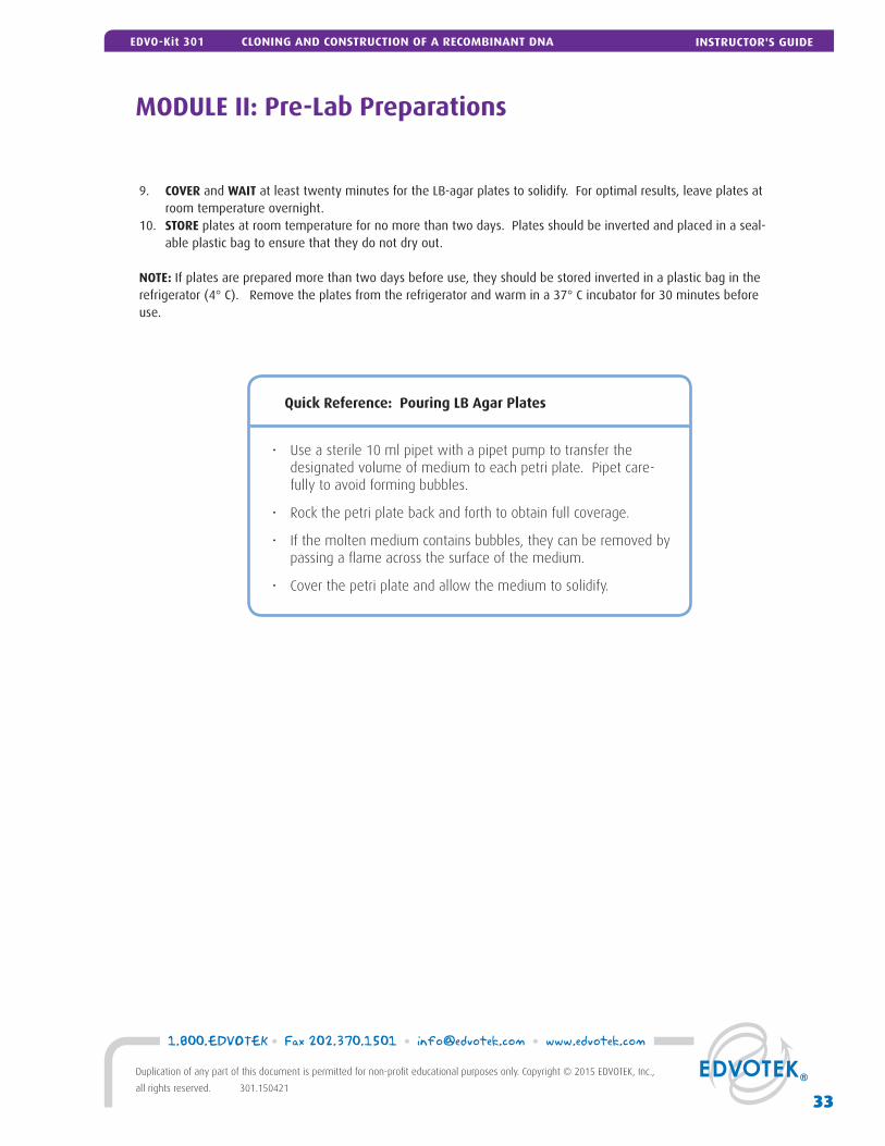
9. COVER and WAIT at least twenty minutes for the LB-agar plates to solidify. For optimal results, leave plates at room temperature overnight.
10. STORE plates at room temperature for no more than two days. Plates should be inverted and placed in a seal-able plastic bag to ensure that they do not dry out.
NOTE: If plates are prepared more than two days before use, they should be stored inverted in a plastic bag in the refrigerator (4° C). Remove the plates from the refrigerator and warm in a 37° C incubator for 30 minutes before use.
• Use a sterile 10 ml pipet with a pipet pump to transfer the designated volume of medium to each petri plate. Pipet care-
fully to avoid forming bubbles.
• Rock the petri plate back and forth to obtain full coverage.
• If the molten medium contains bubbles, they can be removed by passing a fl ame across the surface of the medium.
• Cover the petri plate and allow the medium to solidify.
Quick Reference: Pouring LB Agar Plates
MODULE II: Pre-Lab Preparations
33
1.800.EDVOTEK • Fax 202.370.1501 • [email protected] • www.edvotek.com
Duplication of any part of this document is permitted for non-profi t educational purposes only. Copyright © 2015 EDVOTEK, Inc.,
all rights reserved. 301.150421
INSTRUCTOR'S GUIDEEDVO-Kit 301 CLONING AND CONSTRUCTION OF A RECOMBINANT DNA
MODULE II: Pre-Lab Preparations
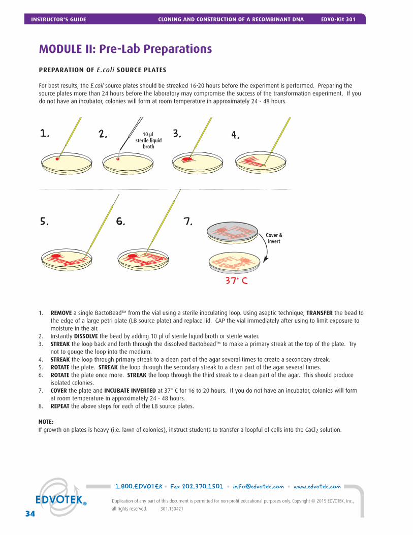
PREPARATION OF E.coli SOURCE PLATES
For best results, the E.coli source plates should be streaked 16-20 hours before the experiment is performed. Preparing the source plates more than 24 hours before the laboratory may compromise the success of the transformation experiment. If you do not have an incubator, colonies will form at room temperature in approximately 24 - 48 hours.
1. 2. 3.
5.
4.10 µl sterile liquid
broth
37° C
6. 7.Cover &Invert
1. REMOVE a single BactoBead™ from the vial using a sterile inoculating loop. Using aseptic technique, TRANSFER the bead to the edge of a large petri plate (LB source plate) and replace lid. CAP the vial immediately after using to limit exposure to moisture in the air.
2. Instantly DISSOLVE the bead by adding 10 μl of sterile liquid broth or sterile water.3. STREAK the loop back and forth through the dissolved BactoBead™ to make a primary streak at the top of the plate. Try
not to gouge the loop into the medium. 4. STREAK the loop through primary streak to a clean part of the agar several times to create a secondary streak.5. ROTATE the plate. STREAK the loop through the secondary streak to a clean part of the agar several times. 6. ROTATE the plate once more. STREAK the loop through the third streak to a clean part of the agar. This should produce
isolated colonies.7. COVER the plate and INCUBATE INVERTED at 37° C for 16 to 20 hours. If you do not have an incubator, colonies will form
at room temperature in approximately 24 - 48 hours.8. REPEAT the above steps for each of the LB source plates.
NOTE: If growth on plates is heavy (i.e. lawn of colonies), instruct students to transfer a loopful of cells into the CaCl2 solution.
1.800.EDVOTEK • Fax 202.370.1501 • [email protected] • www.edvotek.com
34
Duplication of any part of this document is permitted for non-profi t educational purposes only. Copyright © 2015 EDVOTEK, Inc.,
all rights reserved. 301.150421
INSTRUCTOR'S GUIDE CLONING AND CONSTRUCTION OF A RECOMBINANT DNA EDVO-Kit 301
MODULE II: Pre-Lab Preparations
PREPARATION OF CONTROL DNA FOR TRANSFORMATION
This kit includes three control plasmids. Each control plasmid represents a different combination of insert and vector. If the ligation experiment is not successful, the bacteria transformed with the control plasmids can be used for Modules III, IV, and V. Aliquots of plasmid DNA can be prepared the day before the lab and stored at 4° C.
1. Place the tubes of Control DNA for Transformation (E1, E2, and E3) on DNA on ice to thaw. a. E1 represents a single insert in the 5’-3’ orientation b. E2 represents a single insert in the 3’-5’ orientation c. E3 represents a double insert in the 3’-5’ orientation2. Assign each group one of the three control plasmids for transformation. 3. Label fi ve 0.5 ml microcentrifuge tubes with “Control” and the group name/number.4. Before dispensing, tap the tubes of control plasmids until all the samples are at the tapered bottom of the tubes. Using
an automatic micropipet, dispense 12 μl of the control plasmid into the ap-propriately labeled tube. NOTE: Students will use 10 μl for the transformation experiment.
5. Cap the tubes and place them on ice.6. Return the plasmid DNA to the freezer. This DNA can be restriction digested in
Module V if the plasmid preparation in Module IV is unsuccessful.
Day of the Lab:1. Equilibrate waterbaths at 37° C and 42° C; incubator at 37° C.2. Dispense 1 ml of CaCl2 into microcentrifuge tubes for each of the 5 groups and
place on ice. Label tubes CaCl2. Temperature of the CaCl2 is critical. We recom-mend storing the CaCl2 in the refrigerator the night before the experiment to ensure that it is at the correct temperature for transformation.
3. Dispense 1.5 ml of Luria Broth Medium ("Recovery broth") into tubes for each of the 5 groups and keep at room temperature. (Alternatively, the Recovery Broth bottle can be placed at a classroom pipetting station for students to share)
4. Dispense 50 μl ultra-pure water into 0.5 ml microcentrifuge tube for each of the groups. Label tubes H2O.
Each Group Requires:
• Ligation Mixture from Module I• One microcentrifuge tube contain-
ing 12 μl control DNA (E1, E2, or E3)• One microcentrifuge tube contain-
ing 1 ml 0.05M CaCl2• One microcentrifuge tube contain-
ing 50 μl ultra-pure water• One E.coli source plate• Two LB-Kan plates• Two 1.5 ml microcentrifuge tubes• Two sterile inoculating loops• Two sterile 1 ml pipets• Toothpicks• Ice and ice bucket
MODULE III: Pre-Lab Preparations
CULTURING OF kanR TRANSFORMANTS
If ligation was unsuccessful, students can pick a single colony off of the transformation control plate for Modules III, IV, and IV (Growth, Purifi cation, and Restriction Digest).
Kanamycin Medium should be prepared on the day of the Module III laboratory.
1. Thaw the Kanamycin (D).
2. Withdraw 0.25 ml of the Kanamycin with a sterile 1 ml pipet.
3. Add 0.25 ml of Kanamycin to the growth medium. Cap and swirl to mix.
4. Aseptically transfer 10 ml of the medium to each of 5 sterile culture tubes (sterile 50 ml conical tubes). Close the tubes.
(NOTE: The culture tubes have a substantially larger volume than 10 ml to allow for adequate aeration during growth.)
NOTE:New controls may be used in Module II and III. Read carefully before performing experiment.
35
1.800.EDVOTEK • Fax 202.370.1501 • [email protected] • www.edvotek.com
Duplication of any part of this document is permitted for non-profi t educational purposes only. Copyright © 2015 EDVOTEK, Inc.,
all rights reserved. 301.150421
INSTRUCTOR'S GUIDEEDVO-Kit 301 CLONING AND CONSTRUCTION OF A RECOMBINANT DNA
MODULE IV: Pre-Lab Preparations
EXTRACTION OF RECOMBINANT PLASMID DNA
Harvesting the Transformed Cells from Liquid Culture:Cells must be harvested 12-15 hours after the cultures are prepared in Module III. At this point, sterility is no longer important.
If your centrifuge has a rotor that can accommodate the 50 ml conical tubes:1. Harvest the cells by centrifuging the entire culture at 4000 RPM for 5 minutes. 2. Pour off most of the medium, leaving about 500 μl at the bottom of the tube. 3. Resuspend the pellet in the remaining media and transfer the cells into a fresh 1.5 ml microcentrifuge tube. Make sure to label
this tube with the appropriate student group name or number. 4. Centrifuge the tubes again at 4000 RPM for 5 minutes.5. Pour off the supernatant. The pellet should be the size of a match head.6. Drain off residual medium by leaving the tubes inverted on paper towels for a few minutes. Take care not to let the cell pellet
slip out of the tube.7. If Module IV (Plasmid Extraction) will be performed on the same day as the cell harvest (steps 1-6), put the cells on ice or in the
refrigerator until use. If Module IV will be performed at a later date, freeze the cells until they are required.
If your centrifuge DOES NOT have a rotor that can accommodate the 50 ml conical tubes:1. Label fi ve 1.5 ml microcentrifuge tubes with each student group name or number. 2. Transfer 1.5 ml of the bacterial culture to the appropriately labeled microcentrifuge tube.3. Harvest the cells by centrifuging the culture at 4000 RPM for 5 minutes. 4. Pour off the entire volume of medium, leaving the pellet at the bottom of the tube. 5. Transfer another 1.5 ml of the bacterial culture into the appropriate microcentrifuge tube. Centrifuge the tubes again at 4000
RPM for 5 minutes.6. Repeat steps 4 and 5 until the bacterial pellet is the size of a match head.7. Drain off residual medium by leaving the tubes inverted on paper towels for a few minutes. Take care not to let the cell pellet
slip out of the tube.8. If Module IV (Plasmid Extraction) will be performed on the same day as the cell harvest (steps 1-7), put the cells on ice or in the
refrigerator until use. If Module IV will be performed at a later date, freeze the cells until they are required.
PREPARATION OF LYSIS REAGENTS
The reagents can be aliquoted for each group of students. We recom-mend the aliquoting the reagents into sets for each lab group as de-scribed below. Alternatively, the students can share the stock tubes placed in a central location. Note that sharing the tubes increases the risk of a spill or contamination. Set up each pipetting station with a designated 1 ml pipet for the reagent.
1. Prepare the Cell Lysis Solution. (NOTE: This solution should be prepared fresh and kept at room temperature.) The SDS will precipitate out of the lysis solution if it is stored on ice or in the refrigerator.
a. In a small beaker, add all of the Sodium Hydroxide solution (I) to 8 ml of distilled water. Mix. b. Add all of the SDS solution (J) to the beaker. Mix. c. Label this beaker “Cell Lysis Solution”. Cover the beaker and keep at room temperature until use. d. If desired, label fi ve 1.5 ml microcentrifuge tubes “Lysis Solution”. Dispense 500 μl of Cell Lysis Solution into each tube. 2. Label fi ve 1.5 ml microcentrifuge tubes “Tris.” Dispense 100 μl of 1X Tris-EDTA buffer (L) into each tube.3. Label fi ve 1.5 ml microcentrifuge tubes “KOAc.” Dispense 250 μl of Potassium Acetate Solution (M) into each tube.4. Label fi ve 1.5 ml microcentrifuge tubes “TEG.” Dispense 250 μl of Tris-EDTA-Glucose buffer (H) into each tube.5. Label fi ve 0.5 ml microcentrifuge tubes “RNAse.” Dispense 10 μl of RNAse Solution (K) into each tube.6. Each student group will require 95-100% Isopropanol and 70% Ethanol. These reagents can be dispensed into 1.5 ml aliquots if
desired. Cool the 70% Ethanol by placing it on ice 30-60 minutes before use.
Each Group Requires:
• One 1.5 ml microcentrifuge tube containing a cell pellet• One clean 1.5 ml snap-top microcentrifuge tube• One microcentrifuge tube containing 500 μl Cell Lysis Solution• One microcentrifuge tube containing 250 μl Potassium Ac-
etate Neutralization Buffer • One microcentrifuge tube containing 250 μl TEG• One microcentrifuge tube containing 100 μl 1x TE Solution • One microcentrifuge tube containing 10 μl RNAse Solution • Isopropanol - 95-100%• Ethanol - 70%
1.800.EDVOTEK • Fax 202.370.1501 • [email protected] • www.edvotek.com
36
Duplication of any part of this document is permitted for non-profi t educational purposes only. Copyright © 2015 EDVOTEK, Inc.,
all rights reserved. 301.150421
INSTRUCTOR'S GUIDE CLONING AND CONSTRUCTION OF A RECOMBINANT DNA EDVO-Kit 301
MODULE V: Pre-Lab Preparations
RESTRICTION ENZYME ANALYSIS
This kit contains enough restriction enzyme to perform fi ve sets of restriction digests. If the student DNA extractions (Module IV) were successful, the isolated plasmid DNA will be used for restric-tion digest in Module V. However, if the students DNA extractions were unsuccessful, or if you have fewer than fi ve groups, the control plasmid DNA (components E1, E2, E3) can be used for the restriction digest.
1. Place the tubes of Control DNA for Transformation (E1, E2, and E3) on DNA on ice to thaw. Once thawed, tap the tubes of control plasmids until all the samples are at the tapered bottom of the tubes.
2. Using an automatic micropipet, dispense 30 μl of the control plasmid into an appropriately labeled tube. NOTE: Students will use 25 μl for the restriction digest experiment.
3. Return control DNA to freezer.
DAY OF THE LAB:
1. Equilibrate a 37° C waterbath.2. Thaw the Restriction Enzyme Reaction Buffer (O), Restriction Enzyme Dilu-
tion Buffer (P), and Ultra-Pure Water (N) and place on ice.3. Within 30 minutes of starting the Module V experiment, remove the
restriction enzymes from the freezer and place them on ice. Gently tap the tubes on the tabletop or centrifuge them to collect the enzymes at the bottom of the tube.
4. Dilute the restriction enzymes in the Restriction Enzyme Dilution Buffer (P): a. Add 35 μl of ice cold dilution buffer to the tube of EcoRI (S) b. Add 35 μl of ice cold dilution buffer to the tube of ClaI (U) c. Add 75 μl of ice cold dilution buffer to the tube of PvuII (T). Be sure to mix the diluted enzymes well as the concen-
trated enzyme solutions contain glycerol and are very viscous. Change tips between samples to avoid cross-contam-ination. At this point, the enzymes can no longer be stored. They must be used as soon as possible. Keep the tubes on ice until use.
5. Aliquot the following reagents into labeled 0.5 ml microcentrifuge tubes. Store on ice until use. a. 200 μl Ultra-Pure Water (N) b. 40 μl Restriction Enzyme Buffer (O) c. 7 μl Diluted EcoRI d. 14 μl Diluted PvuII e. 7 μl Diluted ClaI
AGAROSE GEL ELECTROPHORESIS
Each group will require one 0.8% gel with 6 sample wells for electrophoresis. See pages 14-15 (Module I-B) and Appendix B (page 48) for detailed preparation information.
Before electrophoresis, each group will receive 10 μl of 10x Gel loading solution, 25 μl Standard DNA fragments (component Q, labeled “1”) for electrophoresis, and 25 μl Supercoiled plasmid vector standards (component R, labeled “2”). These reagents will be distributed when electrophoresis is performed.
Metric rulers and semi-log graph paper (semi-log) will be needed if the students are calculating the restriction fragment sizes by creating a linear standard curve (Appendix C). However, the students should be able to estimate the sizes of the DNA frag-ments by visual inspection of the gel or a photograph of the gel.
NOTE:New controls may be used in Module V. Read carefully before per-forming experiment.
Each Group Requires:
• One purifi ed DNA sample (from Module IV) OR control plasmid DNA
• Five clean 1.5 ml microcentrifuge tubes• One microcentrifuge tube containing 200 μl
UltraPure water• One microcentrifuge tube containing 40 μl
Restriction Enzyme Dilution Buffer• One microcentrifuge tube containing 7 μl
diluted EcoRI• One microcentrifuge tube containing 7 μl
diluted ClaI• One microcentrifuge tube containing 14 μl
diluted PvuII
37
1.800.EDVOTEK • Fax 202.370.1501 • [email protected] • www.edvotek.com
Duplication of any part of this document is permitted for non-profi t educational purposes only. Copyright © 2015 EDVOTEK, Inc.,
all rights reserved. 301.150421
INSTRUCTOR'S GUIDEEDVO-Kit 301 CLONING AND CONSTRUCTION OF A RECOMBINANT DNA
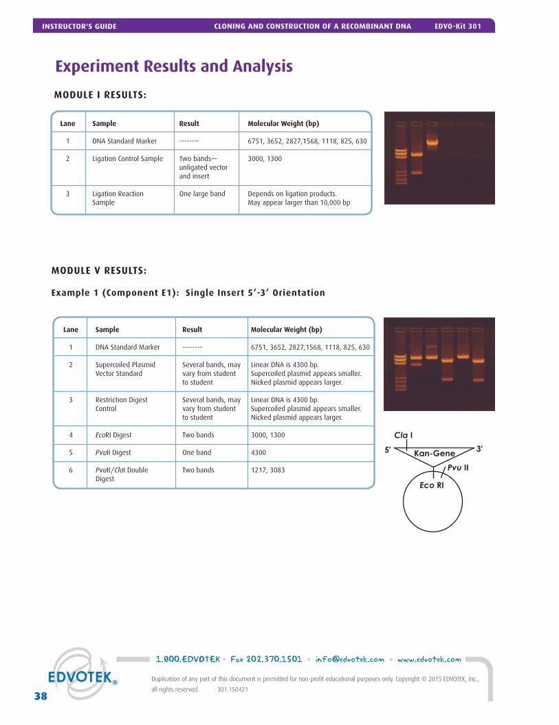
MODULE I RESULTS:
Experiment Results and Analysis
Lane Sample Result Molecular Weight (bp) 1 DNA Standard Marker -------- 6751, 3652, 2827,1568, 1118, 825, 630
2 Ligation Control Sample Two bands— 3000, 1300 unligated vector and insert
3 Ligation Reaction One large band Depends on ligation products. Sample May appear larger than 10,000 bp
MODULE V RESULTS:
Example 1 (Component E1): Single Insert 5’-3’ Orientation
Lane Sample Result Molecular Weight (bp) 1 DNA Standard Marker -------- 6751, 3652, 2827,1568, 1118, 825, 630
2 Supercoiled Plasmid Several bands, may Linear DNA is 4300 bp. Vector Standard vary from student Supercoiled plasmid appears smaller. to student Nicked plasmid appears larger.
3 Restriction Digest Several bands, may Linear DNA is 4300 bp. Control vary from student Supercoiled plasmid appears smaller. to student Nicked plasmid appears larger.
4 EcoRI Digest Two bands 3000, 1300
5 PvuII Digest One band 4300 6 PvuII/ClaI Double Two bands 1217, 3083 Digest
Eco RI
Kan-Gene
Cla I
Pvu II
3'5'
1.800.EDVOTEK • Fax 202.370.1501 • [email protected] • www.edvotek.com
38
Duplication of any part of this document is permitted for non-profi t educational purposes only. Copyright © 2015 EDVOTEK, Inc.,
all rights reserved. 301.150421
INSTRUCTOR'S GUIDE CLONING AND CONSTRUCTION OF A RECOMBINANT DNA EDVO-Kit 301
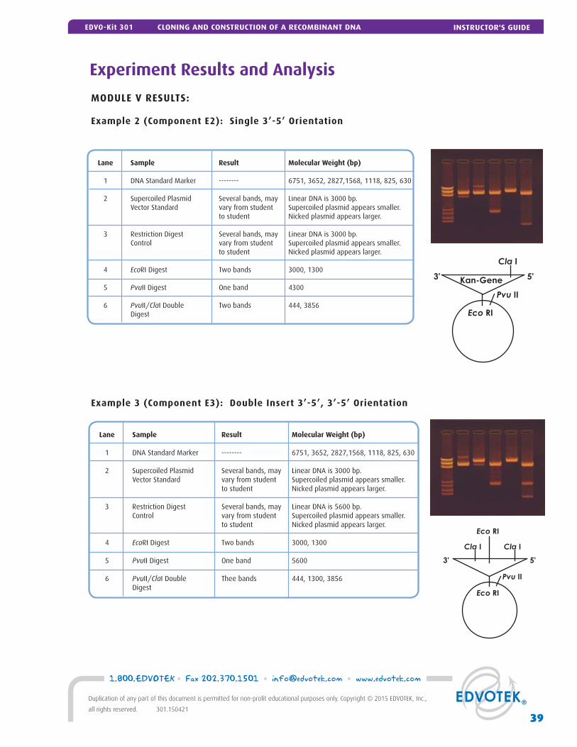
Experiment Results and Analysis
MODULE V RESULTS:
Example 2 (Component E2): Single 3’-5’ Orientation
Lane Sample Result Molecular Weight (bp) 1 DNA Standard Marker -------- 6751, 3652, 2827,1568, 1118, 825, 630
2 Supercoiled Plasmid Several bands, may Linear DNA is 3000 bp. Vector Standard vary from student Supercoiled plasmid appears smaller. to student Nicked plasmid appears larger.
3 Restriction Digest Several bands, may Linear DNA is 3000 bp. Control vary from student Supercoiled plasmid appears smaller. to student Nicked plasmid appears larger.
4 EcoRI Digest Two bands 3000, 1300
5 PvuII Digest One band 4300 6 PvuII/ClaI Double Two bands 444, 3856 Digest Eco RI
Kan-Gene
Cla I
Pvu II
3' 5'
Example 3 (Component E3): Double Insert 3’-5’, 3’-5’ Orientation
Lane Sample Result Molecular Weight (bp) 1 DNA Standard Marker -------- 6751, 3652, 2827,1568, 1118, 825, 630
2 Supercoiled Plasmid Several bands, may Linear DNA is 3000 bp. Vector Standard vary from student Supercoiled plasmid appears smaller. to student Nicked plasmid appears larger.
3 Restriction Digest Several bands, may Linear DNA is 5600 bp. Control vary from student Supercoiled plasmid appears smaller. to student Nicked plasmid appears larger.
4 EcoRI Digest Two bands 3000, 1300
5 PvuII Digest One band 5600 6 PvuII/ClaI Double Thee bands 444, 1300, 3856 Digest
Eco RI
Eco RI
Cla ICla I
Pvu II
3' 5'
39
1.800.EDVOTEK • Fax 202.370.1501 • [email protected] • www.edvotek.com
Duplication of any part of this document is permitted for non-profi t educational purposes only. Copyright © 2015 EDVOTEK, Inc.,
all rights reserved. 301.150421
INSTRUCTOR'S GUIDEEDVO-Kit 301 CLONING AND CONSTRUCTION OF A RECOMBINANT DNA
Please refer to the kit insert for the Answers to
Study Questions
A EDVOTEK® Troubleshooting Guide
B Bulk Preparation of Agarose Gels
C Data Analysis Using a Standard Curve
D LyphoCells™ For Transformation
Material Safety Data Sheets:
Now available for your convenient download on www.edvotek.com
Appendices
41
1.800.EDVOTEK • Fax 202.370.1501 • [email protected] • www.edvotek.com
Duplication of any part of this document is permitted for non-profi t educational purposes only. Copyright © 2015 EDVOTEK, Inc.,
all rights reserved. 301.150421
APPENDICESEDVO-Kit 301 CLONING AND CONSTRUCTION OF A RECOMBINANT DNA
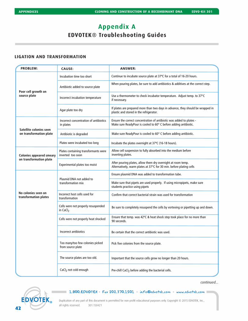
Appendix AEDVOTEK® Troubleshooting Guides
LIGATION AND TRANSFORMATION
continued...
PROBLEM: CAUSE: ANSWER:
Poor cell growth on source plate
Incubation time too short Continue to incubate source plate at 37ºC for a total of 16-20 hours.
Antibiotic added to source plateWhen pouring plates, be sure to add antibiotics & additives at the correct step.
Colonies appeared smeary on transformation plate
Plates containing transformants were inverted too soon
Allow cell suspension to fully absorbed into the medium before inverting plates.
Experimental plates too moistAfter pouring plates, allow them dry overnight at room temp.Alternatively, warm plates at 37°C for 30 min. before plating cells
Incorrect host cells used for transformation
Confirm that correct bacterial strain was used for transformation
Cells were not properly heat shocked Ensure that temp. was 42ºC & heat shock step took place for no more than 90 seconds.
No colonies seen on transformation plates
Plasmid DNA not added to transformation mix
Ensure plasmid DNA was added to transformation tube.
Incorrect incubation temperature Use a thermometer to check incubator temperature. Adjust temp. to 37°C if necessary.
Agar plate too dry If plates are prepared more than two days in advance, they should be wrapped in plastic and stored in the refrigerator.
Make sure that pipets are used properly. If using micropipets, make sure students practice using pipets
Satellite colonies seen on transformation plate
Incorrect concentration of antibioticsin plates
Antibiotic is degraded
Ensure the correct concentration of antibiotic was added to plates - Make sure ReadyPour is cooled to 60° C before adding antibiotic.
Make sure ReadyPour is cooled to 60° C before adding antibiotic.
Incubate the plates overnight at 37ºC (16-18 hours).
Incorrect antibiotics Be certain that the correct antibiotic was used.
Too many/too few colonies picked from source plate
Pick five colonies from the source plate.
The source plates are too old. Important that the source cells grow no longer than 20 hours.
CaCl2 not cold enough Pre-chill CaCl2 before adding the bacterial cells.
Cells were not properly resuspendedin CaCl2
Be sure to completely resuspend the cells by vortexing or pipetting up and down.
Plates were incubated too long
1.800.EDVOTEK • Fax 202.370.1501 • [email protected] • www.edvotek.com
42
Duplication of any part of this document is permitted for non-profi t educational purposes only. Copyright © 2015 EDVOTEK, Inc.,
all rights reserved. 301.150421
APPENDICES CLONING AND CONSTRUCTION OF A RECOMBINANT DNA EDVO-Kit 301
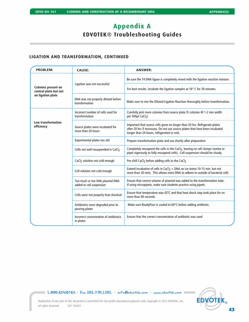
Appendix AEDVOTEK® Troubleshooting Guides
LIGATION AND TRANSFORMATION, CONTINUED
Low transformation efficiency
Incorrect number of cells used for transformation
Source plates were incubated for more than 20 hours
Experimental plates too old
Cells not well resuspended in CaCl2
CaCl2 solution not cold enough
Cell solution not cold enough
Too much or too little plasmid DNA added to cell suspension
Cells were not properly heat shocked
Antibiotics were degraded prior to pouring plates
Incorrect concentration of antibiotics in plates
Carefully pick more colonies from source plate (5 colonies @ 1-2 mm width per 500μl CaCl2)
Important that source cells grow no longer than 20 hrs. Refrigerate plates after 20 hrs if necessary. Do not use source plates that have been incubatedlonger than 24 hours, refrigerated or not).
Prepare transformation plate and use shortly after preparation
Completely resuspend the cells in the CaCl2, leaving no cell clumps (vortex or pipet vigorously to fully resuspend cells). Cell suspension should be cloudy.
Pre-chill CaCl2 before adding cells to the CaCl2
Extend incubation of cells in CaCl2 + DNA on ice (extra 10-15 min. but not more than 30 min). This allows more DNA to adhere to outside of bacterial cell).
Ensure that correct volume of plasmid was added to the transformation tube. If using micropipets, make sure students practice using pipets.
Ensure that temperature was 42ºC and that heat shock step took place for no more than 90 seconds.
Make sure ReadyPour is cooled to 60°C before adding antibiotic.
Ensure that the correct concentration of antibiotic was used
Colonies present oncontrol plate but not on ligation plate
Ligation was not successfulBe sure the T4 DNA ligase is completely mixed with the ligation reaction mixture.
For best results, incubate the ligation samples at 16° C for 30 minutes.
Make sure to mix the Diluted Ligation Reaction thoroughly before transformation.DNA was not properly diluted beforetransformation
PROBLEM: CAUSE: ANSWER:
43
1.800.EDVOTEK • Fax 202.370.1501 • [email protected] • www.edvotek.com
Duplication of any part of this document is permitted for non-profi t educational purposes only. Copyright © 2015 EDVOTEK, Inc.,
all rights reserved. 301.150421
APPENDICESEDVO-Kit 301 CLONING AND CONSTRUCTION OF A RECOMBINANT DNA
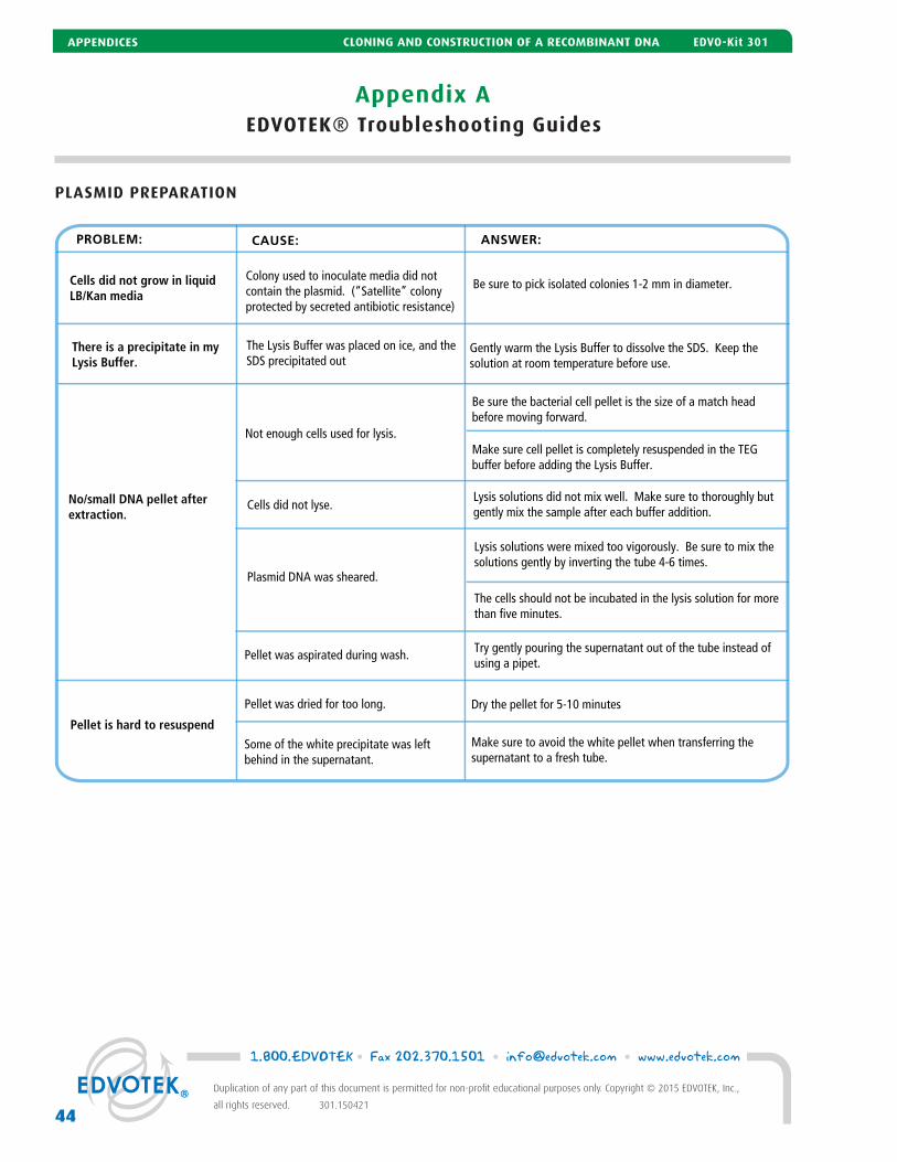
Appendix AEDVOTEK® Troubleshooting Guides
PLASMID PREPARATION
PROBLEM: CAUSE: ANSWER:
Cells did not grow in liquidLB/Kan media
Colony used to inoculate media did not contain the plasmid. (“Satellite” colony protected by secreted antibiotic resistance)
Be sure to pick isolated colonies 1-2 mm in diameter.
No/small DNA pellet after extraction.
Not enough cells used for lysis.
Be sure the bacterial cell pellet is the size of a match head before moving forward.
Make sure cell pellet is completely resuspended in the TEG buffer before adding the Lysis Buffer.
There is a precipitate in my Lysis Buffer.
The Lysis Buffer was placed on ice, and the SDS precipitated out
Gently warm the Lysis Buffer to dissolve the SDS. Keep the solution at room temperature before use.
Cells did not lyse.Lysis solutions did not mix well. Make sure to thoroughly but gently mix the sample after each buffer addition.
Plasmid DNA was sheared.
Lysis solutions were mixed too vigorously. Be sure to mix the solutions gently by inverting the tube 4-6 times.
The cells should not be incubated in the lysis solution for more than five minutes.
Try gently pouring the supernatant out of the tube instead of using a pipet.
Pellet was aspirated during wash.
Dry the pellet for 5-10 minutesPellet was dried for too long.
Make sure to avoid the white pellet when transferring the supernatant to a fresh tube.
Some of the white precipitate was left behind in the supernatant.
Pellet is hard to resuspend
1.800.EDVOTEK • Fax 202.370.1501 • [email protected] • www.edvotek.com
44
Duplication of any part of this document is permitted for non-profi t educational purposes only. Copyright © 2015 EDVOTEK, Inc.,
all rights reserved. 301.150421
APPENDICES CLONING AND CONSTRUCTION OF A RECOMBINANT DNA EDVO-Kit 301
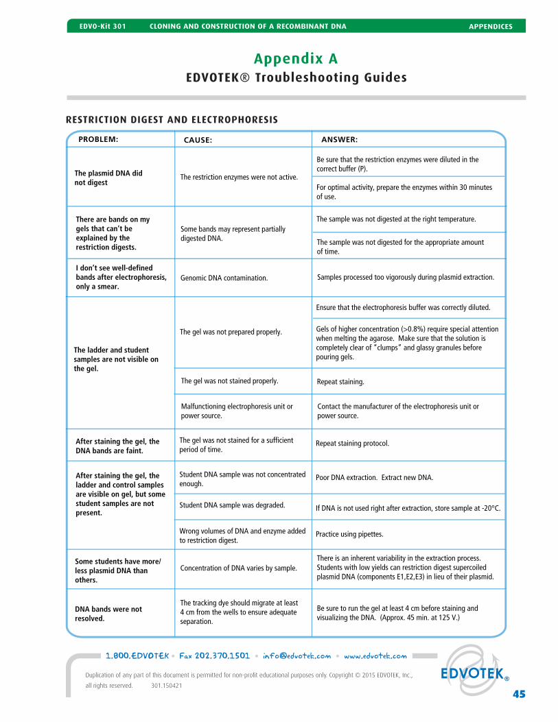
Appendix AEDVOTEK® Troubleshooting Guides
RESTRICTION DIGEST AND ELECTROPHORESIS
PROBLEM: CAUSE: ANSWER:
The plasmid DNA did not digest
The restriction enzymes were not active.
Be sure that the restriction enzymes were diluted in the correct buffer (P).
For optimal activity, prepare the enzymes within 30 minutes of use.
I don’t see well-definedbands after electrophoresis,only a smear.
The ladder and studentsamples are not visible onthe gel.
The gel was not prepared properly.
Ensure that the electrophoresis buffer was correctly diluted.
Gels of higher concentration (>0.8%) require special attentionwhen melting the agarose. Make sure that the solution is completely clear of “clumps” and glassy granules beforepouring gels.
There are bands on mygels that can’t be explained by the restriction digests.
Some bands may represent partiallydigested DNA.
The sample was not digested at the right temperature.
The sample was not digested for the appropriate amount of time.
The gel was not stained properly. Repeat staining.
Malfunctioning electrophoresis unit or power source.
Contact the manufacturer of the electrophoresis unit or power source.
Samples processed too vigorously during plasmid extraction.
After staining the gel, theDNA bands are faint.
The gel was not stained for a sufficientperiod of time.
Repeat staining protocol.
After staining the gel, the ladder and control samplesare visible on gel, but somestudent samples are notpresent.
Student DNA sample was not concentratedenough.
Poor DNA extraction. Extract new DNA.
Student DNA sample was degraded. If DNA is not used right after extraction, store sample at -20°C.
Wrong volumes of DNA and enzyme addedto restriction digest.
Practice using pipettes.
Some students have more/less plasmid DNA than others.
Concentration of DNA varies by sample.There is an inherent variability in the extraction process. Students with low yields can restriction digest supercoiled plasmid DNA (components E1,E2,E3) in lieu of their plasmid.
DNA bands were notresolved.
The tracking dye should migrate at least4 cm from the wells to ensure adequateseparation.
Be sure to run the gel at least 4 cm before staining and visualizing the DNA. (Approx. 45 min. at 125 V.)
Genomic DNA contamination.
45
1.800.EDVOTEK • Fax 202.370.1501 • [email protected] • www.edvotek.com
Duplication of any part of this document is permitted for non-profi t educational purposes only. Copyright © 2015 EDVOTEK, Inc.,
all rights reserved. 301.150421
APPENDICESEDVO-Kit 301 CLONING AND CONSTRUCTION OF A RECOMBINANT DNA

Appendix BBulk Preparation of Agarose Gels
To save time, the electrophoresis buffer and agarose gel solution can be prepared in larger quantities for sharing by the class. Un-used diluted buffer can be used at a later time and solidifi ed agarose gel solution can be remelted.
Bulk Electrophoresis Buffer
Quantity (bulk) preparation for 3 liters of 1x electrophoresis buffer is outlined in Table D.
Batch Agarose Gels (0.8%)
For quantity (batch) preparation of 0.8% agarose gels, see Table E.
1. Use a 500 ml fl ask to prepare the diluted gel buffer.
2. Pour 3.0 grams of UltraSpec-Agarose™ into the prepared buffer. Swirl to disperse clumps.
3. With a marking pen, indicate the level of solution volume on the outside of the fl ask.
4. Heat the agarose solution as outlined previously for individual gel preparation. The heating time will require adjustment due to the larger total volume of gel buffer solution.
5. Cool the agarose solution to 60° C with swirling to promote even dissipation of heat. If evaporation has occurred, add distilled water to bring the solution up to the original volume as marked on the fl ask in step 3.
6. Dispense the required volume of cooled agarose solution for casting each gel. Measure 30 ml for a 7 x 7 cm tray, 50 ml for a 7 x 10 cm tray, and 60 ml for a 7 x 14 cm tray. For this experiment, 7 x 7 cm gels are recommended.
7. Allow the gel to completely solidify. It will become fi rm and cool to the touch after approxi-mately 20 minutes. Then proceed with preparing the gel for electrophoresis.
60˚C
Note: The UltraSpec-Agarose™ kit component is usually labeled with the amount it contains. Please read the label care-fully. If the amount of aga-rose is not specifi ed or if the bottle's plastic seal has been broken, weigh the agarose to ensure you are using the correct amount.
50x Conc.Buffer +
DistilledWater
Total Volume Required
60 ml 2,940 ml 3000 ml (3 L)
Bulk Preparation of Electrophoresis BufferTable
D
Batch Prep of 0.8% UltraSpec-Agarose™Table
EAmt ofAgarose
(g)
ConcentratedBuffer (50X)
(ml)+
DistilledWater(ml)
TotalVolume
(ml)+
3.0 7.5 382.5 390
1.800.EDVOTEK • Fax 202.370.1501 • [email protected] • www.edvotek.com
46
Duplication of any part of this document is permitted for non-profi t educational purposes only. Copyright © 2015 EDVOTEK, Inc.,
all rights reserved. 301.150421
APPENDICES CLONING AND CONSTRUCTION OF A RECOMBINANT DNA EDVO-Kit 301
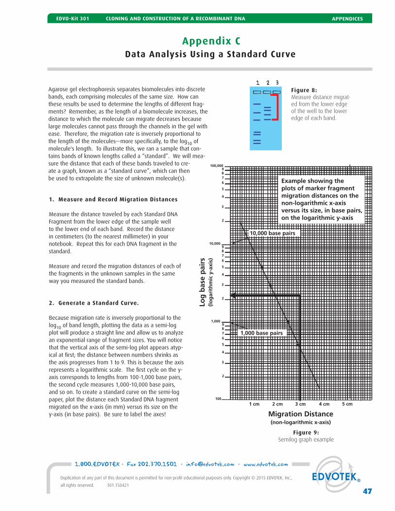
Agarose gel electrophoresis separates biomolecules into discrete bands, each comprising molecules of the same size. How can these results be used to determine the lengths of different frag-ments? Remember, as the length of a biomolecule increases, the distance to which the molecule can migrate decreases because large molecules cannot pass through the channels in the gel with ease. Therefore, the migration rate is inversely proportional to the length of the molecules—more specifi cally, to the log10 of molecule's length. To illustrate this, we ran a sample that con-tains bands of known lengths called a “standard”. We will mea-sure the distance that each of these bands traveled to cre-ate a graph, known as a “standard curve”, which can then be used to extrapolate the size of unknown molecule(s).
1. Measure and Record Migration Distances
Measure the distance traveled by each Standard DNA Fragment from the lower edge of the sample well to the lower end of each band. Record the distance in centimeters (to the nearest millimeter) in your notebook. Repeat this for each DNA fragment in the standard.
Measure and record the migration distances of each of the fragments in the unknown samples in the same way you measured the standard bands.
2. Generate a Standard Curve.
Because migration rate is inversely proportional to the log10 of band length, plotting the data as a semi-log plot will produce a straight line and allow us to analyze an exponential range of fragment sizes. You will notice that the vertical axis of the semi-log plot appears atyp-ical at fi rst; the distance between numbers shrinks as the axis progresses from 1 to 9. This is because the axis represents a logarithmic scale. The fi rst cycle on the y-axis corresponds to lengths from 100-1,000 base pairs, the second cycle measures 1,000-10,000 base pairs, and so on. To create a standard curve on the semi-log paper, plot the distance each Standard DNA fragment migrated on the x-axis (in mm) versus its size on the y-axis (in base pairs). Be sure to label the axes!
Figure 9:Semilog graph example
Figure 8:Measure distance migrat-ed from the lower edge of the well to the lower edge of each band.
1 2 3
Appendix CData Analysis Using a Standard Curve
47
1.800.EDVOTEK • Fax 202.370.1501 • [email protected] • www.edvotek.com
Duplication of any part of this document is permitted for non-profi t educational purposes only. Copyright © 2015 EDVOTEK, Inc.,
all rights reserved. 301.150421
APPENDICESEDVO-Kit 301 CLONING AND CONSTRUCTION OF A RECOMBINANT DNA
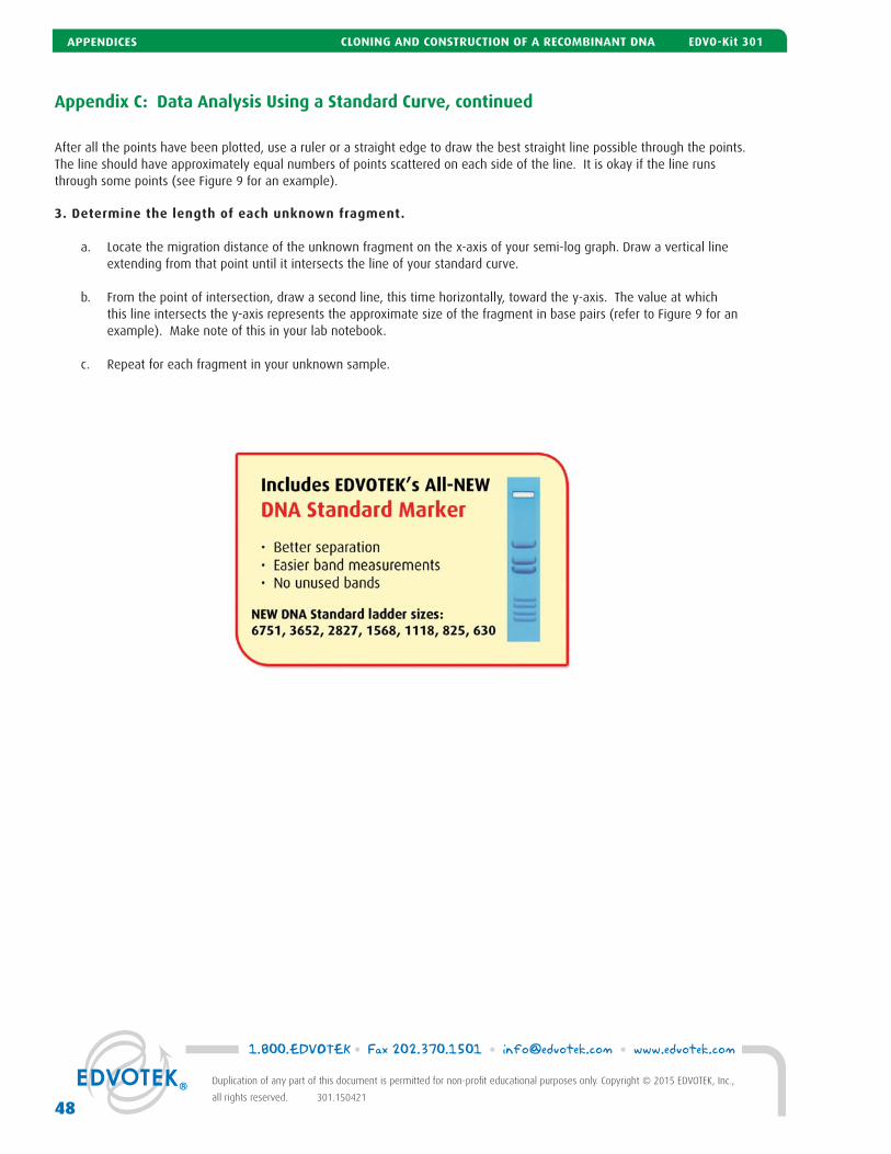
After all the points have been plotted, use a ruler or a straight edge to draw the best straight line possible through the points. The line should have approximately equal numbers of points scattered on each side of the line. It is okay if the line runs through some points (see Figure 9 for an example).
3. Determine the length of each unknown fragment.
a. Locate the migration distance of the unknown fragment on the x-axis of your semi-log graph. Draw a vertical line extending from that point until it intersects the line of your standard curve.
b. From the point of intersection, draw a second line, this time horizontally, toward the y-axis. The value at which this line intersects the y-axis represents the approximate size of the fragment in base pairs (refer to Figure 9 for an example). Make note of this in your lab notebook.
c. Repeat for each fragment in your unknown sample.
Appendix C: Data Analysis Using a Standard Curve, continued
1.800.EDVOTEK • Fax 202.370.1501 • [email protected] • www.edvotek.com
48
Duplication of any part of this document is permitted for non-profi t educational purposes only. Copyright © 2015 EDVOTEK, Inc.,
all rights reserved. 301.150421
APPENDICES CLONING AND CONSTRUCTION OF A RECOMBINANT DNA EDVO-Kit 301
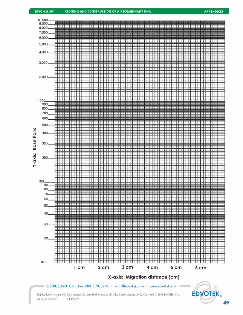
8,000
10,000
7,000
6,000
5,000
4,000
3,000
2,000
9,000
80 70
60
50
40
30
20
10
90 100
1,000
800 700
600
500
400
300
200
900
X-axis: Migration distance (cm)
1 cm 2 cm 3 cm 4 cm 5 cm 6 cm
Y-a
xis:
Ba
se P
airs
49
1.800.EDVOTEK • Fax 202.370.1501 • [email protected] • www.edvotek.com
Duplication of any part of this document is permitted for non-profi t educational purposes only. Copyright © 2015 EDVOTEK, Inc.,
all rights reserved. 301.150421
APPENDICESEDVO-Kit 301 CLONING AND CONSTRUCTION OF A RECOMBINANT DNA
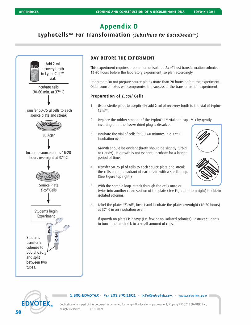
Appendix DLyphoCells™ For Transformation (Substitute for BactoBeads™)
DAY BEFORE THE EXPERIMENT
This experiment requires preparation of isolated E.coli host transformation colonies 16-20 hours before the laboratory experiment, so plan accordingly.
Important: Do not prepare source plates more than 20 hours before the experiment. Older source plates will compromise the success of the transformation experiment.
Preparation of E.coli Cells
1. Use a sterile pipet to aseptically add 2 ml of recovery broth to the vial of Lypho-Cells™.
2. Replace the rubber stopper of the LyphoCell™ vial and cap. Mix by gently inverting until the freeze dried plug is dissolved.
3. Incubate the vial of cells for 30-60 minutes in a 37° C incubation oven.
Growth should be evident (broth should be slightly turbid or cloudy). If growth is not evident, incubate for a longer period of time.
4. Transfer 50-75 μl of cells to each source plate and streak the cells on one quadrant of each plate with a sterile loop. (See Figure top right.)
5. With the sample loop, streak through the cells once or twice into another clean section of the plate (See Figure bottom right) to obtain isolated colonies.
6. Label the plates "E.coli", invert and incubate the plates overnight (16-20 hours) at 37° C in an incubation oven.
If growth on plates is heavy (i.e. few or no isolated colonies), instruct students to touch the toothpick to a small amount of cells.
LB Agar
Source PlateE.coli Cells
Add 2 mlrecovery brothto LyphoCell™
vial.
Incubate cells 30-60 min. at 37° C
Incubate source plates 16-20hours overnight at 37° C
Transfer 50-75 µl cells to eachsource plate and streak
AN
D+
-DN
A
Students beginExperiment
Students transfer 5colonies to 500 µl CaCl2and splitbetween two tubes.
1.800.EDVOTEK • Fax 202.370.1501 • [email protected] • www.edvotek.com
50
Duplication of any part of this document is permitted for non-profi t educational purposes only. Copyright © 2015 EDVOTEK, Inc.,
all rights reserved. 301.150421
APPENDICES CLONING AND CONSTRUCTION OF A RECOMBINANT DNA EDVO-Kit 301