Embed Size (px)
Citation preview

INFECTION AND IMMUNITY,0019-9567/97/$04.0010
Dec. 1997, p. 5142–5148 Vol. 65, No. 12
Copyright © 1997, American Society for Microbiology
Contact-Dependent Disruption of the Host Cell MembraneSkeleton Induced by Trichomonas vaginalis
PIER LUIGI FIORI,* PAOLA RAPPELLI, MARIA FILIPPA ADDIS, FRANCA MANNU,AND PIERO CAPPUCCINELLI
Department of Biomedical Sciences, Division of Experimental and Clinical Microbiology,University of Sassari, Sassari, Italy
Received 5 June 1997/Returned for modification 24 July 1997/Accepted 15 September 1997
This report presents evidence showing that the pathogenetic process of the protozoan parasite Trichomonasvaginalis involves degradation of the target cell membrane skeleton; spectrin, the most representative proteinwithin this structure, has been identified as the main molecular target. Degradation of the target cell spectrinis accomplished only upon contact with the parasite, and immunochemical and immunofluorescence studiesperformed with the erythrocyte as a model demonstrate that degradation of the protein takes place beforetarget cell lysis. A preliminary characterization of the effectors involved has led to the identification of anonsecreted 30-kDa proteinase which is characterized by a high specificity for spectrin. This molecule issuggested as the main effector responsible for cytoskeletal disruption.
Microorganisms employ a combination of strategies in orderto accomplish target cell disruption. Some commonly usedmechanisms are phagocytosis, release of toxic molecules, inva-sion and killing of the target cell through disintegration ofinternal components, and secretion of proteinases for degra-dation and digestion of the host cell. In a recent study on thepathogenetic mechanisms of the protist Trichomonas vaginalis,we demonstrated that the microorganism lyses erythrocytesthrough insertion of ionic channels on the target cell mem-brane (17) under defined conditions (2, 14).
T. vaginalis is a flagellated protozoan parasite responsiblefor one of the most common sexually transmitted diseases inhumans (27). Trichomoniasis affects mostly women, and itsclinical manifestations range from an asymptomatic carrierstate to a severe vaginitis. During protozoan infection, exten-sive damage to vaginal epithelium is observed (24). An in vitrocytopathic effect of T. vaginalis on epithelial cells is well doc-umented (3, 4, 26); the protozoon is also able to lyse erythro-cytes, which represent an important source of iron and fattyacids (29, 35). For our in vitro studies we utilized erythrocytesas a target for the evaluation of the T. vaginalis cytopathiceffect, as these cells are easy to monitor for lysis; moreover,they represent the simplest model for studying membranedamage processes.
The goals of this work were to achieve a better understand-ing of the T. vaginalis pathogenetic mechanism and to shedlight on the relationship between T. vaginalis and the target cellcytoskeleton. This cellular component is a specific target fornumerous pathogens, for which it plays an important role as amechanical means for entering the host cell as well as repre-senting a direct target mediating disruption of the parasitizedcell. In order to lyse the target cell, a close contact between T.vaginalis and target cell membranes is required. Here we de-scribe by scanning electron microscopy (SEM) the occurrenceof important morphological alterations, such as loss of shapeand collapse, in erythrocytes in contact with the parasite sur-
face. Similar alterations have been reported to occur in para-sitized epithelial cell monolayers as well; cell rounding, detach-ment, and cell death have been observed (3, 4, 21, 26). Thisfinding suggests that a pathogenetic mechanism based on cy-toskeletal disruption of the target cell could be involved, which,together with the formation of functional pores, could be re-sponsible for cytotoxicity.
The cytoskeleton of nucleate cells is a complicated lattice-work of microfilaments, intermediate filaments, and microtu-bules that provides motility, shape, and stability to the cell,with complicated interconnections between plasma membranecomponents and the cytoplasmic network. By contrast, eryth-rocytes lack the intracellular network that is characteristic ofnucleate cells, and their shape, deformability, and structuralstability are maintained by the membrane skeleton located justbeneath the plasma membrane. Its relative simplicity has per-mitted its detailed characterization, and during the 1980s ho-mologs of erythrocyte membrane skeleton proteins were foundin a variety of tissues from several species. The well-definedcharacterization of the erythrocyte membrane skeleton and itshomology with nonerythroid structures were the reasons forthe utilization of erythrocytes as a cellular and molecularmodel throughout this work.
Our studies allowed the identification of a T. vaginalis-in-duced disruption of the host cell membrane skeleton, with themost representative protein, spectrin, being the main target.The characterization of the mode and kinetics of the phenom-enon has led to the finding that the parasite accomplishesdegradation of spectrin before lysis of the target cell takesplace.
MATERIALS AND METHODS
Microorganisms and culture conditions. T. vaginalis isolates were obtained asvaginal specimens from women affected by trichomoniasis. After axenization,protozoa were cultured in Diamond’s TYM (13) supplemented with 10% bovineserum, 100 mg of streptomycin sulfate ml21, and 100 IU of penicillin G per mlin a 5% carbon dioxide atmosphere at 37°C. Fresh isolates were stored in liquidnitrogen with 90% serum and 10% dimethyl sulfoxide. Cultured parasites werealways monitored for motility, and only viable organisms (.99%) were used toperform the experiments. Further characterization experiments were performedwith our isolate SS-22 as a representative strain. Escherichia coli and Staphylo-coccus aureus strains, isolated from urinary specimens of patients presenting withurologic infections, were grown in Luria-Bertani medium at 37°C.
* Corresponding author. Mailing address: Department of Biomedi-cal Sciences, Division of Experimental and Clinical Microbiology, Uni-versity of Sassari, Viale S. Pietro 43/B, 07100 Sassari, Italy. Phone:011/39/79/228301. Fax: 011/39/79/212345. E-mail: [email protected].
5142
on July 26, 2018 by guesthttp://iai.asm
.org/D
ownloaded from
Erythrocytes. Group O Rh1 erythrocytes were obtained from healthy humandonors. Cells were either used fresh or stored until needed in glucose and citratebuffer at 4°C; only fresh erythrocytes were used for cytoskeletal integrity assays.Prior to use, erythrocytes were washed three times in isotonic buffer at 400 3 gfor 5 min. For preparation of ghosts, pelleted erythrocytes were washed andincubated in 5 mM phosphate lysis buffer (pH 7.4) supplemented with 1 mg ofleupeptin ml21, 50 mg of Na-p-tosyl-L-lysine chloromethyl ketone (TLCK) ml21,and 10 mM EDTA in order to prevent the degradation of cytoskeletal compo-nents by erythrocyte-endogenous proteinases. Samples were incubated for 5 minon ice and centrifuged at 18,000 3 g in a Sorvall refrigerated centrifuge at 4°C.Supernatants containing the released hemoglobin were discarded, and incuba-tion and centrifugation cycles were repeated until an acceptable elimination ofhemoglobin was achieved. After the last wash, ghosts were collected and usedimmediately.
SEM. For SEM experiments, T. vaginalis organisms were incubated with fresherythrocytes in a ratio of 1:5. After 30 min, the cells were washed with Hankssolution and fixed for 20 to 30 min in 2% glutaraldehyde (electron microscopygrade) in 0.1 M sodium cacodylate buffer, pH 7.2. Cells were washed andpostfixed in 1% osmium tetroxide in sodium cacodylate buffer for 30 min, de-hydrated in a graded series of acetone (20 to 100%), and critical-point dried ina Polaron apparatus with amyl acetate as the intermediate fluid and CO2 as thetransition fluid. Samples were covered with gold in an Edwards S-150 sputtercoater and observed with ISI-130 and JEOL JMS-35 electron microscopes at 15and 30 kV (16).
Cell lysates. Parasite lysates were obtained by sonication in a Branson SonifierB 12. A total of 3 3 106 cells ml21 harvested in the late exponential phase ofgrowth were washed three times in phosphate-buffered saline (PBS), pH 7.4, bycentrifugation at 350 3 g. The pH value was established at over 6.50 to avoid lysisof erythrocytes by protozoan perforins. Washed cells were resuspended to thedesired density in PBS and subjected to five cycles of sonication for 10 s each at50 W in an ice bath. Samples were then centrifuged at 18,000 3 g, and super-natants were filtered through a 0.45-mm-pore-size filter membrane in order toperform the degradation assays.
Secreted proteins. Parasites harvested in the late exponential phase of growthwere washed three times in PBS by centrifugation at 350 3 g. Washed organismswere resuspended to a density of 3 3 106 ml21 in PBS plus 15 mM maltose(PBS-M) (pH 7.4) and incubated at 37°C to allow secretion (14). After 90 min,the parasites were harvested by centrifugation; the recovered supernatant wascentrifuged again at 15,000 3 g and filtered to eliminate whole organisms. ThepH was raised to 7.00 in order to prevent hemolysis. The proteins present in thecell-free supernatant were then concentrated 10 times in a Centricon (AmiconDivision, Beverly, Mass.) unit with a 3-kDa cutoff. Parasites were always moni-tored for motility (.99%) during the incubation period. As determined byenumeration and trypan blue exclusion, these conditions did not result in celllysis or parasite death.
Spectrin extraction. Freshly obtained ghosts were stripped by incubation for 5min on ice with 1 N NaOH. After incubation, the suspension was promptlybrought to neutrality with 1 M HCl. The sample was then centrifuged at 18,000 3g for 20 min, and the supernatant was collected. The released protein wasconcentrated in a Centricon concentrating unit with a 30-kDa cutoff, and theprotein concentration was determined by the Bradford method (9) with theBio-Rad (Hercules, Calif.) protein assay dye (Coomassie brilliant blue), withbovine serum albumin as a standard. Since spectrin integrity is not affected byshort-term storage, the concentrated protein was stored at 220°C until needed.Prior to utilization, it was always tested for purity and integrity by sodium dodecylsulfate-polyacrylamide gel electrophoresis (SDS-PAGE).
Cytoskeletal degradation assays. T. vaginalis organisms from exponentiallygrowing cultures were washed three times in PBS-M and resuspended at aconcentration of 2 3 106 per ml. The suspension was then incubated with freshwashed erythrocytes in a ratio of 1:5, and vials were kept at 37°C to allowerythrocyte lysis. After 60 min, the supernatant fraction, which contained mem-branes from erythrocytes lysed by the microorganisms, was centrifuged severaltimes at 350 3 g to eliminate any remaining cells and checked by light microscopyto ensure clearance of microorganisms. Ghosts were then collected by centrifu-gation at 18,000 3 g for 10 min in a refrigerated centrifuge. After proteinquantification by the Bradford method, membranes were electrophoresed andblotted. The same amount of protein (15 mg per lane), derived from microor-ganism-lysed erythrocytes and control membranes, was loaded in a 10% poly-acrylamide gel. SDS-PAGE and immunoblotting for this and the following ex-periments were performed as described previously (15). Anti-spectrin and anti-actin antibodies were purchased from Sigma (St. Louis, Mo.); anti-2.1 andanti-4.1 protein antibodies were kindly provided by P. Arese (University of Turin,Italy). All antibodies used for immunoblots were tested for absence of cross-reactivity with trichomonal and bacterial proteins.
The same procedure was carried out with S. aureus and a hemolytic E. colistrain, which were incubated with washed erythrocytes in a ratio of 20:1. Mem-branes from erythrocytes lysed in hypotonic buffer (see “Erythrocytes” above),quantified for protein concentration, were used as a control for backgrounddegradation by erythrocyte-endogenous proteinases.
Immunofluorescence assays. Exponentially growing T. vaginalis organismswere washed three times in PBS, resuspended in PBS-M at a concentration of2 3 106 per ml, and incubated at 37°C with washed erythrocytes in a ratio of 1:5.
At different time intervals, sets of 10-ml aliquots were spotted on 10-well mul-titest slides, air dried at 37°C, and fixed with ice-cold methanol. One well of eachset was then incubated with an anti-cytoskeletal protein antibody against spec-trin, actin, and proteins 2.1 and 4.1, respectively. Slides were then processed asdescribed elsewhere (15).
Analysis of membrane skeleton-degrading activity in unlysed erythrocytes. T.vaginalis organisms from exponentially growing cultures were washed three timesin PBS-M, pH 7.4, and resuspended at a concentration of 2 3 106 per ml. Thesuspension was then incubated with fresh washed erythrocytes in a ratio of 1:5 at37°C. Every 20 min an aliquot of the sample was centrifuged at 350 3 g for 5 minin order to collect parasites and unlysed erythrocytes. Intact cells were separatedfrom T. vaginalis by a Ficoll-Hypaque gradient centrifugation at 350 3 g for 20min, washed several times, and then monitored by light microscopy for theabsence of parasites as described previously (17). This procedure allowed theseparation of intact erythrocytes from the already-lysed ones. Once free fromparasites, unlysed erythrocytes were lysed in hypotonic buffer in the presence ofproteinase inhibitors and electrophoresed (25 mg per lane). Immunoblotting wasperformed as described above with anti-spectrin, anti-protein 4.1, anti-protein2.1, and anti-actin antibodies.
Degradation assays on purified spectrin. One hundred microliters of filteredparasite lysates (see “Cell lysates” above) or parasite secretions (see “Secretedproteins” above) obtained from different concentrations of exponentially grow-ing T. vaginalis was incubated at 37°C with 20 mg of purified spectrin (see“Spectrin extraction” above) in the presence of 1 mM dithiothreitol. Controlsconsisted of 20 mg of purified spectrin resuspended in an equal volume ofPBS-M. At different time points, 20-ml aliquots were collected, resuspended inLaemmli buffer (28), and boiled. Samples were then electrophoresed, Westernblotted, and probed with anti-spectrin antibody (Sigma).
Substrate gel electrophoresis. Total T. vaginalis proteins for substrate SDS-PAGE (10, 25, 31) were obtained by pelleting 106 organisms and resuspendingthem in 50 ml of Laemmli buffer. Samples were centrifuged to remove insolublematerial, and the supernatants were resuspended in Laemmli buffer withoutb-mercaptoethanol (40). Secreted protein samples were prepared by resuspend-ing the previously concentrated cell-free supernatant (see “Secreted proteins”above) in Laemmli buffer.
Substrate gels were obtained by including the substrate protein in SDS–10%polyacrylamide gels. The gelatin-degrading activity was evaluated in a substrateconcentration of 1.8 mg per ml, whereas spectrin was copolymerized in a con-centration of 300 mg of protein per ml. After the electrophoretic run, gels weresoaked in a 2.5% Triton X-100 solution for 1 h to allow protein renaturation andincubated for 2 h at 37°C in 100 mM sodium acetate buffer (pH 6.00) containing1 mM dithiothreitol as a reducing agent. After incubation, the gels were stainedwith Coomassie blue. Experiments were performed in triplicate at differentperiods, and identical patterns were obtained.
RESULTS
Electron microscopy reveals morphological alterations inerythrocytes. SEM (Fig. 1) performed at early stages of inter-action between T. vaginalis and erythrocytes revealed distinctmorphological alterations occurring in parasitized erythro-cytes, such as loss of shape and collapse. Such alterations couldbe observed only in erythrocytes in contact with the parasitesurface, suggesting contact dependence. The morphologicalalterations were induced at early times of exposure; after 30min of coincubation, erythrocytes would already display con-sistent abnormalities in shape.
Erythrocytes lysed upon contact with T. vaginalis undergoloss of spectrin. The morphological alterations revealed bySEM in erythrocytes were strongly suggestive of cytoskeletaldamage. Therefore, membranes obtained upon parasite-in-duced lysis were electrophoresed, blotted, and probed withantibodies directed against the main membrane cytoskeletalproteins. As shown in Fig. 2 (lane 2), exposure of erythrocytesto live T. vaginalis induced the complete disappearance ofspectrin from the target cell membrane. Spectrin disappearedin an extremely rapid fashion: after 60 min of erythrocyteexposure to the trichomonad cell, the protein would becomeundetectable. On the other hand, protein 4.1, protein 2.1, andactin (Fig. 2, lanes 3 to 5) were readily detectable after thesame incubation time. In order to assess whether the disap-pearance of spectrin was specifically due to T. vaginalis and notmerely to exposure to a hemolytic pathogen, the same proce-dure was carried out with S. aureus and an E. coli hemolyticstrain. Ghosts obtained by exposure to these pathogens under-
VOL. 65, 1997 CYTOSKELETAL TARGETING BY T. VAGINALIS 5143
on July 26, 2018 by guesthttp://iai.asm
.org/D
ownloaded from

went only a light degradation of spectrin, whose high-molecu-lar-weight fragments were still readily detectable even afterextensive incubation times (Fig. 3, lanes 1 and 2). Erythrocytemembranes obtained by bacterium-mediated lysis displayed aspectrin degradation pattern similar to the one obtained fol-lowing simple erythrocyte lysis in hypotonic buffer (Fig. 3, lane3), suggesting an involvement of erythrocyte-endogenous pro-teinases rather than bacterial proteolytic activity. The findingthat osmotic lysis and exposure to other hemolytic pathogensdo not induce the dramatic disappearance of spectrin observedupon exposure of erythrocytes to T. vaginalis shows that en-dogenous erythrocyte proteinases themselves are not able toinduce the spectrin loss observed upon contact with the para-site.
T. vaginalis-secreted proteins and cell lysates do not affectthe internal membrane cytoskeleton of intact erythrocytes. Inorder to test if the membrane skeleton damage was attribut-able to secreted effectors, washed erythrocytes were exposed toa protozoan-free supernatant obtained upon incubation of par-asites for 90 min in PBS-M. After 3 h, erythrocyte membraneswere electrophoresed and blotted. This experiment revealedthat secreted proteins are not able to perform the cytoskeletaldisruption observed upon direct contact with the parasite: theimmunoblot patterns of spectrin, as well as the protein 4.1,protein 2.1, and actin ones, do not reveal any detectable dif-ference between treated and untreated erythrocytes (data notshown). Incubation of erythrocytes with parasite sonicates ledto exactly the same results obtained upon incubation withsecreted proteins.
Disruption of spectrin occurs before lysis of the target cell.The finding that protozoan-free supernatants and protozoanlysates were ineffective in accomplishing cytoskeletal disrup-tion suggested that the presence of live parasites was a pre-requisite. An immunofluorescence study was performed in or-der to evaluate the kinetics of spectrin degradation insideerythrocytes exposed to live T. vaginalis. Highly motile para-
sites were incubated with erythrocytes in a ratio of 1:5, and themain skeletal proteins were observed at different times. Theparasite/target ratio was optimized in order to ensure thatupon brief incubation most of the parasites would come incontact with the erythrocytes.
Spectrin fluorescence, still readily detectable after 20 and 40min (Fig. 4A and B), was observed to disappear within an hour(Fig. 4C), while protein 4.1, protein 2.1, and actin were detect-able after the same incubation time. Figure 5 shows that fluo-rescence is still detectable after 60 min. Surprisingly, phase-contrast microscopy of the samples (Fig. 4C, right side)
FIG. 1. Erythrocyte alterations in morphology following exposure to T. vaginalis. Scanning electron micrographs at early stages of erythrocyte exposure to T.vaginalis (parasite/target ratio, 1:5) show that erythrocytes adhering to the parasite surface undergo alteration in shape. The arrows indicate collapsed erythrocytes. Bars:A, 4 mm; B, 1 mm.
FIG. 2. Representative experiment showing the extensive degradation ofspectrin upon exposure to T. vaginalis. Exponentially growing parasites wereincubated with erythrocytes in a 1:5 ratio. Membranes from lysed erythrocyteswere collected after 1 h, washed, quantified for protein content, resuspended inLaemmli buffer, and boiled. Samples were then electrophoresed in a 10% poly-acrylamide gel and subjected to Western blotting; nitrocellulose membraneswere probed with antibodies directed against the main erythrocyte cytoskeletalproteins. Lanes 2, 3, 4, and 5 represent the immunoblot patterns of spectrin,protein 2.1, protein 4.1, and actin, respectively, in erythrocyte membranes col-lected after 1 h of exposure to T. vaginalis. The positive control for spectrin,loaded in lane 1, was obtained by electrophoresing an equivalent amount ofproteins from erythrocytes that were not exposed to the parasite. Molecularweights are shown on the left.
5144 FIORI ET AL. INFECT. IMMUN.
on July 26, 2018 by guesthttp://iai.asm
.org/D
ownloaded from

revealed that the loss of spectrin within erythrocytes in contactwith the parasite occurred, although the hemoglobin contentwas still readily detectable, suggesting that target cell lysis wasnot a prerequisite for the disruption of spectrin.
In order to shed light on this phenomenon, sets of erythro-cytes were allowed to interact with T. vaginalis for differentlengths of time and unlysed erythrocytes were collected and
immunoblotted. Blots were then probed with anti-spectrin an-tibodies (Fig. 6a). The results revealed that spectrin is de-graded inside unlysed erythrocytes and that the phenomenonoccurs almost immediately after contact with the parasite. Infact, unlysed erythrocytes collected after 20 min of exposure toT. vaginalis already display a regular cleavage of spectrin intohigh-molecular-weight fragments, and within 40 min the high-molecular-weight fragments are cleaved into lower-weightbands; after 60 min of erythrocyte exposure to the parasite theprotein becomes undetectable, although cell lysis has not oc-curred yet. The same samples were then probed with anti-protein 4.1 (Fig. 6b), protein 2.1, and actin antibodies (data notshown). The results showed that degradation of these proteinsdoes not occur until lysis has taken place.
Degradation of purified spectrin is accomplished in a time-and concentration-dependent fashion. Trichomonal lysatesdisplayed a greater ability to degrade purified spectrin than didparasite supernatants. In fact, lysates derived from the lysis of2 3 106 organisms were able to completely degrade 20 mg ofpurified spectrin within 1 h, while supernatants derived fromthe same number of organisms achieved only a slight degrada-tion of the purified protein in the same time. Using lysatesfrom 2 3 106, 2 3 105, and 2 3 104 T. vaginalis organisms, weobserved a time and concentration dependency of spectrindegradation. In fact, after 1 h the lysate corresponding to 2 3104 organisms was able to degrade only about 50% of the
FIG. 3. Degradation pattern of spectrin obtained upon lysis of erythrocytesby hemolytic pathogens and in hypotonic medium. Erythrocyte membranes wereelectrophoresed in a 10% polyacrylamide gel, Western blotted, and then probedwith an anti-spectrin antibody. Lane K, spectrin pattern obtained upon hypotoniclysis in the presence of proteinase inhibitors; lane 1, E. coli-mediated lysis; lane2, S. aureus-mediated lysis; lane 3, erythrocytes lysed in hypotonic buffer withoutproteinase inhibitors. Molecular weights are shown on the left.
FIG. 4. Immunofluorescence patterns (left) of spectrin in erythrocytes ex-posed to T. vaginalis for 20 min (A), 40 min (B), and 60 min (C). Micrographs ofthe corresponding samples by phase-contrast microscopy are shown on the right.Bar, 8 mm.
FIG. 5. Visualization by indirect immunofluorescence of the main membraneskeleton proteins inside erythrocytes exposed to T. vaginalis for 60 min. Indirectimmunofluorescence patterns of parasitized erythrocytes treated with rabbitpolyclonal antibodies directed against protein 4.1 (A), protein 2.1 (B), and actin(C) are shown on the left. Micrographs of the corresponding samples by phase-contrast microscopy are on the right. Bar, 8 mm.
VOL. 65, 1997 CYTOSKELETAL TARGETING BY T. VAGINALIS 5145
on July 26, 2018 by guesthttp://iai.asm
.org/D
ownloaded from
purified spectrin degraded by lysates obtained from 2 3 106
cells.Substrate SDS-PAGE reveals the presence of a proteinase
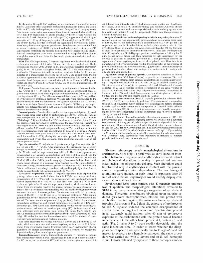
specific for spectrin. Substrate-gel electrophoresis (Fig. 7) wasperformed with spectrin (Fig. 7a) and the generic substrategelatin (Fig. 7b) in order to identify and evaluate the specificityof the protozoan proteinases able to degrade spectrin. Thisanalysis revealed that several proteinases were present whichwere able to degrade spectrin, both in lysates and secretions.The two patterns displayed a difference between cytoplasmicand secreted proteinases: a 30-kDa band was present in lysates
which was never detected in the secreted protein samples (Fig.7). Furthermore, the 30-kDa proteinase seems to be specificfor spectrin, since it was not detectable when gelatin was usedas a substrate. A strong inhibition was observed upon pretreat-ment with TLCK, suggesting a thiol proteinase nature (10, 39).
DISCUSSION
The cytoskeleton is a target for many intracellular microor-ganisms. Some bacterial pathogens, such as Yersinia, are ableto target the cytoskeleton without lysing or entering the cell, bymeans of secreted or directly injected proteins able to interactwith the cellular cytoskeleton (42). This property is not exclu-sive to bacteria: the parasite Trypanosoma cruzi, for example,accomplishes cell invasion by triggering a rearrangement in themembrane skeleton, thus subverting cellular mechanisms andeventually leading to a combination of events which enable theparasite to enter the host cell (41). Moreover, the cytoskeletonplays a significant role in the replication of animal viruses,which interact with cytoskeletal elements inside infected cellsat different stages of replication, often by means of virus-encoded proteinases, as in the case of Moloney murine leuke-mia virus or human immunodeficiency virus (32).
This report presents evidence showing that the host cellcytoskeleton is a target for the protozoan parasite T. vaginalisas well. It is noteworthy, however, that the molecular targetwithin this structure has been identified as a component, spec-trin, of the submembranal cytoskeleton. The most commontargets for microbial pathogens which interact with the hostcell cytoskeleton are in fact components of the cytoplasmicnetwork, mostly actin, although the ability to target the hostcell spectrin has been reported for the intracellular protozoaPlasmodium falciparum and Plasmodium bergei (12).
The degradation of spectrin within the target cell is achievedonly upon exposure to live T. vaginalis parasites. Under theseconditions, spectrin loss from the target cell is extremely fastand effective: in fact, the protein becomes undetectable within60 min. Upon contact, spectrin degradation is achieved withoutlysis of the target cell as a prerequisite, as seen from immuno-fluorescence assay and immunoblotting performed on mem-branes of erythrocytes allowed to interact with the parasite andseparated before lysis. Exposure of the target cell to protozoansecretions and lysates does not induce loss of spectrin withinintact erythrocytes. Moreover, the ability of protozoan lysatesto degrade purified spectrin has been observed to be far moreeffective than the degradation accomplished by secreted pro-teins.
The experiments performed in order to investigate the ef-fectors involved in spectrin disruption led to the identificationof an intracellular (or membrane-associated) 30-kDa protein-ase able to degrade spectrin but not gelatin, suggesting a sub-strate specificity. The involvement of this protein in membraneskeleton disruption can be inferred. That the effectors respon-sible for the disappearance of spectrin could be only one or afew proteinases is in fact suggested by the degradation modeobserved within unlysed erythrocytes, as shown in Fig. 5a. Themolecular structure of spectrin in fact consists of repeateddomains, and the ladder degradation into discrete subunitsobtained within intact erythrocytes is likely the result of onlyone or a few cleavage sites. These findings, together with theobserved high velocity of spectrin degradation and the abilityof the enzyme to degrade spectrin but not a generic substratesuch as gelatin, suggest a narrow specificity for this proteinase.
Several cysteine proteinases have been described in T. vagi-nalis that degrade different substrates (36, 38–40); using gelatinas a substrate, Garber and Lemchuk-Favel described 30-kDa
FIG. 6. Comparison of degradation modes and kinetics of spectrin (a) andprotein 4.1 (b) in unlysed erythrocytes exposed to T. vaginalis and collected at thefollowing times of incubation: lanes 1, 20 min; lanes 2, 40 min; and lanes 3, 1 h.Lanes K, control obtained by hypotonic lysis of erythrocytes in the presence ofproteinase inhibitors. In panel a, lanes 1, 2, and 3, the sample was loaded in a10-times-higher concentration in order to achieve a more detailed pattern ofspectrin degradation fragments.
FIG. 7. Evaluation of proteinase activity by substrate SDS-PAGE with spec-trin (a) and gelatin (b). Lanes: T, parasite lysate obtained in the presence of 1mM TLCK; L, parasite lysate; S, secreted proteins. The arrow in panel a pointsto a 30-kDa proteinase present only in parasite lysates which was able to degradespectrin but not gelatin. The proteinase pattern represented in the figure corre-sponds to that of isolate SS-22. Molecular weights are shown on the right andleft.
5146 FIORI ET AL. INFECT. IMMUN.
on July 26, 2018 by guesthttp://iai.asm
.org/D
ownloaded from

extracellular enzymes (19) that are produced by only a limitednumber of trichomonad isolates (20). In order to rule out anymisinterpretation of the results, for characterization experi-ments we used the SS-22 T. vaginalis isolate, since it lacks thesegelatin-degrading extracellular proteinases. We can thereforeexclude an identity between the spectrin-degrading proteaseand the secreted 30-kDa proteases described by Garber andLemchuk-Favel; nevertheless, we cannot rule out the possibil-ity that the spectrin proteinase is one of the protozoan enzymesalready described by others, using different substrates (39).
Since the 30-kDa spectrin proteinase appears to be nonse-creted, the proteolytic activity might be performed internally tothe target cell following a release from the parasite, which maytake place during the intimate contact that occurs betweentrichomonads and target cells. An intimate contact has beendemonstrated to occur between membranes, with multiplecontact focal points between parasite and host cell and numer-ous cytoplasmic projections interdigitating with the microvilliof the host cell plasma membrane (5, 22, 37). We observed bytransmission electron microscopy that the same intimate con-tact occurs between T. vaginalis and erythrocyte membranes aswell (unpublished data). Such a situation suggests that theeffector delivery might take place through a membrane fusionevent or by release through exocytotic microvesicles. It is alsopossible that a focal secretion of the spectrin protease couldtake place in the microenvironment between target and effec-tor membranes, followed by the translocation of the moleculeinto target cells, as recently described for example for YopE ofYersinia (44) or for diphtheria (34) and ricin toxin (43).
Disruption of spectrin has fundamental implications for thehost cell, since it arranges into a submembrane network largelyresponsible for maintenance of erythrocyte shape, membranestructural integrity, and reversible deformability (23). Spectrinaccounts for 75% of the membrane skeleton protein mass inerythrocytes, and spectrin analogs are widely distributedamong the majority of cell types (6).
We reported recently a T. vaginalis lytic mechanism medi-ated by pore-forming proteins, highly effective on erythrocytes(14, 17). Nucleate cells, though, are more resistant to osmoticlysis by perforins, and therefore an auxiliary mechanism, suchas membrane skeleton disruption, might be needed to accom-plish lysis. Contact-dependent cytotoxicity of T. vaginalis is welldocumented in the literature; on epithelial cell monolayers theparasite exhibits a direct cytotoxicity (3, 4, 21, 26) by inducingalterations in morphology as well as cell rounding, detachment,and cell death. The precise interrelationships between the per-forin-mediated cytolytic mechanism and the proteolytic dem-olition of the submembrane network remain to be elucidated.The two mechanisms, though, need not be mutually exclusive;they may apply to different types of target cells as well as to thesame target cells in a cooperative mechanism directed towardsthe accomplishment of lysis of nucleate cells, which are 6 to100 times more resistant to osmotic lysis by perforins than areerythrocytes.
Spectrin seems to be involved in the association of interme-diate filaments with the plasma membrane of epithelial cellsand cultured fibroblasts (33). The proteolytic demolition ofspectrin within these cells might induce a disaggregation of themembrane skeleton and of its connections with the cytoplasmicactin network, thus leading to an enhanced sensitivity of thenucleate cell to osmotic lysis mediated by the trichomonadperforins. Examples of perforin- or proteinase-mediated cyto-toxicity are numerous, and such association has been observedto occur in other protozoan parasites, such as Entamoeba his-tolytica (30). In cytotoxic T lymphocytes both effectors arecompartmentalized in microvesicles and released into the ex-
tracellular space formed between lymphocyte and target cell(7).
The study of microbial pathogenetic effectors is acquiringincreasing importance as a tool for dissecting the molecularmechanisms of the host. An increasing number of recent find-ings on the molecular interactions between pathogens andtheir targets have provided useful insights into issues in cellbiology (11). The understanding of actin behavior within themembrane skeleton, for example, has been greatly increased bythe study of its interactions with several intracellular patho-gens—Salmonella and Shigella, for example (1, 8, 11, 18). TheT. vaginalis proteinase, being able to target spectrin in a spe-cific fashion, could thus be suggested as an interesting tool forunderstanding the role spectrin plays in other cell types, suchas within epithelial cells, in polarized membrane domains, andin lymphocyte capping, as well as its involvement in synaptictransmission among neuronal cells.
ACKNOWLEDGMENTS
We thank Gianni Monaco for the SEM micrographs. The excellenttechnical assistance of Giuseppe Delogu and Giuseppina Casu isgreatly appreciated.
This work was supported by grants from MURST and CNR(97.04051.CT04).
REFERENCES
1. Adam, T., M. Arpin, M. C. Prevost, P. Gounon, and P. Sansonetti. 1995.Cytoskeletal rearrangments and the functional role of T-plastin during entryof Shigella flexneri into HeLa cells. J. Cell Biol. 129:367–381.
2. Addis, M. F., P. Rappelli, P. Cappuccinelli, and P. L. Fiori. 1997. Extracel-lular release by Trichomonas vaginalis of a NADP1 dependent malic enzymeinvolved in pathogenicity. Microb. Pathog. 23:55–61.
3. Alderete, J. F., M. V. Lehker, and R. Arroyo. 1995. The mechanisms andmolecules involved in cytoadherence and pathogenesis of Trichomonas vagi-nalis. Parasitol. Today 11:70–74.
4. Alderete, J. F., and E. Pearlman. 1984. Pathogenic Trichomonas vaginaliscytotoxicity to cell culture monolayers. Br. J. Vener. Dis. 60:99–105.
5. Arroyo, R., A. Gonzalez-Robles, A. Martınez-Palomo, and J. F. Alderete.1993. Signalling of Trichomonas vaginalis for amoeboid transformation andadhesin synthesis follows cytoadherence. Mol. Microbiol. 7:299–309.
6. Bennett, V., and D. Gilligan. 1993. The spectrin-based membrane skeletonand micron-scale organisation of the plasma membrane. Annu. Rev. CellBiol. 9:27–66.
7. Berke, G. 1991. Lymphocyte-triggered internal target disintegration. Immu-nol. Today 12:396–399.
8. Bliska, J. B., J. E. Galan, and S. Falkow. 1993. Signal transduction in themammalian cell during bacterial attachment and entry. Cell 73:903–920.
9. Bradford, M. M. 1976. A rapid and sensitive method for the quantitation ofmicrogram quantities of protein utilizing the principle of protein-dye bind-ing. Anal. Biochem. 72:248–254.
10. Coombs, G. H., and M. J. North. 1983. An analysis of the proteinases ofTrichomonas vaginalis by polyacrylamide gel electrophoresis. Parasitology86:1–6.
11. Cossart, P., P. Boquet, S. Normark, and R. Rappuoli. 1996. Cellular micro-biology emerging. Science 271:315–316.
12. Deguercy, A., M. Hommel, and J. Schrevel. 1990. Purification and charac-terization of 37-kilodalton proteases from Plasmodium falciparum and Plas-modium bergei which cleave erythrocyte cytoskeletal components. Mol. Bio-chem. Parasitol. 38:233–244.
13. Diamond, L. S. 1957. The establishment of various trichomonads of animalsand man in axenic cultures. J. Parasitol. 43:488–490.
14. Fiori, P. L., P. Rappelli, M. F. Addis, A. Sechi, and P. Cappuccinelli. 1996.Trichomonas vaginalis haemolysis: pH regulates a contact-independentmechanism based on pore-forming proteins. Microb. Pathog. 20:109–118.
15. Fiori, P. L., P. Rappelli, C. Manca, A. Mattana, and P. Cappuccinelli. 1992.Phenotypic variation of surface antigenic determinants in Trichomonas vagi-nalis detected by monoclonal antibodies. Microbiologica 15:227–236.
16. Fiori, P. L., G. Monaco, S. Scappaticci, A. Pugliese, N. Canu, and P. Cap-puccinelli. 1988. Establishment of cell cultures from hydatid cysts of Echi-nococcus granulosus. Int. J. Parasitol. 18:297–305.
17. Fiori, P. L., P. Rappelli, A. M. Rocchigiani, and P. Cappuccinelli. 1993.Trichomonas vaginalis haemolysis: evidence of functional pore formation onred cell membranes. FEMS Microbiol. Lett. 109:13–18.
18. Francis, C. L., T. A. Ryan, B. D. Jones, S. J. Smith, and S. Falkow. 1993.Ruffles induced by Salmonella and other stimuli direct macropynocytosis ofbacteria. Nature 364:639–642.
VOL. 65, 1997 CYTOSKELETAL TARGETING BY T. VAGINALIS 5147
on July 26, 2018 by guesthttp://iai.asm
.org/D
ownloaded from

19. Garber, G. E., and L. T. Lemchuk-Favel. 1989. Characterization and purifi-cation of extracellular proteases of Trichomonas vaginalis. Can. J. Microbiol.35:903–909.
20. Garber, G. E., and L. T. Lemchuk-Favel. 1994. Analysis of extracellularproteases of Trichomonas vaginalis. Parasitol. Res. 80:361–365.
21. Garber, G. E., L. T. Lemchuk-Favel, and W. R. Bowie. 1989. Isolation of acell-detaching factor of Trichomonas vaginalis. J. Clin. Microbiol. 27:1548–1553.
22. Gonzales-Robles, A., A. Lazaro-Haller, M. Espinosa-Castellano, F. Anaya-Velasquez, and A. Martinez-Palomo. 1995. Trichomonas vaginalis: ultrastruc-tural bases of the cytopathic effect. J. Eukaryot. Microbiol. 42:641–651.
23. Goodman, S. R., and K. A. Shiffer. 1983. The spectrin membrane skeleton ofnormal and abnormal human erythrocytes: a review. Am. J. Physiol. 244:C121–C141.
24. Gupta, P. K., and J. K. Frost. 1989. Cytopathology and histopathology offemale genital tract in Trichomonas vaginalis infection, p. 274–290. In B. M.Honigberg (ed.), Trichomonads parasitic in humans. Springer-Verlag, NewYork, N.Y.
25. Heussen, C., and E. B. Dowdle. 1980. Electrophoretic analysis of plasmino-gen activators in polyacrylamide gels containing sodium dodecyl sulfate andcopolymerized substrates. Anal. Biochem. 102:196–202.
26. Krieger, J. N., J. I. Ravdin, and M. F. Rein. 1985. Contact-dependent cyto-pathogenic mechanism of Trichomonas vaginalis. Infect. Immun. 50:778–786.
27. Krieger, J. N. 1981. Urologic aspects of trichomoniasis. Investig. Urol. 18:411–417.
28. Laemmli, U. K. 1970. Cleavage of structural proteins during the assembly ofthe head of bacteriophage T4. Nature 227:680–685.
29. Lehker, M. W., T. H. Chang, D. C. Dailey, and J. F. Alderete. 1990. Specificerythrocyte binding is an additional nutrient acquisition system forTrichomonas vaginalis. J. Exp. Med. 171:2165–2170.
30. Leippe, M. 1997. Amoebapores. Parasitol. Today 13:178–183.31. Lockwood, B. C., M. J. North, K. I. Scott, A. F. Bremmer, and G. H. Coombs.
1987. The use of a highly sensitive electrophoretic method to compare theproteinases of trichomonads. Mol. Biochem. Parasitol. 24:89–95.
32. Luftig, R. B., and L. D. Lupo. 1994. Viral interactions with the host-cellcytoskeleton: the role of retroviral proteinases. Trends Microbiol. 2:178–182.
33. Mangeat, P. H., and K. Burridge. 1984. Immunoprecipitation of noneryth-rocyte spectrin within live cells following microinjection of specific antibod-ies: relation to cytoskeletal structures. J. Cell Biol. 98:1363–1377.
34. Montecucco, C., and E. Papini. 1995. Cell penetration of bacterial proteintoxins. Trends Microbiol. 3:165–168.
35. Muller, M. 1989. Biochemistry of Trichomonas vaginalis, p. 53–83. In B. M.Honigberg (ed.), Trichomonads parasitic in humans. Springer-Verlag, NewYork, N.Y.
36. Neale, K. A., and J. F. Alderete. 1990. Analysis of the proteinases of repre-sentative Trichomonas vaginalis isolates. Infect. Immun. 58:157–162.
37. Nielsen, M. H., and R. Nielsen. 1975. Electron microscopy of Trichomonasvaginalis Donne: interaction with vaginal epithelium in human trichomoni-asis. Acta Pathol. Microbiol. Scand. 83:305–320.
38. North, M. J., J. C. Mottram, and G. H. Coombs. 1990. Cysteine proteinaseof parasitic protozoa. Parasitol. Today 6:270–275.
39. North, M. J., C. D. Robertson, and G. H. Coombs. 1990. The specificity oftrichomonad cysteine proteinases analysed using fluorogenic substrates andspecific inhibitors. Mol. Biochem. Parasitol. 39:183–194.
40. Provenzano, D., and J. F. Alderete. 1995. Analysis of human immunoglob-ulin-degrading cysteine proteinases of Trichomonas vaginalis. Infect. Immun.63:3388–3395.
41. Rodriguez, A., M. Rioult, A. Ora, and N. A. Andrews. 1995. Trypanosomesoluble factor induces IP3 formation, intracellular Ca21 mobilization andmicrofilament rearrangement in host cells. J. Cell Biol. 129:1263–1273.
42. Rosqvist, R., A. Forsberg, and H. Wolf-Watz. 1991. Intracellular targeting ofthe Yersinia YopE cytotoxin in mammalian cells induces actin microfilamentdisruption. Infect. Immun. 59:4562–4569.
43. Sandvig, K., and B. van Deurs. 1996. Endocytosis, intracellular transport,and cytotoxic action of Shiga toxin and ricin. Physiol. Rev. 76:949–966.
44. Straley, S. C., E. Skrzypek, G. V. Plano, and J. B. Bliska. 1993. Yops ofYersinia spp. pathogenic for humans. Infect. Immun. 61:3105–3110.
Editor: P. J. Sansonetti
5148 FIORI ET AL. INFECT. IMMUN.
on July 26, 2018 by guesthttp://iai.asm
.org/D
ownloaded from