Embed Size (px)
Citation preview
Contributing Authors:
Miriam Robbins, DDS, MPHMaureen Romer, DDS, MPASteven Krauss, DDS,MPHEvan Spivack, DDSNancy Dougherty, DMD, MPHRobert Marion, MDKoshi Cherian, MD
Editor:
Funded by the NYS Developmental Disabilities Planning Council
Special Care Dentistry For the General Practice Resident
Practical Training Modules
This educational modular series consists of eight evidence basedPower Point presentations designed to give the general practice resident a global view of dental treatment for people with special needs. Approximately 300 references are listed throughout this work. The eight modules address the most important aspects of clinicalmedicine and dentistry required for treating a patient with special needs. Discussion of access and barriers to dental care, the need for special care dentistry in the pre and post doctoral dental curricula, along with assessment of the competency of participants are included in the modules. Upon completion of the modules, the participantshould have the knowledge to assess a patient with special needs.
The educational package is a previously piloted pre and post test exam. The modules are accompanied by “teacher’s notes” which are visible in each Power Point presentation. This format alternately allows the instructor to assign the series as a self-study project.
continued
Special Care Dentistry For the General Practice Resident
Practical Training Modules
A description of each module follows below:Introduction to Special Patient Care: discusses the definition of disability, the prevalence and incidence of disability, aspects of “normalization”, and the barriers to care. A list of resources is provided for the individual and family.Special Care Dentistry/Legal and Ethical Issues: discusses informed consent and various other types of consent, comprehensive medical history documentation, appropriate use of desensitization and restraint, communication/human rights issues, case law and detailed literature review of restraint.Treatment Modalities/Treatment Planning for Patients with Special Needs: discusses reasons for sedation, hospitalization OR cases, general anesthesia, pharmacological techniques, IV and enteral drugs.Learning Disabilities/Mental Retardation and Down Syndrome: discusses the causes and risk factors, diagnosis and intervention, physical findings and medical concerns, dental and craniofacial characteristics of people with learning disabilities, mental retardation and Down syndrome. Neuromuscular Disorders/Cerebral Palsy and Muscular Dystrophy: discusses types of cerebral palsy, risk factors, oral and dental findings, various forms of muscular dystrophy and treatment planning considerations.Autistic Spectrum Disorders: defines and describes the spectrum of autistic disorders including Pervasive Developmental Disorder and Asperger’s. A recent review of the literature regarding proposed etiologies (i.e.: genetic links, vaccines) is presented, as well as suggestions for behavior management and treatment strategies.Oral Manifestations/Genetic and Congenital Disorders: discusses syndromology definitions, gene and chromosomal abnormalities, craniofacial disorders, dental and orthopedic conditions.Seizure Disorders: discusses definitions of seizures and epilepsy, risk, incidence and prevalence of seizures, classification and treatment of seizures, choice of medication therapies and practical considerations for dental treatment. Pre and post tests and the answer sheets are not included in the module series. Please contact Annette Shafer in the Office of Investigations and Internal Affairs at [email protected] to request a copy and we will forward it to you electronically.
Oral Manifestations of Genetic & Congenital
DisordersDr. Robert W. Marion
Director, Children’s Evaluation and Rehabilitation Center, Rose F. Kennedy Center,
Professor, Departments of Pediatrics and Obstetrics and Gynecology, Albert Einstein College of Medicine,
Chief, Divisions of Genetics and Developmental Medicine, Children’s Hospital at Montefiore,
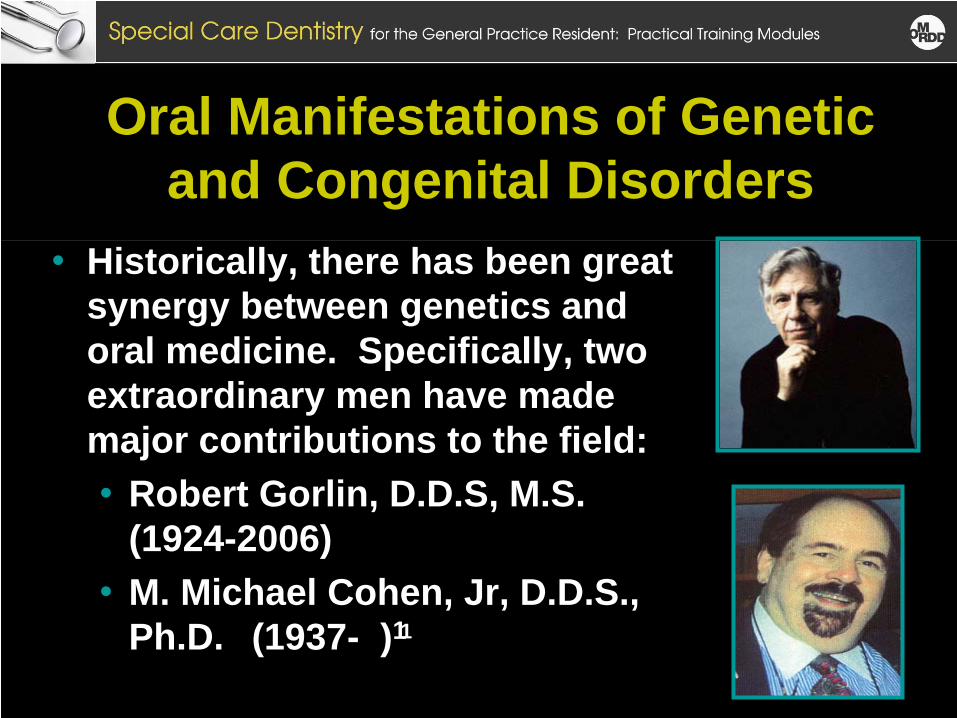
Oral Manifestations of Genetic and Congenital Disorders
• Historically, there has been great synergy between genetics and oral medicine. Specifically, two extraordinary men have made major contributions to the field:• Robert Gorlin, D.D.S, M.S.
(1924-2006)• M. Michael Cohen, Jr, D.D.S.,
Ph.D. (1937- )11
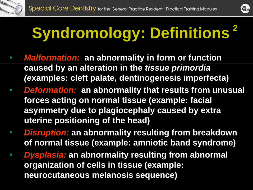
Syndromology: Definitions• Malformation: an abnormality in form or function
caused by an alteration in the tissue primordia (examples: cleft palate, dentinogenesis imperfecta)
• Deformation: an abnormality that results from unusual forces acting on normal tissue (example: facial asymmetry due to plagiocephaly caused by extra uterine positioning of the head)
• Disruption: an abnormality resulting from breakdown of normal tissue (example: amniotic band syndrome)
• Dysplasia: an abnormality resulting from abnormal organization of cells in tissue (example: neurocutaneous melanosis sequence)
2
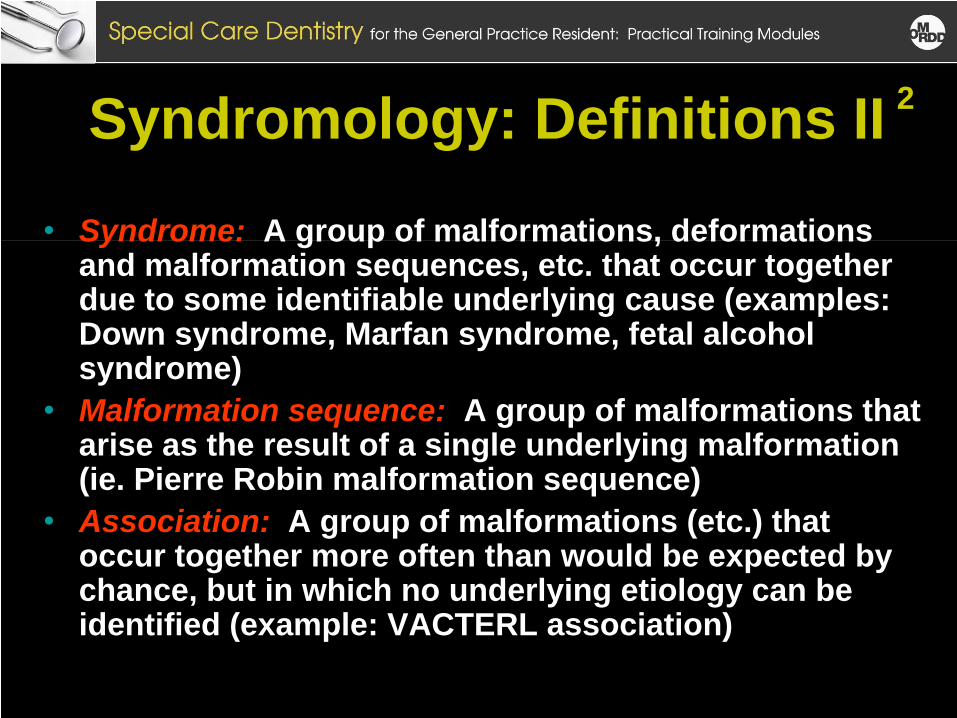
Syndromology: Definitions II
• Syndrome: A group of malformations, deformations and malformation sequences, etc. that occur together due to some identifiable underlying cause (examples: Down syndrome, Marfan syndrome, fetal alcohol syndrome)
• Malformation sequence: A group of malformations that arise as the result of a single underlying malformation (ie. Pierre Robin malformation sequence)
• Association: A group of malformations (etc.) that occur together more often than would be expected by chance, but in which no underlying etiology can be identified (example: VACTERL association)
2
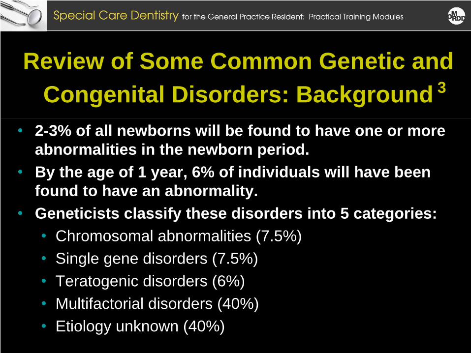
Review of Some Common Genetic and Congenital Disorders: Background
• 2-3% of all newborns will be found to have one or more abnormalities in the newborn period.
• By the age of 1 year, 6% of individuals will have been found to have an abnormality.
• Geneticists classify these disorders into 5 categories:• Chromosomal abnormalities (7.5%)• Single gene disorders (7.5%)• Teratogenic disorders (6%)• Multifactorial disorders (40%)• Etiology unknown (40%)
3
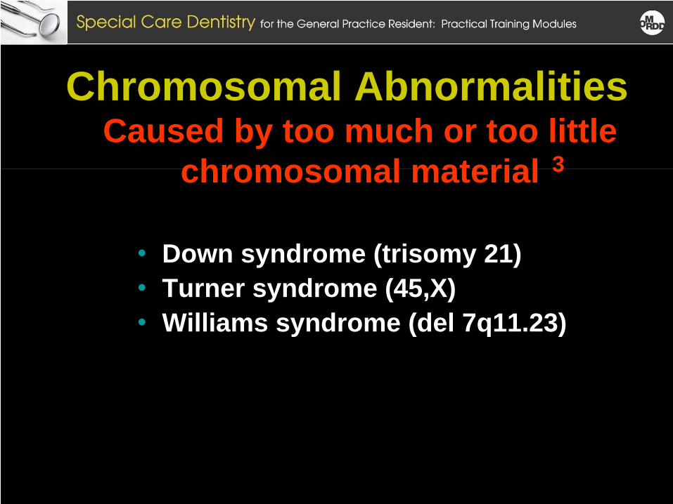
Chromosomal Abnormalities Caused by too much or too little
chromosomal material
• Down syndrome (trisomy 21)• Turner syndrome (45,X)• Williams syndrome (del 7q11.23)
3
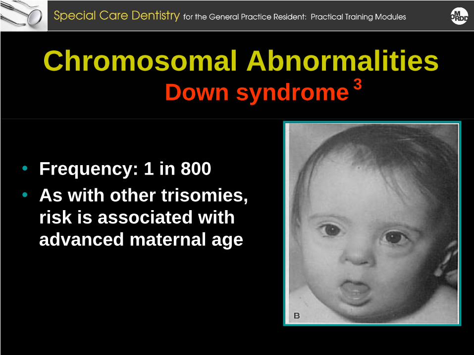
Chromosomal Abnormalities Down syndrome
• Frequency: 1 in 800 • As with other trisomies,
risk is associated with advanced maternal age
3
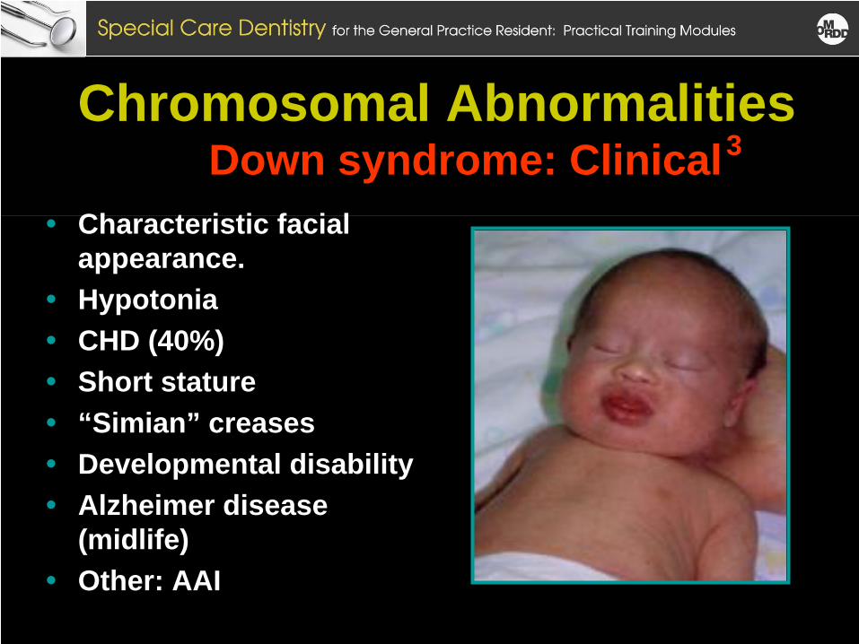
Chromosomal Abnormalities Down syndrome: Clinical
• Characteristic facial appearance.
• Hypotonia• CHD (40%) • Short stature• “Simian” creases • Developmental disability• Alzheimer disease
(midlife)• Other: AAI
3
Chromosomal Abnormalities Down syndrome: Oral & Dental
• Delays and alterations in the sequence of tooth eruption
• Malocclusion (mandibular overjet, posterior cross bite)
• High arched palate• Relative macroglossia • Missing teeth • Fissuring of the tongue, enlargement of
vallate papillae
4
Chromosomal Abnormalities Turner syndrome (45,X)
• Frequency: 1 in 5000• In 1st trimester abortuses: 1 in 11 • Risk not related to advanced maternal age• 1/3 dx’d in newborn, 1/3 in childhood, 1/3 in
adolescence• 50% are due to 45,X karyotype; the others
due to some variation (mosaicism, isochromosome Xq or Xp, etc.)
3
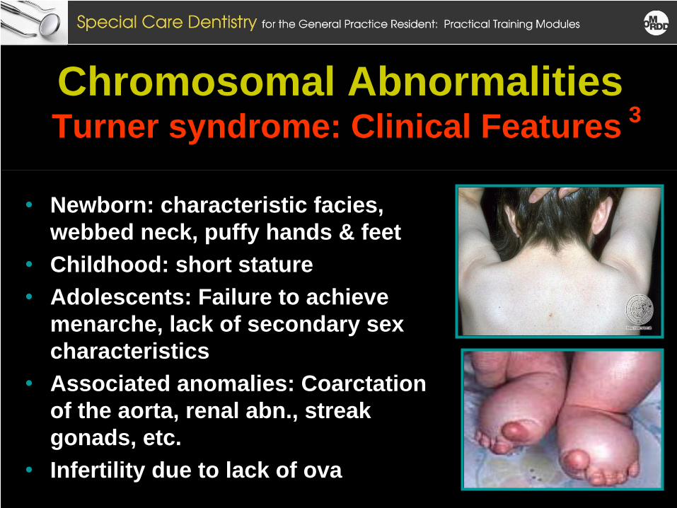
Chromosomal Abnormalities Turner syndrome: Clinical Features
• Newborn: characteristic facies, webbed neck, puffy hands & feet
• Childhood: short stature• Adolescents: Failure to achieve
menarche, lack of secondary sex characteristics
• Associated anomalies: Coarctation of the aorta, renal abn., streak gonads, etc.
• Infertility due to lack of ova
3
Chromosomal Abnormalities Turner syndrome: Oral & Dental• Premature eruption of the teeth• High arched palate• Increased molarization of premolars• Cusp and crown size are reduced• Prior to treatment, may need
prophylactic antibiotics (due to associated cardiac disease)
5
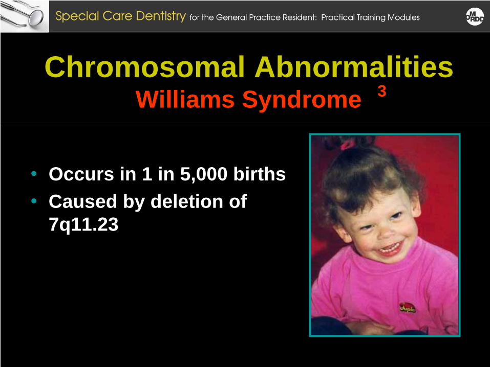
Chromosomal Abnormalities Williams Syndrome
• Occurs in 1 in 5,000 births• Caused by deletion of
7q11.23
3
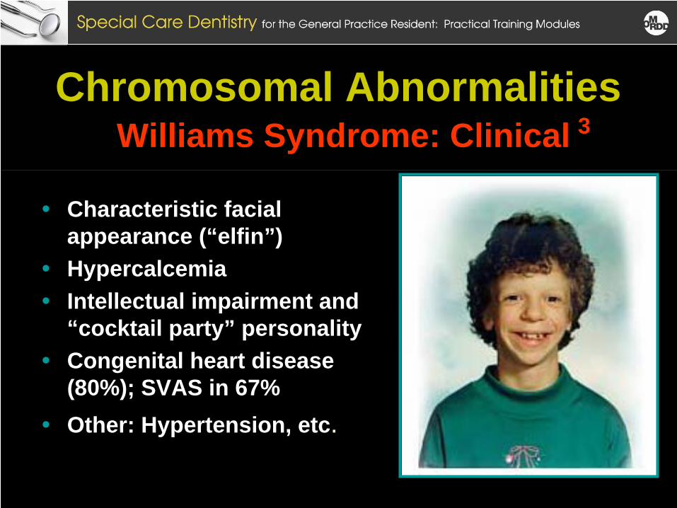
Chromosomal Abnormalities Williams Syndrome: Clinical
• Characteristic facial appearance (“elfin”)
• Hypercalcemia• Intellectual impairment and
“cocktail party” personality• Congenital heart disease
(80%); SVAS in 67%• Other: Hypertension, etc.
3
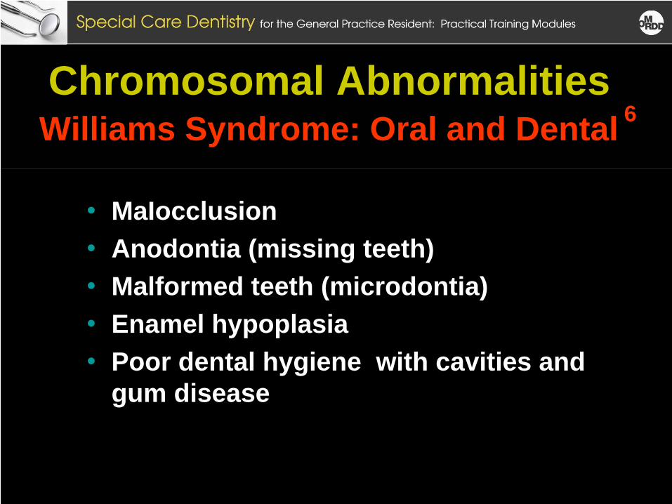
Chromosomal Abnormalities Williams Syndrome: Oral and Dental
• MaIocclusion• Anodontia (missing teeth) • Malformed teeth (microdontia)• Enamel hypoplasia• Poor dental hygiene with cavities and
gum disease
6
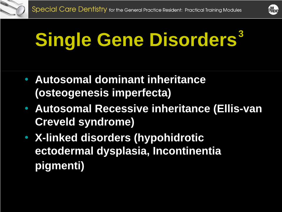
Single Gene Disorders
• Autosomal dominant inheritance (osteogenesis imperfecta)
• Autosomal Recessive inheritance (Ellis-van Creveld syndrome)
• X-linked disorders (hypohidrotic ectodermal dysplasia, Incontinentia pigmenti)
3
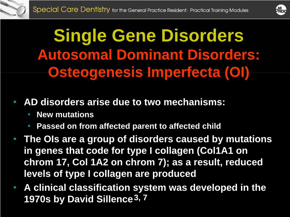
Single Gene Disorders Autosomal Dominant Disorders:
Osteogenesis Imperfecta (OI)
• AD disorders arise due to two mechanisms:• New mutations• Passed on from affected parent to affected child
• The OIs are a group of disorders caused by mutations in genes that code for type I collagen (Col1A1 on chrom 17, Col 1A2 on chrom 7); as a result, reduced levels of type I collagen are produced
• A clinical classification system was developed in the 1970s by David Sillence3, 7
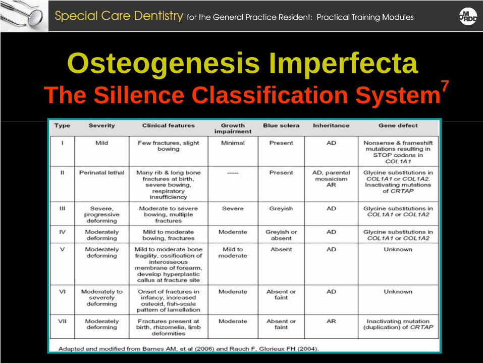
Osteogenesis Imperfecta The Sillence Classification System7
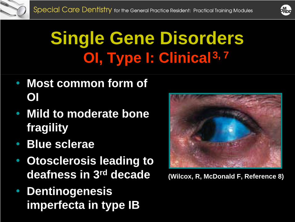
Single Gene Disorders OI, Type I: Clinical
• Most common form of OI
• Mild to moderate bone fragility
• Blue sclerae• Otosclerosis leading to
deafness in 3rd decade • Dentinogenesis
imperfecta in type IB
(Wilcox, R, McDonald F, Reference 8)
3, 7
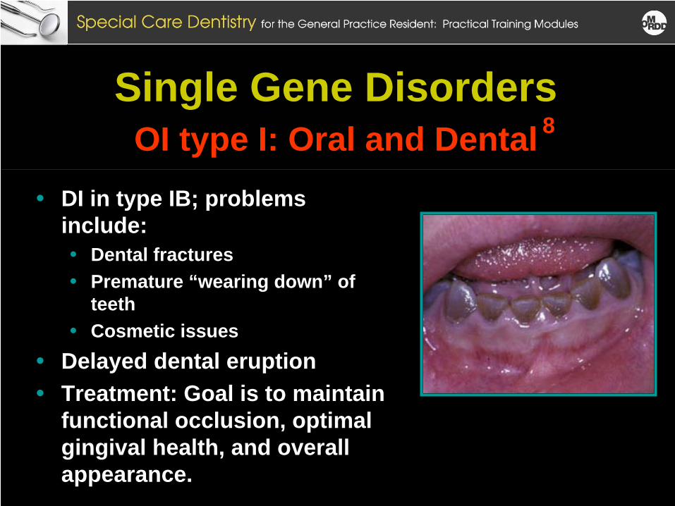
Single Gene Disorders OI type I: Oral and Dental
• DI in type IB; problems include:• Dental fractures• Premature “wearing down” of
teeth• Cosmetic issues
• Delayed dental eruption• Treatment: Goal is to maintain
functional occlusion, optimal gingival health, and overall appearance.
8
Single Gene Disorders Autosomal Recessive Disorders:
Ellis-van Creveld Syndrome (EVCS)
• AR Disorders occur when an offspring inherits two copies of a non-working gene from parents
• EVCS is a rare AR disorder (prevalence=1 in 60,000)• Much more common in the old order Amish population
(“founder effect”)• Caused by mutations in the EVC and EVC2 genes
(function unknown)
3, 9,10
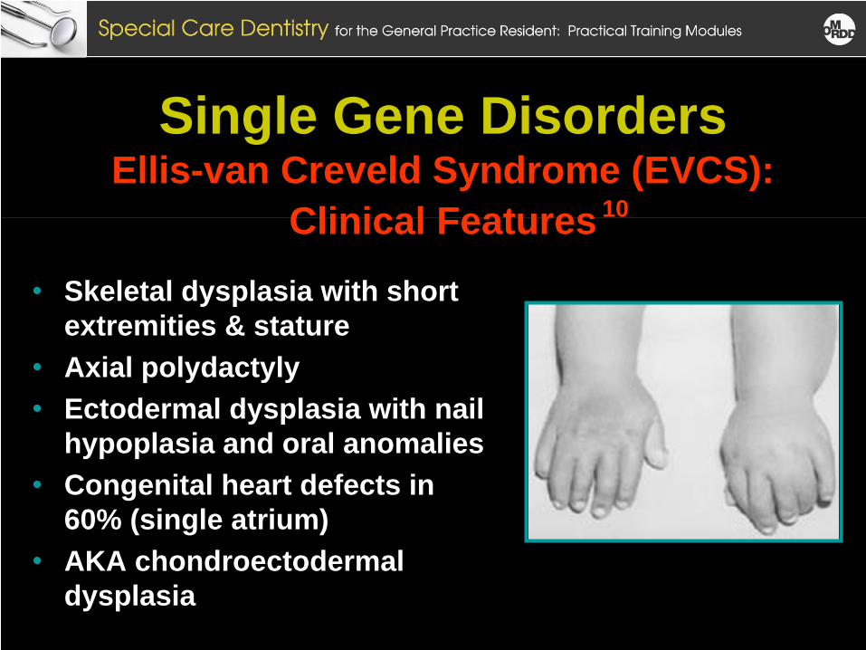
Single Gene Disorders Ellis-van Creveld Syndrome (EVCS):
Clinical Features
• Skeletal dysplasia with short extremities & stature
• Axial polydactyly• Ectodermal dysplasia with nail
hypoplasia and oral anomalies• Congenital heart defects in
60% (single atrium) • AKA chondroectodermal
dysplasia
10
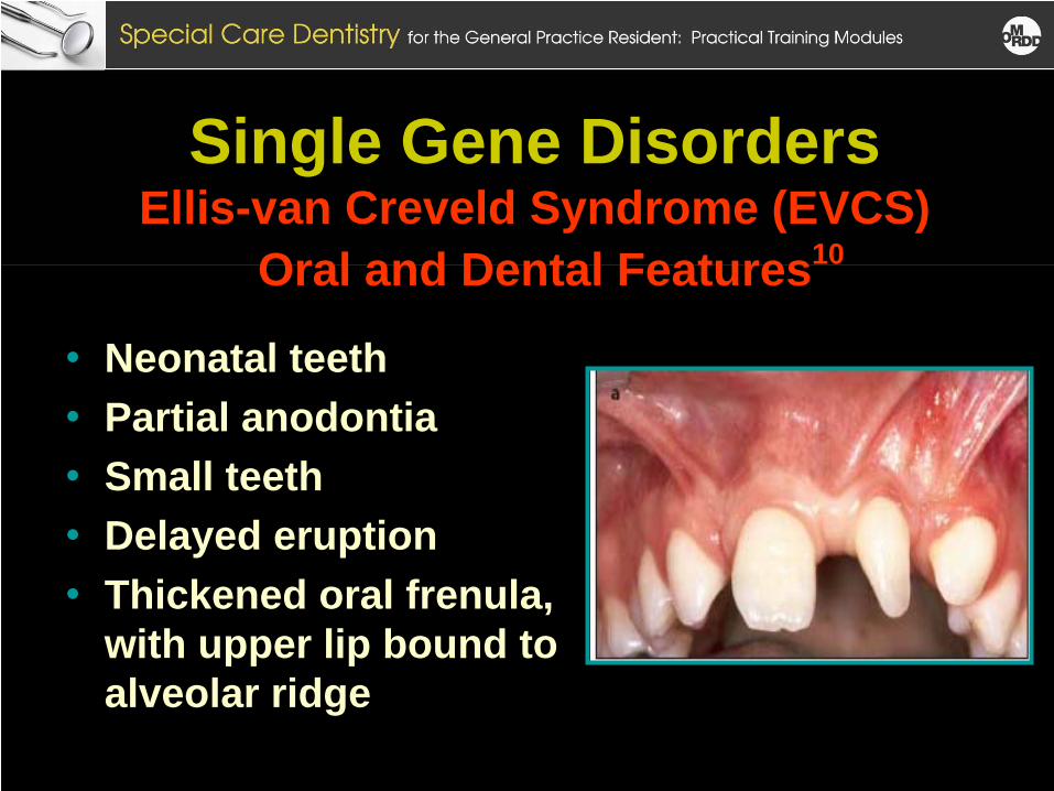
Single Gene Disorders Ellis-van Creveld Syndrome (EVCS)
Oral and Dental Features
• Neonatal teeth• Partial anodontia• Small teeth• Delayed eruption• Thickened oral frenula,
with upper lip bound to alveolar ridge
10
Single Gene Disorders X-Linked Disorders
Hypohidrotic Ectodermal Dysplasia (HED)
• X-linked disorders are caused by mutations on the X chromosome
• HED is an X-linked recessive disorder, passed from carrier mothers to affected sons
• Occurs in 1 in 10,000 newborns (all boys)• Caused by a mutation in the EDA gene (gene
product = Ectodysplasin-A) 3
3
Single Gene Disorders HED: Clinical
• Hypotrichosis • Hypohidrosis• Hypodontia• Wrinkled, hyperpigmental periorbital
skin
13
Single Gene Disorders HED: Oral and Dental I
• May develop only 5 to 7 teeth (canines and 1st
molar) • Teeth are small with conical crowns. • Paucity of saliva (thick)• Carrier females may have minor dental anomalies • Dental treatment must begin at an early age. • Bonding of conical shaped teeth in young
individuals improves esthetics and chewing ability. • Orthodontics may be necessary.
13
Single Gene Disorders HED: Oral and Dental II
• Dental implants in the anterior portion of the mandible are only successful in children >7 y.o.
• Prostheses may need to be replaced every 2.5 yrs. • Because of problems with chewing and swallowing,
dietary counseling may be helpful
13
Teratogenic Disorders
• Teratogens: Substances that have the potential to cause congenital malformation when they come into contact with a developing embryo or fetus.
• Many factors determine what effect a teratogen will have on the developing conceptus. These factors include: • timing of the exposure • length of time of the exposure• species variability• genetic predisposition 14,3
Teratogenic Disorders
• Basically, there are four categories of teratogenic substances:• Infectious agents (TORCH) • Prescription medications (i.e., anticonvulsant
medications)• Non-prescription drugs (including alcohol)• Environmental agents (methyl mercury)
3
Teratogenic Disorders Fetal Alcohol Spectrum Disorder (FASD)
• First described in the US literature by Smith et al., as “fetal alcohol syndrome” in 1973
• However, it is a “spectrum disorder”:• “Full-blown” FAS (typical phenotype, congenital
malformations, developmental anomalies) results from exposure to alcohol all through pregnancy.
• Alcohol exposure during part of the pregnancy may lead to a milder phenotype
• Occurring in app. 1% of the population, FASD is the most common teratogenic disorder in humans
15, 16
17
Teratogenic Disorders FASD: Clinical Features
• Characteristic facial features (short PFs, flat philtrum, hypoplastic cheeks, minor ear anomalies, etc.)
• Skeletal anomalies (cervical spine abn, clinobrachy- dactyly of fifth fingers, abn. palmar crease pattern)
• Cardiac defects (VSD, etc.)• Intrauterine and extrauterine growth restriction• Neurodevelopmental disabilities (microcephaly, MR,
LDs, etc.)
3
Teratogenic Disorders FASD: Oral and Dental
• Mouth breathing (with corresponding dry mouth) • Higher incidence of caries and gingivitis • TMJ disorders, • Cleft lip/palate• Malocclusions• In treating these patients, concern re. congenital
heart disease and seizure disorder.
18
Multifactorial Inheritance (MFI) Cleft lip + palate (CL + P)
• MFI conditions are caused by an interplay of genetic and environmental factors
• In addition to common birth defects (spina bifida, CL + P), MFI conditions include all chronic disorders of childhood & adulthood (asthma, cancer, diabetes, etc.)
• Occurring in 1 in 600-700 livebirths, CL + P is one of the most common MFI birth defects: • Recurrence risk: 2 – 4 %
3
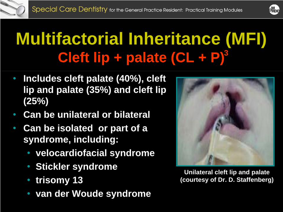
Multifactorial Inheritance (MFI) Cleft lip + palate (CL + P)
• Includes cleft palate (40%), cleft lip and palate (35%) and cleft lip (25%)
• Can be unilateral or bilateral• Can be isolated or part of a
syndrome, including: • velocardiofacial syndrome • Stickler syndrome• trisomy 13• van der Woude syndrome
Unilateral cleft lip and palate (courtesy of Dr. D. Staffenberg)
3
Multifactorial Inheritance (MFI) Cleft lip + palate (CL + P): Oral & Dental
• Pediatric dentist must be part of an integrated, multidisciplinary team in the management of children with CL + P
• Clefting can cause a variety of dental problems throughout the mouth, but mostly in the vicinity of the cleft
• Children need a cohesive long-term plan of preventive and restorative care
Common Craniofacial Disorders Dental and Orthopedic Considerations
Four categories:• Syndromes with craniosynostosis • Syndromes with clefting• Branchial arch disorders• Syndromes with unusual dental, gingival or
periodontal findings1
1
Syndromes with Craniosynostosis• Crouzon syndrome • Apert syndrome• Pfeiffer syndrome• Other
19
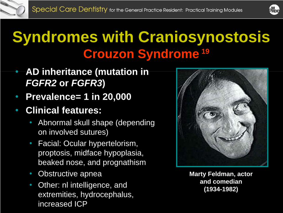
Syndromes with Craniosynostosis Crouzon Syndrome
• AD inheritance (mutation in FGFR2 or FGFR3)
• Prevalence= 1 in 20,000• Clinical features:
• Abnormal skull shape (depending on involved sutures)
• Facial: Ocular hypertelorism, proptosis, midface hypoplasia, beaked nose, and prognathism
• Obstructive apnea • Other: nl intelligence, and
extremities, hydrocephalus, increased ICP
Marty Feldman, actor and comedian
(1934-1982)
19
Syndromes with Craniosynostosis Crouzon Syndrome: Dental & Orthodontic
• Mandibular prognathism with midface hypoplasia• V-shaped maxillary arch• Overcrowding of upper teeth with malocclusions• Narrow, high palate (occasionally cleft)• Occasional oligodontia, macrodontia, peg-shaped,
and widely spaced teeth• The pediatric dentist & orthodontist should function
as part of a multidisciplinary team in planning care of patients with this & other craniosynostosis syndromes.
20
Syndromes with Orofacial Clefting
• Pierre Robin malformation sequence and associated syndromes
• Van der Woude syndrome• Many others
1
Syndromes with Orofacial Clefting: Pierre Robin Malformation Sequence
(PRMS) & Associated Syndromes
• PRMS is a malformation sequence, a series of anomalies caused by a single malformation
• Features of PRMS: micrognathia, glossoptosis, and U- shaped cleft palate
• Primary defect = micrognathia; cleft palate and glossoptosis are secondary to this
• PRMS is a neonatal medical emergency: obstructive apnea due to glossoptosis can be life threatening!
• PRMS can occur as an isolated finding, or can be part of a large number of malformation syndromes
3
Pierre Robin Malformation Sequence (PRMS): Associated Syndromes
• Stickler syndrome: PRMS, high myopia with vitreo- retinal degeneration, flat midface, mild epiphyseal dysplasia; autosomal dominant inheritance
• Velo-cardio-facial syndrome (aka diGeorge): PRMS, characteristic facial appearance, conotruncal cardiac defects, etc.; due to deletion 22q11.2.
• Treacher Collins syndrome (see below)• Many others
3
Pierre Robin Malformation Sequence (PRMS): Dental and Orthopedic Implications
• Micrognathia with retraction of the inferior dental arch (10-12 mm behind the superior arch)
• Mandibular growth usually “catches up” by one year of age
• Obstructive sleep apnea and feeding issues predominate in the neonatal period
• Dental malformations occur in 1/3 of cases• Because of the high association with syndromes, it is
essential that all children with PRS are seen by a geneticist before dental/orthopedic care begins
21
Branchial Arch Disorders
• Treacher Collins syndrome• Hemifacial microsomia (aka oculo-auriculo-
vertebral sequence, Goldenhar syndrome)• Others (Nager syndrome, Miller syndrome,
Townes Brock syndrome)
1
Branchial Arch Disorders: Treacher Collins syndrome (TCS)
• AD inheritance (mutation in TCOF1 gene)• Prevalence = 1 in 10,000 to 1 in 50,000• Clinical features (variable expression):
• Symmetric facial anomalies: micrognathia with extreme shortening of mandible; colobomata of lower eyelid; microtia, macrostomia
• Respiratory: Severe obstructive apnea (due to PRS, choanal atresia/stenosis)
• Usually normal intelligence
22
Branchial Arch Disorders: TCS: Dental & Orthodontic Implications
• Dental anomalies occur in 60%, with 1 to 8 per individual• tooth agenesis (33.3%), • enamel opacities (20%), • ectopic eruption of the maxillary first molars (13.3%)
• Less frequently observed features:• Nasal deformity • High-arched palate • Angle class II anterior open-bite malocclusion
23
Syndromes with Unusual Dental, Gingival or Periodontal Findings
• Zimmerman Laband syndrome and the gingival fibromatosis disorders
• Holoprosencephaly sequence (single central incisor)
1
Syndromes with Unusual Dental, Gingival or Periodontal Findings: Holoprosencephaly Sequence (HPE)
• Like Pierre Robin, HPE is a malformation sequence, a series of malformations caused by a single underlying malformation
• In HPE, the underlying malformation involves damage to the tissue primordia forming the middle of the brain and the middle of the face
• As a result, affected individuals have a spectrum of anomalies including midline brain (alobar, semilobar and lobar HPE) and facial (cyclopia, cephocephaly, midline clefts, single central incisor) anomalies
24
Syndromes with Unusual Dental, Gingival or Periodontal Findings: Holoprosencephaly Sequence (HPE)
• Like PRMS, HPE can occur in isolation or be part of a more complex syndrome:• Isolated HPE can be due to mutations in a series of
genes: SHH, TGIF, SIX3, and ZIC2 (all with AD inheritance)
• Syndromic HPE can be part of chromosomal disorders (trisomy 13), teratogenic disorders (accutane embryopathy, maternal diabetes), or single gene disorders (Kallman syndrome)
• Combined prevalence of 1:10000 to 1:20000.
24
HPE: Clinical Features
• Prognosis is poor for patients at the severe end of the spectrum.
• Any patient with a midline cleft of the lip and/or palate or a single central incisor should have a full evaluation and family history.
• Because of the association with syndromes, any child with these findings should be evaluated by a geneticist before treatment is begun.
24
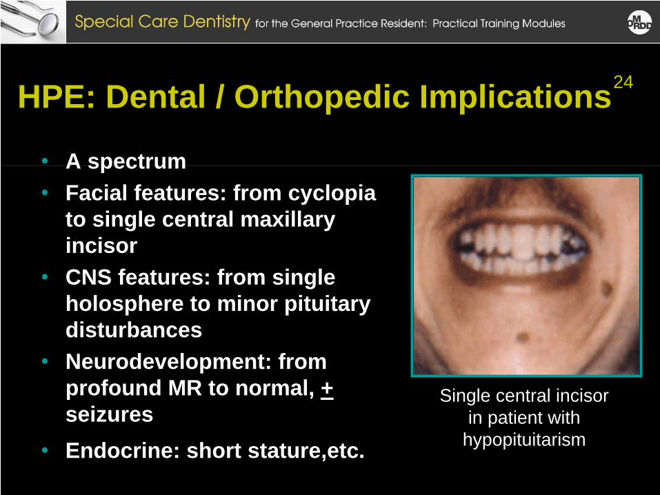
HPE: Dental / Orthopedic Implications
• A spectrum • Facial features: from cyclopia
to single central maxillary incisor
• CNS features: from single holosphere to minor pituitary disturbances
• Neurodevelopment: from profound MR to normal, + seizures
• Endocrine: short stature,etc.
Single central incisor in patient with
hypopituitarism
24
Summary• Dysmorphology is the field of clinical genetics that
deals with syndromology.• Syndromes can be caused by chromosomes
anomalies, single genes mutations, teratogens, or other causes.
• Because many genetic syndromes have oral manifestations, the special care dentist must have a good working knowledge of dysmorphology.
• Syndromes tend to be complex, with multiple manifestations. Because of this, the dentist and orthodontist should function as members of a multidisciplinary team in managing these patients.
Resources for Professionals• OMIM (On-line Mendelian Inheritance in Man)
Provides information about single gene disorders: • www.ncbi.nlm.nih.gov/sites/entrez?db=omim
• Gene Tests: Provides information about testing for (and excellent reviews of) of many disorders:• www.genetests.org
• Genetic Alliance: Provides information and referral information about a large number of disorders• http://www.geneticalliance.org
REFERENCES1. Gorlin RJ, Cohen MM, Hennakam R. Syndromes of the Head
and Neck, Fourth Edition. New York: Oxford University Press, 2001.
2. Jones K. Smith’s Recognizable Patterns of Human Malformations, Sixth Edition. New York: W.B. Saunders, 2005.
3. Levy P, Marion RW: Human Genetics and Dysmorphology; Chapters 47-50. In Kliegman RM, Marcdante KJ, Jenson HB and Behrman RE (eds), Nelson's Essentials of Pediatrics, Fifth Edition. Philadelphia: Elsevier Saunders, 2005, pp 217-242.
4. Nunn, J: Dental manifestations of genetic disorders and treatment challenges. http://www.bzaek.de/list/presse/sym/ nunn_pp.pdf (accessed Feb. 1, 2008)
REFERENCES5. Kusiak A, Sadlak-Nowicka J, Limon J, Kochanska B. Root
morphology of mandibular premolars in 40 patients with Turner syndrome. International Endodontic Journal 2005 38 (11), 822– 826.
6. Tarjan I, Balaton G, Balaton P, Varbiro , Vajo Z. Facial and dental appearance of Williams syndrome. Postgraduate Medical Journal 2003;79:241
7. Sillence DO, Senn A, Danks DH. Genetic heterogeneity in osteogenesis imperfecta. J Med Genet 16:101, 1979.
8. http://www.usc.edu/hsc/dental/PTHL312abc/312b/08/IMGs/25bb.ht ml, accessed 2/1/07)
9. Wilcox RA, McDonald FS. Medical Images: Gray-Blue Sclerae and Osteopenia Secondary to Osteogenesis Imperfecta. Mayo Clinic Proceedings 2007;82:265.
10. http://www.use.ed/hsc/dental/PTHL312abc/312b/08/IMGs//25bb. html, accessed 2/1/08
11. McKusick V, et al. Dwarfism in the Amish. The Ellis-van Creveld syndrome. Bull Hopkins Hosp, 115, 306, 1964.
12. McKusick VA. Ellis-van Creveld syndrome and the Amish. Nat Genet. Mar 2000;24(3):203-4.
13. Wright JT, Grange DK, Richter MK. Hypohidrotic ectodermal dysplasia. Gene Clinics, http://www.genetests.org/servlet/access?db=geneclinics&site=gt &id=8888891&key=V5v5SWTUAUFEr&gry=&fcn=y&fw=yj5V&fil ename=/profiles/x-hed/index.html, accessed February 1, 2008.
14. Gregg NM. Congenital cataracts following German measles in the mother. Trans Ophthalmol Soc Australia, 3:35, 1941
REFERENCES
REFERENCES15. Jones KL, Smith DW, et al. Pattern of malformations in
offspring of chronic alcoholic mothers. Lancet 1:1267, 1973.16. Smith DW: Fetal Alcohol Syndrome. Hosp. Pract. 10:121,
197917. Wattendorf DJ, Muenke M. Fetal Alcohol Spectrum
Disorders. Am Family Phys 2005;72:279-82, 285.18. FAS Physical Abnormalities, on FASLink, Fetal Alcohol
Disorders Society website http://www.faslink.org/physical.htm, accessed 2/1/08
19. Cohen MM, MacLean RW: Craniosynostosis: Diagnosis, Evaluation and Management; Second Edition. New York: Oxford University Press, 2000.
REFERENCES20. Chen H. Crouzon syndrome. Emedicine:
http://www.emedicine.com/PED/topic511.htm, accessed 8/1/08.21. Tewfick TL. Pierre Robin syndrome. In emedicine:
http://www.emedicine.com/ent/topic150.htm. accessed 2/1/08.22. Katsanis SH, Cutting GR. Treacher Collins syndrome. In
Genetests: http://www.genetests.org/servlet/access?db=geneclinics&site=gt& id=8888891&key=seQVrrdJQzTew&gry=&fcn=y&fw=bW69&filena me=/profiles/tcs/index.html, accessed 2/1/08
23. da Silva Dalben G, Costa B, Gomide MR Prevalence of dental anomalies, ectopic eruption and associated oral malformations in subjects with Treacher Collins syndrome. Oral Surg Oral Med Oral Pathol Oral Radiol Endod 2006 101:588-92.
REFERENCES
24. Muenke M, Gropman A. Holoprosencephaly overview. In Genetests: http://www.genetests.org/servlet/access?db=geneclinics&site=gt&i d=8888891&key=seQVrrdJQzTew&gry=&fcn=y&fw=pstB&filenam e=/profiles/hpe-overview/index.html, accessed 2/1/08.
THANK YOUThank you to the Task Force on Special Dentistry Committee for their dedication to this project.
Special thanks to the past and current Chair members of the Task Force on Special Dentistry:
Dr. Alicia BaumanDr. Craig ColasDr. Nancy DoughertyDr. Vincent Filanova
Dr. Gary GoldsteinDr. Roderick MacRaeDr. Edward RigginsDr. Maureen RomerDr. Carl Tegtmeier