Embed Size (px)
Citation preview

Thesis of doctoral (PhD) dissertation
CONVENTIONAL AND MOLECULAR COMPARISONS OF THE TAXONOMY
OF PHOMA-LIKE SPECIES
László Miklós Irinyi
Supervisors: dr. Erzsébet Sándor and dr. György Kövics
University of Debrecen Kálmán Kerpely Doctoral School
Debrecen, 2009

1
Introduction
The pathogenic fungi are responsible for most of plant diseases. Their
described species number is approximately 75.000 at present, which could
represent only 7% of the estimated number of all fungi species. Therefore
there are lots of challenges that mycologist have to face to study and discover
the biodiversity of fungi. The traditional mycological tools are widened with
newer and newer powerful molecular techniques which make possible to
infer phylogenetic relationships in some cases by reconstructing systems
which had been already established. Our taxonomical issue contributes to
the first result of this by the phylogenetic analysis of some complex and
hard identifiable fungal taxa with the help of new molecular tools such as
Phoma-like fungi which contain more than 5000 species. Phoma is a
cosmopolitan genus of coelomycetous fungi. Many species have been
reported from a wide range of hosts and substrates, particularly as pathogens
from plants, as well as soil-borne but predominantly saprophytic and
opportunistic species have also been isolated.
There are several ways in the traditional and modern mycology to
contribute to taxonomical studies of fungi including morphology,
biochemistry, nucleic acid sequences and many others. The three most
commonly discussed species concepts are morphological, biological, and
phylogenetic ones. Since the beginning of mycology, studies of species
concept in fungi have been mainly based on morphological elements. The
most of the species and other taxa of Phoma have so far been determined
on the basis of morphology on standardized media, and gene sequence
analysis was only used as a confirmative or distinctive complement. Thus,
members of the genus are primarily defined by the application of the

2
Morphological Species Recognition (MSR). The weakness of MSR is that
species diagnosed by this often comprise more than one species when
diagnosed by Biological Species Recognition (BSR) or Phylogenetic
Species Recognition (PSR). Biological species concept defines species as
groups of actually or potentially interbreeding natural populations which
are reproductively isolated from other such groups. BSR is acceptable for
many fungi, where sexual reproduction occurs. But there are also fungal
groups, where sexual reproduction has never been recognized. Since strains
of Phoma spp. apparently can not be crossed, the application of the BSR
concept is impracticable. Though, despite reproducing asexually, many
anamorphic fungi including Phoma spp. are known to possess a
surprisingly high level of genetic variation.
The current advances in biochemical and molecular research have
provided mycologists with powerful tools that can be used for delineation
of fungal taxa. The PSR defines species as the smallest aggregation of
populations with a common lineage that share unique, diagnosable
phenotypic characters. According to Taylor et al. (2000) PSR seems to be
well suited for fungi and likely to become very popular with mycologists,
because it can be applied equally both to sexual and to asexual organisms.
They proposed the introduction of Genealogical Concordance Phylogenetic
Species Recognition (GCPSR) for species defining, which could be an
attractive alternative or complement to the morphological species concept,
but has not been widely applied to Phoma spp. yet. It requires the analysis
of several unlinked genes and implies that the phylogenetic position of a
true species is concordant in at least some of them and can not be
contradicted in the others.

3
Up to now the characterization of Phoma and Phoma-like species has
been mostly applied on the basis of morphology, phenotype and
physiology which have often resulted in uncertainty and misidentification.
Recently, Boerema et al. (2004) published Phoma Identification Manual,
based on morphological studies which contain 223 cultural descriptions of
specific and intraspecific taxa of Phoma Sacc.
Till now the molecular-based phylogenetic analysis within the Phoma
genus have only been used for defining phylogenetic relationships among
isolates within one ore closely related species. Therefore our goal has been
to find and apply such phylogentetic markers which are suitable for
inferring phylogenetic relationships – particularly for Phoma-like species
which are hard to identify by traditional way – at species level within the
Phoma genus.
For phylogenetic analysis we have chosen the complete sequence of ITS
region and fragments of tef1 and β-tubulin genes which have already been
proved to be useful as potential markers for phylogenetic studies at other
different fungal taxa.
Ribosomal DNA (rDNA) has long been used as a potential marker for
phylogenetic studies (reviewed in Avise, 2004). Many fungal taxonomy
studies have applied ITS regions and have proved them to be suitable for
resolving relationships both at genus and at species level.
Simultaneously, translation elongation factor 1 subunit alpha (EF1 α)
encoding gene (tef1) has been proved to be a useful gene to resolve
phylogenetic relationships at species level, as well as in deeper divergence
of fungi.
The nucleotide sequences of tubulin encoding gene β-tubulin has
received more and more attention for phylogenetic studies in lower and

4
higher taxonomic levels as well. Voigt et al. (2005) and Fatehi et al. (2003)
used the β-tubulin gene, among others, to analyse the Leptosphaeria
maculans (anamorph: Phoma lingam) species complex, to resolve
molecular relatedness within the Ascochyta pinodes complex. The
phylogenetic analysis of multiple protein-encoding genes with
Genealogical Concordance Phylogenetic Species Recognition
is proposed (Taylor et al., 2000) as a more robust way of determining
and recognising species than analysis based on morphology or mating
behaviour. Our further aim was to apply and compare different character-
based phylogenetic methods (Maximum Likelihood, Maximum Parsimony
and Bayesian method).
Different pathogenic Phoma-like species (Phoma pinodella, Phoma
sojicola, Phyllosticta sojicola, Phoma exigua var. exigua) have been found
on Fabaceae species like soybean (Glycine max). Since these species are
symptomatically and morphologically very similar to each other, it is hard
to differentiate them. Consequently, this has resulted in uncertainty and
misidentification in their taxonomy. Therefore there is need to develop
additional rapid molecular methods to enable accurate identification.

5
Objectives
I compiled and performed my research relating to the conventional and
molecular comparison of the taxonomy of Phoma-like species on the
following conception:
1. Characterization and identification of different isolates of Phoma-like
species based on the accepted conventional morphological concept of
Phoma genus.
2. Looking for phylogenetic markers which could be suitable for
inferring phylogenetic relationships – particularly for Phoma-like species
which are hard to identify by traditional way – at species level within the
Phoma genus.
3. Molecular phylogenetic relatedness studies to infer phylogenetic
relationships at species level within the Phoma genus.
Inferring phylogenetic relationships at species level within the Phoma
genus and Phoma-like species.
4. Applying, assessing and comparing different character-based
phylogenetic methods (Maximum Likelihood, Maximum Parsimony and
Bayesian method) in the characterization of Phoma-like species.
5. Determination the taxonomical status of Phoma-like isolate deposited
as Phyllosticta sojicola isolated from soybean.
6. Re-evaluation on molecular basis of the taxonomic status of soybean
pathogen Phoma pinodella and closely related Phoma sojicola species.
6
Materials and Methods
Morphological analysis
Twenty-two isolates of nine different Phoma-like species were obtained
from reference culture collections (Table 1). Seven of them were isolated
from soybean, the others were collected from different hosts. All cultures
were maintained on oatmeal agar at 5 °C during the study. The species
were characterized morphologically according to Boerema et al. (2004).
The isolates were cultured on oatmeal agar and malt agar incubated at
22°C in dark, and examined 7 days later. For description of colony colours
Rayner’s colour chart (1970) terminology was used. Petri dishes were then
exposed to cycles of 13 h NUV-light, 11 h darkness to stimulate the
formation of pycnidia. 2 weeks later colony descriptions were
complemented and after 3 weeks the morphology of pycnidia, conidia and
other structures, such as chlamydospores, were studied from the OA
cultures. The NaOH spot test was done on MA by addition of a drop of 1N
NaOH on the colony margin, and changes in colour were noted. Conidia
dimensions refer to 30 measurements with oil-immersion at 1250x
magnifition.

7
Table 1 – Isolates used in the phylogenetic analyses GenBank accession no. Isolate
no. Alternative isolate no. Fungal species Host plant Location of
collection Collector Tefa ITSb β-tubulinc
D/035 BT-15 Phoma pinodella Glycine max Hungary I. Walcz EU543973 EU573015 EU541416
D/045 PD82/550 P. pinodella Hordeum vulgare Hungary G.J. Kövics EU543971 EU573025 EU541417
D/046 PD77/165 MYA-411 P. pinodella Pisum
sativum Hungary G.J. Kövics EU543972 EU573024 EU541419
D/095 N.A. P. pinodella P. sativum Hungary L. Gergely EU543970 EU573027 EU541418
D/159 CBS 318.90 PD 81/729 P. pinodella P. sativum Netherlands M.E.
Noordeloos EU595355 EU573028 EU595352
D/063d Ph 58 MYA-408 P. pinodella Petroselinum
crispum Poland J. Marcin-kowska EU543975 EU573012 EU541420
D/054 MYA-406 P. sojicola G. max Hungary G.J. Kövics EU543974 EU573023 EU541434
D/056 CBS 567.97 PD97/2160 P. sojicola G. max Hungary G.J. Kövics EU543976 EU573026 EU541433
D/050 CBS 301.39 Phyllosticta sojicola G. max Germany K. Böning EU595356 EU573029 EU595357
D/075 N.A. P. exigua var. exigua G. max Poland G.J. Kövics EU543982 EU555533 EU541421
D/077 N.A. P. exigua var. exigua G. max Poland G.J. Kövics EU543983 EU573010 EU541422
D/145 N.A. P. exigua Althaea officinalis Hungary G. Nagy - EU573011 EU541425
D/146 N.A. P. exigua Althaea rosae Hungary G. Nagy EU543984 EU573013 EU541427
D/158 ICMP 15330 P. exigua var. exigua
Agapanthus sp. New Zealand M.
Braithwaite EU543981 EU573008 EU541428
D/157 ICMP 13336 P. exigua Cucurbita maxima New Zealand P.G.
Broadhurst EU543980 EU573007 EU541429
D/071 PD 86/73 P. exigua var. linicola
Linum usita-tissimum Hungary G.J. Kövics EU543979 EU573009 EU541423
D/072 PD 75/907 P. plurivora Medicago sativa Australia J. de Gruyter EU552929 EU573018 EU552932
D/155 ICMP 6875 P. plurivora Pennisetum clandestinum New Zealand P.R.
Johnston EU552930 EU573019 EU552931
D/034 AI-416 P. glomerata G. max Hungary G.J. Kövics EU543969 EU573016 EU541424 D/156 ICMP 15788 P. glomerata Yucca sp. New Zealand C.F. Hill EU543968 EU573017 EU541426
D/048 PD 76/1021 P. foveata Chenopodium quinoa Netherlands G. H.
Boerema EU543985 EU573021 EU541431
D/044 PD 77/508 P. multirostrata Phylodendron sp. Netherlands G. H.
Boerema EU543986 EU573022 EU541430
D/144 N.A. Ascochyta rabiei
Cicer arietinum Australia N.A. EU595354 EU595358 EU595353
D/160 CBS 581.83A
Didymella rabiei C. arietinum Syria H.A. van der
Aa EU543978 EU573020 EU541432
AI refers to Agrobotanical Institute, Tápiószele, Hungary BT refers to Fodder-plant Research Institute of Pannon University, Iregszemcse-Bicsérd, Hungary CBS refers to Centraalbureau voor Schimmelcultures, Utrecht, The Netherlands; D refers to the Culture Collection of the Plant Protection Department of University of Debrecen, Hungary ICMP refers to International Collection of Microorganisms from Plants MYA refers to American Type Culture Collection, (ATCC), USA. N.A. = not available PD refers to Plantenziektenkundige Dienst; Dutch Plant Protection Service Collection, The Netherlands
a partial sequence data of the translation elongation factor coding gene (tef1) b 18S ribosomal RNA gene, partial sequence; internal transcribed spacer 1, 5.8S ribosomal
RNA, internal transcribed spacer 2, complete sequence; 26S ribosomal RNA gene, partial sequence
c partial sequence data of the β-tubulin coding gene d D/063: P. pinodella misidentified as ’P. exigua var. exigua’
8
DNA extraction For PCR based methods the studied cultures were grown in 100 ml of malt
broth (MB, containing 2% malt extract) for 48 hours at room temperature
in the dark on a rotary shaker (125 rpm).Mycelium was harvested by
vacuum filtration. Total genomic DNA was extracted from freeze-dried
mycelium and isolated using E.Z.N.A.® TM Fungal DNA Isolation Kit
(Omega Bio-tek, Inc., USA) according to the protocol, followed the
manufacturer’s instructions. DNA concentrations were estimated in
comparison to known standard in agarose gels stained with ethidium
bromide (EtBr).
PCR reaction and DNA sequencing
Amplifications of 50 µl PCR reaction contained 25 µl 2X PCR Master Mix
40-40 pmol each primer, 20-40 ng of DNA and nuclease free water were
run out. Primers used to amplify approx. 520bp of the ITS region
containing the internal transcribed spacer regions 1 and 2, moreover the
5.8S rDNA are based on published composite sequences, SR6R and LR1
(White et al., 1990) with the following amplification protocol: 3 min initial
denaturing at 95 °C, followed by 5 cycles of 1 min at 95 °C, 1 min
annealing at 50 °C, 1 min at 72 °C and 25 cycles of 1 min at 90 °C, 1 min
annealing at 50 °C, 1 min at 72 °C and 15 min final extension at 72 °C.
The large intron (approx. 300bp) of the tef1 gene was amplified by the
EF1-728F and EF1-986R primer pair (Druzhinina & Kubicek, 2005)
according to the previously described protocol with a temperature of 56 °C
rather than 50 °C. Primers Bt2a and Bt2b (Glass & Donaldson, 1995;
O’Donnell & Cigelnik, 1997) were used to amplify a fragment (approx.

9
300bp) of β-tubulin gene and PCR conditions were carried out as described
above with an annealing temperature of 58 °C. PCR was performed in a
Primus thermocycler (MWG Biotech). Amplification products were
subjected to electrophoresis in a 0.7 % agarose gel containing EtBr and
visualized by UV illumination. The PCR products were purified by using
YM-100 Microcon Centrifugal Filter Devices (Millipore). Purified
amplification products were sequenced by MWG Biotech Company in
Germany.
Data analysis
The obtained DNA sequences were aligned first with ClustalX (Thompson
et al., 1997) and manually checked for ambiguities and adjusted when
necessary using Genedoc (Nicholas et al., 1997). Single gaps were treated
either as missing data or as the fifth base and multistate characters as
uncertain. For the Bayesian and Maximum Likelihood analyses, models of
sequence evolution were evaluated for each dataset and model parameter
estimates obtained with Modeltest v.3.7 (Posada & Grandall, 1998). The
character based phylogenetic studies were performed by Paup*4.0b
(Swofford, 2002) and MrBayes (Huelsenbeck, 2000).
Results
Morphology
On the basis of morphological characters we studied different
morphological characters on five Phoma pinodella (D/035, CBS 318.90,
D/095, PD82/550, PD77/165), two Phoma sojicola (D/054, CBS 567.97),
10
three Phoma exigua var. exigua (D/075, D/077, Ph 58), one Phyllosticta
sojicola (D/050) and one Phoma exigua var. linicola (D/071) isolates.
The morphological characters of Phoma pinodella (L.K. Jones) Morgan-
Jones & K.B. Burch and Phoma sojicola (Abramov) Kövics et al. showed
high resemblance and often overlap because of the high variability of the
isolates. Cultural characteristics of certain isolates were very variable even
in standardized conditions. It occurred that the same (P. pinodella
PD77/165) isolates transferred at the same time in the same media, and
cultured at the same temperature even showed different morphological
characters. We have experienced similar variability at other isolates too,
and we observed that the morphological characters may change after
certain number of transfers on media in vitro as well. The P. pinodella
generally produces crystals on malt-extract agar after one week while at P.
sojicola misses this feature. In our study we have not experienced crystal
formation that feature of P. pinodella species at none of the isolates, which
can due to the fact that they can loose this character temporarily during
several transfers on the media. In our experiments, the cultural
characteristics of both species (P. pinodella and P. sojicola) were very
similar without any well identifiable and strong features. The colours of the
two colonies were very variable even in the same isolate in the same
conditions. The size and shape of pycnidia and conidia showed also such a
high similarity that we could not differentiate the two species reliably.
We have encountered similar difficulties differentiating the isolates of
Phyllosticta sojicola Massalongo and Phoma exigua Desm. var. exigua
(syn.: Ascochyta phaseolorum Saccardo). Studying the morphological
features we can assess that there are no significant differences in none of
the cultural characteristics, the size and shape of pycnidia and conidia and

11
that the morphological variability is rather high among isolates. The only
difference we have found between the two isolates was the “E metabolite”
production, which is not a feature of P. sojicola but a frequent character of
P. exigua var. exigua. However, this absence of “E-metabolite” production
might be a result of a genetic mutation.
In pursuance of examination, the taxonomical status of the isolate of
Phoma exigua var. exigua Ph 58 has been found ambiguous on
morphological characters. According to the MSR, the isolate shows high
similarity to P. pinodella except the only difference in the “E-metabolite”
production only. The Ph 58 isolate showed a positive NaOH spot test,
which is a distinctive feature of Phoma exigua var. exigua but nor a
characteristic of P. pinodella. All phylogenetic analyses classified the
isolate Ph 58 in the clade of P. pinodella that is why we suppose that the
isolate Ph 58 has been misidentified as P. exigua var. exigua and actually it
belongs to P. pinodella species.
Since these species and isolates are very similar to each other both
symptomatically and morphologically, and rather variable depending on
strains it is hard to delimit them. Consequently, this has resulted in
uncertainty and misidentification in their taxonomy. Therefore there is
need to develop additional rapid molecular methods to enable accurate
identification.
Molecular features
Sequence analysis of the tef1 gene
The topology of phylogenetic trees (Figs 1, 2, 3) obtained by the analyses
of tef1 sequences by three character based methods (Maximum Parsimony,

12
Maximum Likelihood, Bayesian method) was very similar. One part of the
Phoma isolates (P. pinodella, P. exigua, P. plurivora, P. destructiva, P.
glomerata) were well separated from the other Phoma isolates as well as
the other parts constitute clades.
Parsimony analysis of tef1 revealed 173 parsimony informative sites, 16
polymorphic sites, and 121 sites that were constant among all isolates.
Species represented with more than one isolate (P. pinodella, P. exigua
var. exigua, P. glomerata, P. plurivora) were placed in the same clade.
Interestingly, P foveata and P. multirostrata were also placed in one,
highly supported group (100% PP) which raises the issue of
misidentification on the behalf of depositor.
Besides Phoma isolates, the Ascochyta isolates (Ascochyta lentis /
teleomorph: Didymella lentis) were also clustered in one group which has
proved that the tef1 sequences are well suited for delineating phylogenetic
relationships within the Phoma as well as Ascochyta genera.
The isolates of P. sojicola (MYA-406, EU543974 and PD 97/2160,
EU543976) grouped together with a P. pinodella isolates since their tef1
sequences were completely identical.
The isolate deposited as Phyllosticta sojicola (CBS 301.39, EU595356)
associated with the Phoma exigua var. exigua subgroups, which has
supported the statement that the two species are identical (Kövics et al.,
1999).
The distances between each taxon have proved to be long and different
enough so that we can consider the results or the phylogenetic tree well
established.
The high bootstrap and Bayesian posterior probability values supported
the solidity of the well separated clades and true coherence of the trees.

13
Fig 1. Phylogenetic relationships of Phoma strains inferred by Parsimony analysis of tef1 sequences. The numbers above the lines represent the bootstrap (bootstrap=1000) values. The columns on the right side represent the Phoma sections based on morphological characterization (Boerema et al., 2004). *P. pinodella (Ph 58) misidentified as’P. exigua var. exigua’
Fig 2. Phylogenetic relationships of Phoma strains inferred by Maximum Likelihood analysis of tef1 sequences. The columns on the right side represent the Phoma section based on morphological characterization (Boerema et al., 2004). *P. pinodella (Ph 58) misidentified as ’P. exigua var. exigua’

14
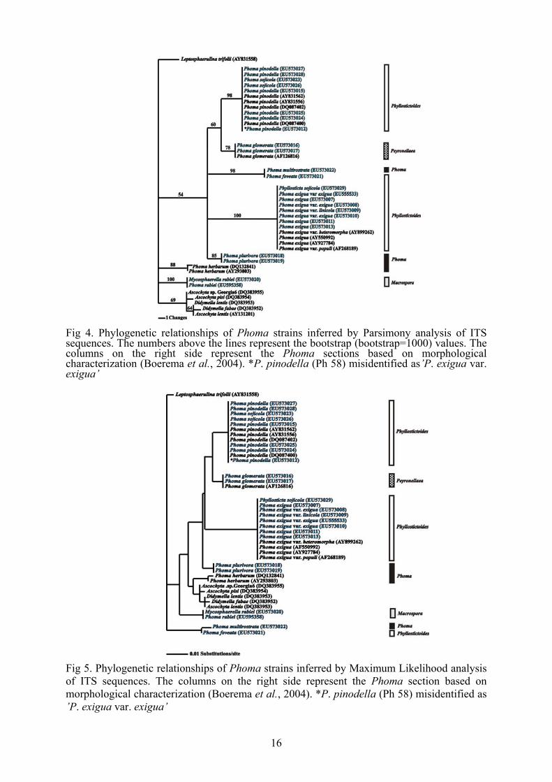
Fig 3. Phylogenetic relationships of Phoma strains inferred by Bayesian analysis of tef1 sequences. The numbers above the lines represent the Bayesian posterior probability values. The columns on the right side represent the Phoma sections based on morphological characterization (Boerema et al., 2004). *P. pinodella (Ph 58) misidentified as’P. exigua var. exigua’ Sequence analysis of the ITS region The obtained phylogenetic trees (Figs 4, 5, 6) were well resolved with
similar clades recovered by tef1 analysis. Parsimony analysis of the ITS
revealed 32 parsimony informative sites, 5 polymorphic sites, and 47 sites
are constant among all isolates.
The differences between the isolates of Phoma and Ascochyta species
were not significant so it is questionable, that the selected ITS region is
suitable for phylogenetic studies at species level within the Phoma genus.
The bootstrap values of Phoma and Ascochyta clades were low 854-69)
therefore it is not possible to draw reliable conclusions for the phylogenetic

15
relationships of the species of the two genera. However the Bayesian
posterior probability values (93-99) were higher than that of the bootstrap.
Species represented with more than one isolate (P. pinodella, P. exigua
var. exigua, P. glomerata, P. plurivora) were placed in the same clade
which supported the solidity of the well separated clades and coherence on
the trees. Interestingly, P. foveata and P. multirostrata were also placed in
one, highly supported group (100% PP and 100% BS) which raises the
issue of misidentification on behalf of the depositor.
The high bootstrap and Bayesian posterior probabilities values of the
two biggest clades (P. pinodella and P. exigua) of the trees confirm that
the two taxa were well separated both from each other as well as other
taxons.
The isolates of P. sojicola (MYA-406, EU543974 and PD 97/2160,
EU543976) groupped together with a P. pinodella isoletes since their ITS
sequences were completely identical.
The isolate deposited as Phyllosticta sojicola (CBS 301.39, EU595356)
associated with the Phoma exigua var. exigua subgroups, which support
the statement that the two species are identical (Kövics et al., 1999).
The high bootstrap and Bayesian posterior probability values supported
the solidity of the well separated clades and the true coherence of the trees.
16
Fig 4. Phylogenetic relationships of Phoma strains inferred by Parsimony analysis of ITS sequences. The numbers above the lines represent the bootstrap (bootstrap=1000) values. The columns on the right side represent the Phoma sections based on morphological characterization (Boerema et al., 2004). *P. pinodella (Ph 58) misidentified as’P. exigua var. exigua’
Fig 5. Phylogenetic relationships of Phoma strains inferred by Maximum Likelihood analysis of ITS sequences. The columns on the right side represent the Phoma section based on morphological characterization (Boerema et al., 2004). *P. pinodella (Ph 58) misidentified as ’P. exigua var. exigua’

17
Fig 6. Phylogenetic relationships of Phoma strains inferred by Bayesian analysis of ITS sequences. The numbers above the lines represent the Bayesian posterior probability values. The columns on the right side represent the Phoma sections based on morphological characterization (Boerema et al., 2004). *P. pinodella (Ph 58) misidentified as’P. exigua var. exigua’ Sequence analysis of the β-tubulin gene
The obtained phylogenetic trees by the analysis of β-tubulin sequences
(Figs 7, 8, 9) bore a resemblance to the tef1 and ITS trees. The trees were
well resolved by Bayesian inference and Parsimony analysis with similar
highly supported clades. Parsimony analysis of β-tubulin revealed 49
parsimony informative sites, 20 polymorphic sites, and 229 sites that were
constant among all isolates.
The Phoma pinodella clade contained the Phoma sojicola sequences
(100% PP and BS support), and Phyllosticta sojicola groupped together

18
with Phoma exigua var. exigua (100% PP and BS support). Interestingly,
the two P. exigua isolates that originated from New Zealand groupped
together both in the β-tubulin and tef1 analyses. Similarly to the tef1
analysis, species represented with more than one isolate, specifically P.
pinodella and P. exigua, were placed in the same highly supported clade.
The high bootstrap and Bayesian posterior probability values supported
the solidity of the well separated clades and the true coherence of the trees.
Fig 7. Phylogenetic relationships of Phoma strains inferred by Bayesian analysis of β-tubulin sequences. The numbers above the lines represent the Bayesian posterior probability values. The columns on the right side represent the Phoma sections based on morphological characterization (Boerema et al., 2004). *P. pinodella (Ph 58) misidentified as’P. exigua var. exigua’

19
Fig 8. Phylogenetic relationships of Phoma strains inferred by Parsimony analysis of β-tubulin sequences. The numbers above the lines represent the bootstrap (bootstrap=1000) values. The columns on the right side represent the Phoma sections based on morphological characterization (Boerema et al., 2004). *P. pinodella (Ph 58) misidentified as’P. exigua var. exigua’
Fig 9. Phylogenetic relationships of Phoma strains inferred by Maximum Likelihood analysis of β-tubulin sequences. The columns on the right side represent the Phoma section based on morphological characterization (Boerema et al., 2004). *P. pinodella (Ph 58) misidentified as ’P. exigua var. exigua’

20
Conclusions and recommendations
The taxonomy of Phoma species has not been made completely clear up to
now. It includes numerous several uncertainties. It is getting more and
more obvious that even the most complex morphological studies can not
give reliable result to the actual taxonomical relatedness of species.
The speciation of the genus Phoma was based on host-alone and later
on, the trend was to study different species of Phoma in pure culture. On
the basis of morphological studies on different culture media, several
species were found to be identical.
In our research we have studied twenty-two isolates of nine different
Phoma-like species on the basis of morphological and molecular
characters. Our morphological studies were carried out on the basis of the
accepted conception of the taxonomy of Phoma species (Boerema et al.,
2004). While the morphological identification of certain isolates was
obvious in other cases it has been charged with uncertainty since the
morphological characters overlap (on the basis of culture size, color and
shape as well as the size of conidia and pycnidia certain taxa can not be
differentiated without any doubt. We observed that the morphological
characters can change after certain number of transfers on media in vitro as
well.
The Phoma sojicola (Abramov) Kövics et al. shows high similarity to
Phoma pinodella (L.K. Jones) Morgan-Jones & K.B. Burch, however
Kövics et al. (1999) have found representative characteristics in culture
morphology, pycnidia production and crystal formation. The P. pinodella
produces crystals on malt-extract agar after one week while the P. sojicola
misses this feature. In our study we could not managed to detect crystal

21
production in neither of the species. According to Boerema et al. (2004)
the crystal production is a contingent feature depending on cultural
conditions and/or other factors (longevity of storage, temperature, number
of transfers). In our experiments, the cultural characteristics of both species
(P. pinodella and P. sojicola) were very similar without any well
identifiable and strong features. The colours of the two colonies were very
variable even in the same isolate in the same conditions. The size and
shape of pycnidia and conidia showed also such a high similarity that we
could not differentiate the two species reliably.
We have encountered similar difficulties differentiating the isolates of
Phyllosticta sojicola Massalongo and Phoma exigua Desm. var. exigua
(syn.: Ascochyta phaseolorum Saccardo). Kövics et al. (1999) have
suggested that the two species were supposedly identical. Studying the
morphological features we can assess that there are no significant
differences in none of the cultural characteristics, the size and shape of
pycnidia and conidia and that the morphological variability is rather high
among isolates. The only difference we have found between the two
isolates was the “E-metabolite” production, which is not a feature of P.
sojicola but a frequent character of P. exigua var. exigua. However it
sometimes might be completely missing in some strains. But this absence
of “E-metabolite” production can be a result of a genetic mutation.
In pursuance of examination, the taxonomical status of the isolate of
Phoma exigua var. exigua Ph 58 has been found ambiguous on
morphological characters. According to the MSR, the isolate shows high
similarity to P. pinodella except the only difference in the “E-metabolite”
production. The Ph 58 isolate showed a positive NaOH spot test which is a
distinctive feature of Phoma exigua var. exigua but does not a

22
characteristic of P. pinodella. All phylogenetic analyses classified the
isolate Ph 58 in the clade of P. pinodella that is why we suppose that the
isolate Ph 58 has been misidentified as P. exigua var. exigua and actually
belongs to P. pinodella species.
Up to now, molecular based phylogenetic analyses within the Phoma
genus have only been used for defining phylogenetic relationships among
isolates within one or closely related species (Fatehi et al., 2003; Mendes-
Pereira et al., 2003; Balmas et al., 2005; Voigt et al., 2005). For the first
time, we have tried to find such molecular markers which can be suitable
for delineating phlyogenetic relationships at species level within the
Phoma genus.
For our studies, we have selected the complete sequence of the ITS
region and fragments of the tef1 (translation elongation factor coding) and
β-tubulin genes which have been used as potential marker in many fungal
taxonomy studies. The phylogenetic analysis of multiple protein-encoding
genes with the GCPSR is proposed as a more robust way of determining
and recognizing species than analysis based on morphology or sexual
recognition (Taylor et al., 2000).
The translation elongation factor 1 subunit alpha (EF1α) encoding gene
(tef1) has been proved to be a useful gene to resolve phylogenetic
relationships at species level, as well as in deeper divergence in fungi.
However, this gene has not been used for the genetic analysis of the Phoma
genus yet. Ribosomal DNA (rDNA) has long been used as a potential
marker for phylogenetic studies (reviewed in Avise, 2004; White et al.,
1990). Many fungal taxonomy studies have applied ITS regions for
resolving relationships at genus and species level. Mendes-Pereira et al.
(2003) used ITS sequences for studying the molecular phylogeny of

23
Leptosphaeria maculans – L. biglobosa species complex. Balmas et al.
(2005) inferred phylogenetic relationships among isolates of Phoma
tracheiphila on the basis of ITS sequences as well as Fatehi et al. (2003) in
Ascochyta pinodes complex. Several studies proved that β-tubulin at the
nucleotide level can be suitable for phylogenetic studies at low taxonomic
levels within Ascomycetes (Baldauf et al., 2000; Yli-Mattila et al., 2004).
Voigt et al. (2005) used β-tubulin gene among others to analyze
Leptosphaeria maculans (anamorph: Phoma lingam) species complex, as
well as Fatehi et al. (2003) to refer molecular relatedness within Ascochyta
pinodes complex.
Species represented with more than one isolates (P. pinodella, P. exigua
var. exigua, P. glomerata, P. plurivora) were placed in the same clade,
which proved the suitability of the examined makers for phylogenetic
studies not only within species (Mendes-Pereira et al., 2003; Balmas et al.,
2005), but also species complex (Fatehi et al., 2003) as well as species
level in Phoma genus. The different Phoma species were well separated
from the closely related species of Ascochyta genus. Since the separation
of the Phoma and Ascochyta species is often problematic in the isolates of
pseudo-Ascochyta species (Fatehi et al., 2003) this new molecular feature
can provide a useful tool for mycologists to identify clearly an unknown
species.
Phylogenetic analysis of the two protein-encoding genes, tef1 and β-
tubulin together with the complete ITS sequences yielded consensus
results. P. sojicola isolates always formed one clade with all the examined
P. pinodella isolates, while representative isolate of Phyllosticta sojicola
groupped together with P. exigua var. exigua. The highest resolution could

24
be revealed with the analysis of the tef1 gene, while sequences of β-tubulin
showed the lowest polymorphism among the examined Phoma species.
On the basis of phylogenetic trees obtained by different character based
methods (Maximum Likelihood, Maximum Parsimony, Bayesian method)
the studied Phoma-like isolates were well separated from each other and
the closely related Ascochyta genus. The topologies of the trees showed
high similarity, which has proved the confidence of different phylogenetic
analyses respectively within the Phoma genus. Comparing the advantages
and disadvantages of the three studied phylogenetic methods we can
conclude the following statements: during the analysis one of the most
important aspects is the speed, which was the strength of the Maximum
Parsimony but it does not offer the opportunity of the selection of the
evolution models which the other two methods do. The most accurate and
confident tree was given by the Maximum Likelihood (ML) analysis since
it takes every possible mutation step into account, which could resulted the
topology of the given tree that is why this method approaches the best the
real evolutionary changes at the expense of the required time and
calculation capacity for the analysis. It took much more time with our data
than the other two methods as well as we could not do bootstrap analysis
because the calculation was outstandingly time-consuming.
On the basis of our experiences, among the different character based
methods the Bayesian one has seemed to be the best choice because of its
easy controllability, speed and high confidence. However we have to
emphasize that the other two methods are also suitable for correct
coherence analysis within the Phoma genus at species level.

25
New results
1. – Twenty-two isolates of nine different Phoma-like species by the
comparison of morphological and molecular features. Seven of them
were isolated from soybean, the others were collected from different
hosts.
2. – The different Phoma species were well separated from the closely
related species of Ascochyta genus. Since the separation of the Phoma
and Ascochyta species is often problematic in the isolates of pseudo-
Ascochyta species (Fatehi et al., 2003) this new molecular feature can
provide a useful tool for mycologists to identify unknown species.
3. – In pursuance of our study we have found such molecular markers
(tef1, ITS, β-tubulin), which have proved suitable for phylogenetic
studies at species level in Phoma genus. Among these, it is the tef1,
which has not been used until now for phylogenetic studies in Phoma
genus, proved to be the most confident marker.
4. – The soybean pathogenic Phoma sojicola (Abramov) Kövics et al. (syn.:
Ascochyta sojicola), which was described as a new combination in 1999
by Kövics et al. But in our present studies the morphological differences
were small and delimitation was made basically on the absence of crystal
production on MA, in contrast to P. pinodella. However this feature
appears to be an unstable character, viz. in our experiments none of the
isolates produced crystals. All phylogenetic relationship analyses
classified Phoma sojicola in the same clades of P. pinodella. Based on
the presented GCPRS and morphological results we suggest the re-
classification of Phoma sojicola as synonymous with P. pinodella
(Irinyi et al., 2009).

26
5. – The isolate originally deposited as Phyllosticta sojicola – the isolate,
which caused a serious disease in Germany in the 1930s – was
groupped with the Phoma exigua var. exigua clade on the basis of all
studied phylogenetic markers. As besides the variability of
morphological characters the Phyllosticta sojicola can be identical to
Phoma exigua var. exigua on the basis of molecular results, we
propose the usage of Phyllosticta sojicola species name as a
synonymous of Phoma exigua var. exigua.
6. – We extended the molecular gene bank database (NCBI) with the tef1,
ITS and β-tubulin sequences of twenty-two isolates of nine Phoma-
like species.
7. – We contributed to clarify the plant pathology of symptomatically
identical diseases that damage on Fabaceae species. Damage can be
caused, beside others, by
Phoma exigua Desm. var. exigua (syn.: Ascochyta phaseolorum
Saccardo) (new synonym Phyllosticta sojicola Massal.);
Phoma pinodella (L.K. Jones) Morgan-Jones & K.B. Burch (new
synonyms: Phoma sojicola (Abramov) Kövics et al., Ascochyta
sojicola Abramov (as, „A. sojaecola”).
Summary
In our research we have studied twenty-two isolates of nine different
Phoma-like species on the basis of morphological and molecular
characters. The isolates were characterized and identified morphologically
in details according to an accepted concept based on standardized in vitro
physiological and morphological characters of Phoma-like species.

27
Different pathogenic Phoma-like species (Phoma pinodella, Phoma
sojicola, Phyllosticta sojicola, Phoma exigua var. exigua) have been found
on Fabaceae species, including soybean (Glycine max). Since these species
are very similar to each other both symptomatically and morphologically,
and show strain-dependent variability it is hard to delimit them.
Consequently, this has resulted in uncertainity and misidentification in
their taxonomy. Therefore there is a need to develop additional rapid
molecular methods to enable accurate identification.
Phoma sojicola (Abramov) Kövics et al. highly resembles
morphologically to Phoma pinodella (L.K. Jones) Morgan-Jones & K.B.
Burch. According to Kövics et al. (1999) there are profound morphological
differences in cultural characteristics, pycnidia production and crystal
formation between the two aforementioned species. However, in our
studies, crystal formation was not observed in either of the species which
can be attributed to that this feature may depend on cultural conditions
and/or other factors (longevity of storage, temperature, number of transfer).
On the basis of morphological characters, the Phyllosticta sojicola
showed great similarity to Phoma exigua var. exigua. The sporadical
incidence of P. sojicola on soybeans and the examination of living cultures
makes its identification difficult.
We have selected phylogenetic markers (tef1, ITS, β-tubulin) that were
suitable for phylogenetic studies and identification at species level in the
Phoma genus. Phylogenetic studies of the two protein-encoding genes tef1
and β-tubulin, together with the ITS sequences yielded consensual results.
The highest resolution could be revealed by the analysis of tef1 gene,
whereas sequences of ITS and β-tubulin showed the lowest polymorphism
among the examined Phoma species.

28
According to the phylogenetic trees based on molecular markers, the
Phoma species were well separated from the closely related Ascochyta
taxa: Ascochyta rabiei /teleomorph: Didymella rabiei/, Ascochyta lentis
/teleomorph: Didymella lentis/. As the identification of Phoma and
Ascochyta genera based on morphological characteristics is often
confusable, these new phylogenetic markers can be useful tools for
mycologists to identify unknown species.
The species represented by more than one isolate were classified in the
same subgroup (P. pinodella, P. exigua, P. glomerata, P. plurivora), which
prove that the molecular sequences are well suited for delineating
phylogenetic relationships within the Phoma genus.
For phylogenetic analysis, three different character-based methods were
used (Maximum Likelihood, Maximum Parsimony, Bayesian method) to
study the advantages and disadvantages of each marker. In our experience,
the most suitable method seemed to be the Bayesian analysis due to its
complexity, reliability and fastness.
Soybean pathogenic P. sojicola (syn.: Ascochyta sojicola) was described
as a new combination (comb. nov.) in 1999 by Kövics et al. In our
presented performed studies the morphological differences were small and
delimitation was made basically on the absence of crystal production on
MA, in contrast to P. pinodella. However, this feature appears to be an
unstable character, viz. in our experiments none of the isolates produced
crystals. All phylogenetic relationship analyses classified Phoma sojicola
in the same clades of P. pinodella. Based on the presented genealogical
concordance phylogenetic species recognition and morphological results,
we suggest the re-classicfication of Phoma sojicola as synonymous with P.
pinodella (Irinyi et al., 2009).

29
The morphological characters of the Phyllosticta sojicola isolate (CBS
301.39, deposited by Böning in 1939) discussed in this study, together with
its molecular features, proved that Phyllosticta sojicola is synonymous
with Phoma exigua var. exigua. This supports our hypothesis based on
examined type material, that P. sojicola on leaves of Glycine max
(Massalongo, 1900) represents the plurivorous P. exigua var. exigua.
References
Baldauf, S. L., Roger, A. J., Wenk-Siefert, I., Doolittle, W. F. (2000). A kingdom-level phylogeny of eukaryotes based on combined protein data. Science 290: 972-977.
Balmas, V., Scherm, B., Ghignone, S., Salem, A.O.M., Cacciola, S.O., Migheli, Q. (2005). Characterisation of Phoma tracheiphila by RAPD-PCR, microsatellite-primed PCR and ITS rDNA sequencing and development of species primers for in planta PCR detection. European Journal of Plant Pathology 111: 235-247.
Boerema, G.H., de Gruyter, J., de, Noordeloos, M.E., Hamers, M.E.C. (2004). Phoma identification manual. Differentiation of species and infra-specific taxa in culture. CABI Publishing, CAB International Wallingford, Oxfordshire, UK.
Druzhinina, I., Kubicek, C.P. (2005). Species concepts and biodiversity in Trichoderma and Hypocrea: from aggregate species to species cluster? J. Zhejiang Univ. Sci. 6B (2): 100-112.
Fatehi, J., Bridge, P.D., Punithalingam, E. (2003). Molecular relatedness within the “Ascochyta pinodes”-complex. Mycopathologia 156: 317-327.
Glass, N.L., Donaldson, G.C. (1995). Development of primer sets designed for use with the PCR to amplify conserved genes from filamentous Ascomycetes. Applied and Environmental Microbiology 1323-1330.
Huelsenbeck J.P., Ronquist F. (2001). MRBAYES: Bayesian inference of phylogenetic trees. Bioinformatics 17: 754-755.
Irinyi L., Kövics, G.J., Sándor, E. (2009). Taxonomic re-evaluation of Phoma-like soybean pathogenic fungi. Mycological Research 113: 249-260.
30
Kövics, G.J., de Gruyter, J., van der Aa, H.A. (1999). Phoma sojicola comb. nov. and other hyaline-spored coelomycetes pathogenic on soybean. Mycological Research 103: 1065-1070.
Mendes-Pereira, E., Balesdent, M.-H., Brun, H., Rouxel, T. (2003). Molecular phylogeny of the Leptosphaeria maculans-L. biglobosa species complex. Mycological Research 107: 1287-1304.
Nicholas, K.B., Nicholas, H.B.Jr., Deerfield, D.W. II. (1997). GeneDoc: Analysis and Visualization of Genetic Variation, Embnew. News 4: 14.
Posada, D., Grandall K.A. (1998). Modeltest: Testing the model of DNA substitution. Bioinformatics 14: 817-818.
Rayner, R.W. (1970). A mycological color chart. Commonwealth Mycological Institute, Kew, Surrey, and British Mycological Society.
Swofford, D.L. (2002). PAUP: Phylogenetic Analysis Using Parsimony (and other methods). Version 4b10. Sinauer Associates, Sunderland, Massachusetts.
Taylor, J.W., Jacobson, D.J., Kroken, S., Kasuga, T., Geiser, D.M., Hibbett, D.S., Fisher, M.C. (2000). Phylogenetic species recognition and species concepts in fungi. Fungal Genetics and Biology 31: 21-32.
Thompson, J.D., Gibson, T.J., Plewniak, F., Jeanmougin, F., Higgins, D.G. (1997). The ClustalX windows interface: flexible strategies for multiple sequence alignment aided by quality analysis tools. Nucleic Acids Research 24: 4876-4882.
Voigt, K., Cozijnsen, A. J., Kroymann, J., Pöggeler, S., Howlett, B.J. (2005). Phylogenetic relationships between members of the crucifer pathogenic Leptosphaeria maculans species complex as shown by mating type (MAT1-2), actin and β-tubulin sequences. Molecular Phylogenetics and Evolution 37: 541-557.
White, T.J., Bruns, T., Lee, S., Taylor, J. (1990). Amplification and direct sequencing of fungal ribosomal RNA genes for phylogenetics. pp. 315-322. In: PCR protocols. A guide to methods and applications. Innis, M.A., Gelfand, D.H., Sninsky, J.J., White, T.J. (Eds.) Academic Press, Inc., New York.
Yli-Mattila, T., Mach, R.L., Alekhina, I.A., Bulat, S.A., Koskinen, S., Kullnig-Gradinger, C.M., Kubicek, C.P., Klemsdal, S.S. (2004). Phylogenetic relationships of Fusarium langsethiae to Fusarium poae and Fusarium sporotrichioides as inferred by IGS, ITS, β-tubulin sequences and UP-PCR hybridization analysis. International Journal of Food Microbiology 95: 267-285.

31
László Irinyi’s publication list
Referred Articles in English:
Irinyi L. – Kövics, G.J. – Sándor, E. (2009). Taxonomic re-evaluation of
Phoma-like soybean pathogenic fungi. Mycological Research 113: 249-260.
Impact faktor: 1.861 Irinyi, L. – Sándor, E. (2008). Bayesian inference in the phylogeny of
Phoma taxons. Cereal Research Communications 36: 1061-1064. Impact faktor: 1.190
Book chapter in English
Irinyi, L. – Gade, A.K. – Kövics, G.J. – Rai, M.K. – Sándor, E. (2009).
Morphology and Molecular Biology of Phoma. pp. 171-204. In: Current advances in molecular mycology. Gherbawy, Y., Mach, R.L., Rai, M.K. (Eds.) Nova Science Publishers, Inc., New York, USA.
Referred Articles in Hungarian:
Irinyi, L. – Kövics, G.J. – Sándor, E. (2008). Phoma fajok filogenetikai vizsgálata maximum likelihood analízissel. Agrártudományi Közlemények 2008/30: 37-46.
Irinyi, L. – Kövics, G.J. – El-Naggar, M. – Sándor, E. (2007). Phoma fajok filogenetikai vizsgálata. Agrártudományi Közlemények, Különszám 2007/26: 100-107. Conference issues:
Irinyi, L., Kövics, G.J., Sándor, E. (2009). Phoma-szerű gombák filogenetikai vizsgálata Bayesian analízissel. 70-76. pp. in: XIX. Keszthelyi Növényvédelmi Fórum, Keszthely, 2009. február 4-6. Pannon Egyetem, Keszthely.
Irinyi, L. – Kövics, G.J. – Sándor, E. (2008). Phylogenetic estimation of Phoma-like fungus by Bayesian approaches. Acta Microbiologica et Immunologica Hungarica 55: 199. ISSN: 1217-8950.
Irinyi, L. – Kövics, G.J. – Sándor, E. (2008). Szóján előforduló Phoma-szerű gombák filogenetikai vizsgálata Bayesian módszerrel. 78-97. pp. in: Kövics Gy.J. - Dávid I. /szerk./ (2008): 13. Tiszántúli

32
Növényvédelmi Fórum. Előadások – Proceedings. Debrecen, 2008. október 15-16. Debreceni Egyetem, Debrecen.
Irinyi, L. – Kövics, G.J. – Sándor, E. (2008). Bayesian módszer alkalmazása a Phoma taxonok filogenetikai vizsgálatában. 4-12. pp. in: XVIII. Keszthelyi Növényvédelmi Fórum, Keszthely, 2008. január 30.-február 1. Pannon Egyetem, Keszthely.
Irinyi, L. – Kövics, G.J. – Sándor, E. (2007). A phylogenetic study on different Phoma species. Acta Microbiologica et Immunologica Hungarica 54: 51.
Irinyi, L. – Kövics, G.J. – Sándor, E. (2007). Szóján előforduló Phoma-szerű gombák filogenetikai vizsgálata. 107-127. pp. in: Kövics Gy.J. - Dávid I. /szerk./ (2007): 12. Tiszántúli Növényvédelmi Fórum. Előadások – Proceedings. Debrecen, 2007. október 17-18. Debreceni Egyetem, Debrecen.
Irinyi, L. – Kövics, G.J. – Rai, M.K. – Sándor, E. (2006). Studies of evolutionary relationships of Phoma species based on phylogenetic markers. pp. 99-113. In: 4th International Plant Protection Symposium at Debrecen University, Recent Developments of IPM. Proceedings. Kövics, G.J. – Dávid, I. (Eds.). Debrecen University Centre for Agricultural Science, Faculty of Agriculture. 18-19 October, 2006, Debrecen.
Irinyi, L. – Kövics, G.J. – Sándor, E. (2006). New phylogenetic marker for the classification of Phoma species. The 4th International Symposium „Natural resources and sustainable development”. (Ed.: M.T. Teodor). University of Oradea, Oradea 10-11 October, 2006. 253-260.
Irinyi, L. – Kövics, G.J. – Sándor, E. (2006). A study of the utility of translation elongation factor 1 as a phylogenetic marker for Phoma genus. Acta Microbiologica et Immunologica Hungarica 53: 279-280.
Irinyi, L. – Kövics, G.J. – Sándor, E. (2006). Classification of Phoma species using new phylogenetic marker. Analele Universittăţii Din Oradea, Fascicula Agricultură – Horticultură, Editura Universittăţii Din Oradea, Volume XII, Anul 12: 91-97.
Deshmukh, P. – Rai, M.K. – Kövics, G.J. – Irinyi, L. – Sándor, E. (2006). Phoma – Can these fungi be used as biocontrol agents and sources of secondary metabolites? (A review). pp. 224-232. In: 4th International Plant Protection Symposium at Debrecen University, Recent Developments of IPM. Proceedings. Kövics, G.J. – Dávid, I. (Eds.). Debrecen University Centre for Agricultural Science, Faculty of Agriculture. 18-19 October, 2006, Debrecen.