Embed Size (px)
Citation preview
REVIEW
CURRENTOPINION Permissive hypercapnia: what to remember
Copyright
www.co-anesthesiology.com
a a,b a,b,c
Maya Contreras , Claire Masterson , and John G. LaffeyPurpose of review
Hypercapnia is a central component of diverse respiratory disorders, while ‘permissive hypercapnia’ isfrequently used in ventilatory strategies for patients with severe respiratory failure. This review will presentdata from recent studies relating to hypercapnia, focusing on issues that are of importance toanesthesiologists caring for the surgical and/or critically ill patient.
Recent findings
Protective ventilatory strategies involving permissive hypercapnia are widely used in patients with severerespiratory failure, particularly in acute respiratory distress syndrome, status asthmaticus, chronic obstructivepulmonary disease and neonatal respiratory failure. The physiologic effects of hypercapnia are increasinglywell understood, and important recent insights have emerged regarding the cellular and molecularmechanisms of action of hypercapnia and acidosis. Acute hypercapnic acidosis is protective in multiplemodels of nonseptic lung injury. These effects are mediated in part through inhibition of the NF-kBpathway. Hypercapnia-mediated NF-kB inhibition may also explain several deleterious effects, includingdelayed epithelial wound healing and decreased bacterial killing, which has been demonstrated to causeworse lung injury in prolonged untreated pneumonia models.
Summary
The mechanisms of action of hypercapnia and acidosis continue to be elucidated, and this knowledge iscentral to determining the safety and therapeutic utility of hypercapnia in protective lung ventilatorystrategies.
Keywords
acidosis, acute lung injury, acute respiratory distress syndrome, hypercapnia, mechanical ventilation
aDepartment of Anesthesia, St Michael’s Hospital, bCritical Illness andInjury Research Centre, Keenan Research Centre for BiomedicalScience, St. Michael’s Hospital and cDepartments of Anesthesia andPhysiology, University of Toronto, Toronto, Canada
Correspondence to John G. Laffey, MD, FCAI, Department of Anesthe-sia, Critical Illness and Injury Research Centre, Keenan Research Centrefor Biomedical Science, St Michael’s Hospital, University of Toronto,Toronto, Canada. Tel: +1 416 864 5071; e-mail: [email protected]
Curr Opin Anesthesiol 2015, 28:26–37
DOI:10.1097/ACO.0000000000000151
INTRODUCTION
Permissive hypercapnia (PHC) results from lungprotective mechanical ventilation approaches,whereby elevated arterial CO2 is accepted to mini-mize ventilator-induced lung injury (VILI). Theseapproaches have been demonstrated to improve theoutcome from acute respiratory distress syndrome(ARDS) [1,2]. Ventilation strategies incorporatingPHC are also well described in other diseases leadingto acute respiratory failure in adults and children,including severe asthma and chronic obstructivepulmonary disease (COPD). Paralleling these devel-opments is a growing body of knowledge regardingthe mechanisms of action – both beneficial anddeleterious – of hypercapnia and its associatedacidosis, and extensive clinical experience attestingto the benign clinical profile of moderate hyper-capnia, can be used to help guide the rational use ofPHC at the bedside in the patient with severerespiratory failure.
This study reviews the physiology of hypercap-nia, discusses the insights gained to date from basicscientific studies of hypercapnia and acidosis and
© 2015 Wolters Kluwer
considers the potential clinical implications of thesefindings for the management of patients with acutelung injury. The experimental and clinical studies ofspecial interest, published within the annual periodof review, have been highlighted.
PHYSIOLOGY OF HYPERCAPNIA
Hypercapnia exerts multiple physiologic effects ondifferent organs, particularly the pulmonary, cardio-vascular and cerebrovascular systems.
Health, Inc. All rights reserved.
Volume 28 � Number 1 � February 2015
KEY POINTS
� Protective ventilatory strategies, which reduce lungstretch, require tolerance of ‘permissive’ hypercapniaand have improved outcome from ARDS. Evidence alsosupports the use of permissive hypercapnia strategies inacute severe asthma and chronic obstructive airwaysdisease.
� The physiologic effects of hypercapnia are increasinglywell understood, while important recent insights haveemerged regarding the cellular and molecularmechanisms of action of hypercapnia and acidosis.
� The protective effects of acute hypercapnic acidosis indiverse preclinical models are mediated through potenteffects on the host immune system, with key effectsmediated through inhibition of the NF-kB pathway.Hypercapnia-mediated NF-kB inhibition may alsoexplain several deleterious effects, including delayedepithelial wound healing and decreased bacterialkilling.
� A clear understanding of the effects and mechanisms ofaction of hypercapnia is central to determining itssafety and therapeutic utility. When using permissivehypercapnia the clinician must decide for each specificpatient what the appropriate trade-off is between thebenefits of avoiding higher tidal volumes and thecost – and benefits – of the associated hypercapnia.
� The potential for extracorporeal CO2 removal techno-logies to facilitate even greater reductions in tidal andminute ventilation is clear, but awaits definitive studies.
Permissive hypercapnia: what to remember Contreras et al.
Pulmonary
Moderate hypercapnia improves arterial oxygen-ation in both normal [3–5] and diseased lungs[6,7] by reducing ventilation–perfusion heterogen-eity. An important recent experimental studysuggests that CO2 directly affects lung complianceby modulating actin–myosin interactions [8
&&
].Moderate hypercapnia increases, whereas hypocap-nia reduces lung parenchymal compliance, direct-ing ventilation to underventilated lung regions (lowventilation–perfusion) with higher alveolar pCO2,resulting in better ventilation–perfusion matching.Hypercapnia may also increase lung compliancethrough increased alveolar surfactant secretionand more effective surface tension-lowering proper-ties of surfactants under acidic conditions [9].
CO2 tensions – both alveolar and systemic –appear to modulate airway resistance. Hypocapniacauses bronchoconstriction [10
&&
], whereas hyper-capnia has been shown to increase [11,12], decrease[13] or have little net effect [14] on lung resistance.These variable responses appear to result fromcontrasting effects of alveolar hypercapnia, which
Copyright © 2015 Wolters Kluwe
0952-7907 Copyright � 2015 Wolters Kluwer Health, Inc. All rights rese
directly relaxes small bronchi, and systemic hyper-capnia that indirectly can cause vagal nerve-mediated central airway constriction [10
&&
,12].The effects of hypercapnia on the diaphragm are
complex. Older studies suggest that hypercapnicacidosis (HCA) impairs diaphragmatic contractilityand worsens diaphragmatic fatigue in spontaneouslybreathing individuals [15]. In recent studies, in whichminute ventilation is controlled, HCA preserved dia-phragmatic contractility and prevented prolongedventilation-induced diaphragmatic dysfunction[16
&&
] by reducing diaphragmatic myosin loss andinflammation [17
&&
]. The clinical impact of hyper-capnia on diaphragmatic function, especially withregard to weaning from mechanical ventilation, hasyet to be elucidated.
Systemic hemodynamics and tissueoxygenationHCA enhances tissue perfusion and oxygenation,through multiple mechanisms. HCA increases car-diac output (CO), improves lung mechanics andventilation–perfusion matching, increases periph-eral perfusion and enhances peripheral tissue hemo-globin oxygen unloading (Bohr effect). Hypercapniaincreases CO through increased sympathoadrenalactivity despite directly decreasing myocardial con-tractility [18]. Indeed, CO2 increases cardiac indexby 10–15% by each 10 mmHg of PaCO2 increase[19,20], subcutaneous and muscle tissue oxygentension in both animals and humans [19–24]. Incontrast, even a short period of hypocapnic alkalosissignificantly reduces CO [20,25], portal blood flow,gut perfusion and splanchnic oxygen delivery [25].Hypoventilation-induced HCA preserves hemo-dynamics in uncompensated experimental hemor-rhagic shock [26].
Much attention has focused recently on thepotential for hypercapnia-mediated enhanced tissueperfusion to reduce postoperative wound infection.Fleischmann et al. [22] have shown in a small studythat intraoperative hypercapnia was associatedwith significantly higher colon tissue oxygenation.Similar observations have been reported in morbidlyobese surgical patients [23]. However, a recentmulticenter randomized controlled trial (RCT),including 1206 patients undergoing colon surgery,failed to demonstrate clear benefits of intraoperativehypercapnia in surgical site infection (SSI) comparedwith normocapnia [27
&&
].
Cerebrovascular regulation
Carbon dioxide is a key regulator of cerebrovasculartone. For each 1 mmHg change in PaCO2, there is a 1to 2 ml/100 g/min change in global cerebral blood
r Health, Inc. All rights reserved.
rved. www.co-anesthesiology.com 27
Thoracic anesthesia
flow [28]. Indeed, decreases in the reactivity of thecerebral vasculature to CO2 may be a useful predic-tor of stroke risk [29
&&
]. These effects are mediated byextracellular pH rather than by direct changes inPaCO2 [30]. Mechanisms leading to cerebral vaso-dilatation or relaxation differ between adults andneonates. In adults, hypercapnia-induced vasodila-tation is mediated, in part, by nitric oxide, whereasin neonates, the main mediators are prostaglandins[28]. These mediators then activate K-ATP and K-Cachannels through intracellular second messengers(cGMP/cAMP) resulting in decreased intracellular-Ca2þ and vasodilation [31].
HCA-mediated increases in cerebral blood floware a clear concern in the setting of reduced intra-cranial compliance. Indeed, traditional managementof traumatic brain injury frequently included sus-tained hypocapnia to reduce cerebral blood volumeand control raised intracranial pressure [32]. How-ever, accumulating evidence has challenged thisconcept [33]. Sustained hypocapnia reduces cerebralO2 supply [34] and increases brain ischemia [35],increases vasospasm risk [36,37] and worsensneuronal excitability [38], thereby potentiatingseizures [39]. More recent studies have shown thatprehospital severe hypocapnia in traumatic braininjury patients worsens the outcome [40–42].
HYPERCAPNIA IN PRECLINICAL DISEASEMODELS
Key insights into the effects of hypercapnia andacidosis – potentially beneficial and harmful – haveemerged from preclinical models, in which it ispossible to independently alter CO2 tension andventilation.
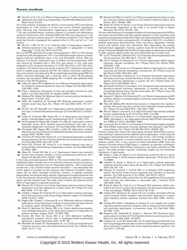
Ventilation-induced lung injury and repair
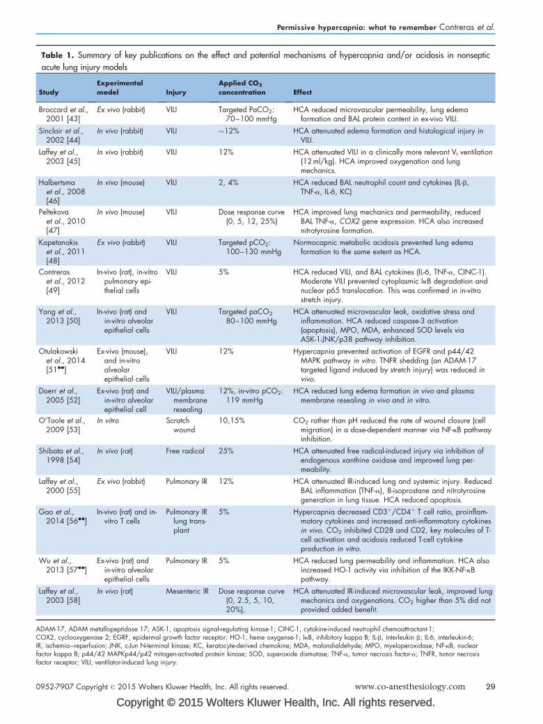
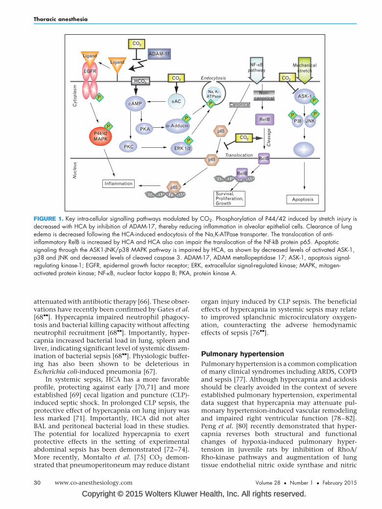
Substantial evidence demonstrates that moderatehypercapnia directly reduces VILI (Table 1) [43–50,51
&&
,52–55,56&&
,57&&
,58]. Studies using clinicallymore relevant (Vt) have further underlined thepotential for hypercapnia to protect against mech-anical stretch [46–49]. The biologic response tocyclic stretch occurs through mechanosensors thattransmit signals from the deformed extracellularmatrix to the interior of the cell [49,50]. A recentstudy has demonstrated that HCA prevents thestretch-induced activation of p44/42 MAP-kinase[51
&&
,59,60] (Fig. 1). Furthermore, hypercapniamarkedly reduced apoptosis, oxidative stress andinflammation by inhibiting the downward acti-vation of the signal-regulating kinase 1 JNK/p38MAP-kinase pathway in alveolar epithelial cells[50]. HCA also reduces stretch-induced lung
Copyright © 2015 Wolters Kluwer
28 www.co-anesthesiology.com
inflammation and improves lung mechanics byinhibiting IkB-a degradation and nuclear p65 trans-location [49] (Fig. 1). The question whether theprotective effect of HCA is mediated through CO2
directly or pH in the context of VILI is still unknown.A recent study comparing the effect of HCA withnormocapnic metabolic acidosis found that meta-bolic acidosis exerted similar protection against VILIas HCA [48].
Of potential concern, hypercapnia may retardlung epithelial and cellular repair following stretch-induced injury. Doerr et al. [52] demonstrated firstthat HCA impairs plasma membrane resealing inVILI. HCA also delays epithelial wound closure inmultiple pulmonary cell lines by reducing NF-kB-dependent epithelial cell migration [53].
Lung ischemia–reperfusion injury
Lung ischemia–reperfusion is a key mechanism ofinjury in diverse clinical situations, including lungtransplantation, pulmonary embolism and ARDS.HCA has been demonstrated to attenuate ische-mia–reperfusion-induced lung injury [54] by pre-serving endothelial capillary barrier function andreducing lipid peroxidation, peroxynitrite pro-duction and apoptosis in lung tissue [55,58,61](Table 1). The dose–response characteristic of hyper-capnia and its efficacy in pulmonary as well assystemic ischemia–reperfusion-induced lung injuryis well described [55,58,61]. Recent insights into theprotective mechanisms of HCA include the demon-stration that hypercapnia suppressed T-cell functionin post-lung transplantation [56
&&
]. Hypercapniaalso attenuated ischemia–reperfusion-inducedNF-kB pathway activation and reduced lung inflam-mation and apoptosis [62], through mechanismsinvolving NF-kB inhibition and upregulation ofthe potent antioxidant enzyme, hemeoxygenase-1[57
&&
].
Sepsis
The potential for HCA to impair the host immuneresponse in the setting of sepsis has raised seriousconcerns (Table 2) [63–67,68
&&
,69–75]. Accumulat-ing data suggest that hypercapnia may result in netbenefit or harm depending on the site and durationof bacterial infection, the use of antibiotic therapyand whether the acidosis induced by hypercapnia isbuffered or not. In pneumonia models, HCA isprotective in early [64] and more established infec-tions [65]. In contrast, hypercapnia may be harmfulin prolonged, untreated pneumonia, likely byreducing neutrophil-mediated and macrophage-mediated bacterial killing. This effect is completely
Health, Inc. All rights reserved.
Volume 28 � Number 1 � February 2015
Copyright © 2015 Wolters Kluwer Health, Inc. All rights reserved.
Table 1. Summary of key publications on the effect and potential mechanisms of hypercapnia and/or acidosis in nonseptic
acute lung injury models
StudyExperimentalmodel Injury
Applied CO2
concentration Effect
Broccard et al.,2001 [43]
Ex vivo (rabbit) VILI Targeted PaCO2:70–100 mmHg
HCA reduced microvascular permeability, lung edemaformation and BAL protein content in ex-vivo VILI.
Sinclair et al.,2002 [44]
In vivo (rabbit) VILI �12% HCA attenuated edema formation and histological injury inVILI.
Laffey et al.,2003 [45]
In vivo (rabbit) VILI 12% HCA attenuated VILI in a clinically more relevant Vt ventilation(12ml/kg). HCA improved oxygenation and lungmechanics.
Halbertsmaet al., 2008[46]
In vivo (mouse) VILI 2, 4% HCA reduced BAL neutrophil count and cytokines (IL-b,TNF-a, IL-6, KC)
Peltekovaet al., 2010[47]
In vivo (mouse) VILI Dose response curve(0, 5, 12, 25%)
HCA improved lung mechanics and permeability, reducedBAL TNF-a, COX2 gene expression. HCA also increasednitrotyrosine formation.
Kapetanakiset al., 2011[48]
Ex vivo (rabbit) VILI Targeted pCO2:100–130 mmHg
Normocapnic metabolic acidosis prevented lung edemaformation to the same extent as HCA.
Contreraset al., 2012[49]
In-vivo (rat), in-vitropulmonary epi-thelial cells
VILI 5% HCA reduced VILI, and BAL cytokines (IL-6, TNF-a, CINC-1).Moderate VILI prevented cytoplasmic IkB degradation andnuclear p65 translocation. This was confirmed in in-vitrostretch injury.
Yang et al.,2013 [50]
In-vivo (rat) andin-vitro alveolarepithelial cells
VILI Targeted paCO2
80–100 mmHgHCA attenuated microvascular leak, oxidative stress and
inflammation. HCA reduced caspase-3 activation(apoptosis), MPO, MDA, enhanced SOD levels viaASK-1-JNK/p38 pathway inhibition.
Otulakowskiet al., 2014[51&&]
Ex-vivo (mouse),and in-vitroalveolarepithelial cells
VILI 12% Hypercapnia prevented activation of EGFR and p44/42MAPK pathway in vitro. TNFR shedding (an ADAM-17targeted ligand induced by stretch injury) was reduced invivo.
Doerr et al.,2005 [52]
Ex-vivo (rat) andin-vitro alveolarepithelial cell
VILI/plasmamembraneresealing
12%, in-vitro pCO2:119 mmHg
HCA reduced lung edema formation in vivo and plasmamembrane resealing in vivo and in vitro.
O’Toole et al.,2009 [53]
In vitro Scratchwound
10,15% CO2 rather than pH reduced the rate of wound closure (cellmigration) in a dose-dependent manner via NF-kB pathwayinhibition.
Shibata et al.,1998 [54]
In vivo (rat) Free radical 25% HCA attenuated free radical-induced injury via inhibition ofendogenous xanthine oxidase and improved lung per-meability.
Laffey et al.,2000 [55]
Ex vivo (rabbit) Pulmonary IR 12% HCA attenuated IR-induced lung and systemic injury. ReducedBAL inflammation (TNF-a), 8-isoprostane and nitrotyrosinegeneration in lung tissue. HCA reduced apoptosis.
Gao et al.,2014 [56&&]
In-vivo (rat) and in-vitro T cells
Pulmonary IRlung trans-plant
5% Hypercapnia decreased CD3þ/CD4þ T cell ratio, proinflam-matory cytokines and increased anti-inflammatory cytokinesin vivo. CO2 inhibited CD28 and CD2, key molecules of T-cell activation and acidosis reduced T-cell cytokineproduction in vitro.
Wu et al.,2013 [57&&]
Ex-vivo (rat) andin-vitro alveolarepithelial cells
Pulmonary IR 5% HCA reduced lung permeability and inflammation. HCA alsoincreased HO-1 activity via inhibition of the IKK-NF-kBpathway.
Laffey et al.,2003 [58]
In vivo (rat) Mesenteric IR Dose response curve(0, 2.5, 5, 10,20%),
HCA attenuated IR-induced microvascular leak, improved lungmechanics and oxygenations. CO2 higher than 5% did notprovided added benefit.
ADAM-17, ADAM metallopeptidase 17; ASK-1, apoptosis signal-regulating kinase-1; CINC-1, cytokine-induced neutrophil chemoattractant-1;COX2, cyclooxygenase 2; EGRF, epidermal growth factor receptor; HO-1, heme oxygense-1; IkB, inhibitory kappa B; IL-b, interleukin b; IL-6, interleukin-6;IR, ischemia–reperfusion; JNK, c-Jun N-terminal kinase; KC, keratocyte-derived chemokine; MDA, malondialdehyde; MPO, myeloperoxidase; NF-kB, nuclearfactor kappa B; p44/42 MAPKp44/p42 mitogen-activated protein kinase; SOD, superoxide dismutase; TNF-a, tumor necrosis factor-a; TNFR, tumor necrosisfactor receptor; VILI, ventilator-induced lung injury.
Permissive hypercapnia: what to remember Contreras et al.
0952-7907 Copyright � 2015 Wolters Kluwer Health, Inc. All rights reserved. www.co-anesthesiology.com 29
Endocytosis
Translocation
Cle
avag
e
Nuc
leus
Cyt
opla
sm
sACcAMP
PKA
Apoptosis
PKC ERK 1/2
P44/42MAPK
Inflammation
Survival,Proliferation,Growth
HCO3
ADAM-17
NF-κBpathway
Mechanicalstretch
ASK-1Non-
canonicalCanonical
Na, K-ATPase
ReIB
CO2
CO2
CO2
P38 JNK
p65
p65
p65
ReIB
ReIB
CO2
Ligand
EGFR
P
P
PP
P
P
P
α-Adducin
P
Ligand
FIGURE 1. Key intra-cellular signalling pathways modulated by CO2. Phosphorylation of P44/42 induced by stretch injury isdecreased with HCA by inhibition of ADAM-17, thereby reducing inflammation in alveolar epithelial cells. Clearance of lungedema is decreased following the HCA-induced endocytosis of the Na,K-ATPase transporter. The translocation of anti-inflammatory RelB is increased by HCA and HCA also can impair the translocation of the NF-kB protein p65. Apoptoticsignaling through the ASK1-JNK/p38 MAPK pathway is impaired by HCA, as shown by decreased levels of activated ASK-1,p38 and JNK and decreased levels of cleaved caspase 3. ADAM-17, ADAM metallopeptidase 17; ASK-1, apoptosis signal-regulating kinase-1; EGFR, epidermal growth factor receptor; ERK, extracellular signal-regulated kinase; MAPK, mitogen-activated protein kinase; NF-kB, nuclear factor kappa B; PKA, protein kinase A.
Thoracic anesthesia
attenuated with antibiotic therapy [66]. These obser-vations have recently been confirmed by Gates et al.[68
&&
]. Hypercapnia impaired neutrophil phagocy-tosis and bacterial killing capacity without affectingneutrophil recruitment [68
&&
]. Importantly, hyper-capnia increased bacterial load in lung, spleen andliver, indicating significant level of systemic dissem-ination of bacterial sepsis [68
&&
]. Physiologic buffer-ing has also been shown to be deleterious inEscherichia coli-induced pneumonia [67].
In systemic sepsis, HCA has a more favorableprofile, protecting against early [70,71] and moreestablished [69] cecal ligation and puncture (CLP)-induced septic shock. In prolonged CLP sepsis, theprotective effect of hypercapnia on lung injury wasless marked [71]. Importantly, HCA did not alterBAL and peritoneal bacterial load in these studies.The potential for localized hypercapnia to exertprotective effects in the setting of experimentalabdominal sepsis has been demonstrated [72–74].More recently, Montalto et al. [75] CO2 demon-strated that pneumoperitoneum may reduce distant
Copyright © 2015 Wolters Kluwer
30 www.co-anesthesiology.com
organ injury induced by CLP sepsis. The beneficialeffects of hypercapnia in systemic sepsis may relateto improved splanchnic microcirculatory oxygen-ation, counteracting the adverse hemodynamiceffects of sepsis [76
&&
].
Pulmonary hypertension
Pulmonary hypertension is a common complicationof many clinical syndromes including ARDS, COPDand sepsis [77]. Although hypercapnia and acidosisshould be clearly avoided in the context of severeestablished pulmonary hypertension, experimentaldata suggest that hypercapnia may attenuate pul-monary hypertension-induced vascular remodelingand impaired right ventricular function [78–82].Peng et al. [80] recently demonstrated that hyper-capnia reverses both structural and functionalchanges of hypoxia-induced pulmonary hyper-tension in juvenile rats by inhibition of RhoA/Rho-kinase pathways and augmentation of lungtissue endothelial nitric oxide synthase and nitric
Health, Inc. All rights reserved.
Volume 28 � Number 1 � February 2015
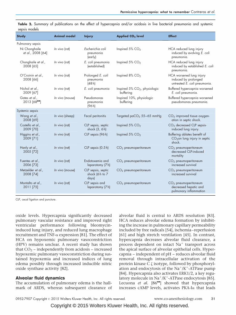
Table 2. Summary of publications on the effect of hypercapnia and/or acidosis in live bacterial pneumonia and systemic
sepsis models
Study Animal model Injury Applied CO2 level Effect
Pulmonary sepsis
Ni Chonghaileet al., 2008 [64]
In vivo (rat) Escherichia colipneumonia(early)
Inspired 5% CO2 HCA reduced lung injuryinduced by evolving E. colipneumonia.
Chonghaile et al.,2008 [65]
In vivo (rat) E. coli pneumonia(established)
Inspired 5% CO2 HCA reduced lung injuryinduced by established E. colipneumonia.
O’Croinin et al.,2008 [66]
In vivo (rat) Prolonged E. colipneumonia(48 h)
Inspired 8% CO2 HCA worsened lung injuryinduced by prolongeduntreated E. coli pneumonia.
Nichol et al.,2009 [67]
In vivo (rat) E. coli pneumonia Inspired 5% CO2, physiologicbuffering
Buffered hypercapnia worsenedE. coli pneumonia.
Gates et al.,2013 [68&&]
In vivo (mouse) Pseudomonaspneumonia(96 h)
Inspired 10%, physiologicbuffering
Buffered hypercapnia worsenedpseudomonas pneumonia.
Systemic sepsis
Wang et al.,2008 [69]
In vivo (sheep) Fecal peritonitis Targeted paCO2 55–65 mmHg CO2 improved tissue oxygen-ation in septic shock.
Costello et al.,2009 [70]
In vivo (rat) CLP sepsis, septicshock (3, 6 h)
Inspired 5% CO2 CO2 decreased CLP sepsis-induced lung injury.
Higgins et al.,2009 [71]
In vivo (rat) CLP sepsis (96h) Inspired 5% CO2 Buffering ablates benefit ofCO2on lung injury in septicshock.
Hanly et al.,2005 [72]
In vivo (rat) CLP sepsis (0.5h) CO2 pneumoperitoneum CO2 pneumoperitoneumdecreased CLP-inducedmortality
Fuentes et al.,2006 [73]
In vivo (rat) Endotoxemia andlaparotomy (7h)
CO2 pneumoperitoneum CO2 pneumoperitoneumincreased survival
Metzelder et al.,2008 [74]
In vivo (mouse) CLP sepsis, septicshock (6 h to 7days)
CO2 pneumoperitoneum CO2 pneumoperitoneumincreased survival
Montalto et al.,2011 [75]
In vivo (rat) CLP sepsis andlaparotomy (7h)
CO2 pneumoperitoneum CO2 pneumoperitoneumdecreased hepatic andpulmonary inflammation
CLP, cecal ligation and puncture.
Permissive hypercapnia: what to remember Contreras et al.
oxide levels. Hypercapnia significantly decreasedpulmonary vascular resistance and improved rightventricular performance following bleomycin-induced lung injury, and reduced lung macrophagerecruitment and TNF-a expression [81]. The effect ofHCA on hypoxemic pulmonary vasoconstriction(HPV) remains unclear. A recent study has shownthat CO2 – independently from acidosis – increasedhypoxemic pulmonary vasoconstriction during sus-tained hypoxemia and increased indices of lungedema possibly through increased inducible nitricoxide synthase activity [82].
Alveolar fluid dynamicsThe accumulation of pulmonary edema is the hall-mark of ARDS, whereas subsequent clearance of
Copyright © 2015 Wolters Kluwe
0952-7907 Copyright � 2015 Wolters Kluwer Health, Inc. All rights rese
alveolar fluid is central to ARDS resolution [83].HCA reduces alveolar edema formation by inhibit-ing the increase in pulmonary capillary permeabilityincluded by free radicals [54], ischemia–reperfusion[61] and high stretch ventilation [45]. In contrast,hypercapnia decreases alveolar fluid clearance, aprocess dependent on intact Naþ transport acrossthe apical surface of alveolar epithelial cells. Hyper-capnia – independent of pH – reduces alveolar fluidremoval through intracellular activation of theprotein kinase C z isotype, followed by phosphoryl-ation and endocytosis of the Naþ/Kþ-ATPase pump[84]. Hypercapnia also activates ERK1/2, a key regu-latory molecule in Naþ/Kþ-ATPase endocytosis [85].Lecuona et al. [86
&&
] showed that hypercapniaincreases cAMP levels, activates PKA-Ia that leads
r Health, Inc. All rights reserved.
rved. www.co-anesthesiology.com 31
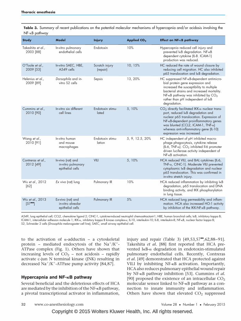
Table 3. Summary of recent publications on the potential molecular mechanisms of hypercapnia and/or acidosis involving the
NF-kB pathway
Study Model Injury Applied CO2 Effect on NF-kB pathway
Takeshita et al.,2003 [88]
In-vitro pulmonaryendothelial cells
Endotoxin 10% Hypercapnia reduced cell injury andprevented IkB degradation. NF-kBdependent cytokine (IL-8. ICAM-1)production was reduced.
O’Toole et al.,2009 [53]
In-vitro SAEC, HBE,A549 cells
Scratch injury(repair)
10, 15% HC reduced the rate of wound closure byreducing cell migration. HC also inhibitedp65 translocation and IkB degradation.
Helenius et al.,2009 [89]
Dorsophila and in-vitro S2 cells
Sepsis 13, 20% HC suppressed NF-kB-dependent antimicro-bial protein gene expression andincreased the susceptibility to multiplebacterial strains and increased mortality.NF-kB pathway was inhibited by CO2
rather than pH independent of IkBdegradation.
Cummins et al.,2010 [90]
In-vitro six differentcell lines
Endotoxin stimu-lated
5, 10% CO2 directly facilitated IKK-a nuclear trans-port, reduced IkB degradation andnuclear p65 translocation. Expression ofNF-kB-dependent proinflammatory geneswas blunted (CCL2, ICAM-1, TNF-a)whereas anti-inflammatory gene (IL-10)expression was increased.
Wang et al.,2010 [91]
In-vitro humanand mousemacrophages
Endotoxin stimu-lation
5, 9, 12.5, 20% HC independent of pH inhibited macro-phage phagocytosis, cytokine release(IL-6, TNF-a). CO2 inhibited Il-6 promoterdriven luciferase activity independent ofNF-kB activation.
Contreras et al.,2012 [49]
In-vivo (rat) andin-vitro pulmonaryepithelial cells
VILI 5, 10% HCA reduced VILI, and BAL cytokines (IL-6,TNF-a, CINC-1). Moderate VILI preventedcytoplasmic IkB degradation and nuclearp65 translocation. This was confirmed inin-vitro stretch injury.
Wu et al., 2012[62]
Ex vivo (rat) lung Pulmonary IR 10% HCA reduced inflammation by inhibiting IkBdegradation, p65 translocation and DNAbinding activity, and IKK phosphorylationin lung tissue.
Wu et al., 2013[57&&]
Ex-vivo (rat) andin-vitro alveolarepithelial cells
Pulmonary IR 5% HCA reduced lung permeability and inflam-mation. HCA also increased HO-1 activityby inhibition of the IKK-NF-kB pathway.
A549, lung epithelial cell; CCL2, chemokine ligand 2; CINC-1, cytokine-induced neutrophil chemoattractant-1; HBE, human bronchial cells; IkB, inhibitory kappa B;ICAM-1, intercellular adhesion molecule 1; IKK-a, inhibitory kappa B kinase complex-a; IL-10, interleukin-10; IL-8, interleukin-8; NF-kB, nuclear factor kappa B;S2, Schneider 2 cells (Drosophila melanogaster cell line); SAEC, small airway epithelial cell.
Thoracic anesthesia
to the activation of a-adductin – a cytoskeletalprotein – mediated endocytosis of the Naþ/Kþ-ATPase complex (Fig. 1). Others have shown thatincreasing levels of CO2 – not acidosis – rapidlyactivate c-jun N terminal kinase (JNK) resulting indecreased Naþ/Kþ-ATPase pump activity [84,87].
Hypercapnia and NF-kB pathway
Several beneficial and the deleterious effects of HCAare mediated by the inhibition of the NF-kB pathway,a pivotal transcriptional activator in inflammation,
Copyright © 2015 Wolters Kluwer
32 www.co-anesthesiology.com
injury and repair (Table 3) [49,53,57&&
,62,88–91].Takeshita et al. [88] first reported that HCA pre-vented IkB-a degradation in endotoxin-stimulatedpulmonary endothelial cells. Recently, Contreraset al. [49] demonstrated that HCA protected againstVILI by inhibiting NF-kB activation. Importantly,HCA also reduces pulmonary epithelial wound repairby NF-kB pathway inhibition [53]. Cummins et al.[90] proposed the existence of an intracellular CO2
molecular sensor linked to NF-kB pathway as a con-nection to innate immunity and inflammation.Others have shown that elevated CO2 suppressed
Health, Inc. All rights reserved.
Volume 28 � Number 1 � February 2015
Permissive hypercapnia: what to remember Contreras et al.
host defence by inhibiting NF-kB-dependent antimi-crobial peptide gene expression in Drosophila result-ing in increased mortality to bacterial infection [89].High levels of CO2 have also been shown to inhibitIL-6, TNF-a induction and phagocytosis in endo-toxin-stimulated macrophages [91]. In the two latterstudies, hypercapnia inhibited the NF-kB pathwaywithout affecting IkB-a degradation, suggesting thatother pathways or regulatory steps may have beeninvolved in mediating the immunosuppressive effectof hypercapnia.
HYPERCAPNIA IN THE CLINICALCONTEXT
Hypercapnia is frequently encountered in thesetting of acute respiratory failure, both as a con-sequence of the disease process and as a result ofstrategies to minimize the potential of mechanicalventilation to stretch and further injure the lung.
Acute respiratory distress syndrome
To date, there have been no clinical trials examiningthe direct effect of hypercapnia on patients withARDS. The potential of PHC to improve ARDSpatients’ survival as part of a protective ventilationstrategy was suggested first by Hickling et al. [92,93].Subsequently, two RCTs comparing ‘traditional’versus low Vt showed improved survival in patientswith ARDS [1,2]. The secondary analysis of theARMA trial suggested that patients with moderateHCA on study day 1 had significantly less odds ratioof death at 28 days in the setting of higher – but notlower – Vt [94]. Because the primary aim of thesetrials was to investigate the effect of low stretchventilation on ARDS, the direct relationshipbetween PHC and lung protection remains to bedetermined. In a recent pilot study, a combinationof stepwise recruitment–derecruitment with PHCwas compared with lung protective ventilation[95]. This ‘open lung’ strategy resulted in signifi-cantly better lung compliance, systemic oxygen-ation in a 7-day period. However, arterial CO2 andpH were not different between the two groups,suggesting that the achieved benefits were morelikely related to better recruitment maneuver inthe open-lung strategy group than to PHC per se.
Asthma
The utility of PHC in status asthmaticus wasreported first by Darioli in 1984 [96]. Subsequentstudies also confirmed that lowering minute venti-lation, in conjunction with longer expiratorytime, significantly reduces dynamic hyperinflation
Copyright © 2015 Wolters Kluwe
0952-7907 Copyright � 2015 Wolters Kluwer Health, Inc. All rights rese
[97,98]. In these studies, arterial CO2 was kept inten-tionally at moderately elevated levels (63, 68mmHg), whereas extremely high arterial CO2 levels(150–200 mmHg) were also well tolerated in caseseries involving more severe presentations ofasthma [99]. In spite of lack of RCTs to guide mech-anical ventilation in status asthmaticus, PHC hasbeen frequently used in patients with severe asthmaadmitted to ICUs both in Europe [100] and in NorthAmerica [101].
Chronic obstructive pulmonary disease
Respiratory failure during COPD exacerbations is adirect result of an acute increase in airway narrow-ing, with increased respiratory workload, similarlyto acute severe asthma. Although noninvasiveventilation is the first-line ventilation strategy inpatients with COPD exacerbations [102], extremerespiratory muscle fatigue, CO2 retention-induced‘coma’ may necessitate invasive ventilation. Theprimary aim of mechanical ventilation in this set-ting is to reduce over-inflation and prevent VILI byreducing minute ventilation, decreasing inspira-tory–expiratory ratio and increasing inspiratoryflow rate. PHC is a useful approach to achievingthese goals [103].
Neonatal respiratory failure
Advances in perinatal medical care and ventilatorysupport have reduced mortality in high-risk new-borns [104]. Prolonged mechanical ventilation,however, remains an important cause of pulmonarycomplications, such asbronchopulmonarydysplasia.Early observational studies suggested that PHC maylower the risk for bronchopulmonary dysplasia inpremature infants. Mariani et al. [105] first reportedthat ventilation strategies allowing higher PaCO2
levels (45–55 versus 35–45 mmHg) in preterminfants, in the first 96 h of life, result in faster weaningfrom mechanical ventilation. Subsequently, a largermulticenter RCT compared PHC with conventionalventilation with dexamethasone or placebo using a2�2 factorial design [106]. Although the trial wasstopped due to adverse events in the dexamethasonegroups, PHC decreased need for assisted ventilationat 36-week gestational age from 16% to just 1%. Thepotential of PHC to cause intracranial hemorrhageand adverse neurological outcomes in prematureinfants is a significant concern. Although an earlymeta-analysis of PHC in newborn infants demon-strated some trends toward decreased incidence ofintracranial hemorrhage in the PHC group [107], in arecent study higher ranges of hypercapnia (PaCO2:55–65 mmHg) were associated with a significant
r Health, Inc. All rights reserved.
rved. www.co-anesthesiology.com 33
Thoracic anesthesia
increase in combined mental impairment and deathin extremely preterm infants [108]. These dataindicate that more research is needed to determinethe optimal range of hypercapnia to balance thebenefits and potential harms of PHC in preterminfants.
EXTRA-CORPOREAL CO2 REMOVAL: THEFUTURE?
In recent years, new-generation extracorporeal CO2
removal (ECCO2-R) devices have been developedthat offer lower resistance to blood flow, have smallpriming volumes and have much more effective gasexchange [109]. These devices may further facilitatelung protective ventilation by allowing greaterreductions in Vt and plateau pressures in patientswith severe ARDS, while avoiding the potential forsevere hypercapnia – beyond levels that are gener-ally well tolerated by patients under current PHCapproaches. The rationale for ECCO2-R derives fromstudies demonstrating that lung hyperinflation stilloccurs in approximately 30% of ARDS patientsdespite lung protective ventilation strategies andthe potential to further decrease mortality by reduc-ing plateau pressures [110]. In a recent proof-of-concept study, Terragni et al. [111] demonstratedthat ECCO2-R could improve pulmonary protectionand decrease pulmonary cytokine concentrations byallowing very low Vt ventilation (3.5–5 ml/kg ofPBW) in patients with ARDS. Most recently, Beinet al. [112
&&
] demonstrated the feasibility of com-bining ECCO2-R with a tidal volume strategy of3 ml/kg in 79 patients with established ARDS.
CONCLUSION
Protective ventilatory strategies involving PHCare widely used in patients with severe respiratoryfailure, particularly in ARDS, status asthmaticus,COPD and neonatal respiratory failure. Thephysiologic effects of hypercapnia are increasin-gly well understood, whereas important recentinsights have emerged regarding the cellular andmolecular mechanisms of action of hypercapniaand acidosis. Acute HCA is protective in multiplemodels of nonseptic lung injury. These effects aremediated by potent effects on the host immunesystem, with key effects mediated by inhibition ofthe NF-kB pathway. Hypercapnia-mediated NF-kBinhibition may also explain several deleteriouseffects, including delayed epithelial wound heal-ing and decreased bacterial killing, which has beendemonstrated to cause worse lung injury in pro-longed untreated pneumonia models. The poten-tial for extracorporeal CO2 removal technologies to
Copyright © 2015 Wolters Kluwer
34 www.co-anesthesiology.com
facilitate even greater reductions in tidal andminute ventilation is clear, but awaits definitivestudies.
Acknowledgements
None.
Financial support and sponsorship
J.G.L. is supported by operating grants from the Cana-dian Institutes of Health Research and PhysiciansServices Incorporated and by a Merit award from theDepartment of Anesthesia at the University of Toronto.C.M. is funded by a grant from the European RespiratorySociety.
Conflicts of interest
None.
REFERENCES AND RECOMMENDEDREADINGPapers of particular interest, published within the annual period of review, havebeen highlighted as:
& of special interest&& of outstanding interest1. The Acute Respiratory Distress Syndrome Network. Ventilation with lowertidal volumes as compared with traditional tidal volumes for acute lung injuryand the acute respiratory distress syndrome. N Engl J Med 2000;342:1301–1308.
2. Amato MB, Barbas CS, Medeiros DM, et al. Effect of a protective-ventilationstrategy on mortality in the acute respiratory distress syndrome. N Engl J Med1998; 338:347–354.
3. Brogan TV, Hedges RG, McKinney S, et al. Pulmonary NO synthase inhibi-tion and inspired CO2: effects on V’/Q’ and pulmonary blood flow distribu-tion. Eur Respir J 2000; 16:288–295.
4. Brogan TV, Robertson HT, Lamm WJ, et al. Carbon dioxide added late ininspiration reduces ventilation-perfusion heterogeneity without causingrespiratory acidosis. J Appl Physiol 2004; 96:1894–1898.
5. Swenson ER, Robertson HT, Hlastala MP. Effects of inspired carbon dioxideon ventilation-perfusion matching in normoxia, hypoxia, and hyperoxia. Am JRespir Crit Care Med 1994; 149:1563–1569.
6. Keenan RJ, Todd TR, Demajo W, Slutsky AS. Effects of hypercarbia onarterial and alveolar oxygen tensions in a model of gram-negative pneumonia.J Appl Physiol 1990; 68:1820–1825.
7. Sinclair SE, Kregenow DA, Starr I, et al. Therapeutic hypercapnia andventilation-perfusion matching in acute lung injury: low minute ventilationvs inspired CO2. Chest 2006; 130:85–92.
8.&&
Emery MJ, Eveland RL, Min JH, et al. CO2 relaxation of the rat lungparenchymal strip. Respir Physiol Neurobiol 2013; 186:33–39.
Lung parenchymal strips from rats were exposed to normoxic hypocapnia (pCO2:20 mmHg, pH: 7.68) or hypercapnia (pCO2: 53mmHg, pH: 7.26) and length–tension curves were determined. Increasing CO2 from hypocapnic to hypercapniccondition induced parenchymal relaxation, and this effect was reversed with actin-myosin blocking agent. This is the first study to demonstrate that hypercapniaincreases lung parenchymal compliance, providing a mechanism by which alveolarpCO2 may modulate ventilation–perfusion matching.
9. Wildeboer-Venema F. The influences of temperature and humidity upon theisolated surfactant film of the dog. Respir Physiol 1980; 39:63–71.
10.&&
Lele EE, Hantos Z, Bitay M, et al. Bronchoconstriction during alveolarhypocapnia and systemic hypercapnia in dogs with a cardiopulmonarybypass. Respir Physiol Neurobiol 2011; 175:140–145.
This study investigates the separate effect of alveolar and systemic CO2 on lungtissue and airways in dogs using cardiopulmonary bypass. Alveolar hypercapniahad no effect on pulmonary mechanics, whereas severe hypocapnia (15.2 mmHg)resulted in peripheral bronchoconstriction and worsened ventilation–perfusionmatching. Systemic hypercapnic acidosis caused vagal nerve-mediated centralairway constriction. This study highlights that the biologic effect of CO2 and/or pHmay convey distinct physiologic responses in the lung depending on the site ofaction, which is alveolar versus systemic.11. Waldron MA, Fisher JT. Differential effects of CO2 and hypoxia on bronch-
omotor tone in the newborn dog. Respir Physiol 1988; 72:271–282.12. D’Angelo E, Calderini IS, Tavola M. The effects of CO2 on respiratory
mechanics in anesthetized paralyzed humans. Anesthesiology 2001;94:604–610.
Health, Inc. All rights reserved.
Volume 28 � Number 1 � February 2015
Permissive hypercapnia: what to remember Contreras et al.
13. Sterling GM, Holst PE, Nadel JA. Effect of CO2 and pH on bronchocon-striction caused by serotonin vs. acetylcholine. J Appl Physiol 1972;32:39–43.
14. Rodarte JR, Hyatt RE. Effect of acute exposure to CO2 on lung mechanics innormal men. Respir Physiol 1973; 17:135–145.
15. Jonville S, Delpech N, Denjean A. Contribution of respiratory acidosis todiaphragmatic fatigue at exercise. Eur Respir J 2002; 19:1079–1086.
16.&&
Jung B, Sebbane M, Goff CL, et al. Moderate and prolonged hypercapnicacidosis may protect against ventilator-induced diaphragmatic dysfunction inhealthy piglet: an in vivo study. Crit Care 2013; 17:R15.
The authors randomized mechanically ventilated piglets to receive normocapniaor hypercapnia (paCO2: 55–70 mmHg) for 72 h. To assess in-vivo diaphragmaticcontractile force, transdiaphragmatic pressure (Pdi) was determined every 12 h.PDI decreased gradually by 25% in the normocapnia group, whereas HCA wasassociated with preserved diaphragmatic contractility. Although the mechanismof HCA was not explored in this study, the results provide potentially importantpiece of evidence on the beneficial effect of HCA in mechanically ventilatedpatients.17.&&
Schellekens WJ, van Hees HW, Kox M, et al. Hypercapnia attenuatesventilator-induced diaphragm atrophy and modulates dysfunction. Crit Care2014; 18:R28.
This study provides novel mechanistic insights into the biologic effect of HCA ondiaphragmatic function with particular relevance to prolonged mechanical ventila-tion. Male rats were ventilated for 18 h under normocapnic or hypercapniccondition. Control group included nonventilated animals. Hypercapnia preventedmyosin loss and muscle atrophy evidenced by reduced concentration of myosinheavy chain concentration in isolated muscle fibers and less reduction in cross-sectional area of muscle fibers in ventilated rats. HCA was also associated withmore preserved contractile function. Diaphragmatic inflammation and proteolysiswere inhibited by HCA.18. Tang WC, Weil MH, Gazmuri RJ, et al. Reversible impairment of myocardial
contractility due to hypercarbic acidosis in the isolated perfused rat heart.Crit Care Med 1991; 19:218–224.
19. Akca O, Doufas AG, Morioka N, et al. Hypercapnia improves tissue oxygena-tion. Anesthesiology 2002; 97:801–806.
20. Mas A, Saura P, Joseph D, et al. Effect of acute moderate changes in PaCO2on global hemodynamics and gastric perfusion. Crit Care Med 2000;28:360–365.
21. Akca O, Sessler DI, Delong D, et al. Tissue oxygenation response to mildhypercapnia during cardiopulmonary bypass with constant pump output. Br JAnaesth 2006; 96:708–714.
22. Fleischmann E, Herbst F, Kugener A, et al. Mild hypercapnia increasessubcutaneous and colonic oxygen tension in patients given 80% inspiredoxygen during abdominal surgery. Anesthesiology 2006; 104:944–949.
23. Hager H, Reddy D, Mandadi G, et al. Hypercapnia improves tissue oxygena-tion in morbidly obese surgical patients. Anesth Analg 2006; 103:677–681.
24. Schwartges I, Schwarte LA, Fournell A, et al. Hypercapnia induces aconcentration-dependent increase in gastric mucosal oxygenation in dogs.Intensive Care Med 2008; 34:1898–1906.
25. Guzman JA, Kruse JA. Splanchnic hemodynamics and gut mucosal-arterialPCO(2) gradient during systemic hypocapnia. J Appl Physiol 1985, 1999;87:1102–1106.
26. Taghavi S, Jayarajan SN, Ferrer LM, et al. Permissive hypoventilation’ in aswine model of hemorrhagic shock. J Trauma Acute Care Surg 2014;77:14–19.
27.&&
Akca O, Kurz A, Fleischmann E, et al. Hypercapnia and surgical site infection:a randomized trial. Br J Anaesth 2013; 111:759–767.
SSI is inversely related to tissue oxygenation. Furthermore, mild hypercapnia hasbeen shown to improve tissue oxygenation in a number of experimental andhuman studies. The investigators hypothesized that moderate hypercapnia wouldreduce SSI compared with normocapnia in patients undergoing elective colonsurgery. Patients were randomly assigned to intraoperative normocapnia(ETCO2: 35 mmHg, n¼623) or hypercapnia (ETCO2: 50 mmHg, n¼592).The primary outcome was SSI rate within 30 postoperative days. The SSI ratewas 13.3% in the normocapnia and 11.2% in the hypercapnia group (P¼0.29).The study was stopped after recruiting 1206 patients due to small treatmenteffect.28. Brian JE Jr. Carbon dioxide and the cerebral circulation. Anesthesiology
1998; 88:1365–1386.29.&&
Reinhard M, Schwarzer G, Briel M, et al. Cerebrovascular reactivity predictsstroke in high-grade carotid artery disease. Neurology 2014; 83:1424–1431.
This meta-analysis, including nine studies and 754 patients, assessed theusefulness of transcranial Doppler (TCD) CO2 reactivity for the prediction ofpatients with symptomatic or asymptomatic severe carotid artery stenosis orocclusion. In a multiple regression model, reduced CO2 activity on TCD wasindependently associated with an increase in ipsilateral ischemic stroke (hazardratio: 3.69, P<0.0001). Risk prediction for asymptomatic, as well as sympto-matic carotid stenosis, was significant and associated with a hazard ratio of 1.64and 1.95, respectively, per every 10% of reduction in CO2 reactivity. TCD CO2
reactivity may represent a simple investigation tool to assess stroke risk inpatients with carotid stenosis. This should be further tested in prospectivestudies.
Copyright © 2015 Wolters Kluwe
0952-7907 Copyright � 2015 Wolters Kluwer Health, Inc. All rights rese
30. Nakahata K, Kinoshita H, Hirano Y, et al. Mild hypercapnia induces vaso-dilation via adenosine triphosphate-sensitive Kþ channels in parenchymalmicrovessels of the rat cerebral cortex. Anesthesiology 2003; 99:1333–1339.
31. Nnorom CC, Davis C, Fedinec AL, et al. Contributions of KATP and KCachannels to cerebral arteriolar dilation to hypercapnia in neonatal brain.Physiol Rep 2014; 2:e12127. [Epub ahead of print]. doi: 10.14814/phy2.12127.
32. Raichle ME, Posner JB, Plum F. Cerebral blood flow during and afterhyperventilation. Arch Neurol 1970; 23:394–403.
33. Curley G, Kavanagh BP, Laffey JG. Hypocapnia and the injured brain:evidence for harm. Crit Care Med 2011; 39:229–230.
34. Diringer MN, Videen TO, Yundt K, et al. Regional cerebrovascular andmetabolic effects of hyperventilation after severe traumatic brain injury.J Neurosurg 2002; 96:103–108.
35. Coles JP, Fryer TD, Coleman MR, et al. Hyperventilation following head injury:effect on ischemic burden and cerebral oxidative metabolism. Crit Care Med2007; 35:568–578.
36. Ito H, Ibaraki M, Kanno I, et al. Changes in the arterial fraction of humancerebral blood volume during hypercapnia and hypocapnia measured bypositron emission tomography. J Cereb Blood Flow Metab 2005; 25:852–857.
37. Ito H, Kanno I, Iida H, et al. Arterial fraction of cerebral blood volume inhumans measured by positron emission tomography. Ann Nucl Med 2001;15:111–116.
38. Huttunen J, Tolvanen H, Heinonen E, et al. Effects of voluntary hyper-ventilation on cortical sensory responses. Electroencephalographicand magnetoencephalographic studies. Exp Brain Res 1999; 125:248–254.
39. Bergsholm P, Gran L, Bleie H. Seizure duration in unilateral electro-convulsive therapy. The effect of hypocapnia induced by hyperventilationand the effect of ventilation with oxygen. Acta Psychiatr Scand 1984;69:121–128.
40. Caulfield EV, Dutton RP, Floccare DJ, et al. Prehospital hypocapnia and pooroutcome after severe traumatic brain injury. J Trauma 2009; 66:1577–1582;discussion 1583.
41. Davis DP, Heister R, Poste JC, et al. Ventilation patterns in patients withsevere traumatic brain injury following paramedic rapid sequence intubation.Neurocrit Care 2005; 2:165–171.
42. Warner KJ, Cuschieri J, Copass MK, et al. The impact of prehospitalventilation on outcome after severe traumatic brain injury. J Trauma 2007;62:1330–1336; discussion 1336–1338.
43. Broccard AF, Hotchkiss JR, Vannay C, et al. Protective effects of hypercapnicacidosis on ventilator-induced lung injury. Am J Respir Crit Care Med 2001;164:802–806.
44. Sinclair SE, Kregenow DA, Lamm WJ, et al. Hypercapnic acidosis isprotective in an in vivo model of ventilator-induced lung injury. Am J RespirCrit Care Med 2002; 166:403–408.
45. Laffey JG, Engelberts D, Duggan M, et al. Carbon dioxide attenuatespulmonary impairment resulting from hyperventilation. Crit Care Med2003; 31:2634–2640.
46. Halbertsma FJ, Vaneker M, Pickkers P, et al. Hypercapnic acidosis attenuatesthe pulmonary innate immune response in ventilated healthy mice. Crit CareMed 2008; 36:2403–2406.
47. Peltekova V, Engelberts D, Otulakowski G, et al. Hypercapnic acidosisin ventilator-induced lung injury. Intensive Care Med 2010; 36:869–878.
48. Kapetanakis T, Siempos II, Metaxas EI, et al. Metabolic acidosis may be asprotective as hypercapnic acidosis in an ex-vivo model of severe ventilator-induced lung injury: a pilot study. BMC Anesthesiol 2011; 11:8.
49. Contreras M, Ansari B, Curley G, et al. Hypercapnic acidosis attenuatesventilation-induced lung injury by a nuclear factor-kappaB-dependent me-chanism. Crit Care Med 2012; 40:2622–2630.
50. Yang WC, Song CY, Wang N, et al. Hypercapnic acidosis confers anti-oxidant and antiapoptosis effects against ventilator-induced lung injury. LabInvest 2013; 93:1339–1349.
51.&&
Otulakowski G, Engelberts D, Gusarova GA, et al. Hypercapnia attenuatesventilator induced lung injury via a disintegrin and metalloprotease-17.J Physiol 2014; 592:4507–4521.
The paper demonstrated hypercapnia inhibits stretch-induced injury via a mechan-ism involving inhibition of the matrix metalloprotease ADAM17, which in turndecreases activation of p44/42 MAPK activation that occurs in response tostretch-induced lung injury.52. Doerr CH, Gajic O, Berrios JC, et al. Hypercapnic acidosis impairs plasma
membrane wound resealing in ventilator-injured lungs. Am J Respir Crit CareMed 2005; 171:1371–1377.
53. O’Toole D, Hassett P, Contreras M, et al. Hypercapnic acidosis attenuatespulmonary epithelial wound repair by an NF-kappaB dependent mechanism.Thorax 2009; 64:976–982.
54. Shibata K, Cregg N, Engelberts D, et al. Hypercapnic acidosis may attenuateacute lung injury by inhibition of endogenous xanthine oxidase. Am J RespirCrit Care Med 1998; 158 (5 Pt 1):1578–1584.
55. Laffey JG, Tanaka M, Engelberts D, et al. Therapeutic hypercapnia reducespulmonary and systemic injury following in vivo lung reperfusion. Am J RespirCrit Care Med 2000; 162:2287–2294.
r Health, Inc. All rights reserved.
rved. www.co-anesthesiology.com 35
Thoracic anesthesia
56.&&
Gao W, Liu D, Li D, et al. Effects of hypercapnia on T cells in lung ischemia/reperfusion injury after lung transplantation. Exp Biol Med (Maywood) 2014.[Epub ahead of print]
This study aimed to investigate the effects of normocapnia, HCA and bufferedhypercapnia on T-cell function in in-vivo and in-vitro ischemia–reperfusioninjury following lung transplantation. Hypercapnia decreased CD3þ/CD4þ
T cell ratio, proinflammatory cytokines, whereas it increased anti-inflammatorycytokines. Furthermore, CO2 inhibited CD28 and CD2, key molecules of T cellactivation, whereas acidosis reduced T cell cytokine production. This is the firststudy that directly investigated the separate effect of CO2 and pH on T cellfunction.57.&&
Wu SY, Li MH, Ko FC, et al. Protective effect of hypercapnic acidosis inischemia-reperfusion lung injury is attributable to upregulation of hemeoxygenase-1. PLoS One 2013; 8:e74742.
Wu et al. [57&&] implicated the protein hemeoxygenase-1 (HO-1) in HC-inducedprotection by NF-kB suppression. HO-1 is an antioxidant enzyme shown to protectagainst ischemia–reperfusion injury in the lung (Constantin, 2012). After theinduction of ischemia–reperfusion injury in isolated rat lung preparations, HCAwas induced by ventilation with a 10% CO2 gas mixture. In vitro, cells weresubjected to 24 h of hypoxia followed by 4 h of NC (5% CO2) or HC (10% CO2)incubation. HC without injury was shown to induce HO-1 expression and proteinlevels in isolated lungs compared with NC. The indices of ischemia–reperfusioninjury were shown to be reduced by HC at a physiologic level (decreased PAP andedema, improved pathology) and a molecular level in which NF-kB pathwayactivation was decreased. siRNA targeting HO-1 in A549 cells abolished HCAinhibition of the NF-kB pathway.58. Laffey JG, Jankov RP, Engelberts D, et al. Effects of therapeutic hypercapnia
on mesenteric ischemia-reperfusion injury. Am J Respir Crit Care Med 2003;168:1383–1390.
59. Pugin J. Molecular mechanisms of lung cell activation induced by cyclicstretch. Crit Care Med 2003; 31 (Suppl 4):S200–S206.
60. Gillespie PG, Walker RG. Molecular basis of mechanosensory transduction.Nature 2001; 413:194–202.
61. Laffey JG, Engelberts D, Kavanagh BP. Buffering hypercapnic acidosisworsens acute lung injury. Am J Respir Crit Care Med 2000; 161:141–146.
62. Wu SY, Wu CP, Kang BH, et al. Hypercapnic acidosis attenuates reperfu-sion injury in isolated and perfused rat lungs. Crit Care Med 2012; 40:553–559.
63. Curley G, Contreras MM, Nichol AD, et al. Hypercapnia and acidosis insepsis: a double-edged sword? Anesthesiology 2010; 112:462–472.
64. Ni Chonghaile M, Higgins BD, Costello JF, Laffey JG. Hypercapnic acidosisattenuates severe acute bacterial pneumonia-induced lung injury by aneutrophil-independent mechanism. Crit Care Med 2008; 36:3135–3144.
65. Chonghaile MN, Higgins BD, Costello J, Laffey JG. Hypercapnic acidosisattenuates lung injury induced by established bacterial pneumonia. Anesthe-siology 2008; 109:837–848.
66. O’Croinin DF, Nichol AD, Hopkins N, et al. Sustained hypercapnic acidosisduring pulmonary infection increases bacterial load and worsens lung injury.Crit Care Med 2008; 36:2128–2135.
67. Nichol AD, O’Cronin DF, Howell K, et al. Infection-induced lung injury isworsened after renal buffering of hypercapnic acidosis. Crit Care Med 2009;37:2953–2961.
68.&&
Gates KL, Howell HA, Nair A, et al. Hypercapnia impairs lung neutrophilfunction and increases mortality in murine pseudomonas pneumonia. Am JRespir Cell Mol Biol 2013; 49:821–828.
In this study, prolonged exposure (96 h) to 10% environmental CO2 resulted in asignificantly higher mortality rate in mice with Pseudomonas aeruginosa pneumo-nia than in animals exposed to room air. Physiologic buffering – that is, placing theanimals into a CO2 chamber before bacterial inoculation – was associated withsimilar degree of mortality as CO2 exposure after bacterial inoculation. Hyper-capnia did not affect neutrophil recruitment; however, it impaired neutrophilphagocytosis and bacterial killing capacity. Hypercapnia increased bacterial loadin the lung. Systemic dissemination of bacterial sepsis was reflected in increasedspleen and liver bacterial load. This study further confirms the potential harmfuleffect of hypercapnia on bacterial pneumonia.69. Wang Z, Su F, Bruhn A, et al. Acute hypercapnia improves indices of tissue
oxygenation more than dobutamine in septic shock. Am J Respir Crit CareMed 2008; 177:178–183.
70. Costello J, Higgins B, Contreras M, et al. Hypercapnic acidosis attenuatesshock and lung injury in early and prolonged systemic sepsis. Crit Care Med2009; 37:2412–2420.
71. Higgins BD, Costello J, Contreras M, et al. Differential effects of bufferedhypercapnia versus hypercapnic acidosis on shock and lung injury inducedby systemic sepsis. Anesthesiology 2009; 111:1317–1326.
72. Hanly EJ, Bachman SL, Marohn MR, et al. Carbon dioxide pneumoperito-neum-mediated attenuation of the inflammatory response is independent ofsystemic acidosis. Surgery 2005; 137:559–566.
73. Fuentes JM, Hanly EJ, Aurora AR, et al. CO2 abdominal insufflationpretreatment increases survival after a lipopolysaccharide-contaminatedlaparotomy. J Gastrointest Surg 2006; 10:32–38.
74. Metzelder M, Kuebler JF, Shimotakahara A, et al. CO2 pneumoperitoneumincreases survival in mice with polymicrobial peritonitis. Eur J Pediatr Surg2008; 18:171–175.
Copyright © 2015 Wolters Kluwer
36 www.co-anesthesiology.com
75. Montalto AS, Bitto A, Irrera N, et al. CO2 pneumoperitoneum impact on earlyliver and lung cytokine expression in a rat model of abdominal sepsis. SurgEndosc 2011; 26:984–989.
76.&&
Stubs CC, Picker O, Schulz J, et al. Acute, short-term hypercapnia improvesmicrovascular oxygenation of the colon in an animal model of sepsis.Microvasc Res 2013; 90:180–186.
The aim of this study was to investigate the effects of acute hypercapnia and HCA onthe colonic microcirculation and early cytokine response in colon ascendens stentperitonitis-induced polymicrobial sepsis. Rats were randomized after 24 h sepsis andventilated for 120 min with normocapnia (45�5 mmHg) or HCA (75�5 mmHg) orbuffered hypercapnia (thromethamine). Colon wall tissue oxygenation, hemody-namics and cytokine levels were determined. Both hypercapnia and acidosisimproved tissue oxygenation; however, cytokine levels did not differ among thegroups. This study highlights that the effect of hypercapnia on inflammatory responsein milder systemic sepsis models may not be discernible due to milder injury.77. Lai YC, Potoka KC, Champion HC, et al. Pulmonary arterial hypertension: the
clinical syndrome. Circ Res 2014; 115:115–130.78. Ooi H, Cadogan E, Sweeney M, et al. Chronic hypercapnia inhibits hypoxic
pulmonary vascular remodeling. Am J Physiol Heart Circ Physiol 2000;278:H331–338.
79. Masood A, Yi M, Lau M, et al. Therapeutic effects of hypercapnia on chroniclung injury and vascular remodeling in neonatal rats. Am J Physiol Lung CellMol Physiol 2009; 297:L920–930.
80. Peng G, Ivanovska J, Kantores C, et al. Sustained therapeutic hypercapniaattenuates pulmonary arterial Rho-kinase activity and ameliorates chronichypoxic pulmonary hypertension in juvenile rats. Am J Physiol Heart CircPhysiol 2012; 302:H2599–2611.
81. Sewing AC, Kantores C, Ivanovska J, et al. Therapeutic hypercapnia preventsbleomycin-induced pulmonary hypertension in neonatal rats by limitingmacrophage-derived tumor necrosis factor-alpha. Am J Physiol Lung CellMol Physiol 2012; 303:L75–87.
82. Ketabchi F, Ghofrani HA, Schermuly RT, et al. Effects of hypercapnia and NOsynthase inhibition in sustained hypoxic pulmonary vasoconstriction. RespirRes 2012; 13:7.
83. Ware LB, Matthay MA. Alveolar fluid clearance is impaired in the majority ofpatients with acute lung injury and the acute respiratory distress syndrome.Am J Respir Crit Care Med 2001; 163:1376–1383.
84. Briva A, Vadasz I, Lecuona E, et al. High CO2 levels impair alveolar epithelialfunction independently of pH. PLoS One 2007; 2:e1238.
85. Welch LC, Lecuona E, Briva A, et al. Extracellular signal-regulated kinase(ERK) participates in the hypercapnia-induced Na,K-ATPase downregula-tion. FEBS Lett 2010; 584:3985–3989.
86.&&
Lecuona E, Sun H, Chen J, et al. Sznajder JI: Protein kinase A-Ialpha regulatesNa,K-ATPase endocytosis in alveolar epithelial cells exposed to high CO(2)concentrations. Am J Respir Cell Mol Biol 2013; 48:626–634.
Previous studies have shown that hypercapnia increases Na/K-ATPase endocy-tosis resulting in impaired alveolar epithelial edema reabsorption. In this study, theinvestigators demonstrated that extreme levels of CO2 (120 mmHg) resulted inNa/K-ATPase endocytosis in alveolar epithelial cells by activation of the solubleadenylyl cyclase. This, in turn, caused increased intracellular cAMP levels andactivation of protein kinase A (PKA) type Ia. a-adducin, an important cytoskeletoncomponent central to Na/K-ATPase endocytosis, was further activated by PKAtype Ia. This study provides important mechanistic insights into the intracellularmechanism of hypercapnia.87. Vadasz I, Dada LA, Briva A, et al. Evolutionary conserved role of c-Jun-N-
terminal kinase in CO(2)-induced epithelial dysfunction. PLoS One 2012;7:e46696.
88. Takeshita K, Suzuki Y, Nishio K, et al. Hypercapnic acidosis attenuatesendotoxin-induced nuclear factor-kB activation. Am J Respir Cell Mol Biol2003; 29:124–132.
89. Helenius IT, Krupinski T, Turnbull DW, et al. Elevated CO2 suppressesspecific Drosophila innate immune responses and resistance to bacterialinfection. Proc Natl Acad Sci U S A 2009; 106:18710–18715.
90. Cummins EP, Oliver KM, Lenihan CR, et al. NF-kappaB links CO2 sensing toinnate immunity and inflammation in mammalian cells. J Immunol 2010;185:4439–4445.
91. Wang N, Gates KL, Trejo H, et al. Elevated CO2 selectively inhibits inter-leukin-6 and tumor necrosis factor expression and decreases phagocytosisin the macrophage. FASEB J 2010; 24:2178–2190.
92. Hickling KG, Henderson SJ, Jackson R. Low mortality associated with lowvolume pressure limited ventilation with permissive hypercapnia in severeadult respiratory distress syndrome. Intensive Care Med 1990; 16:372–377.
93. Hickling KG, Walsh J, Henderson S, Jackson R. Low mortality rate in adultrespiratory distress syndrome using low-volume, pressure-limited ventilationwith permissive hypercapnia: a prospective study. Crit Care Med 1994;22:1568–1578.
94. Kregenow DA, Rubenfeld G, Hudson L, Swenson ER. Permissive hyper-capnia reduces mortality with 12 ml/kg tidal volumes in acute lung injury. Am JResp Crit Care Med 2003; 167:A616.
95. Hodgson CL, Tuxen DV, Davies AR, et al. A randomised controlled trial of anopen lung strategy with staircase recruitment, titrated PEEP and targeted lowairway pressures in patients with acute respiratory distress syndrome. CritCare 2011; 15:R133.
Health, Inc. All rights reserved.
Volume 28 � Number 1 � February 2015
Permissive hypercapnia: what to remember Contreras et al.
96. Darioli R, Perret C. Mechanical controlled hypoventilation in status asthma-ticus. Am Rev Respir Dis 1984; 129:385–387.
97. Leatherman JW, McArthur C, Shapiro RS. Effect of prolongation of expiratorytime on dynamic hyperinflation in mechanically ventilated patients with severeasthma. Crit Care Med 2004; 32:1542–1545.
98. Tuxen DV, Williams TJ, Scheinkestel CD, et al. Use of a measurement ofpulmonary hyperinflation to control the level of mechanical ventilation inpatients with acute severe asthma. Am Rev Respir Dis 1992; 146 (5 Pt 1):1136–1142.
99. Mutlu GM, Factor P, Schwartz DE, Sznajder JI. Severe status asthmaticus:management with permissive hypercapnia and inhalation anesthesia. CritCare Med 2002; 30:477–480.
100. Gupta D, Keogh B, Chung KF, et al. Characteristics and outcome foradmissions to adult, general critical care units with acute severe asthma:a secondary analysis of the ICNARC Case Mix Programme Database. CritCare 2004; 8:R112–R121.
101. Peters JI, Stupka JE, Singh H, et al. Status asthmaticus in the medicalintensive care unit: a 30-year experience. Respir Med 2012; 106:344–348.
102. Lightowler JV, Wedzicha JA, Elliott MW, Ram FS. Noninvasive positivepressure ventilation to treat respiratory failure resulting from exacerbationsof chronic obstructive pulmonary disease: Cochrane systematic review andmeta-analysis. BMJ 2003; 326:185.
103. MacIntyre N, Huang YC. Acute exacerbations and respiratory failure inchronic obstructive pulmonary disease. Proc Am Thorac Soc 2008;5:530–535.
104. Fanaroff AA, Stoll BJ, Wright LL, et al. Trends in neonatal morbidity andmortality for very low birthweight infants. Am J Obstet Gynecol 2007;196:147.e1–147.e8.
105. Mariani G, Cifuentes J, Carlo WA. Randomized trial of permissive hyper-capnia in preterm infants. Pediatrics 1999; 104 (5 Pt 1):1082–1088.
Copyright © 2015 Wolters Kluwe
0952-7907 Copyright � 2015 Wolters Kluwer Health, Inc. All rights rese
106. Carlo WA, Stark AR, Wright LL, et al. Minimal ventilation to prevent bronch-opulmonary dysplasia in extremely-low-birth-weight infants. J Pediatr 2002;141:370–374.
107. Woodgate PG, Davies MW. Permissive hypercapnia for the prevention ofmorbidity and mortality in mechanically ventilated newborn infants. CochraneDatabase Syst Rev 2001; CD002061.
108. Thome UH, Carroll W, Wu TJ, et al. Outcome of extremely preterm infantsrandomized at birth to different PaCO2 targets during the first seven days oflife. Biol Neonate 2006; 90:218–225.
109. MacLaren G, Combes A, Bartlett RH. Contemporary extracorporeal mem-brane oxygenation for adult respiratory failure: life support in the new era.Intensive Care Med 2012; 38:210–220.
110. Hager DN, Krishnan JA, Hayden DL, Brower RG. Network ACT: tidal volumereduction in patients with acute lung injury when plateau pressures are nothigh. Am J Respir Crit Care Med 2005; 172:1241–1245.
111. Terragni PP, Del Sorbo L, Mascia L, et al. Tidal volume lower than 6 ml/kgenhances lung protection: role of extracorporeal carbon dioxide removal.Anesthesiology 2009; 111:826–835.
112.&&
Bein T, Weber-Carstens S, Goldmann A, et al. Lower tidal volume strategy(approximately 3 ml/kg) combined with extracorporeal CO2 removal versus‘conventional’ protective ventilation (6 ml/kg) in severe ARDS: the prospec-tive randomized Xtravent-study. Intensive Care Med 2013; 39:847–856.
In a feasibility study, 79 ARDS patients were randomized to low Vt ventilation(�3 ml/kg) combined with extracorporeal CO2 elimination or to a ARDSNetstrategy (�6 ml/kg) without the extracorporeal device. The primary outcomewas 28-day and 60-day ventilator-free days. Ventilator-free days within 60 dayswere not different between the two groups. However, a subgroup analysis in morehypoxemic patients (PaO2/FIO2 �150) demonstrated significantly higher ventila-tion-free days with very low Vt ventilation group (40.9�12.8) compared withcontrol (28.2�16.4, P¼0.033).
r Health, Inc. All rights reserved.
rved. www.co-anesthesiology.com 37