Embed Size (px)
Citation preview
Copyright 2009, John Wiley & Sons, Inc.
Chapter 16 The Lymphatic System

Copyright 2009, John Wiley & Sons, Inc.
Introduction Maintaining physical health requires continuous combat against
harmful agents in our internal and external environments. Despite constant exposure to a variety of pathogens disease-
producing microbes such as bacteria and viruses, most people remain healthy.
Immunity or resistance is the ability to ward off damage or disease through our defenses.
Lack of resistance is termed susceptibility Immunity or resistance is the ability to ward off damage or
disease through our defenses. Lack of resistance is termed susceptibility. The two general types
of immunity are (1) innate (nonspecific) immunity and (2) adaptive (specific) immunity.
Copyright 2009, John Wiley & Sons, Inc.
InnateInnate (nonspecific) immunity: defenses that are present from birth
and are always available to protect us against disease. Does not involve specific recognition of a microbe Innate immunity does not have a memory component. Components are the first line of defense (skin and mucous
membranes) and the second line of defense (natural killer cells and phagocytes, inflammation, fever, and antimicrobial substances).
Adaptive (specific) immunity: defenses that involve specific recognition of a microbe.
Is based on a specific response to a specific microbe; it adapts or adjusts to handle a single type of invader.
Adaptive immunity is slower to respond but it does have a memory component.
Adaptive immunity involves lymphocytes called T lymphocytes (T cells) and B lymphocytes (B cells).
Copyright 2009, John Wiley & Sons, Inc.
Lymphatic System
The lymphatic system consists of lymph, vessels that transport the lymph, organs containing lymphatic tissue, and red bone marrow, including lymphocytes.
The lymphatic system assists in circulating body fluids and helps defend the body against disease-causing agents.
Blood plasma is filter through blood capillary walls to form interstitial fluid. After interstitial fluid passes into lymphatic vessels, it is called lymph.
Interstitial fluid and lymph are very similar. Interstitial fluid is found between cells, lymph is located within
lymphatic vessels and lymphatic tissue. Lymphatic tissue is a specialized form of reticular connective,
that contains large numbers of lymphocytes.
Copyright 2009, John Wiley & Sons, Inc.
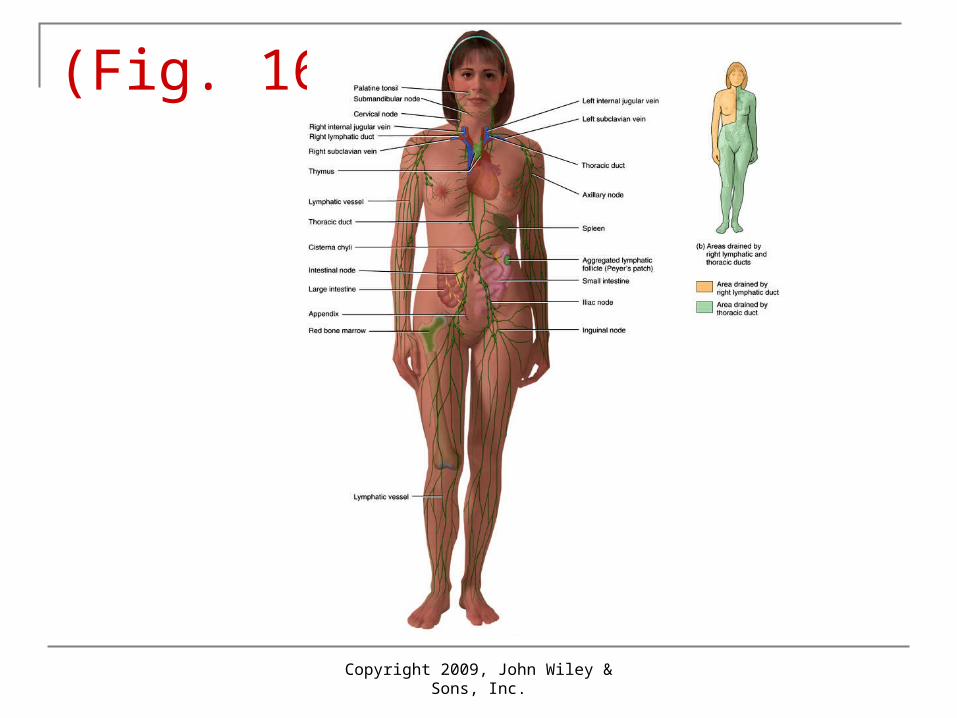
(Fig. 16.1)
Copyright 2009, John Wiley & Sons, Inc.
Lymphatic Capillaries
Copyright 2009, John Wiley & Sons, Inc.
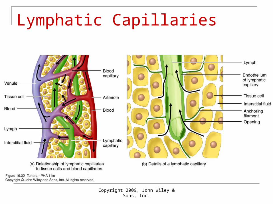
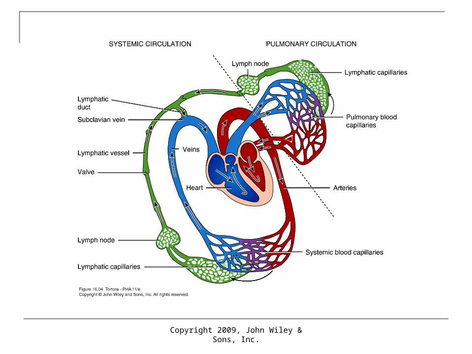
Lymphatic Vessels and Lymph Circulation Lymphatic vessels begin as lymphatic capillaries. The vessels are closed at one end and located in the spaces
between cells.
Lymphatic capillaries unite to form larger lymphatic vessels, which
resemble veins in structure but have thinner walls and more valves.
At intervals along the lymphatic vessels, lymph flows through lymph
nodes.
Tissues that lack lymphatic capillaries include avascular tissues
(such as cartilage, the epidermis, and the cornea of the eye), the
central nervous system, portions of the spleen, and red bone
marrow.
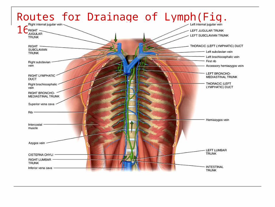
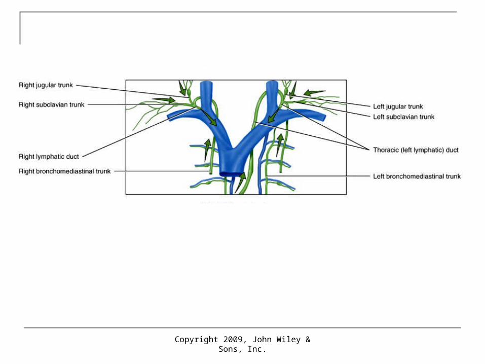
Routes for Drainage of Lymph(Fig. 16.3)
Copyright 2009, John Wiley & Sons, Inc.
Copyright 2009, John Wiley & Sons, Inc.
Lymph Flow1. Skeletal muscle pump. The “milking action” of skeletal muscle contractions
compresses lymphatic vessels and forces lymph toward the junction of the internal jugular and subclavian veins.
2. Respiratory pump. Lymph flow is also maintained by pressure
changes that occur during inhalation (breathing in). Lymph flows from the abdominal region, where the pressure is higher, toward the thoracic region, where it is lower. When the pressures reverse during exhalation (breathing out), the valves prevent backflow of lymph.
Copyright 2009, John Wiley & Sons, Inc.

Copyright 2009, John Wiley & Sons, Inc.
Lymphatic Organs and TissuesLymphatic organs and tissues, are widely distributed throughout the
body, they are classified into two groups based on their functions.
Primary lymphatic organs are the sites where stem cells divide and become immunocompetent, the red bone marrow and the thymus.
The secondary lymphatic organs and tissues are the sites where most immune responses occur. They include lymph nodes, the spleen, and lymphatic nodules .
Copyright 2009, John Wiley & Sons, Inc.
Thymus The thymus a bilobed organ located in the mediastinum between
the sternum and the aorta. Each thymic lobule consists of a cortex and medulla. The cortex is composed of T cells and dendritic cells, epithelial
cells, and macrophages. Immature T cells migrate from red bone marrow to the cortex of the
thymus, where they proliferate and begin to mature.

Copyright 2009, John Wiley & Sons, Inc.
Thymus Because of its high content of lymphoid tissue and a rich blood
supply, the thymus has a reddish appearance in a living body. With age, fatty infiltrations replace the lymphoid tissue and it takes
on more of the yellowish color, giving the false impression of reduced size.
In infants, the thymus has a mass of about 70 g (2.3 oz). As a person reaches maturity, the functional portion of the gland is
reduced considerably, and in old age the functional portion may weigh only 3 g (0.1 oz). Before the thymus atrophies, it populates the secondary lymphatic organs and tissues with T cells.
Copyright 2009, John Wiley & Sons, Inc.
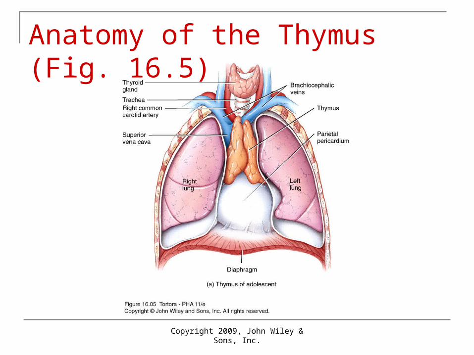
Anatomy of the Thymus (Fig. 16.5)
Copyright 2009, John Wiley & Sons, Inc.
Lymph Node
Located along lymphatic vessels are about 600 bean-shaped lymph nodes. Large groups of lymph nodes are present near the mammary glands and in
the axillae and groin. Lymph nodes are 1–25 mm (0.04–1 in.) long and, are covered by a capsule
of dense connective tissue. The parenchyma of a lymph node is divided into a superficial cortex and a
deep medulla. Within the outer cortex are aggregates of B cells called lymphatic nodules
(follicles). A lymphatic nodule consisting chiefly of B cells is called a primary lymphatic nodule.
Copyright 2009, John Wiley & Sons, Inc.
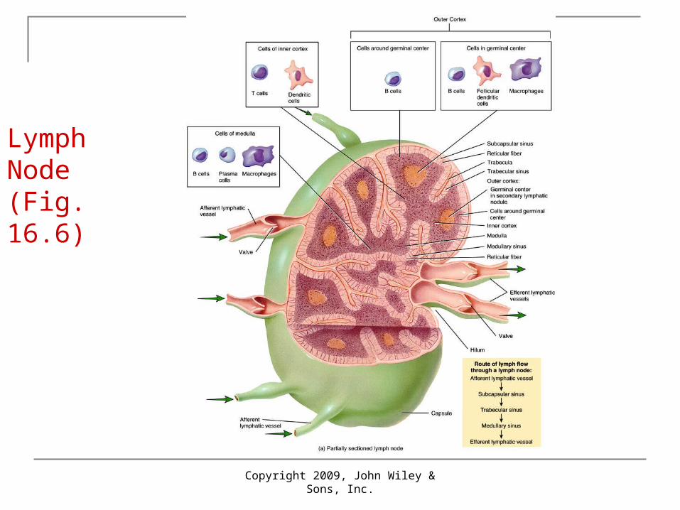
Lymph Node (Fig. 16.6)
Copyright 2009, John Wiley & Sons, Inc.
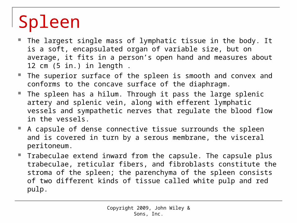
Spleen The largest single mass of lymphatic tissue in the body. It is a soft,
encapsulated organ of variable size, but on average, it fits in a person’s open hand and measures about 12 cm (5 in.) in length .
The superior surface of the spleen is smooth and convex and conforms to the concave surface of the diaphragm.
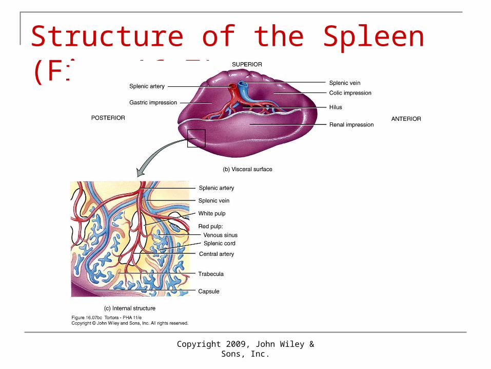
The spleen has a hilum. Through it pass the large splenic artery and splenic vein, along with efferent lymphatic vessels and sympathetic nerves that regulate the blood flow in the vessels.
A capsule of dense connective tissue surrounds the spleen and is covered in turn by a serous membrane, the visceral peritoneum.
Trabeculae extend inward from the capsule. The capsule plus trabeculae, reticular fibers, and fibroblasts constitute the stroma of the spleen; the parenchyma of the spleen consists of two different kinds of tissue called white pulp and red pulp.
Copyright 2009, John Wiley & Sons, Inc.
Structure of the Spleen (Fig. 16.7)
Copyright 2009, John Wiley & Sons, Inc.
Lymphatic Nodules
Lymphatic nodules are masses of lymphatic tissue that are not surrounded by a capsule.
They are scattered throughout the lamina propria of mucous membranes lining the gastrointestinal, urinary, reproductive tracts, and the respiratory airways, lymphatic nodules in these areas are also referred to as mucosa-associated lymphatic tissue (MALT).
Although many lymphatic nodules are small and solitary, some occur in multiple large aggregations in specific parts of the body.
Among these are the tonsils in the pharyngeal region and the aggregated lymphatic follicles (Peyer’s patches) in the ileum of the small intestine.
Aggregations of lymphatic nodules also occur in the appendix.