Embed Size (px)
Citation preview
Copyright 2010, John Wiley &
Sons, Inc.
Chapter 15
The Cardiovascular System: The Heart
Copyright 2010, John Wiley &
Sons, Inc.
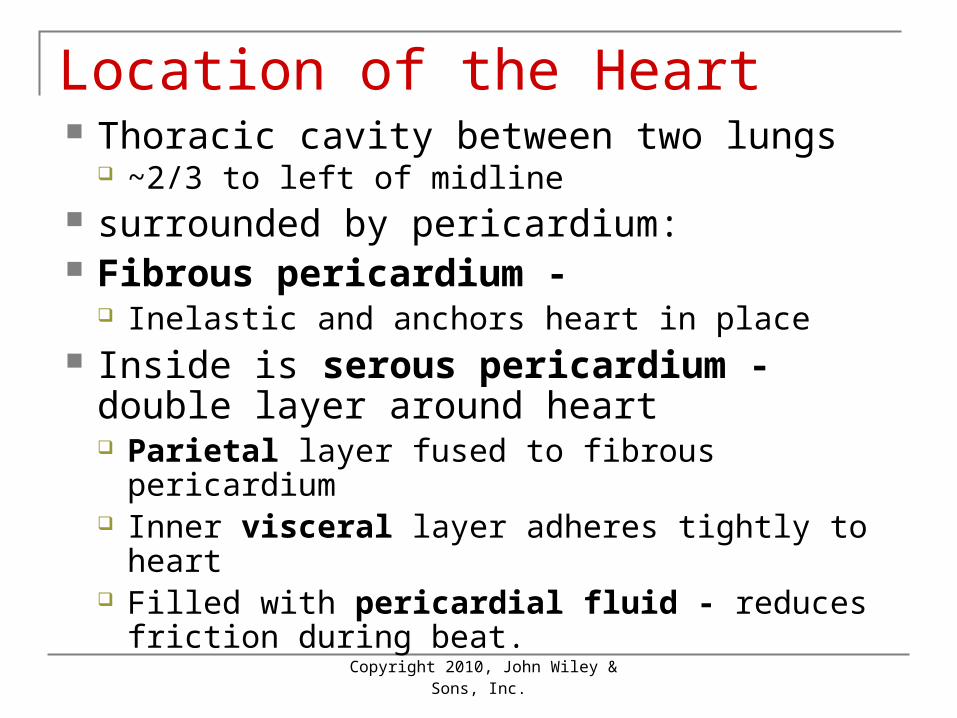
Location of the Heart Thoracic cavity between two lungs
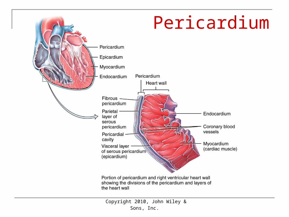
~2/3 to left of midline surrounded by pericardium: Fibrous pericardium -
Inelastic and anchors heart in place Inside is serous pericardium - double
layer around heart Parietal layer fused to fibrous pericardium Inner visceral layer adheres tightly to heart Filled with pericardial fluid - reduces friction
during beat.
Copyright 2010, John Wiley &
Sons, Inc.
Position of the Heart

Copyright 2010, John Wiley &
Sons, Inc.
Position of the Heart
Copyright 2010, John Wiley &
Sons, Inc.
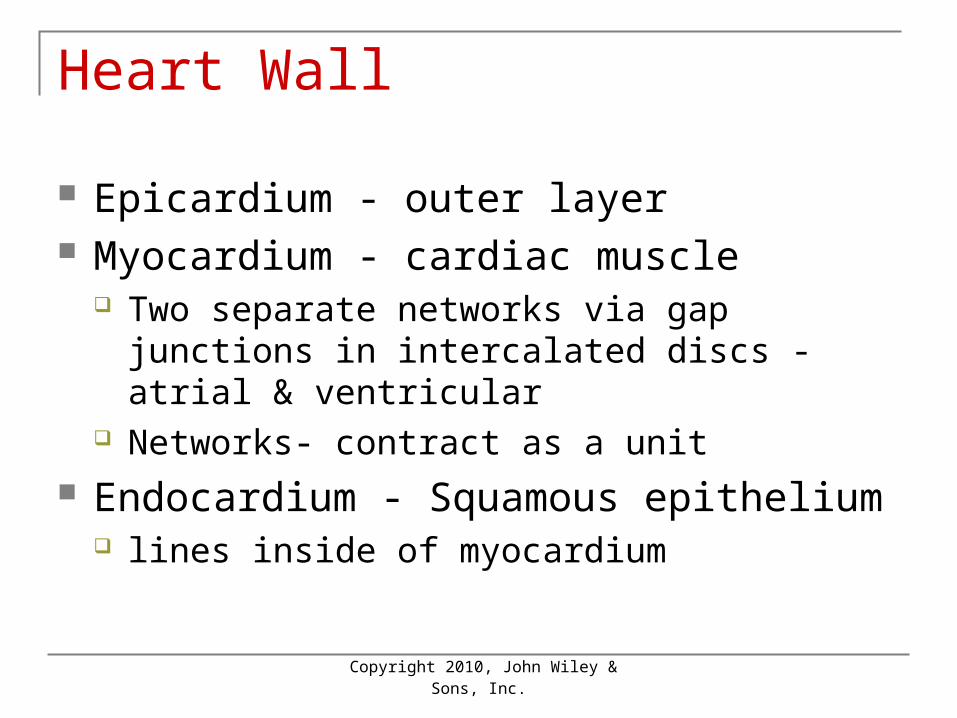
Heart Wall
Epicardium - outer layer Myocardium - cardiac muscle
Two separate networks via gap junctions in intercalated discs - atrial & ventricular
Networks- contract as a unit Endocardium - Squamous epithelium
lines inside of myocardium
Copyright 2010, John Wiley &
Sons, Inc.
Pericardium
Copyright 2010, John Wiley &
Sons, Inc.
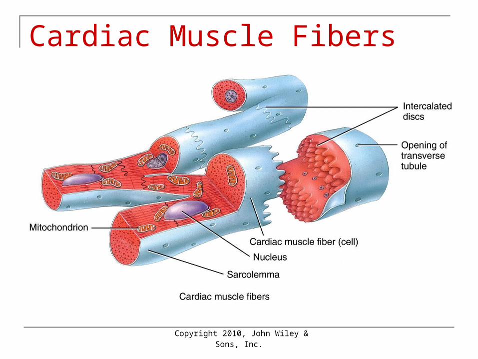
Cardiac Muscle Fibers
Copyright 2010, John Wiley &
Sons, Inc.
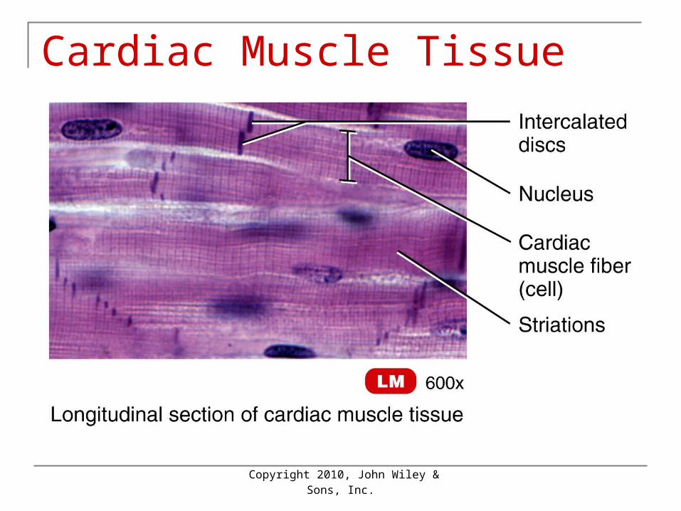
Cardiac Muscle Tissue
Copyright 2010, John Wiley &
Sons, Inc.
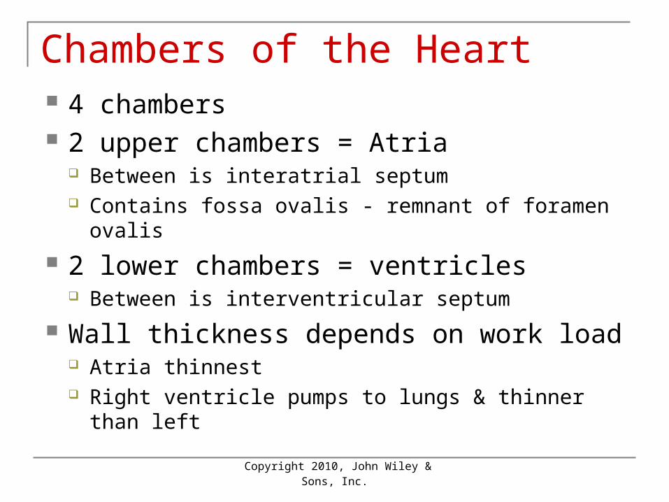
Chambers of the Heart 4 chambers 2 upper chambers = Atria
Between is interatrial septum Contains fossa ovalis - remnant of foramen ovalis
2 lower chambers = ventricles Between is interventricular septum
Wall thickness depends on work load Atria thinnest Right ventricle pumps to lungs & thinner than left
Copyright 2010, John Wiley &
Sons, Inc.
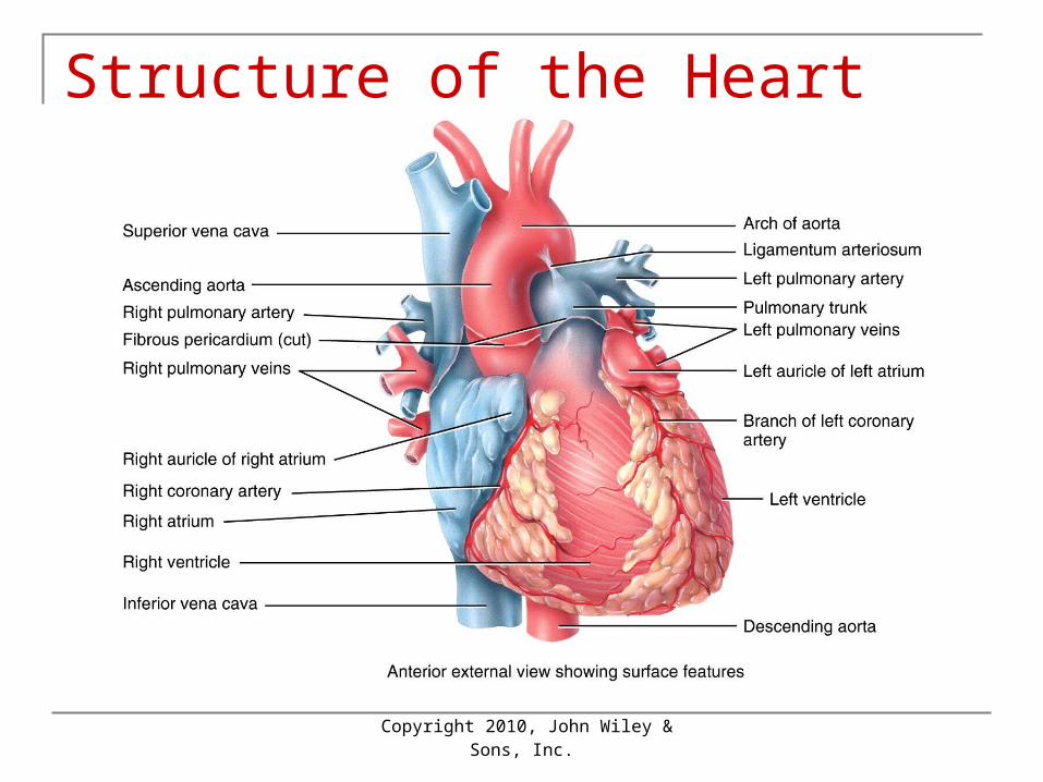
Structure of the Heart
Copyright 2010, John Wiley &
Sons, Inc.
Structure of the Heart
Copyright 2010, John Wiley &
Sons, Inc.
Structure of the Heart
Copyright 2010, John Wiley &
Sons, Inc.
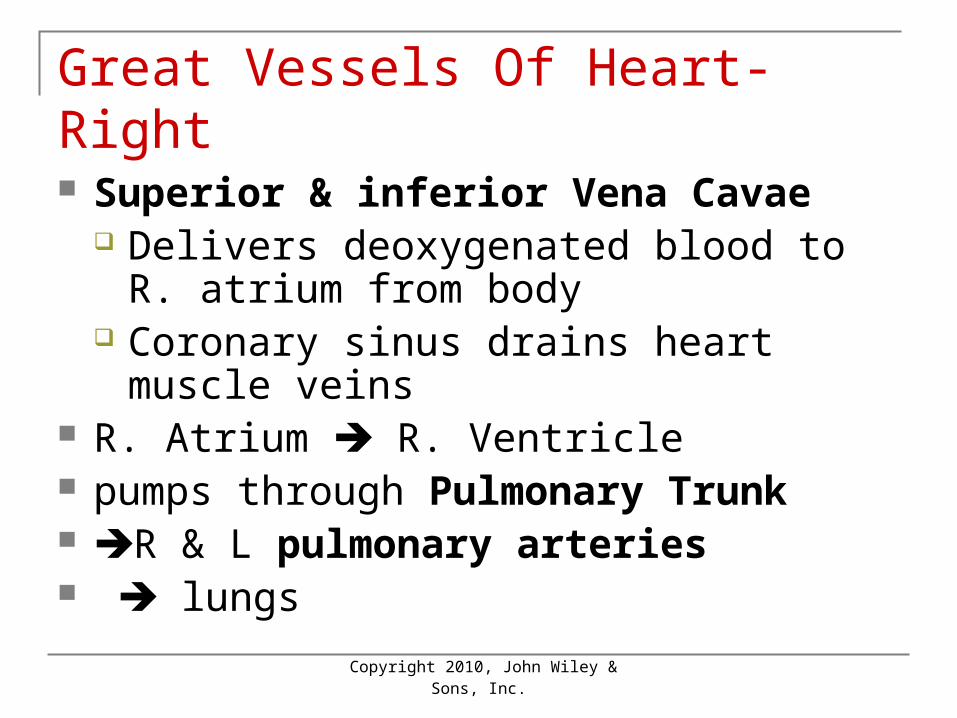
Great Vessels Of Heart-Right
Superior & inferior Vena Cavae Delivers deoxygenated blood to R. atrium
from body Coronary sinus drains heart muscle veins
R. Atrium R. Ventricle pumps through Pulmonary Trunk R & L pulmonary arteries lungs
Copyright 2010, John Wiley &
Sons, Inc.
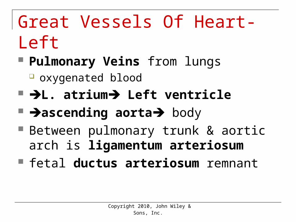
Great Vessels Of Heart-Left
Pulmonary Veins from lungs oxygenated blood
L. atrium Left ventricle ascending aorta body Between pulmonary trunk & aortic arch is
ligamentum arteriosum fetal ductus arteriosum remnant
Copyright 2010, John Wiley &
Sons, Inc.
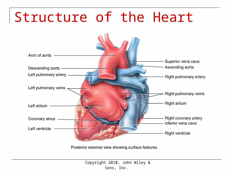
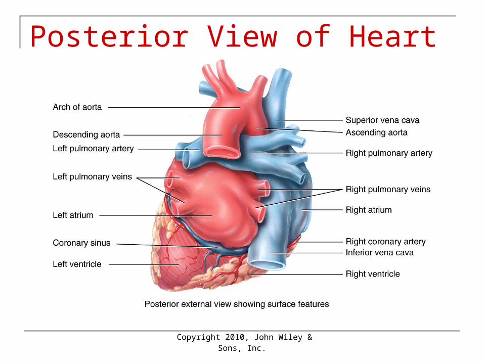
Posterior View of Heart
Copyright 2010, John Wiley &
Sons, Inc.
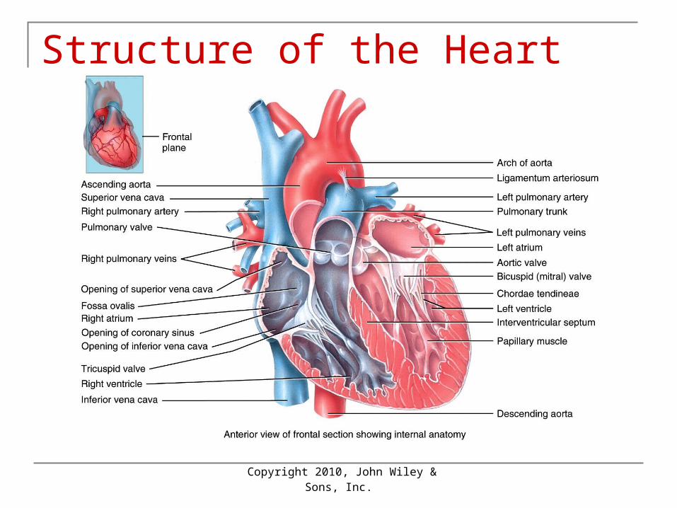
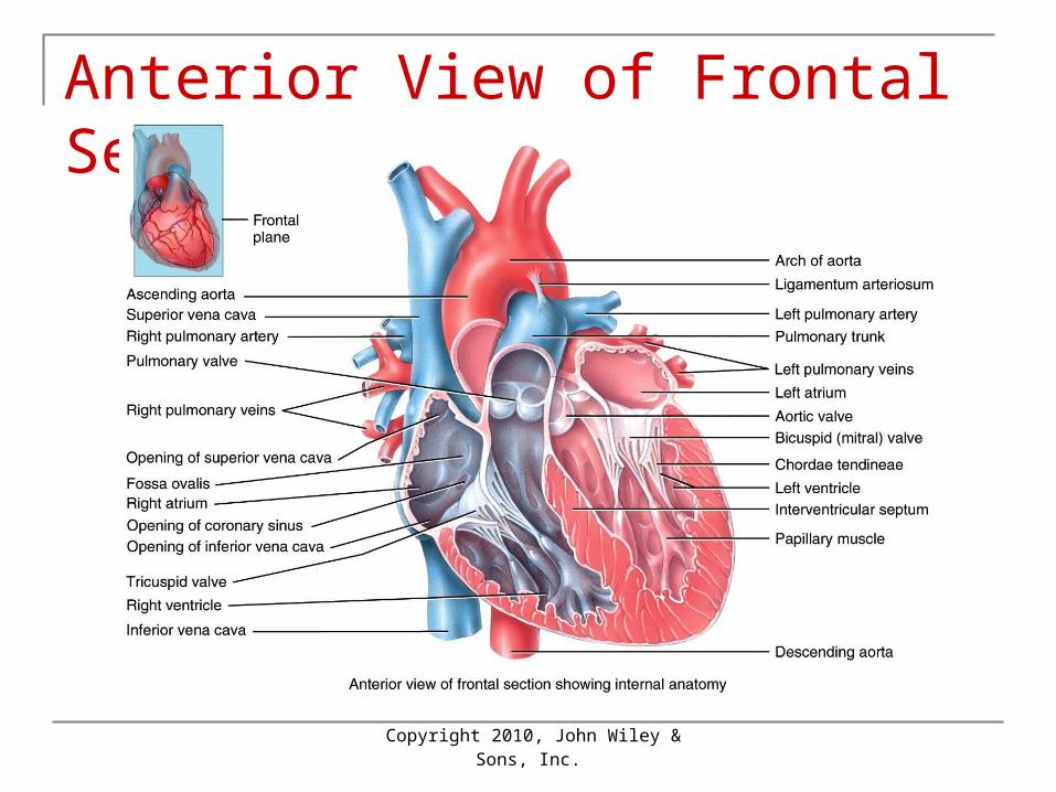
Anterior View of Frontal Section
Copyright 2010, John Wiley &
Sons, Inc.
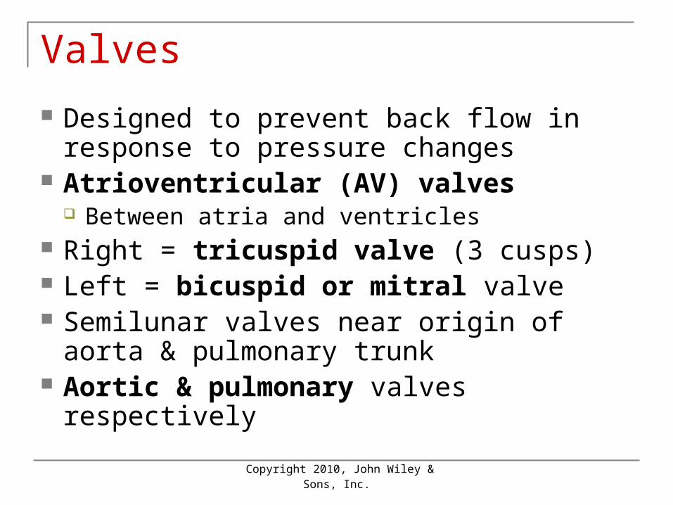
Valves
Designed to prevent back flow in response to pressure changes
Atrioventricular (AV) valves Between atria and ventricles
Right = tricuspid valve (3 cusps) Left = bicuspid or mitral valve Semilunar valves near origin of aorta &
pulmonary trunk Aortic & pulmonary valves respectively
Copyright 2010, John Wiley &
Sons, Inc.
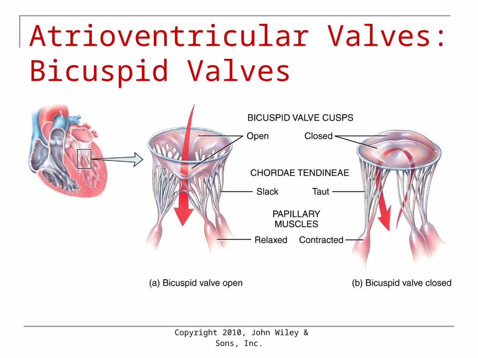
Atrioventricular Valves: Bicuspid Valves
Copyright 2010, John Wiley &
Sons, Inc.
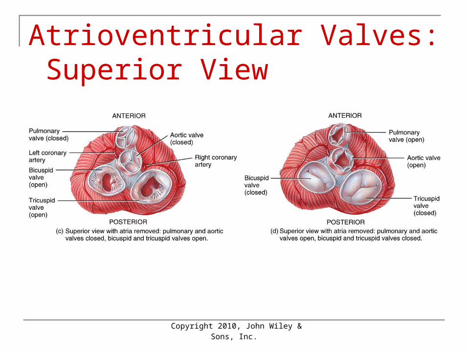
Atrioventricular Valves: Superior View
Copyright 2010, John Wiley &
Sons, Inc.
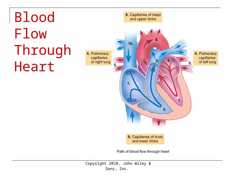
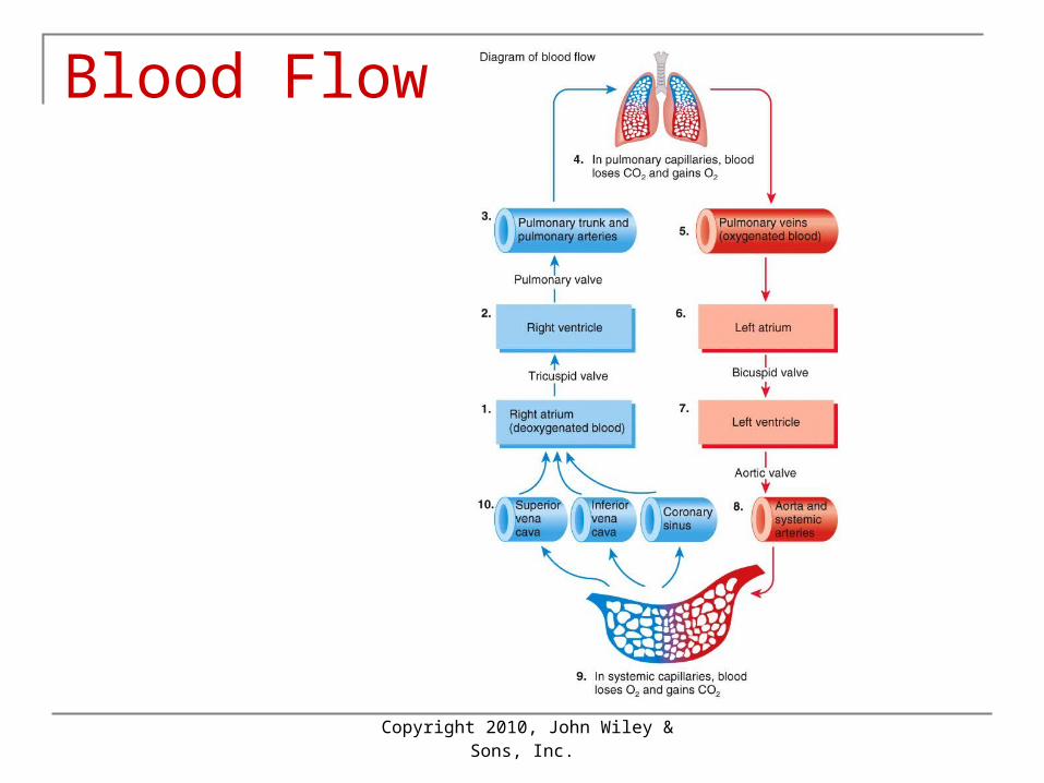
Blood Flow Through Heart
Copyright 2010, John Wiley &
Sons, Inc.
Blood Flow
Copyright 2010, John Wiley &
Sons, Inc.
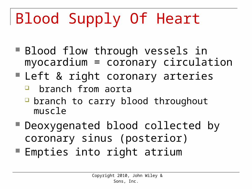
Blood Supply Of Heart
Blood flow through vessels in myocardium = coronary circulation
Left & right coronary arteries branch from aorta branch to carry blood throughout muscle
Deoxygenated blood collected by coronary sinus (posterior)
Empties into right atrium
Copyright 2010, John Wiley &
Sons, Inc.
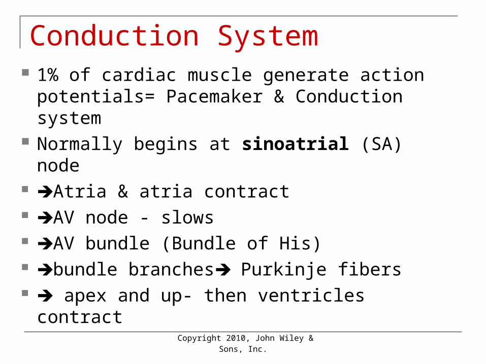
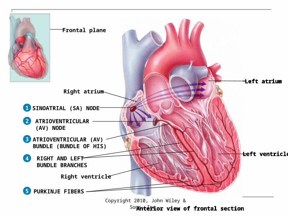
Conduction System 1% of cardiac muscle generate action
potentials= Pacemaker & Conduction system
Normally begins at sinoatrial (SA) node Atria & atria contract AV node - slows AV bundle (Bundle of His) bundle branches Purkinje fibers apex and up- then ventricles contract
Copyright 2010, John Wiley &
Sons, Inc.
Pacemaker
Depolarize spontaneously sinoatrial node ~100times /min also AV node ~40-60 times/min in ventricle ~20-35 /min Fastest one run runs the heart = pacemaker Normally the sinoatrial node
Copyright 2010, John Wiley &
Sons, Inc.
Frontal plane
Right atrium
Right ventricle
Left atrium
Left ventricle
Anterior view of frontal section
Frontal plane
Left atrium
Left ventricle
Anterior view of frontal section
SINOATRIAL (SA) NODE1
Right atrium
Right ventricle
Frontal plane
Left atrium
Left ventricle
Anterior view of frontal section
SINOATRIAL (SA) NODE
ATRIOVENTRICULAR(AV) NODE
1
2
Right atrium
Right ventricle
Frontal plane
Left atrium
Left ventricle
Anterior view of frontal section
SINOATRIAL (SA) NODE
ATRIOVENTRICULAR(AV) NODE
ATRIOVENTRICULAR (AV)BUNDLE (BUNDLE OF HIS)
1
2
3
Right atrium
Right ventricle
Frontal plane
Left atrium
Left ventricle
Anterior view of frontal section
SINOATRIAL (SA) NODE
ATRIOVENTRICULAR(AV) NODE
ATRIOVENTRICULAR (AV)BUNDLE (BUNDLE OF HIS)
RIGHT AND LEFTBUNDLE BRANCHES
1
2
3
4
Right atrium
Right ventricle
Frontal plane
SINOATRIAL (SA) NODE
ATRIOVENTRICULAR(AV) NODE
Left atrium
Left ventricle
Anterior view of frontal section
ATRIOVENTRICULAR (AV)BUNDLE (BUNDLE OF HIS)
RIGHT AND LEFTBUNDLE BRANCHES
PURKINJE FIBERS
1
2
3
4
5
Right atrium
Right ventricle
Copyright 2010, John Wiley &
Sons, Inc.
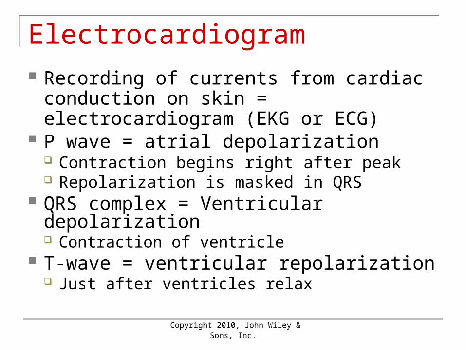
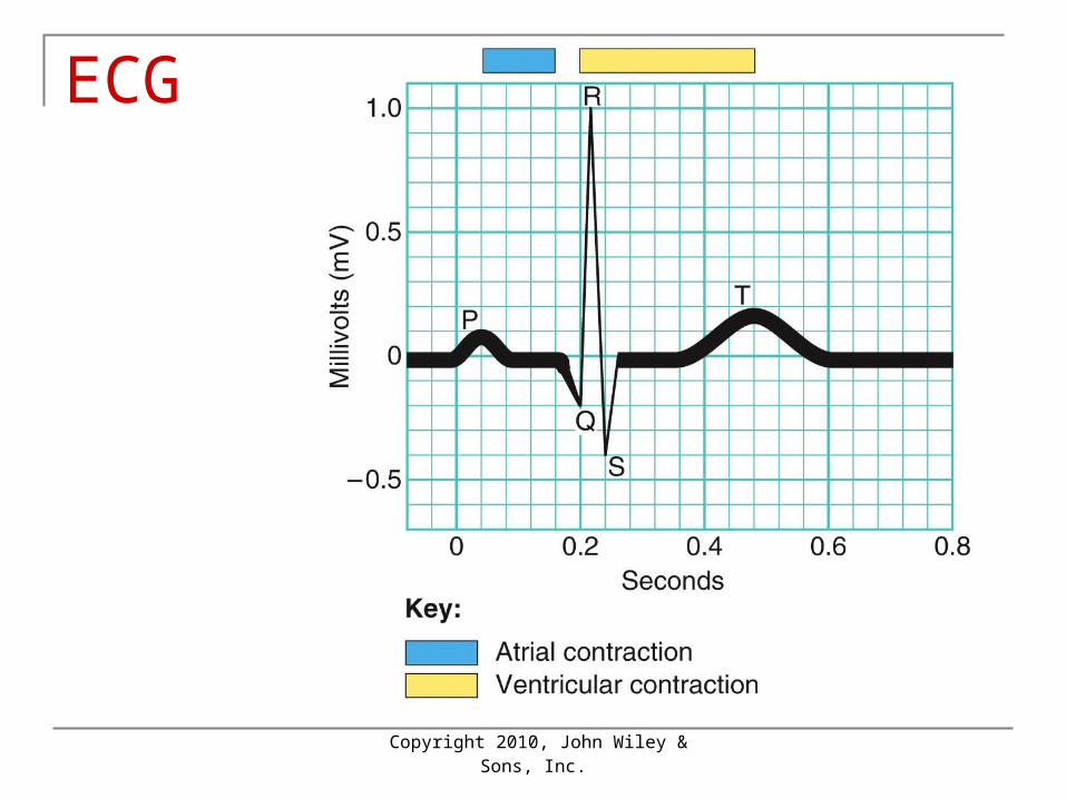
Electrocardiogram Recording of currents from cardiac
conduction on skin = electrocardiogram (EKG or ECG)
P wave = atrial depolarization Contraction begins right after peak Repolarization is masked in QRS
QRS complex = Ventricular depolarization Contraction of ventricle
T-wave = ventricular repolarization Just after ventricles relax
Copyright 2010, John Wiley &
Sons, Inc.
ECG
Copyright 2010, John Wiley &
Sons, Inc.
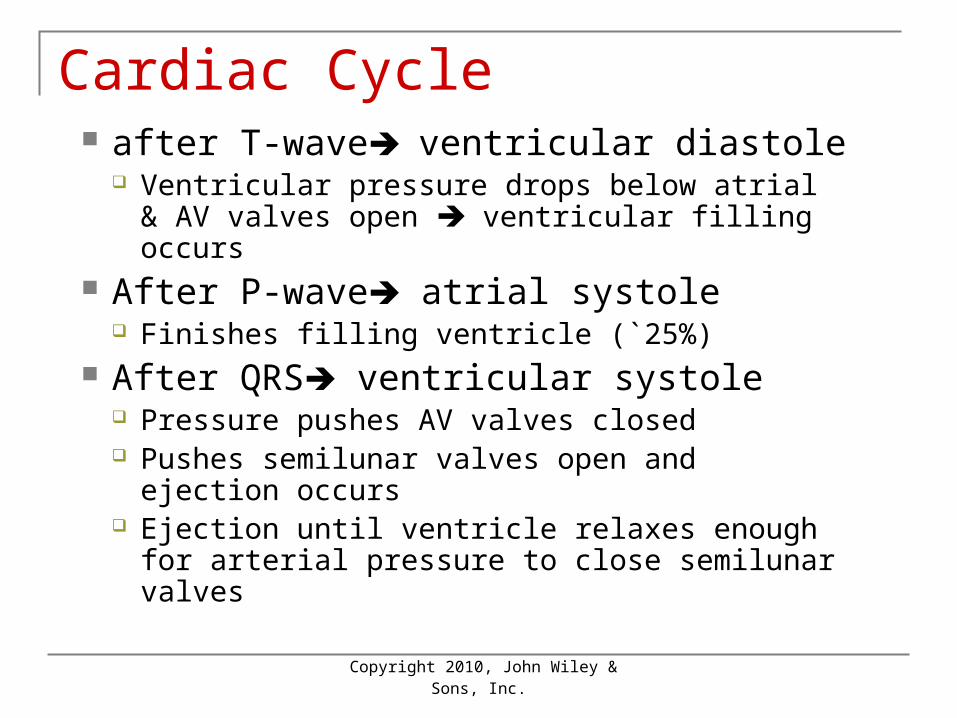
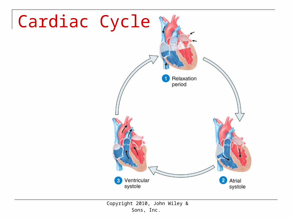
Cardiac Cycle after T-wave ventricular diastole
Ventricular pressure drops below atrial & AV valves open ventricular filling occurs
After P-wave atrial systole Finishes filling ventricle (`25%)
After QRS ventricular systole Pressure pushes AV valves closed Pushes semilunar valves open and ejection
occurs Ejection until ventricle relaxes enough for
arterial pressure to close semilunar valves
Copyright 2010, John Wiley &
Sons, Inc.
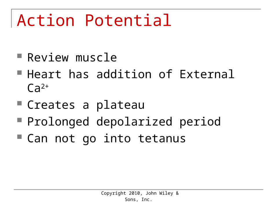
Action Potential
Review muscle Heart has addition of External Ca2+
Creates a plateau Prolonged depolarized period Can not go into tetanus
Copyright 2010, John Wiley &
Sons, Inc.
Cardiac Cycle
Copyright 2010, John Wiley &
Sons, Inc.
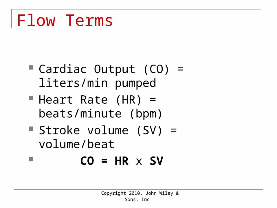
Flow Terms
Cardiac Output (CO) = liters/min pumped
Heart Rate (HR) = beats/minute (bpm) Stroke volume (SV) = volume/beat CO = HR x SV
Copyright 2010, John Wiley &
Sons, Inc.
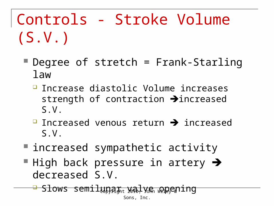
Controls - Stroke Volume (S.V.)
Degree of stretch = Frank-Starling law Increase diastolic Volume increases strength
of contraction increased S.V. Increased venous return increased S.V.
increased sympathetic activity High back pressure in artery decreased
S.V. Slows semilunar valve opening
Copyright 2010, John Wiley &
Sons, Inc.
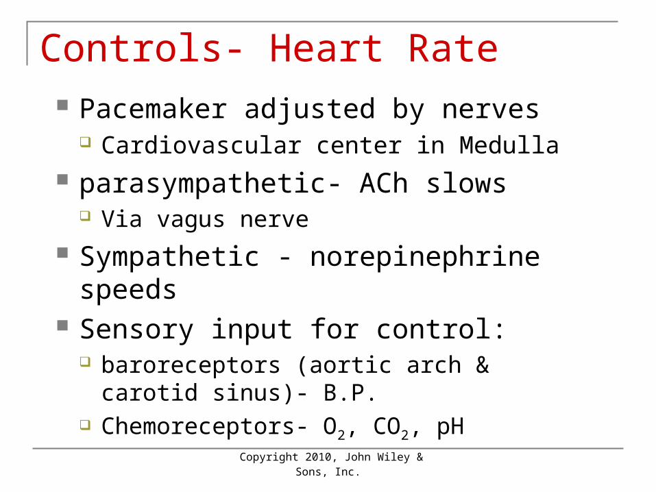
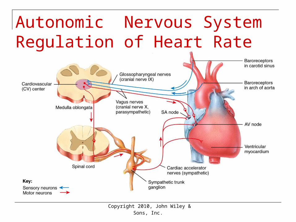
Controls- Heart Rate Pacemaker adjusted by nerves
Cardiovascular center in Medulla parasympathetic- ACh slows
Via vagus nerve Sympathetic - norepinephrine speeds Sensory input for control:
baroreceptors (aortic arch & carotid sinus)- B.P.
Chemoreceptors- O2, CO2, pH
Copyright 2010, John Wiley &
Sons, Inc.
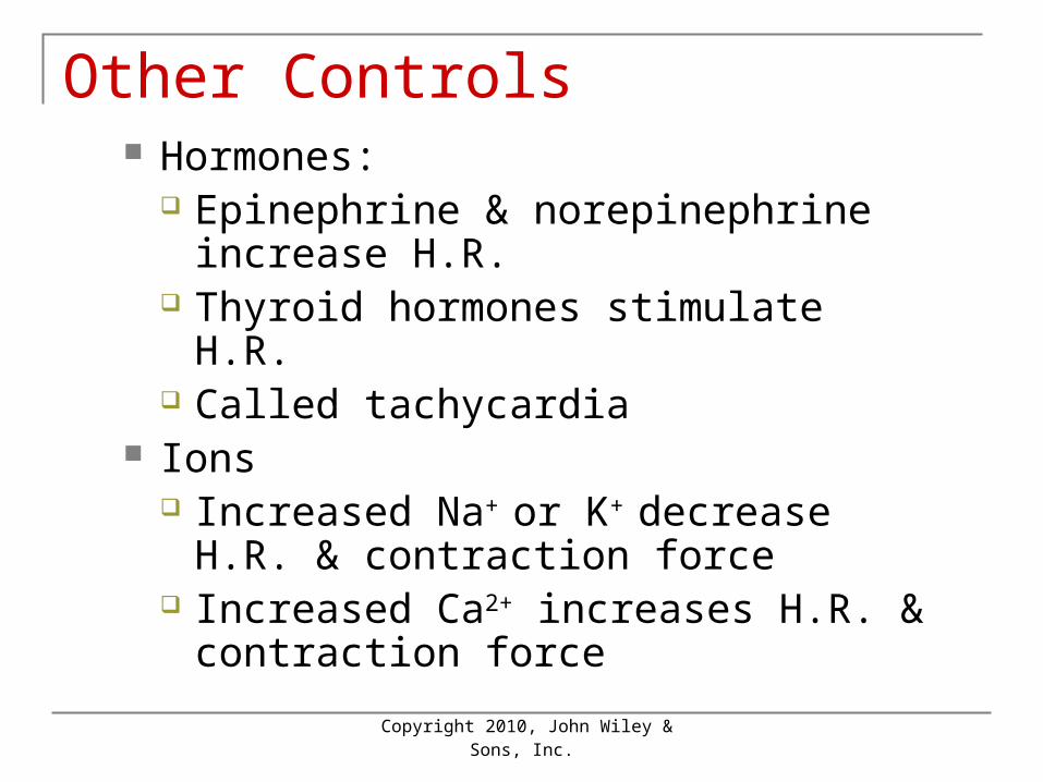
Other Controls Hormones:
Epinephrine & norepinephrine increase H.R.
Thyroid hormones stimulate H.R. Called tachycardia
Ions Increased Na+ or K+ decrease H.R. &
contraction force Increased Ca2+ increases H.R. &
contraction force
Copyright 2010, John Wiley &
Sons, Inc.
Autonomic Nervous System Regulation of Heart Rate
Copyright 2010, John Wiley &
Sons, Inc.
Exercise and the Heart
Aerobic exercise (longer than 20 min) strengthens cardiovascular system
Well trained athlete doubles maximum C.O.
Resting C.O. about the same but resting H.R. decreased
Copyright 2010, John Wiley &
Sons, Inc.
End of Chapter 15
Copyright 2010 John Wiley & Sons, Inc.All rights reserved. Reproduction or translation of this work beyond that permitted in section 117 of the 1976 United States Copyright Act without express permission of the copyright owner is unlawful. Request for further information should be addressed to the Permission Department, John Wiley & Sons, Inc. The purchaser may make back-up copies for his/her own use only and not for distribution or resale. The Publishers assumes no responsibility for errors, omissions, or damages caused by the use of theses programs or from the use of the information herein.