Embed Size (px)
Citation preview
Copyright 2010, John Wiley & Sons, Inc.
Chapter 3
Cells
Copyright 2010, John Wiley & Sons, Inc.
Cell Structure
Plasma membrane Cytoplasm: cytosol + organelles Nucleus
Copyright 2010, John Wiley & Sons, Inc.
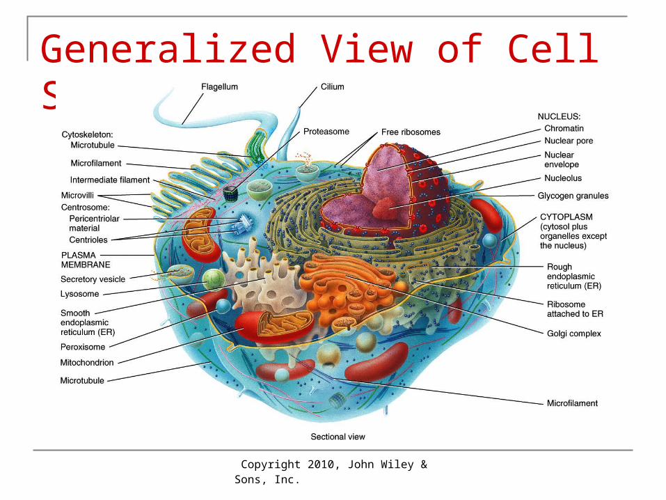
Generalized View of Cell Structure
Copyright 2010, John Wiley & Sons, Inc.
Cell Membrane
Phospholipid bilayer Cholesterol Proteins (integral and peripheral) Attached carbohydrates (glycolipids and
gycoproteins)
Copyright 2010, John Wiley & Sons, Inc.
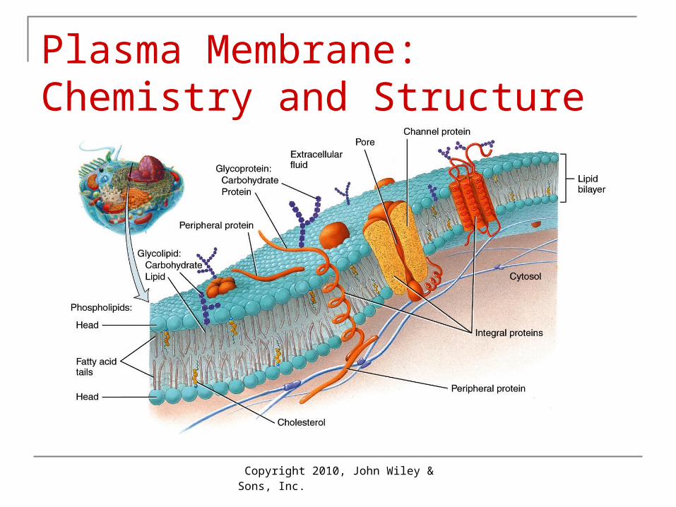
Plasma Membrane: Chemistry and Structure

Copyright 2010, John Wiley & Sons, Inc.
Membrane Function
Barrier between inside and outside of cell Controls entry of materials: transport Receives chemical and mechanical signals Transmits signals between intra- and extra-
cellular spaces Note variety of proteins in Figure 3-2.
Copyright 2010, John Wiley & Sons, Inc.
Terminology: Body Fluid Pools Intracellular (ICF)
Within cells: 2/3 of total Extracellular (ECF):
Between cells = Interstitial In blood vessels = Plasma In lymphatic vessels = Lymphatic
Copyright 2010, John Wiley & Sons, Inc.
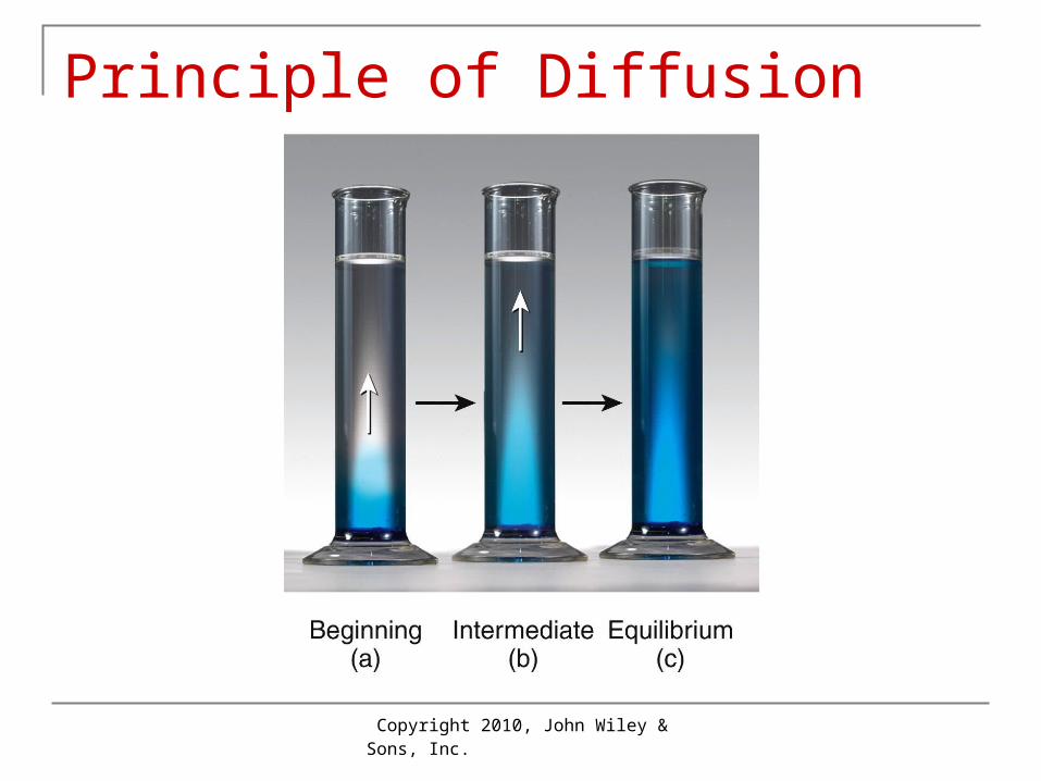
Terminology: Solutions
Solvent: the liquid doing the dissolving Usually water
Solute: the dissolved material (particles or gas)
Concentration Amount of solute in a given amount of solvent
Concentration gradient Difference in concentration between 2 areas of
solution
Copyright 2010, John Wiley & Sons, Inc.
Principle of Diffusion
Copyright 2010, John Wiley & Sons, Inc.
Passive Transport: Simple Diffusion Requirements for simple diffusion
Concentration gradient of solute present Solute can diffuse across a membrane if
membrane is present Pathways of simple diffusion:
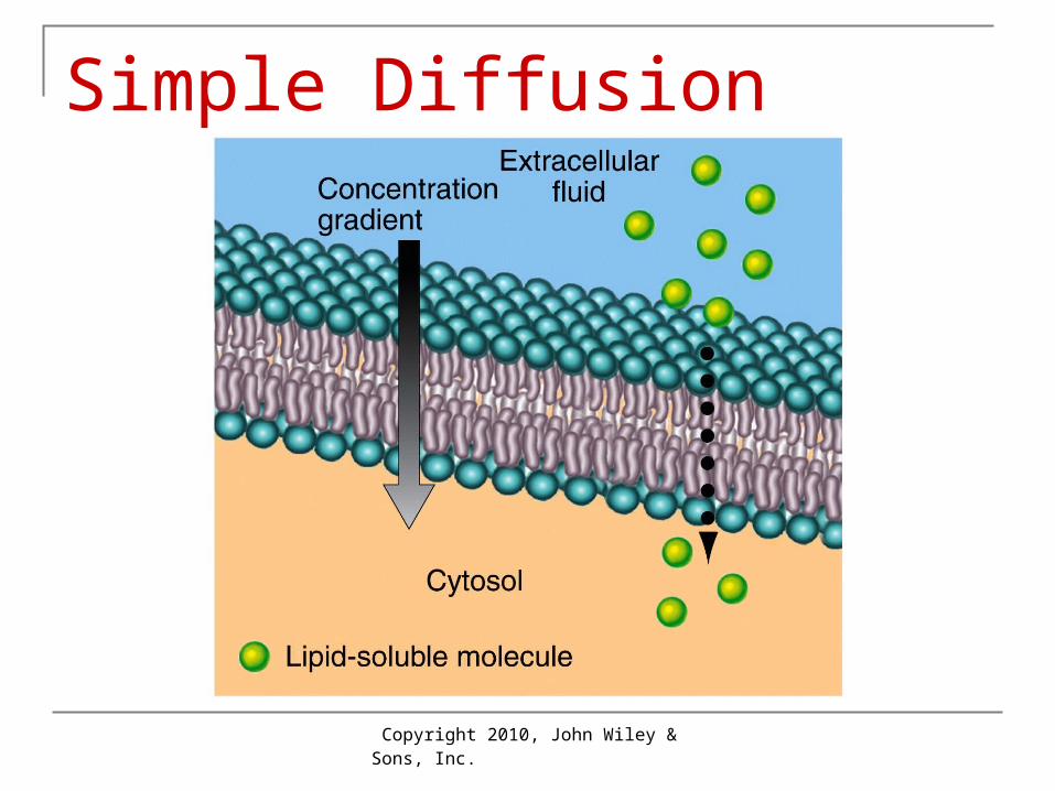
Pass across lipid bilayer if lipid-soluble (O2, CO2, N2, fatty acids, steroids, fat-soluble vitamins), or if polar molecules (H2O, urea)
Pass through ion channels (which may be gated: gates open and close) if ions such as K+, Ca2+, Cl–
Copyright 2010, John Wiley & Sons, Inc.
Simple Diffusion
Copyright 2010, John Wiley & Sons, Inc.
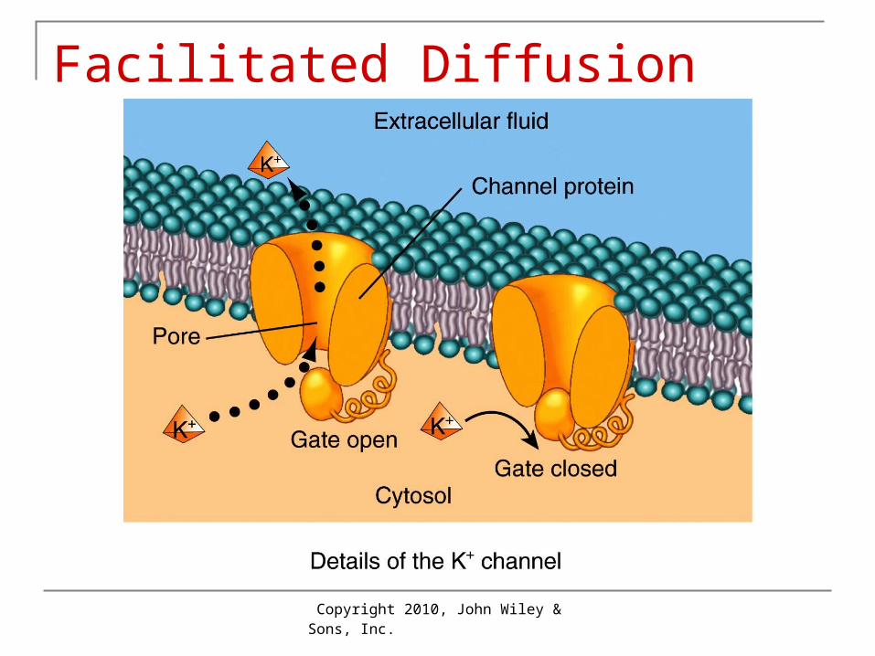
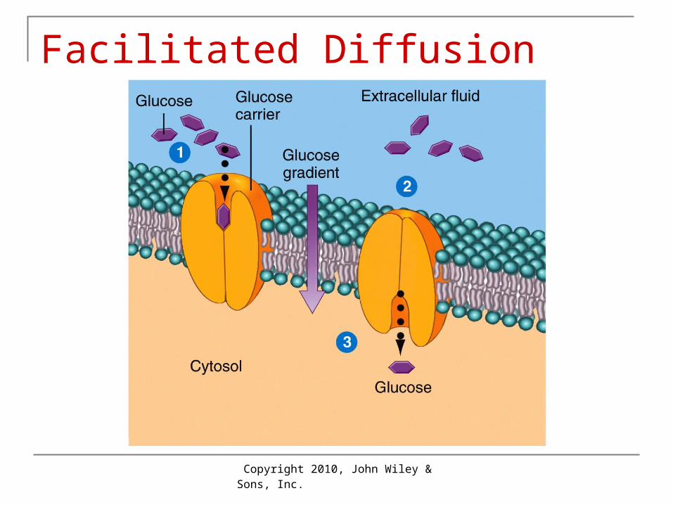
Facilitated Diffusion Requires a carrier in membrane but not
ATP Solute goes down concentration gradient Maximum transport speed depends on
number of carriers insulin increases number of carriers for
glucose in plasma membrane
Copyright 2010, John Wiley & Sons, Inc.
Facilitated Diffusion
Copyright 2010, John Wiley & Sons, Inc.
Facilitated Diffusion
Copyright 2010, John Wiley & Sons, Inc.
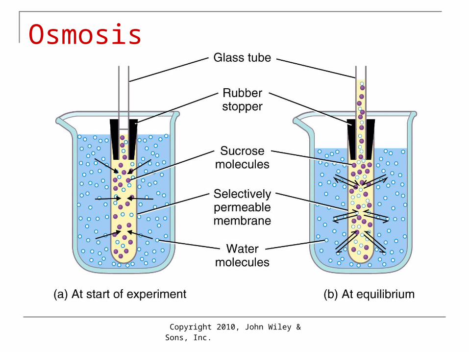
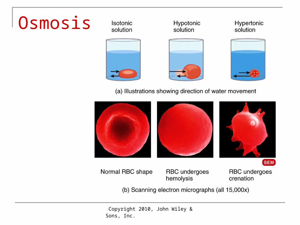
Osmosis Diffusion of water across selectively
permeable membrane: Permeable to solvent Impermeable to solute
Types of solutions surrounding human RBCs Isotonic: solution outside RBC has same
concentration of solute as RBC: 0.9% NaCl Hypotonic: solution outside of RBC has lower
concentration: 0% NaCl hemolysis Hypertonic: solution outside of RBC has higher
concentration: 4% NaCl crenation
Copyright 2010, John Wiley & Sons, Inc.
Osmosis
Copyright 2010, John Wiley & Sons, Inc.
Osmosis
Copyright 2010, John Wiley & Sons, Inc.
Active Transport Requires a carrier (called a pump) Requires energy (ATP) Can transport up a concentration gradient Critical for moving important ions Major active transport in most cells is
sodium-potassium (Na+/K+) pump
Copyright 2010, John Wiley & Sons, Inc.
Copyright 2009 John Wiley & Sons, Inc. 19
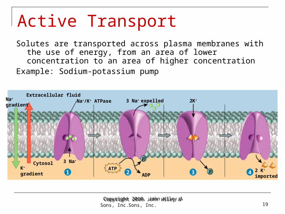
Active TransportSolutes are transported across plasma membranes with the use of
energy, from an area of lower concentration to an area of higher concentration
Example: Sodium-potassium pump
1
3 Na+
K+
gradient
Cytosol
1
3 Na+ expelled
3 Na+
ADPP
P
2 K+
imported
K+
gradient
Na+
gradientNa+/K+ ATPase
Extracellular fluid
Cytosol
2K+
ATP2 3 4
Copyright 2010, John Wiley & Sons, Inc.
Transport in Vesicles
Requires energy (ATP) Involves small membrane sac Endocytosis: importing materials into cell
Phagocytosis: ingestion of particles such as bacteria into white blood cells (WBCs)
Pinocytosis: ingestion of fluid Exocytosis: exporting materials
Copyright 2010, John Wiley & Sons, Inc.
Transport Across the Plasma Membrane
Transport Across the Plasma MembraneInteractions Animation
You must be connected to the internet to run this animation.
Copyright 2010, John Wiley & Sons, Inc.
Cell Organelles: Table 3.2
Cytoskeleton Flagella, cilia & centrioles Endoplasmic reticulum Golgi apparatus Mitochondrion Nucleus, nucleolus, nuclear envelope Vesicles, e.g. lysosome
Copyright 2010, John Wiley & Sons, Inc.
Cytoplasm Cell contents Includes organelles and cytosol Excludes nucleus
Copyright 2010, John Wiley & Sons, Inc.
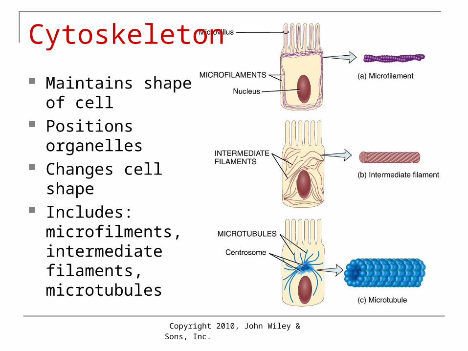
Cytoskeleton
Maintains shape of cell
Positions organelles Changes cell shape Includes:
microfilments, intermediate filaments, microtubules
Copyright 2010, John Wiley & Sons, Inc.
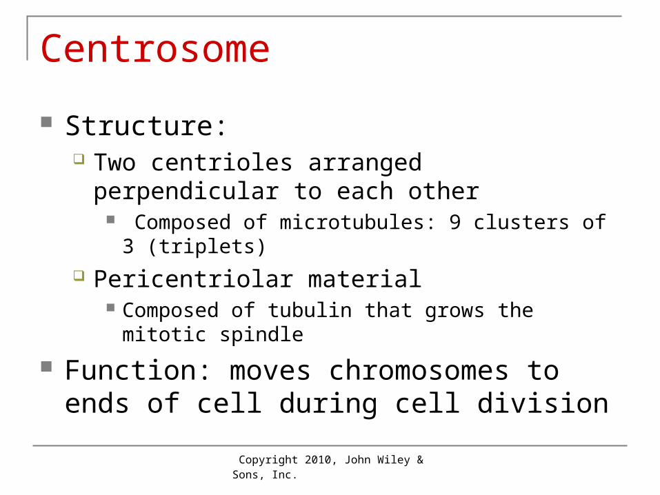
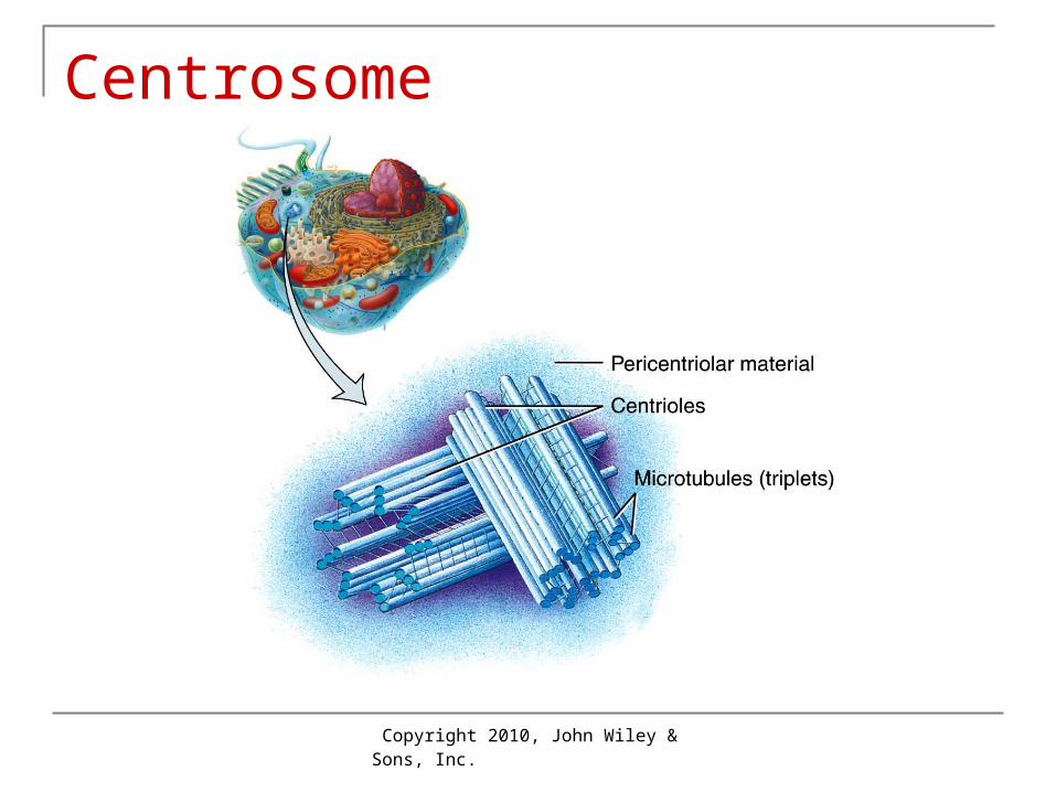
Centrosome
Structure: Two centrioles arranged perpendicular to each
other Composed of microtubules: 9 clusters of 3 (triplets)
Pericentriolar material Composed of tubulin that grows the mitotic spindle
Function: moves chromosomes to ends of cell during cell division
Copyright 2010, John Wiley & Sons, Inc.
Centrosome
Copyright 2010, John Wiley & Sons, Inc.
Cilia and Flagella
Specialized for motion Flagellum: single tail like structure on sperm
Propels sperm forward in reproductive tract Cilia: in groups
Found in respiratory system: move mucus
Copyright 2010, John Wiley & Sons, Inc.
Ribosomes Made within the nucleus (in nucleolus) Sites of protein synthesis (on E.R. or freely
within cytoplasm) Consist of ribosomal RNA (rRNA) + proteins Contain large and small subunits Can be attached to endoplasmic reticulum or
free in cytosol
Copyright 2010, John Wiley & Sons, Inc.
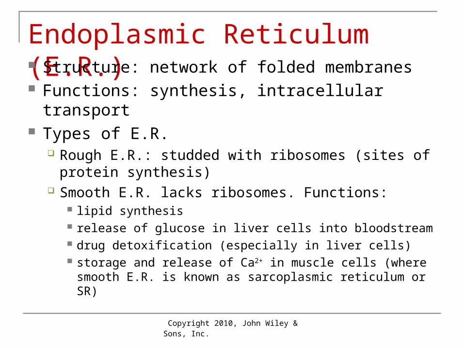
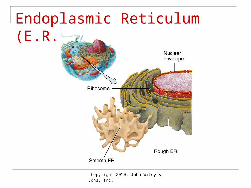
Endoplasmic Reticulum (E.R.) Structure: network of folded membranes Functions: synthesis, intracellular transport Types of E.R.
Rough E.R.: studded with ribosomes (sites of protein synthesis)
Smooth E.R. lacks ribosomes. Functions: lipid synthesis release of glucose in liver cells into bloodstream drug detoxification (especially in liver cells) storage and release of Ca2+ in muscle cells (where
smooth E.R. is known as sarcoplasmic reticulum or SR)
Copyright 2010, John Wiley & Sons, Inc.
Endoplasmic Reticulum (E.R.)
Copyright 2010, John Wiley & Sons, Inc.
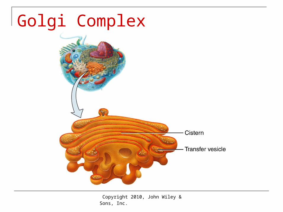
Structure: Flattened membranes (cisterns) with bulging
edges (like stacks of pita bread) Functions:
Modify proteins glycoproteins and lipoproteins that:
Become parts of plasma membranes Are stored in lysosomes, or Are exported by exocytosis
Golgi Complex
Copyright 2010, John Wiley & Sons, Inc.
Golgi Complex
Copyright 2010, John Wiley & Sons, Inc.
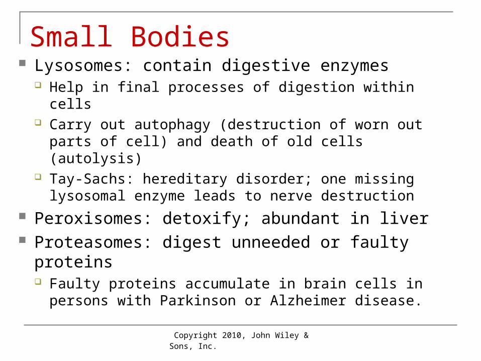
Small Bodies Lysosomes: contain digestive enzymes
Help in final processes of digestion within cells Carry out autophagy (destruction of worn out parts of
cell) and death of old cells (autolysis) Tay-Sachs: hereditary disorder; one missing
lysosomal enzyme leads to nerve destruction Peroxisomes: detoxify; abundant in liver Proteasomes: digest unneeded or faulty
proteins Faulty proteins accumulate in brain cells in persons
with Parkinson or Alzheimer disease.
Copyright 2010, John Wiley & Sons, Inc.
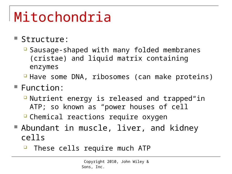
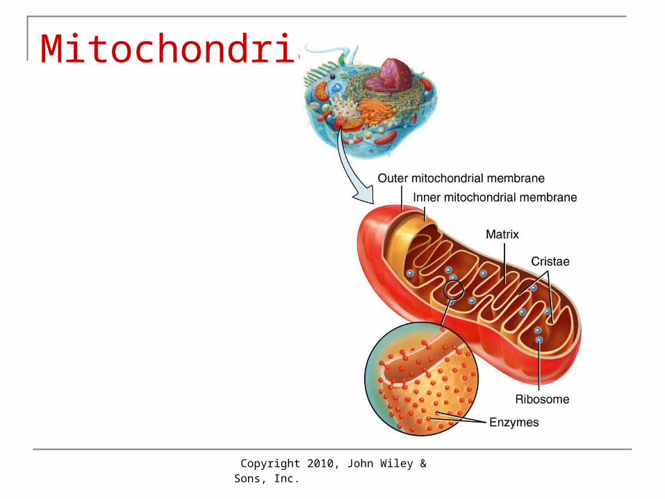
Mitochondria Structure:
Sausage-shaped with many folded membranes (cristae) and liquid matrix containing enzymes
Have some DNA, ribosomes (can make proteins) Function:
Nutrient energy is released and trapped in ATP; so known as “power houses of cell”
Chemical reactions require oxygen Abundant in muscle, liver, and kidney cells
These cells require much ATP
Copyright 2010, John Wiley & Sons, Inc.
Mitochondria
Copyright 2010, John Wiley & Sons, Inc.
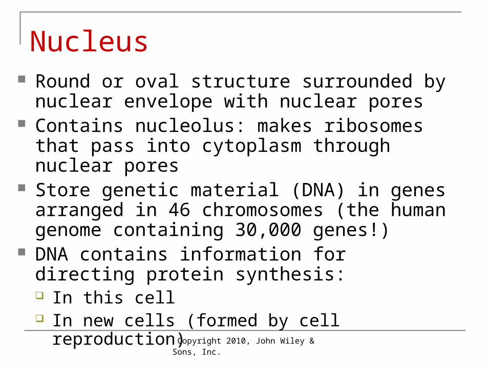
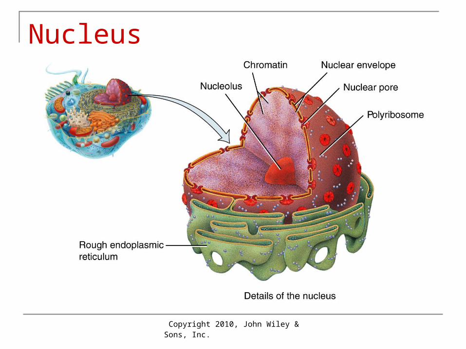
Nucleus Round or oval structure surrounded by nuclear
envelope with nuclear pores Contains nucleolus: makes ribosomes that pass
into cytoplasm through nuclear pores Store genetic material (DNA) in genes arranged
in 46 chromosomes (the human genome containing 30,000 genes!)
DNA contains information for directing protein synthesis: In this cell In new cells (formed by cell reproduction)
Copyright 2010, John Wiley & Sons, Inc.
Nucleus
Copyright 2010, John Wiley & Sons, Inc.
Protein Synthesis 2 steps:
Nuclear = transcription Cytoplasmic = translation
Copyright 2010, John Wiley & Sons, Inc.
Protein Synthesis: Transcription
Copyright 2010, John Wiley & Sons, Inc.
Protein Synthesis: Transcription
Copyright 2010, John Wiley & Sons, Inc.
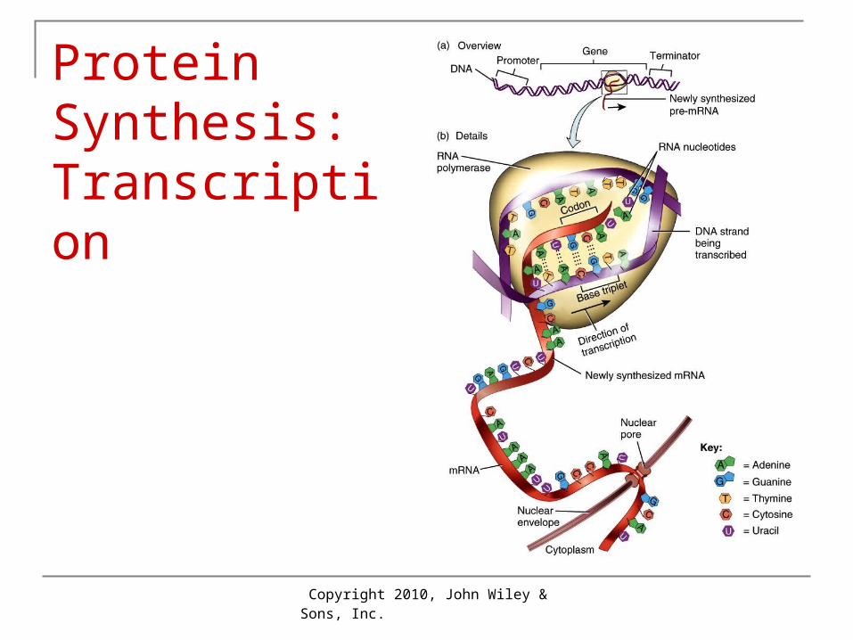
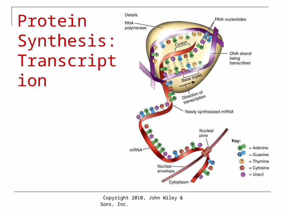
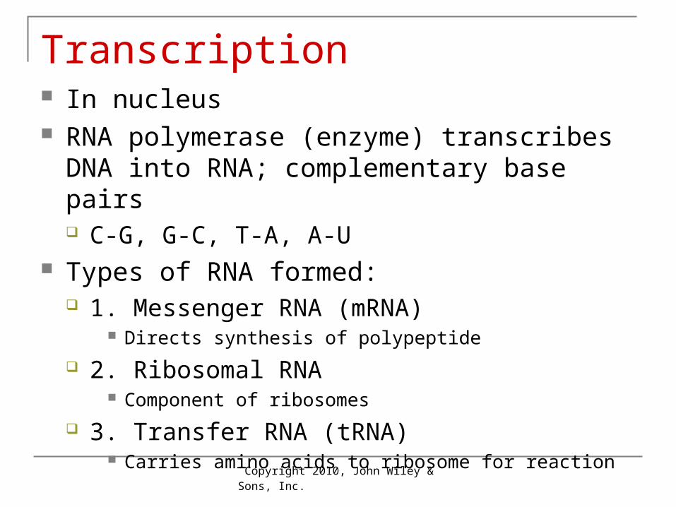
Transcription In nucleus RNA polymerase (enzyme) transcribes DNA into
RNA; complementary base pairs C-G, G-C, T-A, A-U
Types of RNA formed: 1. Messenger RNA (mRNA)
Directs synthesis of polypeptide
2. Ribosomal RNA Component of ribosomes
3. Transfer RNA (tRNA) Carries amino acids to ribosome for reaction
Copyright 2010, John Wiley & Sons, Inc.
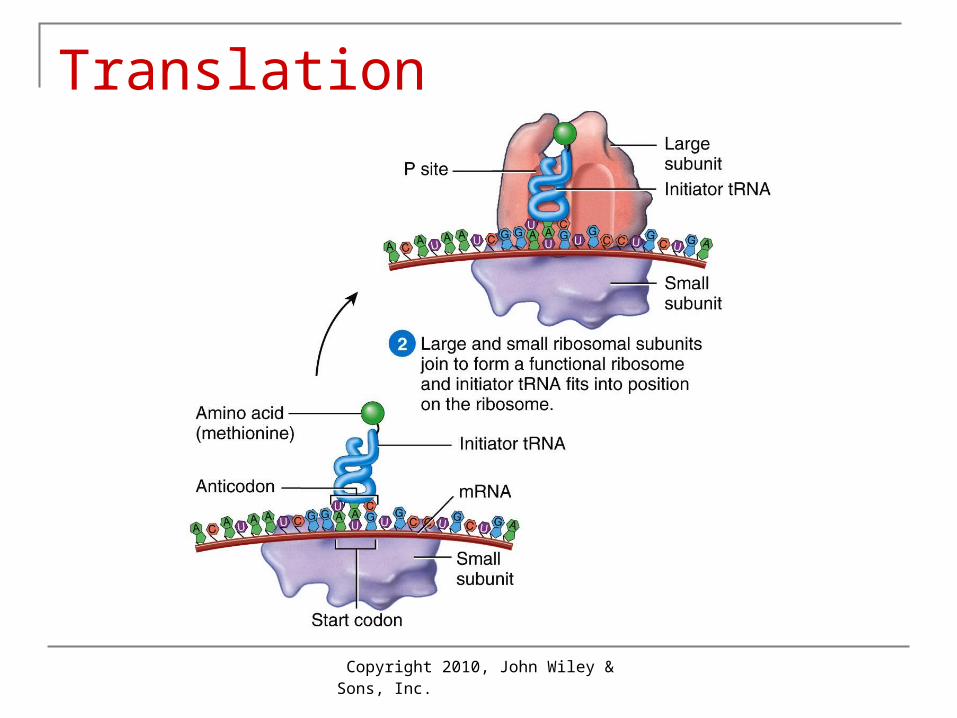
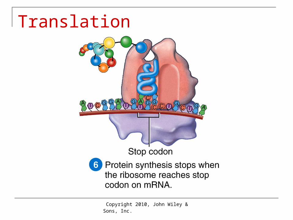
Translation
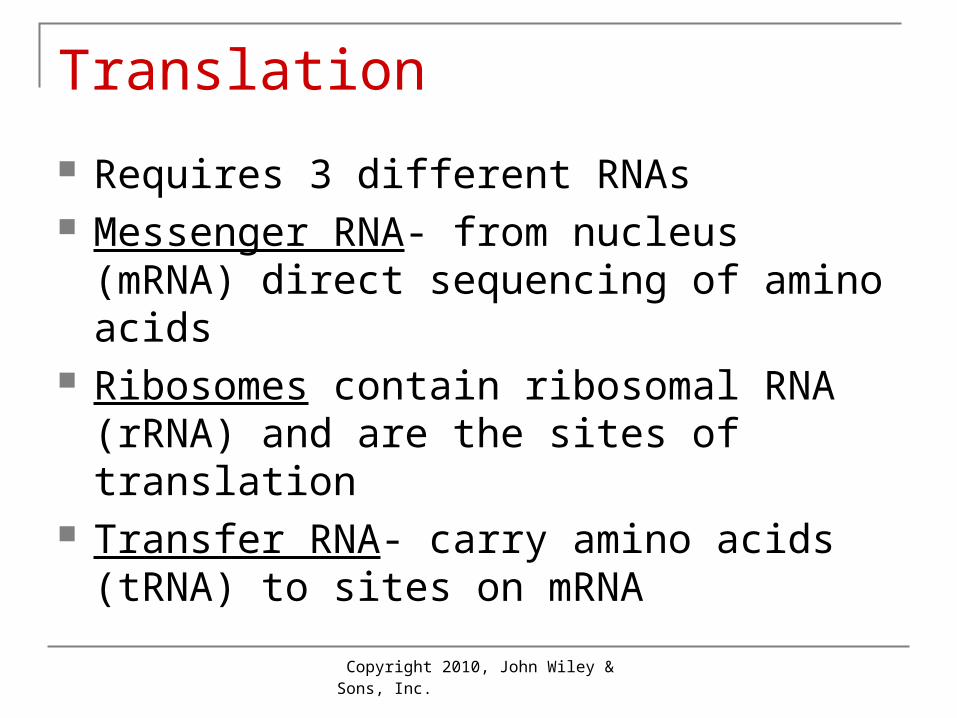
Requires 3 different RNAs Messenger RNA- from nucleus (mRNA)
direct sequencing of amino acids Ribosomes contain ribosomal RNA (rRNA)
and are the sites of translation Transfer RNA- carry amino acids (tRNA) to
sites on mRNA
Copyright 2010, John Wiley & Sons, Inc.
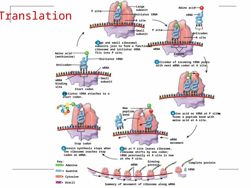
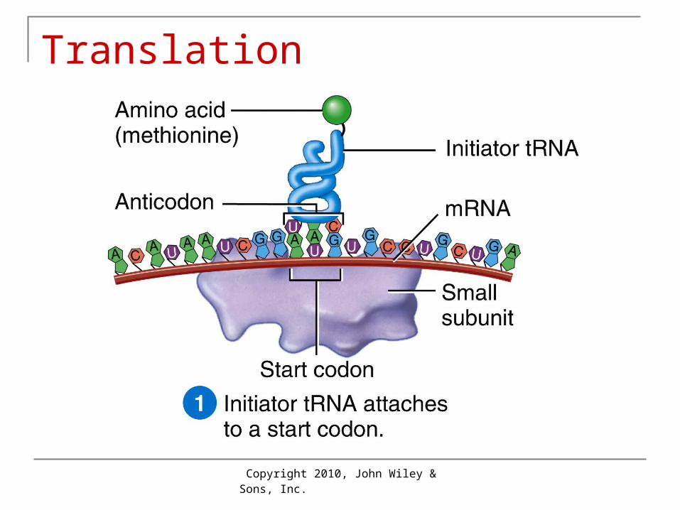
1
Key:
Initiator tRNA attaches to astart codon.
Amino acid(methionine)
AnticodonmRNA
mRNAbindingsite
Initiator tRNA
Start codon
Smallsubunit
= Adenine
= Guanine
= Cytosine
= Uracil
UUU G GGG G GAAAAA
U A CC UCUA
ACUC
1
Key:
Initiator tRNA attaches to astart codon.
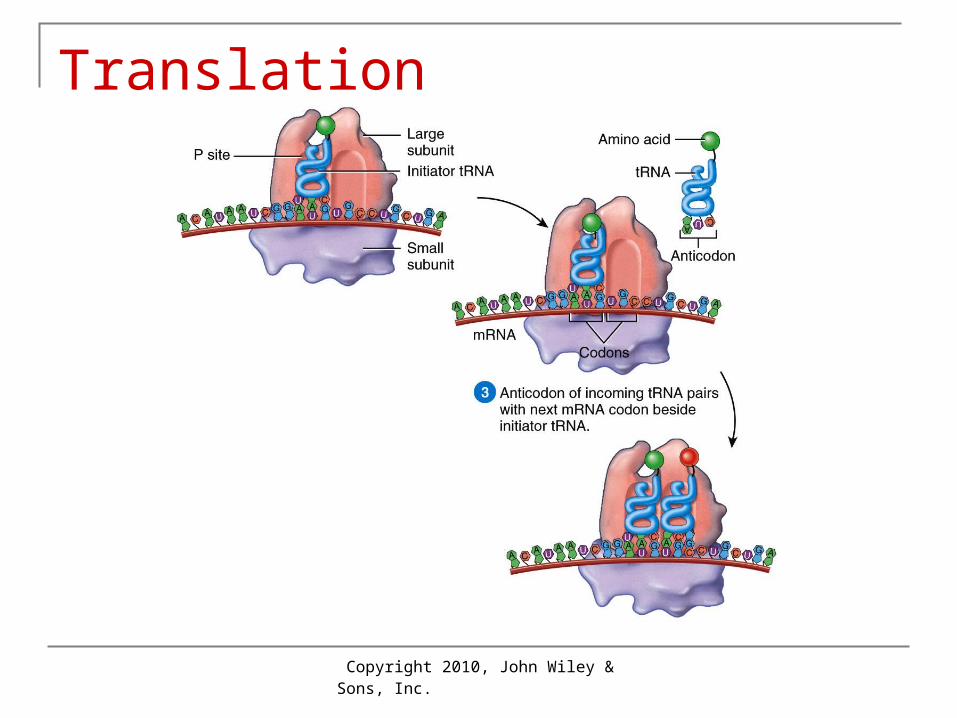
Large and small ribosomalsubunits join to form a functionalribosome and initiator tRNAfits into P site.Amino acid
(methionine)
AnticodonmRNA
mRNAbindingsite
Initiator tRNA
Start codon
Smallsubunit
Initiator tRNA
Smallsubunit
LargesubunitP site
A site
= Adenine
= Guanine
= Cytosine
= Uracil
UUU G GGG G GAAAAA
U A CC UCUA
ACUC
GAUUU GG AAAAACC G GUCU ACU
U A CG
2
1
Key:
Initiator tRNA attaches to astart codon.
Large and small ribosomalsubunits join to form a functionalribosome and initiator tRNAfits into P site.Amino acid
(methionine)
Amino acid
Anticodon
Anticodon
Codons
mRNA
mRNA
mRNAbindingsite
Initiator tRNA
tRNA
Start codon
Smallsubunit
Initiator tRNA
Smallsubunit
LargesubunitP site
A site
Anticodon of incoming tRNA pairswith next mRNA codon at A site.
P site
A site
= Adenine
= Guanine
= Cytosine
= Uracil
UUU G GGG G GAAAAA
U A CC UCUA
ACUC
GAUUU GG AAAAACC G GUCU ACU
U A CG
A UC
UUU GGG G GAAAAAC UCU
ACU
C
U A CGA2
3
1
Key:
Initiator tRNA attaches to astart codon.
Large and small ribosomalsubunits join to form a functionalribosome and initiator tRNAfits into P site.Amino acid
(methionine)
Amino acid
Anticodon
Anticodon
Codons
mRNA
mRNA
mRNAbindingsite
Initiator tRNA
tRNA
Start codon
Smallsubunit
Initiator tRNA
Smallsubunit
LargesubunitP site
A site
Anticodon of incoming tRNA pairswith next mRNA codon at A site.
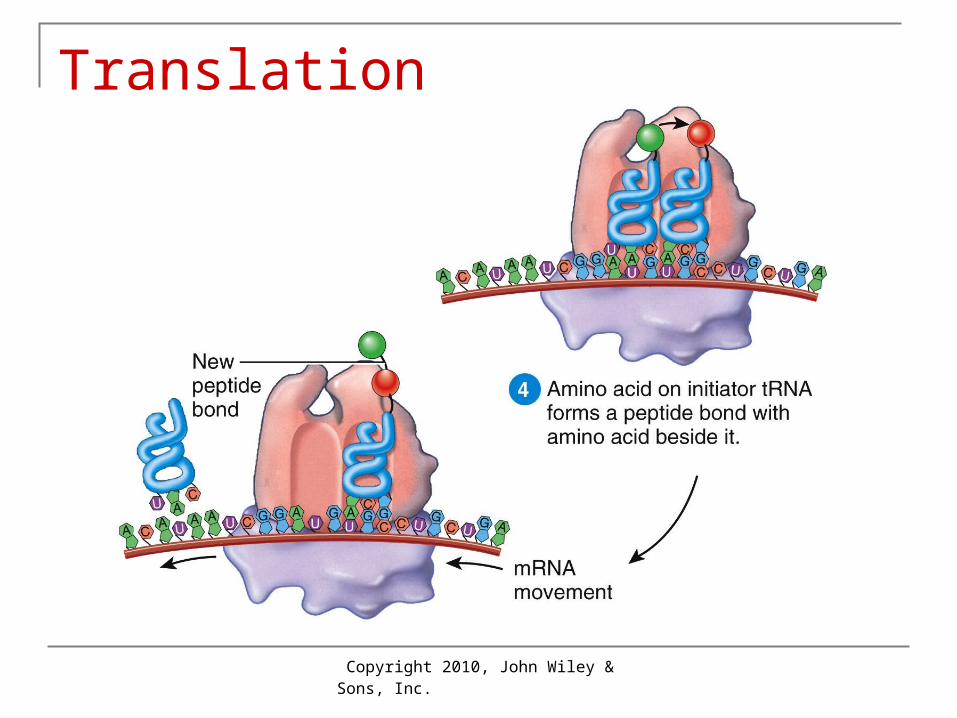
Amino acid on tRNA at P siteforms a peptide bond withamino acid at A site.
P site
A site
= Adenine
= Guanine
= Cytosine
= Uracil
UUU G GGG G GAAAAA
U A CC UCUA
ACUC
GAUUU GG AAAAACC G GUCU ACU
U A CG
A UC
UUU GGG G GAAAAAC UCU
ACU
C
U A CGA
CGAUUUU GGG G GAAAAA
C UCU AC
U A C A UC
2
3
4
1
Key:
Initiator tRNA attaches to astart codon.
Large and small ribosomalsubunits join to form a functionalribosome and initiator tRNAfits into P site.Amino acid
(methionine)
Amino acid
Anticodon
Anticodon
Codons
mRNA
mRNA
mRNAmovement
mRNAbindingsite
Initiator tRNA
tRNA
Start codon
Smallsubunit
Initiator tRNA
Smallsubunit
LargesubunitP site
A site
Anticodon of incoming tRNA pairswith next mRNA codon at A site.
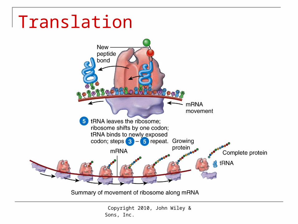
tRNA at P site leaves ribosome,ribosome shifts by one codon;tRNA previously at A site is nowat the P site.
Newpeptidebond Amino acid on tRNA at P site
forms a peptide bond withamino acid at A site.
P site
A site
= Adenine
= Guanine
= Cytosine
= Uracil
UUU G GGG G GAAAAA
U A CC UCUA
ACUC
GAUUU GG AAAAACC G GUCU ACU
U A CG
A UC
UUU GGG G GAAAAAC UCU
ACU
C
U A CGA
CGAUUUU GGG G GAAAAA
C UCU AC
U A C A UC
CGAUUUU
GGG G GAAAAAC UCU
AC
C
U A C A U
2
3
4
5
1
Key:
Initiator tRNA attaches to astart codon.
Large and small ribosomalsubunits join to form a functionalribosome and initiator tRNAfits into P site.Amino acid
(methionine)
Amino acid
Anticodon
Anticodon
Codons
Stop codon
mRNA
mRNA
mRNAmovement
mRNAbindingsite
Initiator tRNA
tRNA
Start codon
Smallsubunit
Initiator tRNA
Smallsubunit
LargesubunitP site
A site
Anticodon of incoming tRNA pairswith next mRNA codon at A site.
Protein synthesis stops whenthe ribosome reaches stopcodon on mRNA.
tRNA at P site leaves ribosome,ribosome shifts by one codon;tRNA previously at A site is nowat the P site.
Newpeptidebond Amino acid on tRNA at P site
forms a peptide bond withamino acid at A site.
P site
A site
Summary of movement of ribosome along mRNA
mRNA
tRNA
Complete proteinGrowingprotein= Adenine
= Guanine
= Cytosine
= Uracil
UUU G GGG G GAAAAA
U A CC UCUA
ACUC
GAUUU GG AAAAACC G GUCU ACU
U A CG
A UC
UUU GGG G GAAAAAC UCU
ACU
C
U A CGA
CGAUUUU GGG G GAAAAA
C UCU AC
U A C A UC
CGAUUUU
GGG G GAAAAAC UCU
AC
C
U A C A U
CUU G GGG G GAA C UCUA ACU
A U
2
3
4
56
Translation
Copyright 2010, John Wiley & Sons, Inc.
Translation
Copyright 2010, John Wiley & Sons, Inc.
Translation
Copyright 2010, John Wiley & Sons, Inc.
Translation
Copyright 2010, John Wiley & Sons, Inc.
Translation
Copyright 2010, John Wiley & Sons, Inc.
Translation
Copyright 2010, John Wiley & Sons, Inc.
Translation
Copyright 2010, John Wiley & Sons, Inc.
Somatic Cell Division In all body cells except gametes Interphase
Period of growth and development of cell Preparation for reproduction: DNA synthesis
Mitotic Phase = division of nucleus 4 phases
Cytokinesis= division of cytoplasm
Copyright 2010, John Wiley & Sons, Inc.
1
Pericentriolar material
NucleolusNuclear envelopeChromatinPlasma membraneCytosol
(a) INTERPHASE
CentriolesCentrosome:
all at 700xLM
1
LateEarly
Pericentriolar material
NucleolusNuclear envelopeChromatinPlasma membraneCytosol
Chromosome(two chromatidsjoined atcentromere
(a) INTERPHASE
(b) PROPHASE
CentriolesCentrosome:
Fragments ofnuclear envelope
Mitotic spindle(microtubules)
Kinetochore
2
all at 700xLM
Centromere
1
Pericentriolar material
NucleolusNuclear envelopeChromatinPlasma membraneCytosol
Metaphase plate
(a) INTERPHASE
CentriolesCentrosome:
(c) METAPHASE
2
3
LateEarly (b) PROPHASE
Fragments ofnuclear envelope
Mitotic spindle(microtubules)
Kinetochore
all at 700xLM
Chromosome(two chromatidsjoined atcentromere
Centromere
1
EarlyLate(d) ANAPHASE
Pericentriolar material
NucleolusNuclear envelopeChromatinPlasma membraneCytosol
Chromosome
(a) INTERPHASE
CentriolesCentrosome:
(c) METAPHASE
2
3
4
Cleavage furrow
LateEarly (b) PROPHASE
Fragments ofnuclear envelope
Mitotic spindle(microtubules)
Kinetochore
Metaphase plate
all at 700xLM
Chromosome(two chromatidsjoined atcentromere
Centromere
1
EarlyLate(d) ANAPHASE
Pericentriolar material
NucleolusNuclear envelopeChromatinPlasma membraneCytosol
(a) INTERPHASE
CentriolesCentrosome:
Cleavage furrow
(e) TELOPHASE
(c) METAPHASE
2
3
4
5
Cleavage furrow
LateEarly (b) PROPHASE
Fragments ofnuclear envelope
Mitotic spindle(microtubules)
Kinetochore
Metaphase plate
Chromosome
all at 700xLM
Chromosome(two chromatidsjoined atcentromere
Centromere
1
EarlyLate(d) ANAPHASE
Pericentriolar material
NucleolusNuclear envelopeChromatinPlasma membraneCytosol
(a) INTERPHASE
CentriolesCentrosome:
(f) IDENTICAL CELLS IN INTERPHASE
Cleavage furrow
(e) TELOPHASE
(c) METAPHASE
Cleavage furrow
2
3
4
5
6
LateEarly (b) PROPHASE
Fragments ofnuclear envelope
Mitotic spindle(microtubules)
Kinetochore
Metaphase plate
Chromosome
all at 700xLM
Centromere
Chromosome(two chromatidsjoined atcentromere
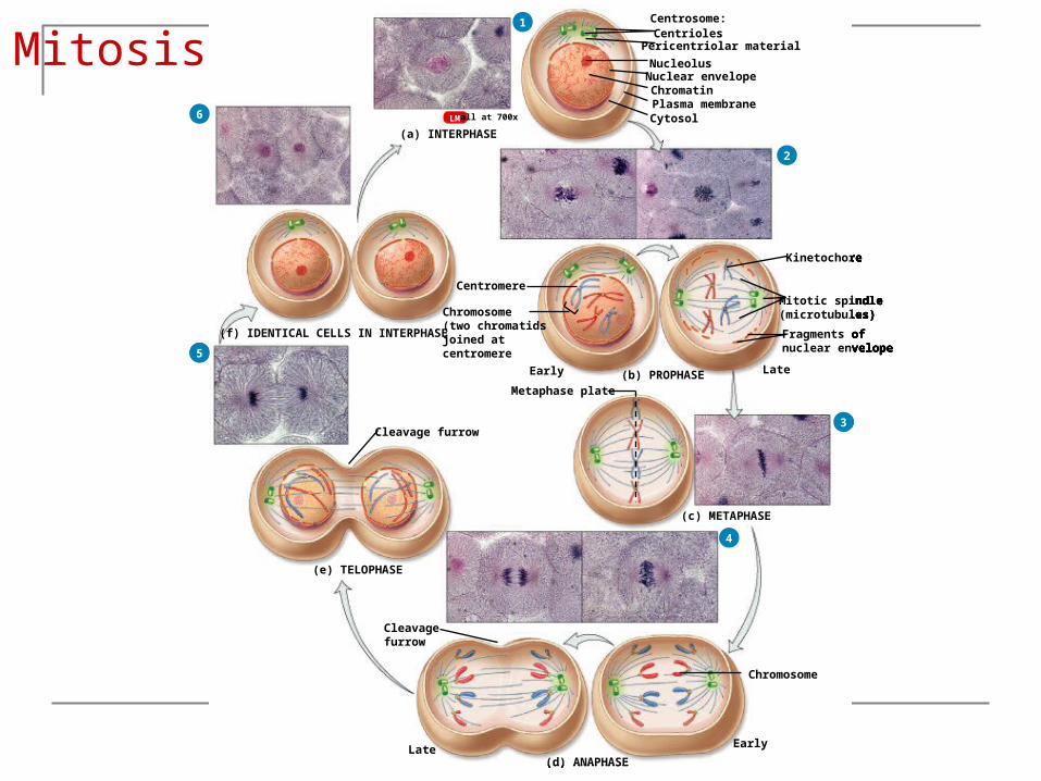
Mitosis
Copyright 2010, John Wiley & Sons, Inc.
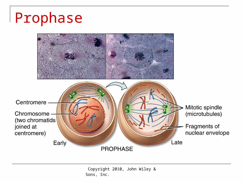
Prophase Chromatin condenses into pairs of
chromatids connected at centromeres Centrosomes form the mitotic spindle
(composed of microtubules) that extends from pole to pole of the cell Some chemotherapy drugs fight cancer cells by
inhibiting formation of the mitotic spindle Nuclear envelope and nucleolus break down
Copyright 2010, John Wiley & Sons, Inc.
Prophase
Copyright 2010, John Wiley & Sons, Inc.
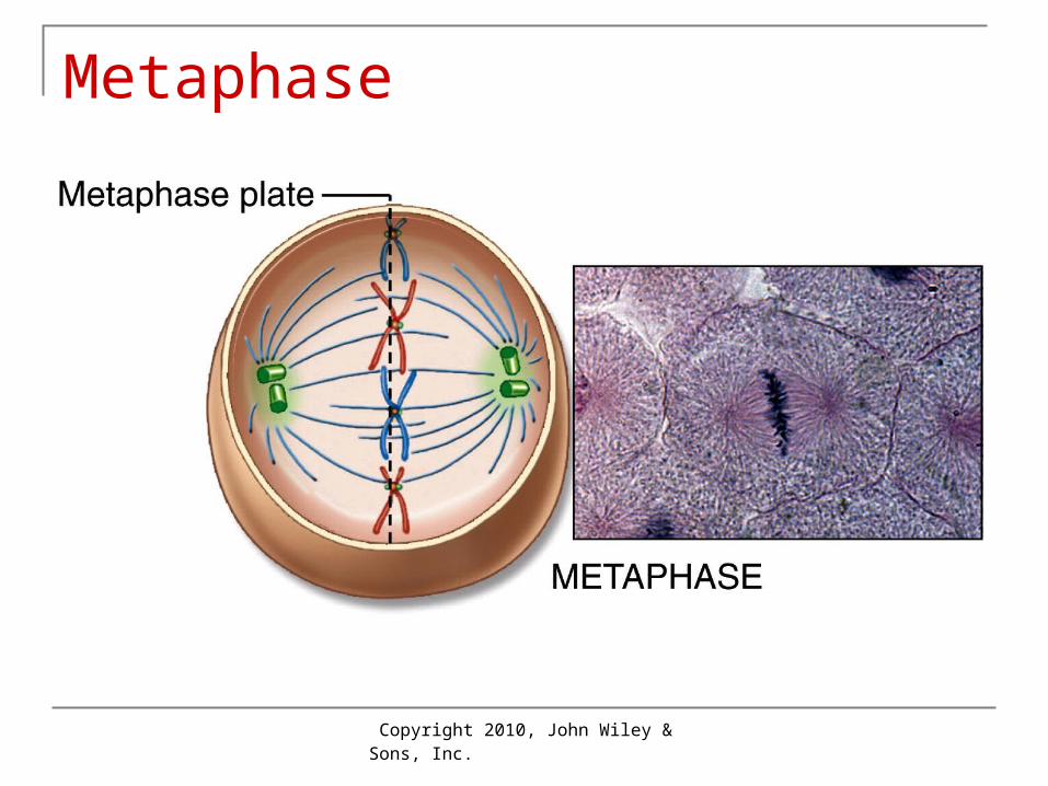
Metaphase
Centromeres of chromatid pairs are aligned along microtubules at the center (“equator”) of the metaphase plate
Copyright 2010, John Wiley & Sons, Inc.
Metaphase
Copyright 2010, John Wiley & Sons, Inc.
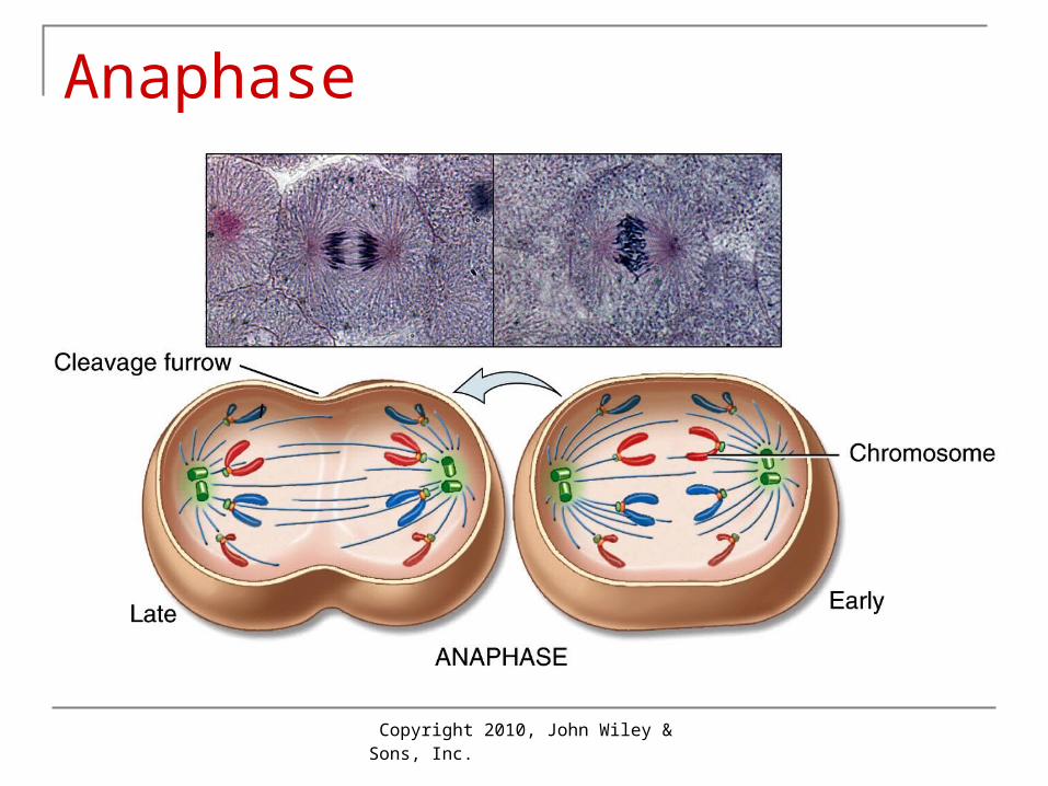
Anaphase
Centromeres split, separating “sister chromatids” (chromosomes)
Chromosomes are pulled to opposite ends of spindle by microtubules of the mitotic spindle
Cytokinesis (division of cytoplasm) begins by the formation of a cleavage furrow
Copyright 2010, John Wiley & Sons, Inc.
Anaphase
Copyright 2010, John Wiley & Sons, Inc.
Telophase
Chromosomes revert to threadlike chromatin Nuclear envelope and nucleolus reappear Mitotic spindle breaks up Cytokinesis is completed
Copyright 2010, John Wiley & Sons, Inc.
Cellular Diversity Because structure determines function, cells
differ in structure related to their functions. Nerve cells may reach several feet in length to
carry nerve impulses from spinal cord to toe Muscle cells can produce effective contractions
because they are cylindrical or spindle-shaped Microvilli increase surface area of intestinal cells
to maximize absorptive ability Most cells are microscopic; the diameter of
the largest human cell (an oocyte) can barely be seen with the unaided eye.
Copyright 2010, John Wiley & Sons, Inc.
Aging A number of factors contribute to aging:
May be programmed genetically Decreased rate of mitosis; nerve cells and skeletal
muscle cells cannot be replaced Telomeres (DNA at tips of chromosomes)
Telomeres shorten with aging Progeria (rapid aging): profound telomere shortening
Protein damage by glucose cross-links Free radicals damage. (See Focus on Wellness:
action of antioxidants to reduce effects of free radicals.)
Autoimmune responses
Copyright 2010, John Wiley & Sons, Inc.
End of Chapter 3
Copyright 2010 John Wiley & Sons, Inc.All rights reserved. Reproduction or translation of this work beyond that permitted in section 117 of the 1976 United States Copyright Act without express permission of the copyright owner is unlawful. Request for further information should be addressed to the Permission Department, John Wiley & Sons, Inc. The purchaser may make back-up copies for his/her own use only and not for distribution or resale. The Publishers assumes no responsibility for errors, omissions, or damages caused by the use of theses programs or from the use of the information herein.