Embed Size (px)
Citation preview
Coronally advanced flap andcombination therapy for rootcoverage. Clinical strategiesbased on scientific evidenceand clinical experiencePIERPAOLO CORTELLINI & GIOVANPAOLO PINI PRATO
Gingival recession is defined as the displacement of
the soft tissue margin apical to the cemento–en-
amel junction (5) and is a frequent clinical feature
in populations with both good (69, 113) and poor
(9, 69, 131) standards of oral hygiene. Localized loss
of attachment with gingival recession is located
mainly at the interdental spaces in patients with
plaque-induced periodontal inflammation and at
the buccal surfaces of teeth in patients with high
standards of oral hygiene (69, 113) and may affect
single or multiple root surfaces. It has historically
been associated with mechanical factors such as
traumatic toothbrushing impacting on predisposed
thin soft tissues (108), even though a recent
systematic review (99) concluded that data to
support or refute the association between tooth-
brushing and gingival recession are still inconclu-
sive. This is a severe !black hole" in our knowledge,
because knowing the cause of recession would
greatly help in the planning of an appropriate
clinical approach directed to improve the prognosis
of this type of periodontal lesion. In fact, prognosis
is defined as !prediction of the future course of a
disease in terms of disease outcomes following its
onset and ⁄ or treatment" and might be positively
modified if the causative agents are controlled; in
other words, if periodontitis is properly treated
and if the traumatic toothbrushing technique is
corrected.
A long-standing debate in the scientific community
relates to whether it is possible to halt the progres-
sion of gingival recession defects by modifying the
oral hygiene habits of patients and ⁄ or by applying
mucogingival surgical procedures (6). A long-term
clinical study recently reported that shallow
recessions showed a tendency for further apical
displacement of the gingival margin in highly moti-
vated patients with high standards of oral hygiene
and enrolled in a stringent supportive periodontal
care system (4–6 months) over a period ranging from
10 to 27 years (1). In the same population of patients,
contralateral grafted sites showed stability or even a
coronal shift of the gingival margin over the same
time frame. The study concluded that untreated
gingival recessions show a negative prognosis over
time in spite of good patient motivation, while the
prognosis is improved after applying mucogingival
procedures. Gingival recession remains a highly
prevalent problem (57, 105) and potentially impacts
on both esthetics and dentine hypersensitivity. Pa-
tients therefore commonly ask about treatment op-
tions for both single and multiple buccal recession
defects.
The ultimate goal of root-coverage procedures is
the complete resolution of the recession defect, with
minimal probing depths after treatment, along with a
nice chromatic and texture integration of the cover-
ing tissues with the adjacent resident soft tissues (20,
21, 31, 78, 102). Clinicians are challenged to achieve
outcomes that meet these exacting standards, and
therefore need a sound, clinically oriented and sci-
entifically supported decision-making process to
plan the therapeutic approach, to predict the out-
come and, finally, to achieve it.
158
Periodontology 2000, Vol. 59, 2012, 158–184
Printed in Singapore. All rights reserved
! 2012 John Wiley & Sons A/S
PERIODONTOLOGY 2000
During the last three decades, several surgical
techniques have been proposed to treat single and
multiple gingival recessions. In the 1970s and 1980s,
the main treatment goals were achieving recession
reduction and increasing keratinized tissue. The
proposed surgical techniques were pedicle flaps
(laterally or coronally positioned) and free gingival
grafts. During the 1980s and 1990s, new approaches,
such as bilaminar techniques or regenerative proce-
dures, were proposed to achieve the goal of complete
root coverage. In the last decade, because of the ever-
increasing esthetic demands from patients, surgical
techniques have been further developed to obtain
complete root coverage associated with a perfect
integration of the grafted tissue with the adjacent soft
tissues (20, 31).
In broad terms, three different approaches can be
identified from the published literature: (i) the free
gingival graft (117), (ii) the coronally advanced flap
(3), and (iii) combined procedures, based on a coro-
nally advanced flap with tissue ⁄ material interposed
between the flap and the root surface. The most
common of the latter approaches are based on a
coronally advanced flap plus a connective tissue graft
(coronally advanced flap + connective tissue graft)
(63), a nonresorbable barrier (91), a bio-resorbable
barrier (92, 101), enamel matrix derivative (25, 41, 89,
100, 115), platelet-rich gel (62), acellular dermal
matrix (51), or living tissue-engineered human
fibroblast-derived dermal substitute (129).
Although all the proposed techniques have shown
potential for root coverage, meta-analyses from sev-
eral systematic reviews (20, 29, 31, 85, 102) showed
the greatest potential for recession reduction and
complete root coverage when applying coronally
advanced flap or combined procedures. These are
therefore the approaches of choice to date.
The aim of this review was to provide a critical
analysis of clinical studies and controlled clinical
trials performed either with coronally advanced flap
therapy alone or with coronally advanced flap ther-
apy in combination with tissues ⁄ materials, and to
propose sound, clinically oriented and scientifically
supported flow charts to help clinicians in their
decision-making processes for the treatment of
localized and multiple gingival recessions.
Overview
From a methodological point of view, this review will
consider published evidence on coronally advanced
flap therapy, alone, or in combination with tis-
sues ⁄ materials, from a clinical perspective with the
aim of helping clinicians in their decision-making
process. The review will include: (i) analysis of the
potential prognostic factors (patient, tooth ⁄ site and
technique-related factors), (ii) discussion of the
surgical procedures, (iii) comparison of different
coronally advanced flap-based approaches, (iv)
evaluation of the healing dynamics and long-term
stability of outcomes, and (v) a discussion of the
patient-related outcomes and side effects of therapy.
The review will end with referenced flow charts.
Single or multiple gingival recessions and patients"requests for root coverage challenge the clinician"sability to choose the best approach and to predict the
outcome. Ideally, a clinician should first discuss with
the patient the desired ⁄ expected outcome(s), then
select the best option to reach those outcome(s).
Interestingly, very few studies have considered
!patient satisfaction" and this should be the true
outcome of a procedure that mainly addresses the
goal of esthetic improvement. A study was performed
to investigate the perceived esthetic outcomes of
simulated root-coverage procedures using three dif-
ferent groups of !evaluators": patients, dentists and
periodontists (104). Complete root coverage was
perceived as the most desirable outcome by the three
groups.
Similarly, information about changes in dentinal
hypersensitivity, another important outcome for
patients, is seldom reported. A recent multicenter
randomized clinical study reported that coronally
advanced flap therapy alone and coronally advanced
flap therapy associated with a connective tissue graft
were both effective in reducing dental hypersensi-
tivity (37).
Most studies have reported surrogate outcomes,
such as complete root coverage, amount of root
coverage, per cent root coverage and changes in the
amount of keratinized tissue. Therefore, this review
will focus on patient outcomes when available, but
will mainly use surrogate outcomes to draw conclu-
sions.
An additional problem has to be highlighted and
should be taken into account when comparing results
from different studies: understanding how !complete
root coverage" or !per cent root coverage" are defined
by different authors. The issue is especially relevant
when teeth with large abrasion cavities and ⁄ or deepsteps involving the cemento–enamel junction are
included (Fig. 1A–C). In these instances, the cemen-
to–enamel junction is no longer detectable and the
record of root coverage becomes a guess. There is, in
fact, a tendency to declare a root to be !completely
159
Coronally advanced flap and combination therapy for root coverage
covered" when the gingival margin reaches a position
that the clinician !feels" is the maximum possible
coverage obtainable in that specific case. This might
in reality reflect the true maximum potential out-
come, but it still is not an !objective" measure.
Potential prognostic factors
Prognostic factors are defined as the characteristics
of a particular patient that can be used to predict,
with greater accuracy, the patient"s eventual out-
come. Prognostic factors do not predict the outcome
completely, but do influence the outcome of treat-
ment (64). Potential prognostic factors for root cov-
erage can be divided into three different categories:
patient-related factors, tooth ⁄ site-related factors and
technique-related factors.
Patient-related factors
Few articles in the periodontal literature are available
that debate the possible influences of age, gender, race
and systemic disease on the outcomes of root coverage
procedures. There is weak evidence that poor oral
hygiene will negatively influence the success of root
coverage (19). Similarly, there is little information on
the influence of traumatic toothbrushing in the
recurrence of recession after treatment (127). Smoking
is a controversial issue. Some papers report less
favorable outcomes in terms of root coverage in
smokers (78, 81, 114, 124, 133), whereas other studies
do not find differences between smokers and non-
smokers (4, 16, 50, 54, 73, 120). A recent systematic
review (28) concluded that smoking may negatively
influence gingival recession reduction and clinical
attachment gain, and smokers may exhibit fewer sites
with complete root coverage. In reality, most of the
studies cited in the review by Chambrone et al. (28)
were not designed to test the influence of smoking on
root coverage and do not provide a comparison be-
tween smokers and nonsmokers.
Tooth ⁄ site-related factors
Interestingly, many of the tooth ⁄ site-specific factors
that are believed to be, and are frequently cited as,
relevant prognostic factors, have never been tested in
sound clinical studies. For example, there is no
information as to whether tooth position (buccal or
lingual), tooth vitality and depth of the vestibule
might influence the outcome of mucogingival pro-
cedures. Limited, and often conflicting, information
is available on the results of root coverage procedures
performed on different tooth types or on maxillary or
mandibular teeth (14, 73, 81, 82).
Cervical dental caries and ⁄ or abrasions are often
associated with gingival recessions. Various ap-
proaches have been attempted to treat gingival
recession associated with cervical lesions, and
excellent clinical results have been achieved, both in
terms of root coverage and cosmetic outcomes (42,
44, 72, 76, 90), showing that superficial caries lesions
or abrasion defects do not seem to impair the pos-
sibility to cover a root.
Root curvature might potentially influence the
outcome of root coverage. This hypothesis is based
on the size of the avascular area, which is larger in
prominent root surfaces. A study (107), performed to
compare the root curvature of four different dental
morphotypes (central incisors, lateral incisors, cus-
pids and bicuspids), showed statistically significant
differences among the tested teeth. To date, no
studies have reported a difference in root coverage in
different morphotypes. However, given the hypothe-
sis of an impact of root curvature on outcomes, it
would be of interest to test such an influence in a
controlled study.
The level of interdental periodontal support (77) is
universally recognized to be of paramount impor-
A B C
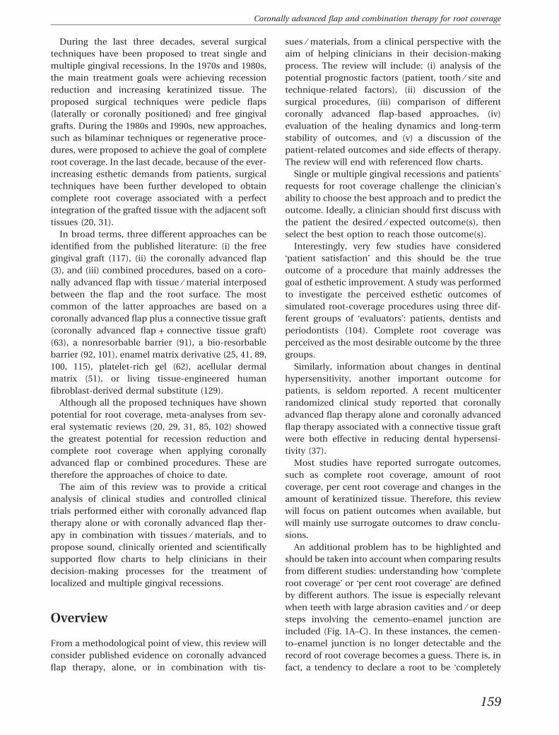
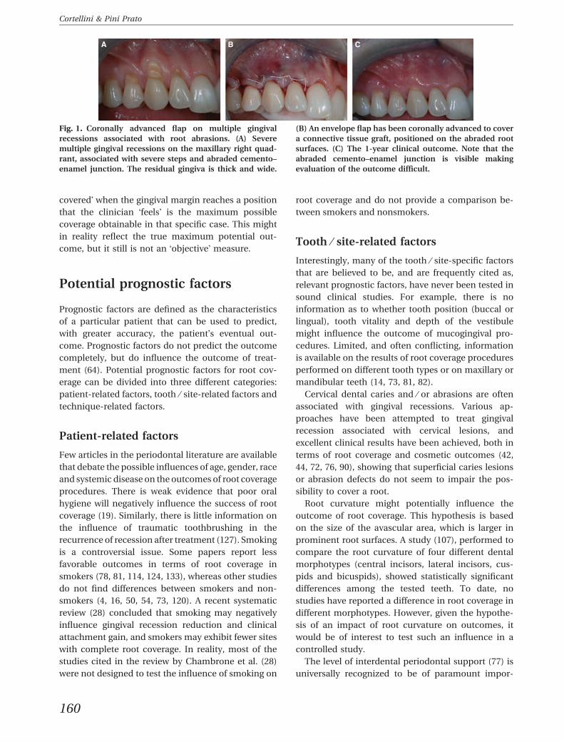
Fig. 1. Coronally advanced flap on multiple gingivalrecessions associated with root abrasions. (A) Severemultiple gingival recessions on the maxillary right quad-rant, associated with severe steps and abraded cemento–enamel junction. The residual gingiva is thick and wide.
(B) An envelope flap has been coronally advanced to covera connective tissue graft, positioned on the abraded rootsurfaces. (C) The 1-year clinical outcome. Note that theabraded cemento–enamel junction is visible makingevaluation of the outcome difficult.
160
Cortellini & Pini Prato
tance for the outcome of root coverage and is one of
the clinical !indicators" generally used to predict
outcome. According to the Miller classification,
Class I and II type defects, in which the interdental
bone support is intact, have the best potential for
complete root coverage. Conversely, only partial
root coverage is thought to be achievable in Miller
Class III and IV type defects: these are associated
with some (from mild to severe) loss of interdental
bone support. This hypothesis (or is it a dogma?),
however, has been challenged in a recent study (8)
on Miller Class III recessions. The authors reported
complete root coverage in 38% of patients treated
with a modified tunnel ⁄ connective tissue graft
technique, with or without the additional use of
enamel matrix derivative. Evidence on treating
Miller Class III and IV defects is both scarce and
weak and does not provide any clear indications on
the potential of interproximal bone loss to impact
on root coverage.
The dimension of the interdental papilla was also
investigated in terms of total area and height (apico-
coronal dimension). Two published studies reached
completely different outcomes. One study, on 33
Miller Class I recessions treated with a coronally ad-
vanced flap, demonstrated that the area of the
interdental papillae adjacent to the recession defect
does not influence the amount of recession reduction
and the likelihood of complete root coverage. On the
other hand, the height of the papilla does influence
complete root coverage: the shorter the papilla, the
greater the probability of obtaining complete root
coverage (106). Other authors have hypothesized that
short papillae could favor coverage because they are
normally associated with a flat and thick gingival
biotype (88). A second study compared two root-
coverage techniques: subepithelial connective tissue
graft and acellular dermal matrix allograft. The study
reported significant, positive correlations between
papilla height and width, and mean root coverage:
the higher and wider the papilla, the greater the ob-
served mean root coverage. In addition, a papilla
height of 5 mm was consistently associated with
complete coverage of the root using both surgical
approaches (47).
The amount and thickness of keratinized tissue is
generally thought to influence the outcome of root
coverage: thick tissues and large amounts of residual
keratinized tissue are !perceived" as favorable. Many
clinicians select a coronally advanced flap or a sliding
flap when the residual keratinized tissue is well rep-
resented, or place a graft under the flap when kera-
tinized tissue is insufficient in thickness and width
(3, 127). However, there is limited evidence to sup-
port this approach.
A clinical study (10) tested the influence of flap
thickness following coronally advanced flap proce-
dures. The results indicate that flap thickness is sig-
nificantly (P < 0.0001) associated with root coverage.
A flap thickness of >0.8 mm was associated with
complete root coverage, while a flap thickness of
<0.8 mmwas associated with partial root coverage. In
addition, linear regression analysis showed that with
each increase in thickness of 0.1 mm, recession was
reduced by approximately 0.2 mm in all treated sites.
Therefore, 0.8 mm can be considered as the critical
flap thickness above which the expected clinical
outcome should be complete root coverage when
using a coronally advanced flap alone.
Another study (136) evaluated the relationship be-
tween root coverage and the baseline amount of
keratinized tissue in laterally positioned and coro-
nally advanced flaps. Multiple logistic regression
analysis showed a statistically significant relationship
between complete root coverage and the amount of
keratinized tissue lateral to the gingival defects: the
greater the amount of keratinized tissue, the greater
the percentage of root coverage.
Many studies and recent systematic reviews
showed the importance of baseline recession depth
in the treatment outcome. The results of the meta-
analyses of controlled and randomized clinical trials
published by Roccuzzo et al. (102) and Clauser et al.
(31) showed a relationship between the initial reces-
sion depth and the final outcome of the surgical
procedure, reporting that !greater baseline recession
depths were always associated with decreased com-
plete root coverage".
Technique-related factors
Root surface
There is a general consensus in the scientific com-
munity that treatment (particularly mechanical
treatment) of the exposed root surface is an impor-
tant component of root-coverage procedures. Various
mechanical and ⁄ or chemical approaches have been
reported in the periodontal literature.
Mechanical root instrumentation (such as root
planing or root surface debridement) is first aimed to
remove the microbial biofilm and has been at-
tempted with hand and machine-driven instruments.
It is important to remember that most Miller Class I
and II recession defects are caused by toothbrushing
161
Coronally advanced flap and combination therapy for root coverage
trauma in patients with good oral hygiene. These
recessions are normally associated with low levels of
plaque, the presence of clinically healthy gingiva and
clean root surfaces. Therefore, the relevance of
planing the root surface might be questioned, and
more conservative approaches should be adopted
(128). A recent randomized, controlled split-mouth
clinical study (139) was performed to compare the
efficacy of hand and ultrasonic instrumentation in
combination with coronally advanced flap therapy in
11 patients with bilateral Miller Class I single reces-
sions. Control root surfaces were planed with
curettes, while test roots were instrumented with
ultrasonic piezoelectric devices. Hand and ultrasonic
root instrumentation were equally effective in terms
of root coverage and clinical attachment gain at
6 months postsurgery.
A randomized controlled clinical study compared
two mechanical treatment modalities: root planing
with curettes vs. polishing with a rubber cup and
prophylaxis paste (93). The experimental population
consisted of 10 patients with bilateral similar Miller
Class I and II single recessions treated with the cor-
onally advanced flap procedure. At 3 months" re-
evaluation, the difference in terms of recession
reduction between the test and control groups was
not statistically significant. In addition, residual
hypersensitivity was experienced only in sites treated
with root planing. This study suggests that planing of
the exposed root surface may be not necessary when
shallow recessions caused by traumatic toothbrush-
ing are treated with the coronally advanced flap
procedure in patients with high levels of oral hygiene.
Heavy mechanical root instrumentation has been
suggested to modify the root surface with the aim of
achieving different end results, such as minimizing
cementum toxicity (13), smoothing irregularities and
grooves in the exposed surface (128), removing root
caries lesions (42) and reducing the convexity of the
root and the mesio–distal distance between the
interproximal spaces (55, 76). Saletta et al. (107)
measured the root curvature before and after
mechanical instrumentation: vigorous root planing
(40 curette strokes) did not substantially modify root
curvature, only slightly reduced (3%) the mesio–dis-
tal dimensions and slightly flattened (6%) the root
surface. Therefore, the use of vigorous root planing is
questionable and none of the cited studies report
evidence to prove a beneficial influence of extensive
root instrumentation on the outcomes of root
coverage (128).
The adjunctive effects of different chemical agents,
such as citric acid (18, 68), tetracycline-HCl (49),
fibrin glue associated with tetracycline-HCl (123) and
sodium hypochlorite (87), in combination with scal-
ing and root planing, have been tested in animal and
clinical studies. These agents have been used to re-
move the smear layer produced by root instrumen-
tation, to expose the collagen fibrils of the dentin
matrix facilitating the formation of new connective
tissue attachment and to remove cytopathic sub-
stances from infected cementum that inhibit human
gingival fibroblast growth. Two systematic reviews
(85, 102) concluded that there are no significant dif-
ferences in terms of root coverage between sites
treated with root planing alone and sites treated with
combined chemical ⁄ mechanical treatment. There-
fore, chemical root surface conditioning cannot be
considered as beneficial for root coverage.
A particular root surface-conditioning approach
consists of chemical treatment of the exposed root
surface with ethylenediaminetetraacetic acid (EDTA)
before the application of enamel matrix derivative.
This approach is part of the clinical protocol for en-
amel matrix derivative application suggested by the
manufacturer, even if its efficacy is unknown (25, 41,
46, 74, 79, 89, 115).
A classification of dental surface defects associated
with gingival recession (cervical dental caries and ⁄ orabrasions) has recently been published (97). This
classification is based on the evaluation of two mor-
phological conditions that may be observed on hard
dental tissues associated with the occurrence of gin-
gival recession: the presence (Class A) or absence
(Class B) of an identifiable cemento–enamel junction;
and the presence (+) or absence ()) of a dental sur-
face discrepancy (step). The study was carried out on
1,010 recession defects. Only 469 had an identifiable
cemento–enamel junction without any associated
step (Class A): 46%), while 144 sites showed an
identifiable cemento–enamel junction associated
with a root surface step (Class A+: 14%), 244 had an
unidentifiable cemento–enamel junction with a step
(Class B+: 24%) and 153 had an unidentifiable
cemento–enamel junction without any associated
step (Class B): 15%).
The high prevalence of sites with an unidentifiable
cemento–enamel junction and ⁄ or with a step re-
quire the adoption of clinical strategies to overcome
these problems. In daily practice, clinicians should
first identify the coronal limit of the potential root
coverage: this becomes difficult when the cemento–
enamel junction is not identifiable. Predicting and
measuring the true and surrogate outcomes becomes
impossible if the reference point (i.e. the cemento–
enamel junction) is not present. A potential solution
162
Cortellini & Pini Prato
might be the !reconstruction" of the cemento–enamel
junction with restorative dentistry prior to surgery
(22, 138). The cemento–enamel junction can be
reconstructed with composite resin mimicking a
!normal cemento–enamel junction" (Fig. 2A–D). The
methods suggested by the cited authors are obviously
based on a !guess" of the shape and position of the
pre-existing cemento–enamel junction, but finally
provide a stable and detectable !reference" for both
the clinician and the patient. Another potential
solution is the reconstruction of the cemento–enamel
junction after the complete healing of the coronally
positioned gingival margin (Fig. 3A–F).
The presence of a step might impair the stabiliza-
tion of the flap ⁄ graft on a flat or concave root
surface, thereby requiring a modified treatment
A B
C D
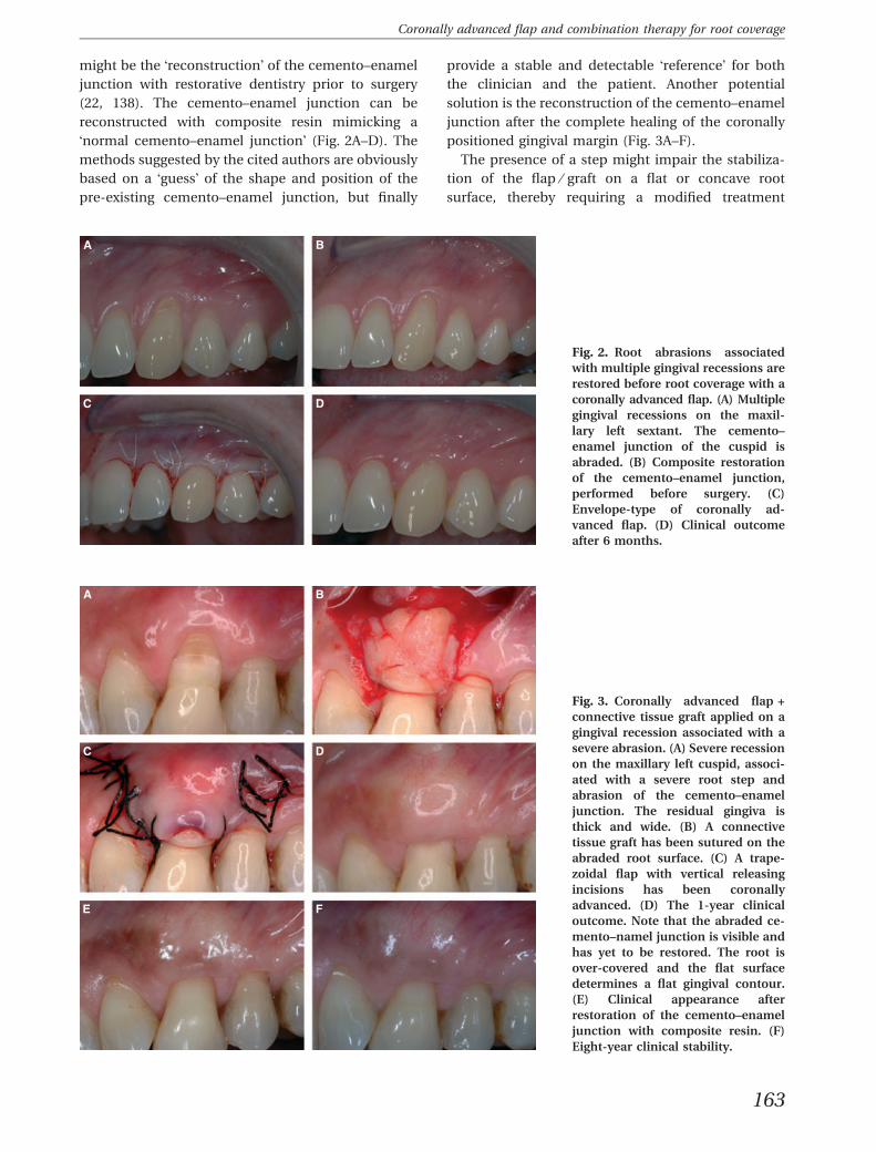
Fig. 2. Root abrasions associatedwith multiple gingival recessions arerestored before root coverage with acoronally advanced flap. (A) Multiplegingival recessions on the maxil-lary left sextant. The cemento–enamel junction of the cuspid isabraded. (B) Composite restorationof the cemento–enamel junction,performed before surgery. (C)Envelope-type of coronally ad-vanced flap. (D) Clinical outcomeafter 6 months.
A B
C D
E F
Fig. 3. Coronally advanced flap +connective tissue graft applied on agingival recession associated with asevere abrasion. (A) Severe recessionon the maxillary left cuspid, associ-ated with a severe root step andabrasion of the cemento–enameljunction. The residual gingiva isthick and wide. (B) A connectivetissue graft has been sutured on theabraded root surface. (C) A trape-zoidal flap with vertical releasingincisions has been coronallyadvanced. (D) The 1-year clinicaloutcome. Note that the abraded ce-mento–namel junction is visible andhas yet to be restored. The root isover-covered and the flat surfacedetermines a flat gingival contour.(E) Clinical appearance afterrestoration of the cemento–enameljunction with composite resin. (F)Eight-year clinical stability.
163
Coronally advanced flap and combination therapy for root coverage
approach. Some authors propose the use of barrier
membranes on the abraded root surface: the root
concavity under a bent barrier is perceived as a
benefit because it provides extra space for peri-
odontal regeneration (90). Other authors suggest the
application of a thick connective tissue graft posi-
tioned to fill the root concavity and finally covered
with a coronally advanced flap (22, 75). Lucchesi et al.
(70) proposed the reconstruction of the abraded
root surface with glass ionomer composite combined
with a coronally advanced flap approach. Two ran-
domized clinical studies compared coronally
advanced flap plus connective tissue graft (109) or
coronally advanced flap alone (110) positioned over a
carefully planed root surface against the coronally
advanced flap plus connective tissue graft or
coronally advanced flap alone positioned over glass
ionomer restorations applied during surgery to
completely fill the root abrasion. According to the
authors, both surgical procedures provide similar soft
tissue coverage either on planed or restored root
surfaces. The presence of a restoration does not
necessarily prevent root coverage but also does
not improve the outcome.
Soft tissue
Soft tissue handling is another factor affecting clinical
outcomes in mucogingival surgery. Design of the
flap, mesio–distal extension, vertical releasing inci-
sions, split-thickness or full-thickness elevation, ten-
sion of the flap and coronal positioning of the flap
should all be planned by the surgeon before surgery.
One of the relevant aspects strictly associated with
flap design is the preservation of a sufficient vascular
system to ensure survival of the flap and in particular
of the marginal gingiva, which is the farthest part of
the flap from the base of the pedicle and lies on an
avascular root surface. Wound healing of pedicle
flaps on exposed root surfaces depends on the pa-
tency of the blood vessels and on anastomoses be-
tween capillaries of the flap ⁄ recipient area and ⁄ orcapillaries of connective tissue graft and recipient
area ⁄ flap. In order to minimize circulatory altera-
tions during root coverage procedures Mormann &
Ciancio (80) suggested that !flaps should be broad
enough at their base to include major gingival vessels
and flap preparations to cover avascular areas should
not be too thin so that more blood vessels are in-
cluded in them". A clinical study (10) reported that
thick gingiva was consistently associated with better
outcomes in terms of recession reduction and com-
plete root coverage in sites treated with a coronally
advanced flap. The authors speculated that thick
marginal tissues could be associated with a more
stable vascular system. Another study (136) con-
cluded that wider residual keratinized tissue was
associated with greater root coverage in sites treated
with laterally positioned, coronally advanced flaps.
The importance of blood supply during healing
has been stressed by Burkhardt & Lang (17), who
evaluated, in a split-mouth study, the degree of
vascularization of connective tissue grafts following
the creation of double papilla flaps using microsur-
gical or macrosurgical approaches. The angiographic
evaluation performed immediately after the surgical
treatment revealed better vascularization of micro-
surgically treated sites compared with macrosurgi-
cally treated sites. The authors assumed that !thesharper and the finer surgical blades, together with
finer suture material used in the microsurgical
approach, were responsible for the reduced tissue
damage". Similar conclusions were drawn by Franc-
etti et al. (43).
The vascularization of the pedicle flap when per-
forming a coronally advanced flap can be further
improved if vertical releasing incisions are avoided.
Zucchelli & De Sanctis (134) proposed a surgical
technique to treat multiple adjacent recession defects
based on an envelope type of flap without vertical
releasing incisions. The authors reported excellent
clinical results in terms of complete root coverage
and optimal esthetic integration of the covering tis-
sue. The same authors have published a study com-
paring coronally advanced flap therapy, with and
without vertical releasing incisions, in the treatment
of multiple recessions (140). Both coronally advanced
flap techniques were effective in reducing recession
depth but the envelope type of coronally advanced
flap (without vertical releasing incisions) was asso-
ciated with an increased probability of achieving
complete root coverage and with a better postoper-
ative course. Nevertheless, a recent systematic review
reported that data on this issue are still insufficient
(27).
An angiographic study on humans supports the
hypothesis that the best clinical outcomes, in terms
of root coverage, are achievable when the flap is
passively adapted and sutured without tension over
the exposed root surface (80). Vestibule depth, root
prominence, presence of frena and recession depth
may influence the passive surgical shift of the cor-
onally advanced flap towards the cemento–enamel
junction. If the flap is not completely released, the
sutures are positioned to overcome the residual
tension to stabilize the flap at the cemento–enamel
164
Cortellini & Pini Prato
junction. As a consequence, sutures that are too
tight may damage the residual vascular system of
the flap: vessel patency is reduced and neo-vascu-
larization is impaired. In addition, the residual ten-
sion of the flap could favor a postoperative apical
shift of the gingival margin during the early phase of
healing.
This hypothesis was confirmed by a randomized
controlled clinical study (94) performed to measure
the tension of the coronally advanced flap before
suturing and to compare the reduction in recession
following coronally advanced flap therapy with or
without tension. The statistical analysis showed that
minimal flap tension (ranging from 0.0 to 0.4 g) fa-
vored recession reduction, while higher tension of the
flap (ranging from 4 to 7 g) was associated with lower
recession reduction.
The position of the gingival margin in relation to
the cemento–enamel junction was proven to be an
important factor in achieving complete root coverage
with coronally advanced flap therapy. In a pilot
study, Pini Prato et al. (93) positioned and sutured
the gingival margin 2 mm coronal to the cemento–
enamel junction, obtaining complete root coverage.
Recently, the same group (95) investigated the
influence of the postsurgical position of the gingival
margin relative to the cemento–enamel junction on
the clinical outcomes of coronally advanced flap
therapy. Coronal displacement of the flap of ‡2 mm
was associated with complete root coverage in 100%
of the patients. The results of the logistic regression
showed that the greater the coronal displacement
of the flap, the greater the recession reduction (P <
0.0001) and the greater the probability of obtaining
complete root coverage (P = 0.0003).
Operator skill is another important factor that can
affect the outcomes of periodontal surgery. A con-
sistent center effect has been demonstrated in several
studies of periodontal regeneration in intrabony de-
fects (36, 111, 121, 122). Regarding root coverage, a
recent multicenter study, comparing coronally
advanced flap therapy with coronally advanced
flap + connective tissue graft therapy in single
recession defects, revealed relevant differences be-
tween centers (37). In this study, the center effect was
significant in spite of the fact that surgery was per-
formed by skilled periodontists who were specifically
trained and calibrated to perform the surgical ap-
proaches tested; in addition, the patient population
was well balanced and carefully selected according to
stringent entry criteria and randomization processes,
and the procedures were conducted within a com-
mon and strict protocol. The center effect should be
taken into account to explain, at least in part, the high
degree of variability frequently observed when stud-
ies performed by different clinicians or groups of
clinicians are evaluated and statistically analysed, as
observed in the meta-analyses from four systematic
reviews on root coverage procedures (20, 31, 85, 102).
Coronally advanced flaps, with or without the use of a
graft, are technique-sensitive procedures that require
specific and refined training and a high level of skills
to be properly applied.
Surgical procedures
Coronally advanced flap
Single recessions
The coronally advanced flap is based on the coronal
shift of soft tissues apical to the exposed root surface
(3). The original procedure was described for cover-
ing isolated gingival recessions. The design of the flap
included !vertical incisions lateral to the recessed area
beginning at a point apical to the papilla tip and
extending well into the alveolar mucosa" (3). A sul-
cular incision and sharp dissection close to the
periosteum allowed a split-thickness flap elevation to
be performed, reaching the alveolar mucosa. Epi-
thelium was removed from the papillae adjacent to
the recession and the flap was coronally positioned
and stabilized with interproximal sutures and apico–
coronal interrupted sutures to close the vertical
releasing incisions. The area was dressed with a
periodontal pack.
This overall design of the coronally advanced flap
has been developed over time with relevant modifi-
cations ⁄ improvements coming from animal and
human research. Following the suggestion of Mor-
mann & Ciancio (80), Pini Prato et al. (91) described a
flap with divergent releasing incisions to obtain a
broad base that included major gingival vessels. The
design of the vertical incisions was a !golf club design"to achieve enough mesio–distal extension of the
coronal part of the flap and obtain perfect adaptation
to the cemento–enamel junction and the interproxi-
mal vascular recipient bed. The starting point of the
vertical incisions should be determined before sur-
gery (134): the amount (in mm) of coronal shift of the
gingiva necessary to cover the exposed root will
indicate the distance from each papilla tip and the
starting point of the vertical incisions. This accurate
design will allow for a perfect adaptation of the coronal
part of the flap to the interdental recipient bed. Pini
Prato et al. (91) also suggested a full-thickness elevation
165
Coronally advanced flap and combination therapy for root coverage
of the gingiva. The clinical study of Baldi et al. (10)
proved the relevance of gingival thickness, as thick
gingiva was consistently associated with improved
outcomes. Flap elevation should therefore be
performed through a buccal intrasulcular incision to
the bone crest followed by a full-thickness flap
elevation beyond the mucogingival junction. Then,
sharp horizontal dissection of the periosteum
reaching the vertical incisions has to be performed for
flap mobilization. Pini Prato et al. (94) demonstrated
that flap tension is key to root coverage: tension-free
flaps have a higher chance of achieving complete root
coverage. Effort should be made to obtain complete
relaxation of the flap through proper apical under-
mining of the alveolar mucosa. The relaxed flap has to
be positioned and stabilized to cover the exposed root
surface (Fig. 4A–C). The position of the gingival
margin influences the final outcomes. Pini Prato et al.
(95) showed that complete root coverage following
coronally advanced flap therapy was consistently
obtained when the flap was positioned 1–2 mm
coronal to the cemento–enamel junction. Flap adap-
tation and stabilization can be achieved through
interdental interrupted sutures or sling sutures. The
application of a periodontal pack is today broadly
avoided.
Multiple recessions
When multiple gingival recessions are located on
adjacent teeth, root coverage should be undertaken
with one surgical procedure. The coronally advanced
flap described above can be extended to treat mul-
tiple recession defects. The pedicle flap should be
broad enough to include all of the individual reces-
sion defects and the vertical releasing incisions will
constitute the mesio–distal limits of the flap. Zucch-
elli & De Sanctis (134) proposed a modified technique
to treat multiple recessions; this technique was based
on an envelope flap, aiming to avoid vertical releasing
incisions and to better preserve the vascular system
and reduce potential scars caused by the vertical
incisions (Fig. 5A–D). The design of the envelope flap
requires the involvement of one extra tooth mesial,
and one extra tooth distal, to the treatment area to
allow for sufficient flap mobility. A modified oblique
papilla incision is performed to obtain proper adap-
A B C
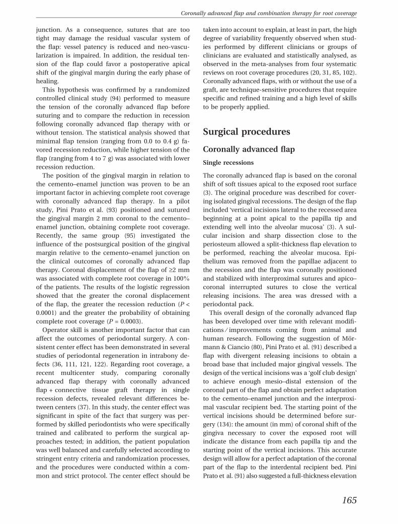
Fig. 4. Coronally advanced flap on a single gingivalrecession associated with thick residual gingiva. (A) Singlegingival recession on the maxillary left bicuspid with thick
residual gingiva. (B) The trapezoidal flap has been suturedcoronally to the cemento–enamel junction. (C) One-yearclinical outcome.
A B
C D
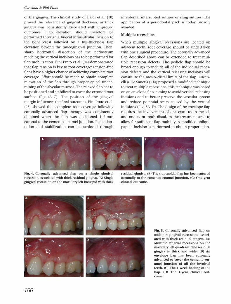
Fig. 5. Coronally advanced flap onmultiple gingival recessions associ-ated with thick residual gingiva. (A)Multiple gingival recessions on themaxillary left quadrant. The residualgingiva is thick and wide. (B) Anenvelope flap has been coronallyadvanced to cover the cemento–en-amel junction of all the involvedteeth. (C) The 1-week healing of theflap. (D) The 1-year clinical out-come.
166
Cortellini & Pini Prato
tation of the surgical papilla to the recipient bed. A
full-thickness flap, followed by a split-thickness
incision beyond the mucogingival junction, is elevated
and coronally positioned to cover the cemento–
enamel junction. A comparison between the coro-
nally advanced flap, with or without vertical incisions
in multiple recessions, demonstrated that both ap-
proaches were effective in providing root coverage,
but the envelope flap was associated with an
increased probability of obtaining complete root
coverage and with a better postoperative result (140).
Combined approaches
Coronally advanced flap therapy has been proposed
in combination with connective tissue graft, barrier
membrane, enamel matrix derivative, acellular der-
mal matrix, platelet concentrated graft and living
tissue-engineered human fibroblast-derived dermal
substitute.
Connective tissue graft
Historically, the use of connective tissue grafts, either
partially (63) or completely (40, 49, 83, 127) covered by
a coronally advanced flap, was suggested. These ap-
proaches consisted of thick and large connective tissue
grafts sutured close to the cemento–enamel junction
and resulted in a high prevalence of complete root
coverage. However, even if consistent root coverage
occurred, often the esthetic appearance of the treated
area was unsatisfactory because of the excessive
thickness of the grafted tissue. Recently, some surgical
modifications to the original technique have been
proposed to improve esthetic outcomes (135). The
size, thickness andpositioning of the connective tissue
graft have been modified accordingly. It has been
proposed that a connective graft, 6 mm larger than the
recession width, should be applied at the cemento–
enamel junction; its apico–coronal dimension is cal-
culated as the distance from the cemento–enamel
junction to the bone crest minus the preoperative
height of the keratinized tissue. An ideal thickness of
the graft, of about 1 mm, was also suggested. The graft
is sutured apical to the cemento–enamel junction at a
distance equal to the height of the preoperative kera-
tinized tissue. It has been speculated that this limited
thickness and size could improve nutritional exchange
between the recipient site, graft and covering pedicle
flap as well as the esthetic outcomes. In a randomized
controlled clinical study, Zucchelli et al. (135) com-
pared the conventional bilaminar approach with the
described novel grafting technique. Outcomes were
similar in terms of root coverage, but esthetics and
perceptions of patients were much more favorable in
the sites treated with the novel approach based on the
use of small grafts. The need to open a second surgical
site to harvest the graft from the palate addsmorbidity
to this approach. This approach can be used to treat
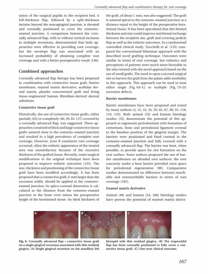
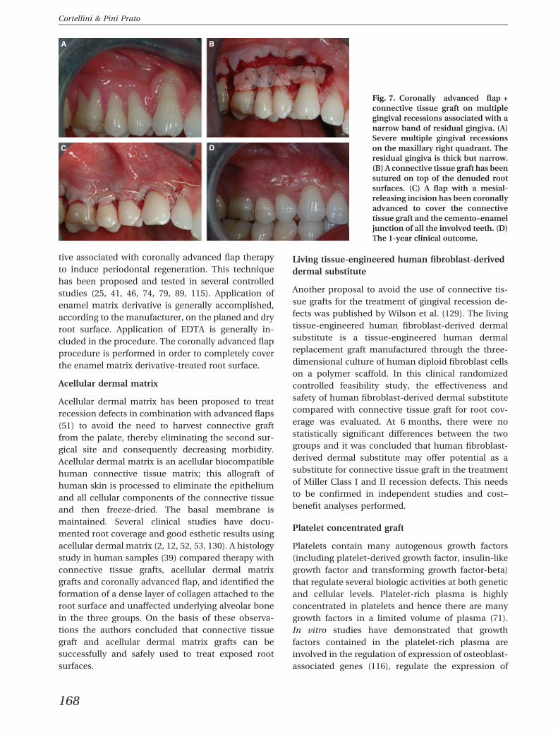
either single (Fig. 6A–C) or multiple (Fig. 7A–D)
recession defects.
Barrier membranes
Barrier membranes have been proposed and tested
by many authors (4, 15, 33, 34, 59, 65, 67, 90, 91, 118,
119, 133). Both animal (33) and human histology
studies (35) demonstrate the potential of this ap-
proach to regenerate periodontium with formation of
cementum, bone and periodontal ligament coronal
to the baseline position of the gingival margin. The
barriers were positioned and fixed coronal to the
cemento–enamel junction and fully covered with a
coronally advanced flap. The barrier was bent, when
possible, to provide space for clot formation on the
root surface. Some authors proposed the use of bar-
rier membranes on abraded root surfaces: the root
concavity under a bent barrier provided extra space
for periodontal regeneration (90). Comparative
studies demonstrated no difference between resorb-
able and nonresorbable barriers in terms of root
coverage (102).
Enamel matrix derivative
Animal (48) and human (24, 100) histology studies
have proven the potential of enamel matrix deriva-
A B C
Fig. 6. Coronally advanced flap + connective tissue grafton a single gingival recession associated with thin residualgingiva. (A) Single gingival recession on the maxillary left
bicuspid with thin residual gingiva. (B) The trapezoidalflap has been coronally positioned to fully cover a con-nective tissue graft. (C) One-year clinical outcome.
167
Coronally advanced flap and combination therapy for root coverage
tive associated with coronally advanced flap therapy
to induce periodontal regeneration. This technique
has been proposed and tested in several controlled
studies (25, 41, 46, 74, 79, 89, 115). Application of
enamel matrix derivative is generally accomplished,
according to the manufacturer, on the planed and dry
root surface. Application of EDTA is generally in-
cluded in the procedure. The coronally advanced flap
procedure is performed in order to completely cover
the enamel matrix derivative-treated root surface.
Acellular dermal matrix
Acellular dermal matrix has been proposed to treat
recession defects in combination with advanced flaps
(51) to avoid the need to harvest connective graft
from the palate, thereby eliminating the second sur-
gical site and consequently decreasing morbidity.
Acellular dermal matrix is an acellular biocompatible
human connective tissue matrix; this allograft of
human skin is processed to eliminate the epithelium
and all cellular components of the connective tissue
and then freeze-dried. The basal membrane is
maintained. Several clinical studies have docu-
mented root coverage and good esthetic results using
acellular dermal matrix (2, 12, 52, 53, 130). A histology
study in human samples (39) compared therapy with
connective tissue grafts, acellular dermal matrix
grafts and coronally advanced flap, and identified the
formation of a dense layer of collagen attached to the
root surface and unaffected underlying alveolar bone
in the three groups. On the basis of these observa-
tions the authors concluded that connective tissue
graft and acellular dermal matrix grafts can be
successfully and safely used to treat exposed root
surfaces.
Living tissue-engineered human fibroblast-derived
dermal substitute
Another proposal to avoid the use of connective tis-
sue grafts for the treatment of gingival recession de-
fects was published by Wilson et al. (129). The living
tissue-engineered human fibroblast-derived dermal
substitute is a tissue-engineered human dermal
replacement graft manufactured through the three-
dimensional culture of human diploid fibroblast cells
on a polymer scaffold. In this clinical randomized
controlled feasibility study, the effectiveness and
safety of human fibroblast-derived dermal substitute
compared with connective tissue graft for root cov-
erage was evaluated. At 6 months, there were no
statistically significant differences between the two
groups and it was concluded that human fibroblast-
derived dermal substitute may offer potential as a
substitute for connective tissue graft in the treatment
of Miller Class I and II recession defects. This needs
to be confirmed in independent studies and cost–
benefit analyses performed.
Platelet concentrated graft
Platelets contain many autogenous growth factors
(including platelet-derived growth factor, insulin-like
growth factor and transforming growth factor-beta)
that regulate several biologic activities at both genetic
and cellular levels. Platelet-rich plasma is highly
concentrated in platelets and hence there are many
growth factors in a limited volume of plasma (71).
In vitro studies have demonstrated that growth
factors contained in the platelet-rich plasma are
involved in the regulation of expression of osteoblast-
associated genes (116), regulate the expression of
A B
C D
Fig. 7. Coronally advanced flap +connective tissue graft on multiplegingival recessions associated with anarrow band of residual gingiva. (A)Severe multiple gingival recessionson the maxillary right quadrant. Theresidual gingiva is thick but narrow.(B) A connective tissue graft has beensutured on top of the denuded rootsurfaces. (C) A flap with a mesial-releasing incision has been coronallyadvanced to cover the connectivetissue graft and the cemento–enameljunction of all the involved teeth. (D)The 1-year clinical outcome.
168
Cortellini & Pini Prato
mineral-associated genes in cementoblasts (112),
modulate the proliferation of periodontal cells in
vitro (86) and stimulate collagen synthesis in peri-
odontal ligament and osteoblastic cells in vitro (61).
Marx et al. (71) demonstrated that a concentration of
1 · 106 platelets ⁄ ml favors the early stages of wound
healing. Several clinical studies showed that platelet-
rich plasma may enhance early graft maturity, bone
density and new bone formation (7, 23, 32, 66, 132).
Different modes of application of platelet concen-
trated graft under a coronally advanced flap have
been proposed in mucogingival surgery. Griffin &
Cheung (45) and Cheung & Griffin (30) used a colla-
gen resorbable sponge soaked with platelet-rich
plasma, and others (58, 62) applied platelet-rich
plasma to a collagen graft, while Huang et al. (56)
applied platelet-rich plasma directly onto the planed
root surface. A coronally advanced flap is performed
in all instances to cover the platelet concentrated
graft or platelet-rich plasma.
Clinical outcomes of coronallyadvanced flap and coronallyadvanced flap-based approaches
A recent meta-analysis from a systematic review (20)
evaluated a total of 794 Miller Class I and II gingival
recession defects in 530 patients from 25 randomized
controlled trials. Coronally advanced flap was se-
lected as the reference treatment and was compared
with the possible combinations (coronally advanced
flap + connective tissue graft, coronally advanced flap +
enamel matrix derivative, coronally advanced flap +
barrier membrane, coronally advanced flap + acel-
lular dermal matrix, coronally advanced flap +
platelet concentrated graft and coronally advanced
flap + human fibroblast-derived dermal substitute).
Complete root coverage was considered as the pri-
mary outcome variable; recession reduction and
keratinized tissue changes were secondary outcomes.
This systematic review confirmed that coronally ad-
vanced flap is a safe and reliable approach and is
consistently associated with complete root coverage
and recession reduction. Results from meta-analyses,
however, showed that two combinations (coronally
advanced flap + connective tissue graft and coronally
advanced flap + enamel matrix derivative) provided
better results than coronally advanced flap alone
and no other therapy provided better results than
coronally advanced flap + connective tissue graft in
terms of complete root coverage and recession
reduction.
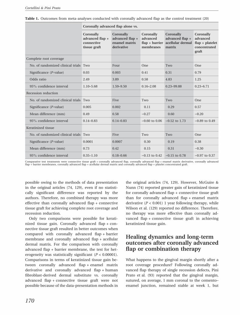
Table 1 presents a summary of the meta-analyses
conducted with coronally advanced flap as the con-
trol treatment. Coronally advanced flap + connective
tissue graft and coronally advanced flap + enamel
matrix derivative were associated with a higher
probability of obtaining complete root coverage and
greater amounts of recession reduction than coro-
nally advanced flap alone. Coronally advanced flap +
barrier membrane, coronally advanced flap + acel-
lular dermal matrix and coronally advanced flap +
platelet concentrated graft were not any more likely
than coronally advanced flap alone to obtain com-
plete root coverage and were not associated with
greater amounts of recession reduction.
For keratinized tissue gain, the adjunctive use of
connective tissue graft or enamel matrix derivative
under a coronally advanced flap was associated with
a gain of keratinized tissue compared with coronally
advanced flap alone, while combinations of coronally
advanced flap + barrier membrane, coronally ad-
vanced flap + acellular dermal matrix and coronally
advanced flap + platelet concentrated graft did not
result in any significant differences compared with
coronally advanced flap alone.
As coronally advanced flap + connective tissue
graft showed the best outcomes, Cairo et al. (20)
performed additional comparisons considering cor-
onally advanced flap + connective tissue graft as the
control surgical procedure (Table 2). Single studies
compared coronally advanced flap + enamel matrix
derivative and coronally advanced flap + human
fibroblast-derived dermal substitute vs. coronally
advanced flap + connective tissue graft, and reported
no significant differences in terms of complete root
coverage. No significant differences in complete
root coverage were reported when comparing coronally
advanced flap + barrier membrane and coronal-
ly advanced flap + acellular dermal matrix vs.
coronally advanced flap + connective tissue graft,
although a trend favoring coronally advanced flap +
connective tissue graft was detected. In terms of
recession reduction, the meta-analyses reported
better results for coronally advanced flap + connec-
tive tissue graft vs. coronally advanced flap + barrier
membrane, but no significant differences were re-
ported vs. coronally advanced flap + acellular dermal
matrix; for this comparison, however, the test for
heterogeneity was statistically significant (P = 0.002).
Comparisons in terms of recession reduction
between coronally advanced flap + enamel matrix
derivative and coronally advanced flap + human
fibroblast-derived dermal substitute vs. coronally
advanced flap + connective tissue graft were not
169
Coronally advanced flap and combination therapy for root coverage
possible owing to the methods of data presentation
in the original articles (74, 129), even if no statisti-
cally significant difference was reported by the
authors. Therefore, no combined therapy was more
effective than coronally advanced flap + connective
tissue graft for achieving complete root coverage and
recession reduction.
Only two comparisons were possible for kerati-
nized tissue gain. Coronally advanced flap + con-
nective tissue graft resulted in better outcomes when
compared with coronally advanced flap + barrier
membrane and coronally advanced flap + acellular
dermal matrix. For the comparison with coronally
advanced flap + barrier membrane, the test for het-
erogeneity was statistically significant (P < 0.00001).
Comparisons in terms of keratinized tissue gain be-
tween coronally advanced flap + enamel matrix
derivative and coronally advanced flap + human
fibroblast-derived dermal substitute vs. coronally
advanced flap + connective tissue graft were not
possible because of the data-presentation methods in
the original articles (74, 129). However, McGuire &
Nunn (74) reported greater gain of keratinized tissue
for coronally advanced flap + connective tissue graft
than for coronally advanced flap + enamel matrix
derivative (P < 0.001) 1 year following therapy, while
Wilson et al. (129) reported no difference. Therefore,
no therapy was more effective than coronally ad-
vanced flap + connective tissue graft in achieving
keratinized tissue gain.
Healing dynamics and long-termoutcomes after coronally advancedflap or combination therapy
What happens to the gingival margin shortly after a
root coverage procedure? Following coronally ad-
vanced flap therapy of single recession defects, Pini
Prato et al. (93) reported that the gingival margin,
sutured, on average, 1 mm coronal to the cemento–
enamel junction, remained stable at week 1, but
Table 1. Outcomes from meta-analyses conducted with coronally advanced flap as the control treatment (20)
Coronally advanced flap alone vs.
Coronallyadvanced flap +connectivetissue graft
Coronallyadvanced flap +enamel matrixderivative
Coronallyadvancedflap + barriermembranes
Coronallyadvanced flap +acellular dermalmatrix
Coronallyadvancedflap + plateletconcentratedgraft
Complete root coverage
No. of randomized clinical trials Two Four One Two One
Significance (P-value) 0.03 0.003 0.41 0.31 0.79
Odds ratio 2.49 3.89 0.58 4.83 1.25
95% confidence interval 1.10–5.68 1.59–9.50 0.16–2.08 0.23–99.88 0.23–6.71
Recession reduction
No. of randomized clinical trials Two Five Two Two One
Significance (P-value) 0.005 0.002 0.11 0.29 0.57
Mean difference (mm) 0.49 0.58 )0.27 0.60 )0.20
95% confidence interval 0.14–0.83 0.14–0.83 )0.60 to 0.06 )0.52 to 1.73 )0.89 to 0.49
Keratinized tissue
No. of randomized clinical trials Two Five Two Two One
Significance (P-value) 0.0001 0.0007 0.30 0.19 0.38
Mean difference (mm) 0.73 0.42 0.15 0.31 )0.30
95% confidence interval 0.35–1.10 0.18–0.66 )0.13 to 0.42 )0.15 to 0.78 )0.97 to 0.37
Comparative test treatments were connective tissue graft + coronally advanced flap, coronally advanced flap + enamel matrix derivative, coronally advancedflap + barrier membranes, coronally advanced flap + acellular dermal matrix and coronally advanced flap + platelet concentrated graft.
170
Cortellini & Pini Prato
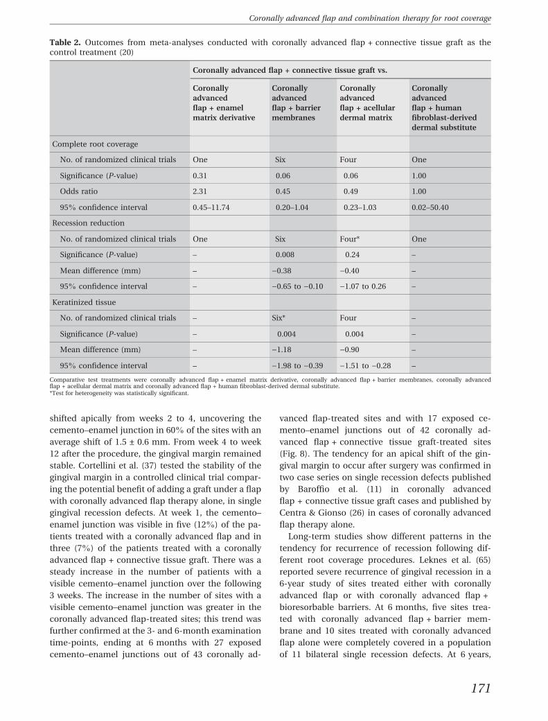
shifted apically from weeks 2 to 4, uncovering the
cemento–enamel junction in 60% of the sites with an
average shift of 1.5 ± 0.6 mm. From week 4 to week
12 after the procedure, the gingival margin remained
stable. Cortellini et al. (37) tested the stability of the
gingival margin in a controlled clinical trial compar-
ing the potential benefit of adding a graft under a flap
with coronally advanced flap therapy alone, in single
gingival recession defects. At week 1, the cemento–
enamel junction was visible in five (12%) of the pa-
tients treated with a coronally advanced flap and in
three (7%) of the patients treated with a coronally
advanced flap + connective tissue graft. There was a
steady increase in the number of patients with a
visible cemento–enamel junction over the following
3 weeks. The increase in the number of sites with a
visible cemento–enamel junction was greater in the
coronally advanced flap-treated sites; this trend was
further confirmed at the 3- and 6-month examination
time-points, ending at 6 months with 27 exposed
cemento–enamel junctions out of 43 coronally ad-
vanced flap-treated sites and with 17 exposed ce-
mento–enamel junctions out of 42 coronally ad-
vanced flap + connective tissue graft-treated sites
(Fig. 8). The tendency for an apical shift of the gin-
gival margin to occur after surgery was confirmed in
two case series on single recession defects published
by Baroffio et al. (11) in coronally advanced
flap + connective tissue graft cases and published by
Centra & Gionso (26) in cases of coronally advanced
flap therapy alone.
Long-term studies show different patterns in the
tendency for recurrence of recession following dif-
ferent root coverage procedures. Leknes et al. (65)
reported severe recurrence of gingival recession in a
6-year study of sites treated either with coronally
advanced flap or with coronally advanced flap +
bioresorbable barriers. At 6 months, five sites trea-
ted with coronally advanced flap + barrier mem-
brane and 10 sites treated with coronally advanced
flap alone were completely covered in a population
of 11 bilateral single recession defects. At 6 years,
Table 2. Outcomes from meta-analyses conducted with coronally advanced flap + connective tissue graft as thecontrol treatment (20)
Coronally advanced flap + connective tissue graft vs.
Coronallyadvancedflap + enamelmatrix derivative
Coronallyadvancedflap + barriermembranes
Coronallyadvancedflap + acellulardermal matrix
Coronallyadvancedflap + humanfibroblast-deriveddermal substitute
Complete root coverage
No. of randomized clinical trials One Six Four One
Significance (P-value) 0.31 0.06 0.06 1.00
Odds ratio 2.31 0.45 0.49 1.00
95% confidence interval 0.45–11.74 0.20–1.04 0.23–1.03 0.02–50.40
Recession reduction
No. of randomized clinical trials One Six Four* One
Significance (P-value) – 0.008 0.24 –
Mean difference (mm) – )0.38 )0.40 –
95% confidence interval – )0.65 to )0.10 )1.07 to 0.26 –
Keratinized tissue
No. of randomized clinical trials – Six* Four –
Significance (P-value) – 0.004 0.004 –
Mean difference (mm) – )1.18 )0.90 –
95% confidence interval – )1.98 to )0.39 )1.51 to )0.28 –
Comparative test treatments were coronally advanced flap + enamel matrix derivative, coronally advanced flap + barrier membranes, coronally advancedflap + acellular dermal matrix and coronally advanced flap + human fibroblast-derived dermal substitute.*Test for heterogeneity was statistically significant.
171
Coronally advanced flap and combination therapy for root coverage
only two sites treated with barriers and one site
treated with coronally advanced flap alone were still
completely covered. Nickles et al. (84) compared
sites treated with coronally advanced flap + a bio-
resorbable barrier to sites treated with coronally
advanced flap + connective tissue graft. After
10 years, stability of root coverage was significantly
better in the sites treated with coronally advanced
flap + connective tissue graft. In other words, the
sites treated with a barrier membrane underwent a
greater apical shift of the gingival margin over
time. A 14-year study of 22 single recession defects
treated with coronally advanced flap alone similarly
reported a consistent apical shift of the gingival
margin in 39% of the cases (96). In a 5-year eval-
uation of a case series treated with the envelope
type of coronally advanced flap on multiple
recession defects, Zucchelli & De Sanctis (137)
reported a slight shift of the gingival margin com-
pared with the 1-year data. The amount of long-
term recurrence, however, was very limited. In fact,
complete root coverage was observed in 88% of
the patients at 1 year, and was reduced to 85% at
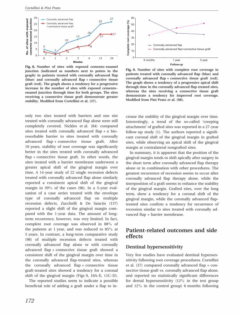
5 years. In contrast, a long-term comparative study
(98) of multiple recession defects treated with
coronally advanced flap alone or with coronally
advanced flap + connective tissue graft showed a
consistent shift of the gingival margin over time in
the coronally advanced flap-treated sites, whereas
the coronally advanced flap + connective tissue
graft-treated sites showed a tendency for a coronal
shift of the gingival margin (Figs 9, 10A–E, 11C–D).
The reported studies seem to indicate a possible
beneficial role of adding a graft under a flap to in-
crease the stability of the gingival margin over time.
Interestingly, a trend of the so-called !creepingattachment" of grafted sites was reported in a 27-year
follow-up study (1). The authors reported a signifi-
cant coronal shift of the gingival margin in grafted
sites, while observing an apical shift of the gingival
margin at contralateral nongrafted sites.
In summary, it is apparent that the position of the
gingival margin tends to shift apically after surgery in
the short term after coronally advanced flap therapy
alone or in combination with other procedures. The
greatest recurrence of recession seems to occur after
coronally advanced flap therapy alone, while the
interposition of a graft seems to enhance the stability
of the gingival margin. Grafted sites, over the long
term, show a tendency for a coronal shift of the
gingival margin, while the coronally advanced flap-
treated sites confirm a tendency for recurrence of
recession similar to sites treated with coronally ad-
vanced flap + barrier membrane.
Patient-related outcomes and sideeffects
Dentinal hypersensitivity
Very few studies have evaluated dentinal hypersen-
sitivity following root coverage procedures. Cortellini
et al. (37) compared coronally advanced flap + con-
nective tissue graft vs. coronally advanced flap alone,
and reported no statistically significant differences
for dental hypersensitivity (12% in the test group
and 12% in the control group) 6 months following
cem
en
to-e
nam
el
jun
ctio
nN
o.
of
site
s w
ith
exp
ose
d
52
1013
16
2527
3 14
78
1717
0
5
10
15
20
25
30
0 1 2 3 4 12 24Weeks
Coronally advanced flap
Coronally advanced flap+connective tissue graft
Fig. 8. Number of sites with exposed cemento–enameljunction (indicated as numbers next to points in thegraph) in patients treated with coronally advanced flap(blue) and coronally advanced flap + connective tissuegraft (red). The graph shows a tendency for a progressiveincrease in the number of sites with exposed cemento–enamel junction through time for both groups. The sitesreceiving a connective tissue graft demonstrate greaterstability. Modified from Cortellini et al. (37).
0
10
20
30
40
50
60
6 months 1 yearFollow-up
5 year
Com
plet
e ro
ot c
over
age
(%)
Coronally advanced flapCoronally advanced flap+connective tissue graft
Fig. 9. Number of sites with complete root coverage inpatients treated with coronally advanced flap (blue) andcoronally advanced flap + connective tissue graft (red).The graph shows a tendency of a progressive apical shiftthrough time in the coronally advanced flap-treated sites,whereas the sites receiving a connective tissue graftdemonstrate a tendency for improved root coverage.Modified from Pini Prato et al. (98).
172
Cortellini & Pini Prato
therapy. McGuire & Nunn (74) reported dentinal
hypersensitivity in only one patient treated with
coronally advanced flap + enamel matrix derivative
and no dentinal hypersensitivity for a coronally ad-
vanced flap + connective tissue graft 1 year following
therapy. Pini Prato et al. (93) reported reduced
hypersensitivity in sites treated with prophylaxis
compared to sites treated with root planing following
coronally advanced flap therapy.
Esthetics
Some studies have evaluated esthetic satisfaction
following therapy. Romagna-Genon (103) compared
coronally advanced flap + barrier membrane vs.
coronally advanced flap + connective tissue graft in a
split-mouth study, and reported that one patient was
not satisfied with either treatment. In a study by
Wang et al. (126) a double esthetic evaluation was
A B
C D
E
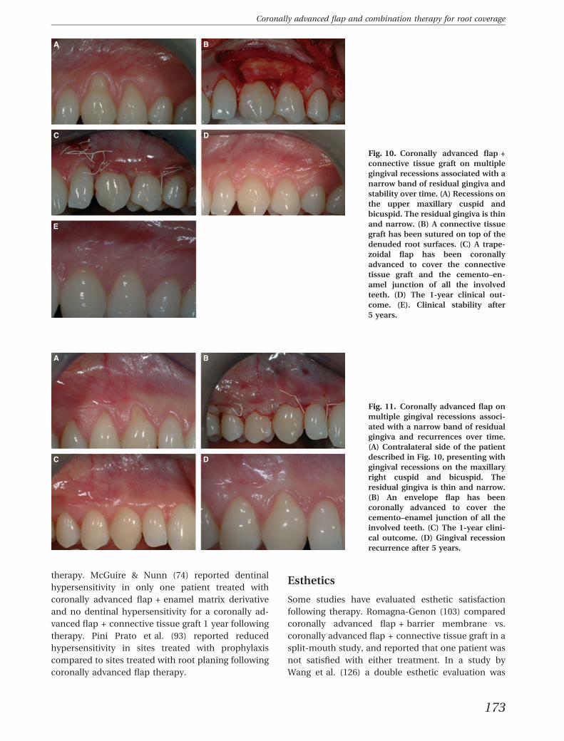
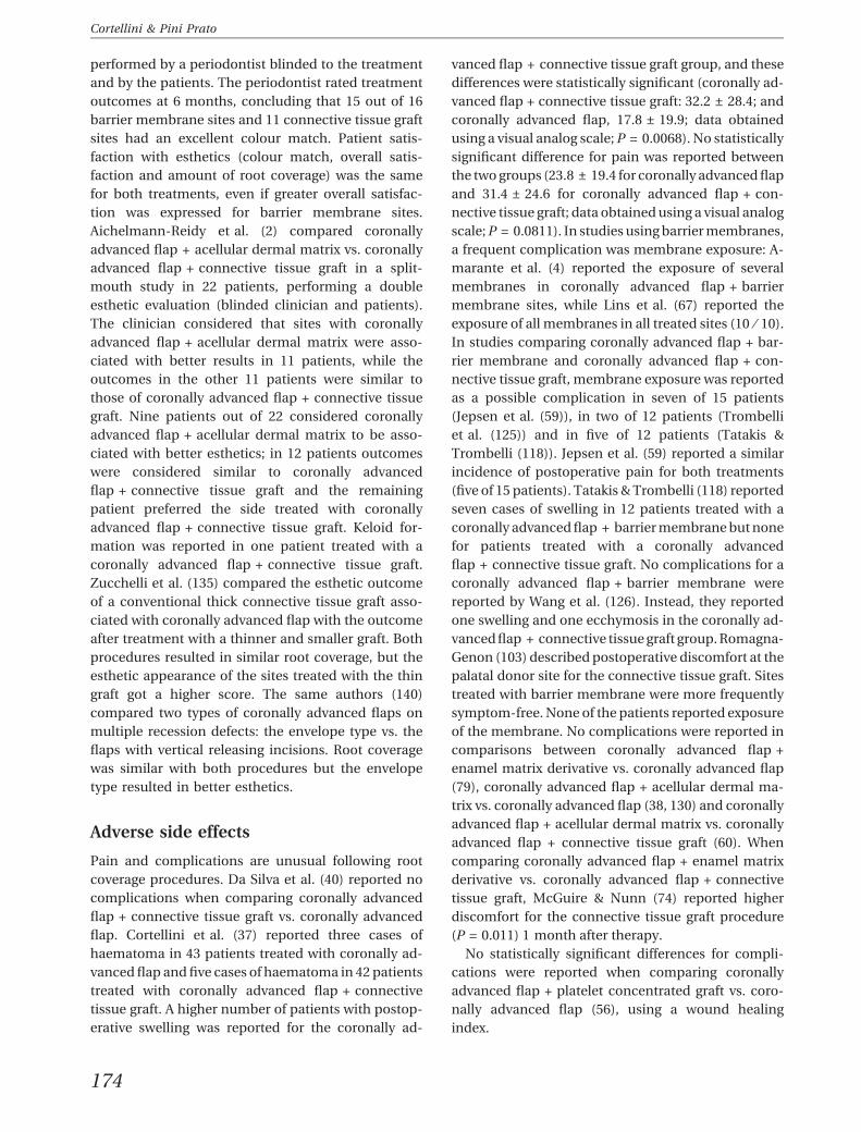
Fig. 10. Coronally advanced flap +connective tissue graft on multiplegingival recessions associated with anarrow band of residual gingiva andstability over time. (A) Recessions onthe upper maxillary cuspid andbicuspid. The residual gingiva is thinand narrow. (B) A connective tissuegraft has been sutured on top of thedenuded root surfaces. (C) A trape-zoidal flap has been coronallyadvanced to cover the connectivetissue graft and the cemento–en-amel junction of all the involvedteeth. (D) The 1-year clinical out-come. (E). Clinical stability after5 years.
A B
C D
Fig. 11. Coronally advanced flap onmultiple gingival recessions associ-ated with a narrow band of residualgingiva and recurrences over time.(A) Contralateral side of the patientdescribed in Fig. 10, presenting withgingival recessions on the maxillaryright cuspid and bicuspid. Theresidual gingiva is thin and narrow.(B) An envelope flap has beencoronally advanced to cover thecemento–enamel junction of all theinvolved teeth. (C) The 1-year clini-cal outcome. (D) Gingival recessionrecurrence after 5 years.
173
Coronally advanced flap and combination therapy for root coverage
performed by a periodontist blinded to the treatment
and by the patients. The periodontist rated treatment
outcomes at 6 months, concluding that 15 out of 16
barrier membrane sites and 11 connective tissue graft
sites had an excellent colour match. Patient satis-
faction with esthetics (colour match, overall satis-
faction and amount of root coverage) was the same
for both treatments, even if greater overall satisfac-
tion was expressed for barrier membrane sites.
Aichelmann-Reidy et al. (2) compared coronally
advanced flap + acellular dermal matrix vs. coronally
advanced flap + connective tissue graft in a split-
mouth study in 22 patients, performing a double
esthetic evaluation (blinded clinician and patients).
The clinician considered that sites with coronally
advanced flap + acellular dermal matrix were asso-
ciated with better results in 11 patients, while the
outcomes in the other 11 patients were similar to
those of coronally advanced flap + connective tissue
graft. Nine patients out of 22 considered coronally
advanced flap + acellular dermal matrix to be asso-
ciated with better esthetics; in 12 patients outcomes
were considered similar to coronally advanced
flap + connective tissue graft and the remaining
patient preferred the side treated with coronally
advanced flap + connective tissue graft. Keloid for-
mation was reported in one patient treated with a
coronally advanced flap + connective tissue graft.
Zucchelli et al. (135) compared the esthetic outcome
of a conventional thick connective tissue graft asso-
ciated with coronally advanced flap with the outcome
after treatment with a thinner and smaller graft. Both
procedures resulted in similar root coverage, but the
esthetic appearance of the sites treated with the thin
graft got a higher score. The same authors (140)
compared two types of coronally advanced flaps on
multiple recession defects: the envelope type vs. the
flaps with vertical releasing incisions. Root coverage
was similar with both procedures but the envelope
type resulted in better esthetics.
Adverse side effects
Pain and complications are unusual following root
coverage procedures. Da Silva et al. (40) reported no
complications when comparing coronally advanced
flap + connective tissue graft vs. coronally advanced
flap. Cortellini et al. (37) reported three cases of
haematoma in 43 patients treated with coronally ad-
vancedflap andfive cases of haematoma in 42 patients
treated with coronally advanced flap + connective
tissue graft. A higher number of patients with postop-
erative swelling was reported for the coronally ad-
vanced flap + connective tissue graft group, and these
differences were statistically significant (coronally ad-
vanced flap + connective tissue graft: 32.2 ± 28.4; and
coronally advanced flap, 17.8 ± 19.9; data obtained
using a visual analog scale;P = 0.0068). No statistically
significant difference for pain was reported between
the twogroups (23.8 ± 19.4 for coronally advancedflap
and 31.4 ± 24.6 for coronally advanced flap + con-
nective tissue graft; data obtainedusing a visual analog
scale;P = 0.0811). In studies usingbarriermembranes,
a frequent complication was membrane exposure: A-
marante et al. (4) reported the exposure of several
membranes in coronally advanced flap + barrier
membrane sites, while Lins et al. (67) reported the
exposure of all membranes in all treated sites (10 ⁄ 10).In studies comparing coronally advanced flap + bar-
rier membrane and coronally advanced flap + con-
nective tissue graft, membrane exposure was reported
as a possible complication in seven of 15 patients
(Jepsen et al. (59)), in two of 12 patients (Trombelli
et al. (125)) and in five of 12 patients (Tatakis &
Trombelli (118)). Jepsen et al. (59) reported a similar
incidence of postoperative pain for both treatments
(five of 15 patients). Tatakis & Trombelli (118) reported
seven cases of swelling in 12 patients treated with a
coronally advancedflap + barriermembranebutnone
for patients treated with a coronally advanced
flap + connective tissue graft. No complications for a
coronally advanced flap + barrier membrane were
reported by Wang et al. (126). Instead, they reported
one swelling and one ecchymosis in the coronally ad-
vancedflap + connective tissuegraft group.Romagna-
Genon (103) described postoperative discomfort at the
palatal donor site for the connective tissue graft. Sites
treated with barrier membrane were more frequently
symptom-free. None of the patients reported exposure
of the membrane. No complications were reported in
comparisons between coronally advanced flap +
enamel matrix derivative vs. coronally advanced flap
(79), coronally advanced flap + acellular dermal ma-
trix vs. coronally advanced flap (38, 130) and coronally
advanced flap + acellular dermal matrix vs. coronally
advanced flap + connective tissue graft (60). When
comparing coronally advanced flap + enamel matrix
derivative vs. coronally advanced flap + connective
tissue graft, McGuire & Nunn (74) reported higher
discomfort for the connective tissue graft procedure
(P = 0.011) 1 month after therapy.
No statistically significant differences for compli-
cations were reported when comparing coronally
advanced flap + platelet concentrated graft vs. coro-
nally advanced flap (56), using a wound healing
index.
174
Cortellini & Pini Prato

Flow charts and discussion
In the era of evidence-based medicine, clinicians are
invited to adopt evidence-based decisions for ther-
apy. However, it is apparent that evidence-based
decision-making is lacking in many areas of peri-
odontal therapy, including decisions about achiev-
ing root coverage at recession defects. The lack of
evidence means that clinical experience is used
when making clinical decisions. The literature sur-
vey performed in this review attempts to provide
clinicians with enough scientific and clinical support
to take proper decisions. References to support the
following flow charts have been reported earlier in
this review.
A reasonable starting point for a flow chart is to set
the outcomes of therapy (Fig. 12). Treatment of
recession is often undertaken to satisfy the esthetic
demands of patients: the main outcome should
therefore be esthetic satisfaction of the patient (37,
74, 93). However, scientific background on this !true"outcome is very scarce. A number of research groups
are now proposing novel !evaluation scales" of
esthetics that need validation and adoption in con-
trolled studies (21, 104): these will probably help
clinicians in the future to choose the proper ap-
proach and acknowledge patient desires. Owing to
this lack of information, we are therefore forced to
make decisions based on !surrogate outcomes".Complete root coverage is the one mostly used, along
with recession reduction. Another commonly re-
Root coverage
CAF+CTG
CAF+EMD
CAF+ADM
CAF+PCG
CAF+BM
CAF
Increased outcomes
Amount of KT
CAF+CTG CAF
Increment over baseline
Reduction over baseline
CAF+ADM CAF+PRC
CAF+BM CAF+EMD
Cost�–benefit
CAF + CTG
CAF+ADM
Increased morbidity and chair-time
CAF+PRC
CAF
CAF+EMD
CAF+BM
Cost�–benefit
CAF+ADM
Increased financial costs
CAF+PRC
CAF
CAF+CTG
CAF+BM
CAF+EMD
Esthetics
CAF + thin CTG CAF + EMD CAF
Long term
CAF+CTG CAF CAF+BM
Greater stability
A B C
D E F
Fig. 12. Objectives of therapy. (A) When the objective oftherapy is root coverage, meta-analyses demonstrate thatthe combinations of coronally advanced flap (CAF) +connective tissue graft (CTG) and coronally advancedflap + enamel matrix derivative (EMD) (green) result in thebest clinical outcomes. (B) When the objective of therapy isincreased keratinized tissue (KT), meta-analyses demon-strate that adding a graft or even enamel matrix derivativeunder a flap (green) results in increased keratinized tissue.The amount of keratinized tissue is slightly reduced overtime when a coronally advanced flap alone or with platelet-rich concentrate (PRC) (red) is used. There is a minorreduction in keratinized tissue when a barrier membrane(BM) or an acellular dermal matrix (ADM) (orange) isadded to a coronally advanced flap. (C) When consideringthe cost–benefit ratio, greater morbidity and chair-time areassociated with some combinations (red and orange) with
respect to a coronally advanced flap, alone or together withenamel matrix derivative (EMD) (green). These results weresupported by controlled studies. (D) When evaluating thecost–benefit ratio, clinicians should consider that increasedfinancial costs are associated with the combinations in redwith respect to a coronally advanced flap, alone or with aconnective tissue graft (green). These results weresupported by expert opinions. (E) According to existingevidence, the three suggested procedures (green) do resultin the best esthetic outcomes. These results were supportedby controlled studies. (F) Considering the long-term sta-bility of therapy, results obtained with coronally advancedflap + connective tissue graft (green) are reportedly morestable over time than results obtained with coronallyadvanced flap or coronally advanced flap + barrier mem-brane (red). These results were supported by controlledstudies. PCG, platelet concentrated graft.
175
Coronally advanced flap and combination therapy for root coverage
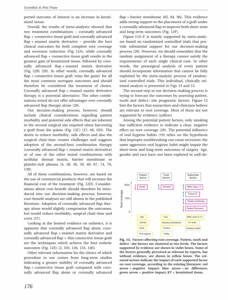
ported outcome of interest is an increase in kerati-
nized tissue.
Overall, the results of meta-analyses showed that
two treatment combinations – coronally advanced
flap + connective tissue graft and coronally advanced
flap + enamel matrix derivative – provide the best
clinical outcomes for both complete root coverage
and recession reduction (Fig. 12A), while coronally
advanced flap + connective tissue graft results in the
greatest gain of keratinized tissue, followed by coro-
nally advanced flap + enamel matrix derivative
(Fig. 12B) (20). In other words, coronally advanced
flap + connective tissue graft !wins the game" for all
the most common surrogate outcomes and should
therefore be considered the treatment of choice.
Coronally advanced flap + enamel matrix derivative
therapy is a potential alternative. The other combi-
nations tested do not offer advantages over coronally
advanced flap therapy alone (20).
Our decision-making process, however, should
include clinical considerations regarding patient
morbidity and potential side effects that are inherent
to the second surgical site required when harvesting
a graft from the palate (Fig. 12C) (37, 40, 103). The
desire to reduce morbidity, side effects and also the
surgical chair-time creates challenges and suggests
adoption of the second-best combination therapy
(coronally advanced flap + enamel matrix derivative)
or of one of the other tested combinations with
acellular dermal matrix, barrier membrane or
platelet-rich plasma (4, 38, 56, 59, 60, 67, 74, 79,
130).
All of these combinations, however, are based on
the use of commercial products that will increase the
financial cost of the treatment (Fig. 12D). Consider-
ations about cost–benefit should therefore be intro-
duced into our decision-making process; however,
cost–benefit analyses are still absent in the published
literature. Adoption of coronally advanced flap ther-
apy alone would slightly compromise the outcomes,
but would reduce morbidity, surgical chair-time and
costs (37).
Looking at the limited evidence on esthetics, it is
apparent that coronally advanced flap alone, coro-
nally advanced flap + enamel matrix derivative and
coronally advanced flap + thin connective tissue graft
are the techniques which achieve the best esthetic
outcomes (Fig. 12E) (2, 103, 126, 135, 140).
Other relevant information for the choice of which
procedure to use comes from long-term studies
indicating a greater stability of coronally advanced
flap + connective tissue graft compared with coro-
nally advanced flap alone or coronally advanced
flap + barrier membrane (65, 84, 96). This evidence
adds strong support to the placement of a graft under
a coronally advanced flap to improve both short-term
and long-term outcomes (Fig. 12F).
Figure 12A–F is mainly supported by meta-analy-
ses based on randomized controlled trials that pro-
vide substantial support for our decision-making
process (20). However, we should remember that the
random assignment of a therapy cannot satisfy the
requirements of each single clinical case. In other
words, the presurgical analysis of every patient
should incorporate information that cannot be fully
exploited by the meta-analytic process of random-
ized controlled trials. This individual, clinically ori-
ented analysis is presented in Figs 14 and 15.
The second step in our decision-making process is
trying to forecast the outcomes by assessing patient,
tooth and defect ⁄ site prognostic factors. Figure 13
lists the factors that researchers and clinicians believe
are relevant to root coverage. Most of these are not
supported by evidence (yellow).
Among the potential patient factors, only smoking
has sufficient evidence to indicate a clear, negative
effect on root coverage (28). The potential influence
of oral hygiene habits (19) relies on the hypothesis
that improper toothbrushing can cause recession: the
same aggressive oral hygiene habit might impair the
short-term and long-term outcomes of surgery. Age,
gender and race have not been explored in well-de-
Roo
t cov
erag
e
Patient factors
Toothfactors
Defect/site factors
Age
Gender
Race
Oral hygiene
Smoke
Type
Maxillary-Mandibular
Position
Vitality
Miller class I, II
Papilla morphology
Vestibule depth
Deeper Recession
KT width
Thicker KT
Abrasion
Miller class III, IV
0
100%
50%
Fig. 13. Factors affecting root coverage. Patient, tooth anddefect ⁄ site factors are clustered at two levels. The factorssupported by evidence are shown in violet boxes. Some ofthe factors generally perceived as relevant by experts, butwithout evidence, are shown in yellow boxes. The col-oured arrows indicate the impact of each supported factoron root coverage, according to the existing literature: redarrow = negative impact; blue arrow = no difference;green arrow = positive impact; KT = keratinized tissue.
176
Cortellini & Pini Prato
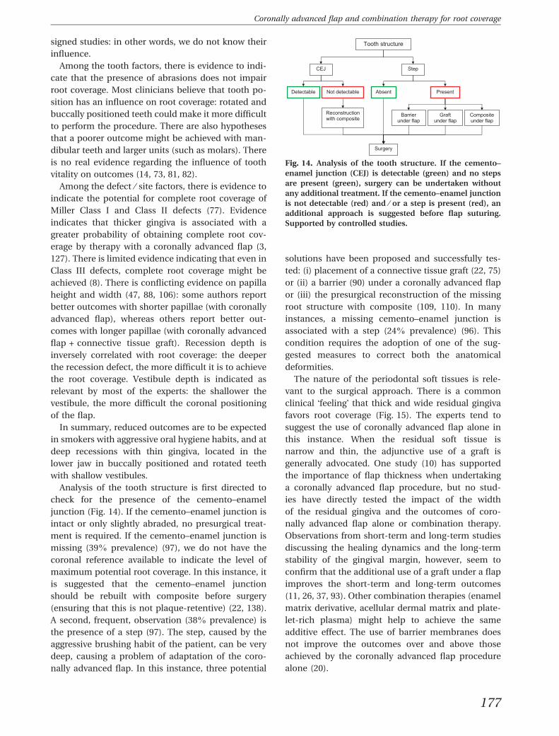
signed studies: in other words, we do not know their
influence.
Among the tooth factors, there is evidence to indi-
cate that the presence of abrasions does not impair
root coverage. Most clinicians believe that tooth po-
sition has an influence on root coverage: rotated and
buccally positioned teeth could make it more difficult
to perform the procedure. There are also hypotheses
that a poorer outcome might be achieved with man-
dibular teeth and larger units (such as molars). There
is no real evidence regarding the influence of tooth
vitality on outcomes (14, 73, 81, 82).
Among the defect ⁄ site factors, there is evidence to
indicate the potential for complete root coverage of
Miller Class I and Class II defects (77). Evidence
indicates that thicker gingiva is associated with a
greater probability of obtaining complete root cov-
erage by therapy with a coronally advanced flap (3,
127). There is limited evidence indicating that even in
Class III defects, complete root coverage might be
achieved (8). There is conflicting evidence on papilla
height and width (47, 88, 106): some authors report
better outcomes with shorter papillae (with coronally
advanced flap), whereas others report better out-
comes with longer papillae (with coronally advanced
flap + connective tissue graft). Recession depth is
inversely correlated with root coverage: the deeper
the recession defect, the more difficult it is to achieve
the root coverage. Vestibule depth is indicated as
relevant by most of the experts: the shallower the
vestibule, the more difficult the coronal positioning
of the flap.
In summary, reduced outcomes are to be expected
in smokers with aggressive oral hygiene habits, and at
deep recessions with thin gingiva, located in the
lower jaw in buccally positioned and rotated teeth
with shallow vestibules.
Analysis of the tooth structure is first directed to
check for the presence of the cemento–enamel
junction (Fig. 14). If the cemento–enamel junction is
intact or only slightly abraded, no presurgical treat-
ment is required. If the cemento–enamel junction is
missing (39% prevalence) (97), we do not have the
coronal reference available to indicate the level of
maximum potential root coverage. In this instance, it
is suggested that the cemento–enamel junction
should be rebuilt with composite before surgery
(ensuring that this is not plaque-retentive) (22, 138).
A second, frequent, observation (38% prevalence) is
the presence of a step (97). The step, caused by the
aggressive brushing habit of the patient, can be very
deep, causing a problem of adaptation of the coro-
nally advanced flap. In this instance, three potential
solutions have been proposed and successfully tes-
ted: (i) placement of a connective tissue graft (22, 75)
or (ii) a barrier (90) under a coronally advanced flap
or (iii) the presurgical reconstruction of the missing
root structure with composite (109, 110). In many
instances, a missing cemento–enamel junction is
associated with a step (24% prevalence) (96). This
condition requires the adoption of one of the sug-
gested measures to correct both the anatomical
deformities.
The nature of the periodontal soft tissues is rele-
vant to the surgical approach. There is a common
clinical !feeling" that thick and wide residual gingiva
favors root coverage (Fig. 15). The experts tend to
suggest the use of coronally advanced flap alone in
this instance. When the residual soft tissue is
narrow and thin, the adjunctive use of a graft is
generally advocated. One study (10) has supported
the importance of flap thickness when undertaking
a coronally advanced flap procedure, but no stud-
ies have directly tested the impact of the width
of the residual gingiva and the outcomes of coro-
nally advanced flap alone or combination therapy.
Observations from short-term and long-term studies
discussing the healing dynamics and the long-term
stability of the gingival margin, however, seem to
confirm that the additional use of a graft under a flap
improves the short-term and long-term outcomes
(11, 26, 37, 93). Other combination therapies (enamel
matrix derivative, acellular dermal matrix and plate-
let-rich plasma) might help to achieve the same
additive effect. The use of barrier membranes does
not improve the outcomes over and above those
achieved by the coronally advanced flap procedure
alone (20).
Tooth structure
CEJ
Detectable
Reconstruction with composite
Step
Barrier under flap
Not detectable Present Absent
Graft under flap
Composite under flap
Surgery
Fig. 14. Analysis of the tooth structure. If the cemento–enamel junction (CEJ) is detectable (green) and no stepsare present (green), surgery can be undertaken withoutany additional treatment. If the cemento–enamel junctionis not detectable (red) and ⁄ or a step is present (red), anadditional approach is suggested before flap suturing.Supported by controlled studies.
177
Coronally advanced flap and combination therapy for root coverage
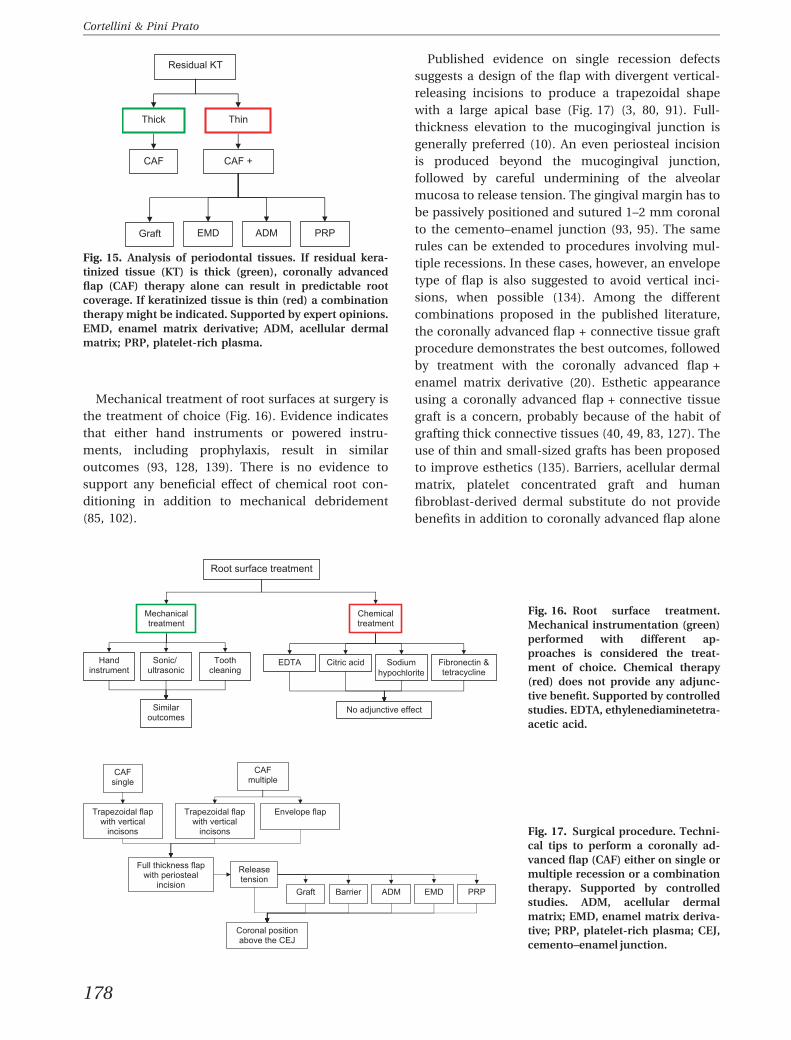
Mechanical treatment of root surfaces at surgery is
the treatment of choice (Fig. 16). Evidence indicates
that either hand instruments or powered instru-
ments, including prophylaxis, result in similar
outcomes (93, 128, 139). There is no evidence to
support any beneficial effect of chemical root con-
ditioning in addition to mechanical debridement
(85, 102).
Published evidence on single recession defects
suggests a design of the flap with divergent vertical-
releasing incisions to produce a trapezoidal shape
with a large apical base (Fig. 17) (3, 80, 91). Full-
thickness elevation to the mucogingival junction is
generally preferred (10). An even periosteal incision
is produced beyond the mucogingival junction,
followed by careful undermining of the alveolar
mucosa to release tension. The gingival margin has to
be passively positioned and sutured 1–2 mm coronal
to the cemento–enamel junction (93, 95). The same
rules can be extended to procedures involving mul-
tiple recessions. In these cases, however, an envelope
type of flap is also suggested to avoid vertical inci-
sions, when possible (134). Among the different
combinations proposed in the published literature,
the coronally advanced flap + connective tissue graft
procedure demonstrates the best outcomes, followed
by treatment with the coronally advanced flap +
enamel matrix derivative (20). Esthetic appearance
using a coronally advanced flap + connective tissue
graft is a concern, probably because of the habit of
grafting thick connective tissues (40, 49, 83, 127). The
use of thin and small-sized grafts has been proposed
to improve esthetics (135). Barriers, acellular dermal
matrix, platelet concentrated graft and human
fibroblast-derived dermal substitute do not provide
benefits in addition to coronally advanced flap alone
Residual KT
Thick
CAF
Thin
CAF +
Graft EMD PRP ADM
Fig. 15. Analysis of periodontal tissues. If residual kera-tinized tissue (KT) is thick (green), coronally advancedflap (CAF) therapy alone can result in predictable rootcoverage. If keratinized tissue is thin (red) a combinationtherapy might be indicated. Supported by expert opinions.EMD, enamel matrix derivative; ADM, acellular dermalmatrix; PRP, platelet-rich plasma.
Root surface treatment
Mechanical treatment
Chemical treatment
Hand instrument
Sonic/ ultrasonic
Tooth cleaning
Citric acid EDTA Sodiumhypochlorite
Fibronectin & tetracycline
No adjunctive effect Similar outcomes
Fig. 16. Root surface treatment.Mechanical instrumentation (green)performed with different ap-proaches is considered the treat-ment of choice. Chemical therapy(red) does not provide any adjunc-tive benefit. Supported by controlledstudies. EDTA, ethylenediaminetetra-acetic acid.
CAF single
Trapezoidal flap with vertical
incisons
CAF multiple
Release tension
Coronal position above the CEJ
Full thickness flap with periosteal
incision
Envelope flap Trapezoidal flap with vertical
incisons
Graft Barrier ADM PRP EMD
Fig. 17. Surgical procedure. Techni-cal tips to perform a coronally ad-vanced flap (CAF) either on single ormultiple recession or a combinationtherapy. Supported by controlledstudies. ADM, acellular dermalmatrix; EMD, enamel matrix deriva-tive; PRP, platelet-rich plasma; CEJ,cemento–enamel junction.
178
Cortellini & Pini Prato
(20). From a surgical standpoint, all these materials
or products are applied on the denuded and debrided
root surface, sutured when indicated and fully cov-
ered with the coronally advanced flap.
Conclusions
It is clear that much relevant information is still
missing in the published literature.
Evidence indicates that the use of a graft under a
coronally advanced flap results in the best short-term
and long-term outcomes in terms of root coverage
and gain in keratinized tissue with the highest prev-
alence of success. It would therefore appear easy to
suggest that clinicians adopt coronally advanced
flap + connective tissue graft for root coverage. On
the other hand, harvesting a graft from the palate
adds morbidity, lengthens surgical chair-time and
requires increased surgical skills. It is evident that
treatment with a coronally advanced flap alone is less
invasive for the patient, and requires less chair-time
and probably less surgical skill. In addition, evidence
shows that coronally advanced flap therapy in many
instances results in complete root coverage and is
stable over time. It would therefore be desirable to
apply coronally advanced flap when indicated.
Unfortunately, the key information – to be able to
forecast when coronally advanced flap will be suc-
cessful – is still missing. Many !experts" support thehypothesis that therapy with coronally advanced flap
alone can be successfully applied when the residual
gingiva is thick and wide. Accordingly, the adjunctive
use of a graft could be restricted to sites with thin and
narrow residual gingiva. A potential alternative is the
use of enamel matrix derivative.
Another indication for placing a graft under a flap is
the presence of a step on the root surface. An alter-
native to placing a connective tissue graft might be the
use of enamelmatrix derivative as this results in better
outcomes than coronally advanced flap alone and is
equally effective as coronally advanced flap + con-
nective tissue graft in terms of root coverage.
If one of the objectives is keratinized tissue gain,
however, clinicians should choose a graft. Other po-
tential alternatives to a graft are acellular dermal
matrix, platelet concentrated graft and human
fibroblast-derived dermal substitute. Evidence, how-
ever, does not indicate an advantage of these ap-
proaches over coronally advanced flap alone. The use
of enamel matrix derivative, acellular dermal matrix,
barrier membrane, platelet concentrated graft and
human fibroblast-derived dermal substitute reduce
morbidity and chair-time, but increases the financial
cost of therapy.
Clinicians should make their decisions after
defining the desired short-term and long-term out-
comes, having completed a careful presurgical anal-
ysis and having discussed with the patient the impact
of morbidity and biologic ⁄ economic costs in addi-
tion to the potential benefits of therapy.
Acknowledgments
This study was supported by the Accademia Toscana
di RicercaOdontostomatologica (ATRO) and the Euro-
peanResearchGrouponPeriodontology (ERGOPerio).
References
1. Agudio G, Nieri M, Rotundo R, Franceschi D, Cortellini P,
Pini Prato GP. Periodontal conditions of sites treated with
gingival-augmentation surgery compared to untreated
contralateral homologous sites: a 10- to 27-year long-term
study. J Periodontol 2009: 80: 1399–1405.
2. Aichelmann-Reidy ME, Yukna RA, Evans GH, Nasr HF,
Mayer ET. Clinical evaluation of acellular allograft dermis
for the treatment of human gingival recession. J Period-
ontol 2001: 72: 998–1005.
3. Allen EP, Miller PD. Coronal positioning of existing gingiva:
short term results in the treatment of shallow marginal
tissue recession. J Periodontol 1989: 60: 316–319.
4. Amarante ES, Leknes KN, Skavland J, Lie T. Coronally
positioned flap procedures with or without a bioabsorbable
membrane in the treatment of human gingival recession.
J Periodontol 2000: 71: 989–998.
5. American Academy of Periodontology. Glossary of Peri-
odontal Terms, 3rd edn. Chicago: The American Academy
of Periodontology, 1992.
6. American Academy of Periodontology. Consensus report on
mucogingival therapy. Proceedings of the World Workshop
in Periodontics. Ann Periodontol 1996: 1: 702–706.
7. Anitua E. Plasma rich in growth factors: preliminary results
of use in the preparation of future sites for implants. Int
J Oral Maxillofac Implants 1999: 14: 529–535.
8. Aroca S, Keglevich T, Nikolidakis D, Gera I, Nagy K, Azzi R,
Etienne D. Treatment of class III multiple gingival reces-
sions: a randomized-clinical trial. J Clin Periodontol 2010:
37: 88–97.
9. Baelum V, Fejerskov O, Karring T. Oral hygiene, gingivitis
and periodontal breakdown in adult Tanzanians. J Peri-
odontal Res 1986: 21: 221–232.
10. Baldi C, Pini Prato GP, Pagliaro U, Nieri M, Saletta D, Muzzi
L, Cortellini P. Coronally advanced flap procedure for root
coverage. Is flap thickness a relevant predictor to achieve
root coverage? A 19-case series. J Periodontol 1999: 70:
1077–1084.
11. Baroffio A, Brusio M, Conforti GP. Clinical performance of
coronally advanced flaps combined with connective tissue
grafts in the treatment of gingival recessions. J Clin Peri-
179
Coronally advanced flap and combination therapy for root coverage
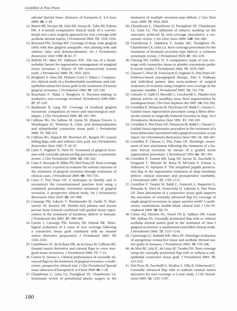
odontol Special Issue: abstracts of Europerio 6, 4–6 June
2009: 36: 1–39.
12. Barros RR, Novaes AB, Grisi MF, Souza SL, Taba MJ, Palioto
DB. A 6-month comparative clinical study of a conven-
tional and a new surgical approach for root coverage with
acellular dermal matrix. J Periodontol 2004: 75: 1350–1356.
13. Bertrand PM, Dunlap RM. Coverage of deep, wide gingival
clefts with free gingival autografts: root planing with and
without citric acid demineralization. Int J Periodontics
Restorative Dent 1988: 8: 64–77.
14. Boltchi FE, Allen EP, Hallmon WW. The use of a bioab-
sorbable barrier for regenerative management of marginal
tissue recession. I. Report of 100 consecutively treated
teeth. J Periodontol 2000: 71: 1641–1653.
15. Borghetti A, Glise JM, Monnet-Corti V, Dejou J. Compara-
tive clinical study of a bioabsorbable membrane and sub-
epithelial connective tissue graft in the treatment of human
gingival recession. J Periodontol 1999: 70: 123–130.
16. Bouchard P, Malet J, Borghetti A. Decision-making in
aesthetics: root coverage revisited. Periodontol 2000 2001:
27: 97–120.
17. Burkhardt R, Lang NP. Coverage of localized gingival
recessions: comparison of micro and macrosurgical tech-
niques. J Clin Periodontol 2005: 32: 287–293.
18. Caffesse RG, De LaRosa M, Garza M, Munne-Travers A,
Mondragon JC, Weltman R. Citric acid demineralization
and subepithelial connective tissue graft. J Periodontol
2000: 71: 568–572.
19. Caffesse RG, Alspach SR, Morrison EC, Burgett FG. Lateral
sliding flaps with and without citric acid. Int J Periodontics
Restorative Dent 1987: 7: 42–57.
20. Cairo F, Pagliaro U, Nieri M. Treatment of gingival reces-
sion with coronally advanced flap procedures: a systematic
review. J Clin Periodontol 2008: 35: 136–162.
21. Cairo F, Rotundo R, Miller PD, Pini Prato GP. Root coverage
esthetic score: a system to evaluate the esthetic outcome of
the treatment of gingival recession through evaluation of
clinical cases. J Periodontol 2009: 80: 705–710.
22. Cairo F, Pini Prato GP. A technique to identify and to
reconstruct the cementoenamel junction level using a
combined periodontal ⁄ restorative treatment of gingival
recession. A prospective clinical study. Int J Periodontics
Restorative Dent 2010: 30: 591–600.
23. Camargo PM, Lekovic V, Weinlaender M, Vasilic N, Mad-
zarevic M, Kenney EB. Platelet-rich plasma and bovine
porous bone mineral combined with guided tissue regen-
eration in the treatment of intrabony defects in humans.
J Periodontal Res 2002: 37: 300–306.
24. Carnio J, Camargo PM, Kenney EB, Schenk RK. Histo-
logical evaluation of 4 cases of root coverage following
a connective tissue graft combined with an enamel
matrix derivative preparation. J Periodontol 2002: 73:
1534–1543.
25. Castellanos AT, de la Rosa MR, de la Garza M, Caffesse RG.
Enamel matrix derivative and coronal flaps to cover mar-
ginal tissue recessions. J Periodontol 2006: 77: 7–14.
26. Centra N, Gionso L. Clinical performance of coronally ad-
vanced flaps in the treatment of gingival recession: a multi-
center, prospective clinical trial. J Clin Periodontol Special
issue: abstracts of Europerio 6, 4–6 June 2009: 36: 1–39.
27. Chambrone L, Lima LA, Pustiglioni FE, Chambrone LA.
Systematic review of periodontal plastic surgery in the
treatment of multiple recession-type defects. J Can Dent
Assoc 2009: 75: 203a–203f.
28. Chambrone L, Chambrone D, Pustiglioni FE, Chambrone
LA, Lima LA. The influence of tobacco smoking on the
outcomes achieved by root-coverage procedures: a sys-
tematic review. J Am Dent Assoc 2009: 140: 294–306.
29. Chambrone L, Sukekava F, Araujo MG, Pustiglioni FE,
Chambrone LA, Lima LA. Root-coverage procedures for the
treatment of localized recession-type defects: a cochrane
systematic review. J Periodontol 2010: 81: 452–478.
30. Cheung WS, Griffin TJ. A comparative study of root cov-
erage with connective tissue in platelet concentrate grafts:
8 month results. J Periodontol 2004: 75: 1678–1687.
31. Clauser C, Nieri M, Franceschi D, Pagliaro U, Pini Prato GP.
Evidence-based mucogingival therapy. Part 2: Ordinary
and individual patient data meta-analyses of surgical
treatment of recession using complete root coverage as the
outcome variable. J Periodontol 2003: 74: 741–756.
32. Consolo U, Zaffe D, Bertoldi C, Ceccherelli G. Platelet-rich
plasma activity on maxillary sinus floor augmentation by
autologous bone. Clin Oral Implants Res 2007: 18: 252–262.
33. Cortellini P, DeSanctis M, Pini Prato GP, Baldi C, Clauser C.
Guided tissue regeneration procedure using a fibrin-fibro-
nectin system in surgically induced recession in dogs. Int J
Periodontics Restorative Dent 1991: 11: 150–163.
34. Cortellini P, Pini Prato GP, DeSanctis M, Baldi C, Clauser C.
Guided tissue regeneration procedure in the treatment of a
bone dehiscence associated with a gingival recession: a case
report. Int J Periodontics Restorative Dent 1991: 11: 460–467.
35. Cortellini P, Clauser C, Pini Prato GP. Histologic assess-
ment of new attachment following the treatment of a hu-
man buccal recession by means of a guided tissue
regeneration procedure. J Periodontol 1993: 64: 387–391.
36. Cortellini P, Tonetti MS, Lang NP, Suvan JE, Zucchelli G,
Vangsted T, Silvestri M, Rossi R, McClain P, Fonzar A,
Dubravec D, Adriaens P. The simplified papilla preserva-
tion flap in the regenerative treatment of deep intrabony
defects: clinical outcomes and postoperative morbidity.
J Periodontol 2001: 72: 1702–1712.
37. Cortellini P, Tonetti M, Baldi C, Francetti L, Rasperini G,
Rotundo R, Nieri M, Franceschi D, Labriola A, Pini Prato
GP. Does placement of a connective tissue graft improve
the outcomes of coronally advanced flap for coverage of
single gingival recessions in upper anterior teeth? A multi-
centre, randomized, double-blind, clinical trial. J Clin Pe-
riodontol 2009: 36: 68–79.
38. Cortes AQ, Martins AG, Nociti FH Jr, Salllum AW, Casati
MZ, Sallum EA. Coronally positioned flap with or without
acellular dermal matrix graft in the treatment of class I
gingival recessions: a randomized controlled clinical study.
J Periodontol 2004: 75: 1137–1144.
39. Cummings LC, Kaldahl WB, Allen EP. Histologic evaluation
of autogenous connective tissue and acellular dermal ma-
trix grafts in humans. J Periodontol 2005: 76: 178–186.
40. da Silva RC, Joly JC, de Lima AF, Tatakis DN. Root coverage
using the coronally positioned flap with or without a sub-
epithelial connective tissue graft. J Periodontol 2004: 75:
413–419.
41. Del Pizzo M, Zucchelli G, Modica F, Villa R, Debernardi C.
Coronally advanced flap with or without enamel matrix
derivative for root coverage: a 2-year study. J Clin Period-
ontol 2005: 32: 1181–1187.
180
Cortellini & Pini Prato
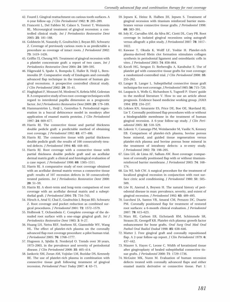
42. Fourel J. Gingival reattachment on carious tooth surfaces. A
4-year follow-up. J Clin Periodontol 1982: 9: 285–289.
43. Francetti L, Del Fabbro M, Calace S, Testori T, Weinstein
RL. Microsurgical treatment of gingival recession: a con-
trolled clinical study. Int J Periodontics Restorative Dent
2005: 25: 181–188.
44. Goldstein M, Nasatzky E, Goultschin J, Boyan BD, Schwartz
Z. Coverage of previously carious roots is as predictable a
procedure as coverage of intact roots. J Periodontol 2002:
73: 1419–1426.
45. Griffin TJ, Cheung WS. Treatment of gingival recession with
a platelet concentrate graft: a report of two cases. Int J
Periodontics Restorative Dent 2004: 24: 589–595.
46. Hagewald S, Spahr A, Rompola E, Haller B, Heijl L, Bern-
imoulin JP. Comparative study of Emdogain and coronally
advanced flap technique in the treatment of human gin-
gival recessions. A prospective controlled clinical study.
J Clin Periodontol 2002: 29: 35–41.
47. Haghighati F, Mousavi M, Moslemi N, Kebria MM, Golestan
B.A comparative studyof two root-coverage techniqueswith
regard to interdental papilla dimension as a prognostic
factor. Int J Periodontics Restorative Dent 2009: 29: 179–189.
48. Hammarstrom L, Heijl L, Gestrelius S. Periodontal regen-
eration in a buccal dehiscence model in monkeys after
application of enamel matrix proteins. J Clin Periodontol
1997: 24: 669–677.
49. Harris RJ. The connective tissue and partial thickness
double pedicle graft: a predictable method of obtaining
root coverage. J Periodontol 1992: 63: 477–486.
50. Harris RJ. The connective tissue with partial thickness
double pedicle graft: the results of 100 consecutively-trea-
ted defects. J Periodontol 1994: 65: 448–461.
51. Harris RJ. Root coverage with a connective tissue with
partial thickness double pedicle graft and an acellular
dermal matrix graft: a clinical and histological evaluation of
a case report. J Periodontol 1998: 69: 1305–1311.
52. Harris RJ. A comparative study of root coverage obtained
with an acellular dermal matrix versus a connective tissue
graft: results of 107 recession defects in 50 consecutively
treated patients. Int J Periodontics Restorative Dent 2000:
20: 51–59.
53. Harris RJ. A short-term and long-term comparison of root
coverage with an acellular dermal matrix and a subepi-
thelial graft. J Periodontol 2004: 75: 734–743.
54. Hirsch A, Attal U, Chai E, Goultschin J, Boyan BD, Schwartz
Z. Root coverage and pocket reduction as combined sur-
gical procedures. J Periodontol 2001: 72: 1572–1579.
55. Holbrook T, Ochsenbein C. Complete coverage of the de-
nuded root surface with a one-stage gingival graft. Int J
Periodontics Restorative Dent 1983: 3: 9–27.
56. Huang LH, Neiva REF, Soehren SE, Giannobile WV, Wang
HL. The effect of platelet-rich plasma on the coronally
advanced flap root coverage procedure: a pilot human trial.
J Periodontol 2005: 76: 1768–1777.
57. Hugoson A, Sjodin B, Norderyd O. Trends over 30 years,
1973–2003, in the prevalence and severity of periodontal
disease. J Clin Periodontol 2008: 35: 405–414.
58. Jankovic SM, Zoran AM, Vojislav LM, Bozidar DS, Kennedy
BE. The use of platelet-rich plasma in combination with
connective tissue graft following treatment of gingival
recession. Periodontal Pract Today 2007: 4: 63–71.
59. Jepsen K, Heinz B, Halben JH, Jepsen S. Treatment of
gingival recession with titanium reinforced barrier mem-
branes versus connective tissue grafts. J Periodontol 1998:
69: 383–391.
60. Joly JC, Carvalho AM, da Silva RC, Ciotti DL, Cury PR. Root
coverage in isolated gingival recessions using autograft
versus allograft: a pilot study. J Periodontol 2007: 78: 1017–
1022.
61. Kawase T, Okuda K, Wolff LF, Yoshie H. Platelet-rich
plasma-derived fibrin clot formation stimulates collagen
synthesis in periodontal ligament and osteoblastic cells in
vitro. J Periodontol 2003: 74: 858–864.
62. Keceli HG, Sengun D, Berberoglu A, Karabulut E. Use of
platelet gel with connective tissue grafts for root coverage:
a randomized-controlled trial. J Clin Periodontol 2008: 35:
255–262.
63. Langer B, Langer L. Subepithelial connective tissue graft
technique for root coverage. J Periodontol 1985: 56: 715–720.
64. Laupacis A, Wells G, Richardson S, Tugwell P. Users" guideto the medical literature V. How to use an article about
prognosis. Evidence based medicine working group. JAMA
1994: 272: 234–237.
65. Leknes KN, Amarante ES, Price DE, Boe OE, Skavland RJ,
Lie T. Coronally positioned flap procedures with or without
a biodegradable membrane in the treatment of human
gingival recession. A 6-year follow-up study. J Clin Peri-
odontol 2005: 32: 518–529.
66. Lekovic V, Camargo PM, Weinlaender M, Vasilic N, Kenney
EB. Comparison of platelet-rich plasma, bovine porous
bone mineral, and guided tissue regeneration versus
platelet-rich plasma and bovine porous bone mineral in
the treatment of intrabony defects: a re-entry study.
J Periodontol 2002: 73: 198–205.
67. Lins LH, de Lima AF, Sallum AW. Root coverage: compar-
ison of coronally positioned flap with or without titanium-
reinforced barrier membrane. J Periodontol 2003: 74: 168–
174.
68. Liu WJ, Solt CW. A surgical procedure for the treatment of
localized gingival recession in conjunction with root sur-
face citric acid conditioning. J Periodontol 1980: 51: 505–
509.
69. Loe H, Anerud A, Boysen H. The natural history of peri-
odontal disease in man: prevalence, severity, and extent of
gingival recession. J Periodontol 1992: 63: 489–495.
70. Lucchesi JA, Santos VR, Amaral CM, Peruzzo DC, Duarte
PM. Coronally positioned flap for treatment of restored
root surfaces: a 6-month clinical evaluation. J Periodontol
2007: 78: 615–623.
71. Marx RE, Carlson ER, Eichstaedt RM, Schimmele SR,
Strauss JE, Georgeff KR. Platelet-rich plasma: growth factor
enhancement for bone grafts. Oral Surg Oral Med Oral
Pathol Oral Radiol Endod 1998: 85: 638–646.
72. Matter J. Free gingival graft and coronally repositioned
flap. A 2-year follow-up report. J Clin Periodontol 1979: 6:
437–442.
73. Maurer S, Hayes C, Leone C. Width of keratinized tissue
after gingivoplasty of healed subepithelial connective tis-
sue grafts. J Periodontol 2000: 71: 1729–1736.
74. McGuire MK, Nunn M. Evaluation of human recession
defects treated with coronally advanced flaps and either
enamel matrix derivative or connective tissue. Part 1:
181
Coronally advanced flap and combination therapy for root coverage
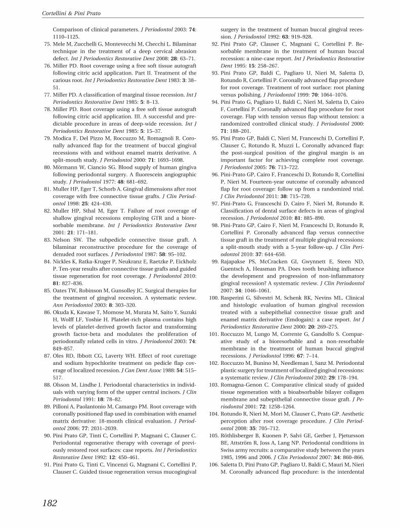
Comparison of clinical parameters. J Periodontol 2003: 74:
1110–1125.
75. Mele M, Zucchelli G, Montevecchi M, Checchi L. Bilaminar
technique in the treatment of a deep cervical abrasion
defect. Int J Periodontics Restorative Dent 2008: 28: 63–71.
76. Miller PD. Root coverage using a free soft tissue autograft
following citric acid application. Part II. Treatment of the
carious root. Int J Periodontics Restorative Dent 1983: 3: 38–
51.
77. Miller PD. A classification of marginal tissue recession. Int J
Periodontics Restorative Dent 1985: 5: 8–13.
78. Miller PD. Root coverage using a free soft tissue autograft
following citric acid application. III. A successful and pre-
dictable procedure in areas of deep-wide recession. Int J
Periodontics Restorative Dent 1985: 5: 15–37.
79. Modica F, Del Pizzo M, Roccuzzo M, Romagnoli R. Coro-
nally advanced flap for the treatment of buccal gingival
recessions with and without enamel matrix derivative. A
split-mouth study. J Periodontol 2000: 71: 1693–1698.
80. Mormann W, Ciancio SG. Blood supply of human gingiva
following periodontal surgery. A fluorescein angiographic
study. J Periodontol 1977: 48: 681–692.
81. Muller HP, Eger T, Schorb A. Gingival dimensions after root
coverage with free connective tissue grafts. J Clin Period-
ontol 1998: 25: 424–430.
82. Muller HP, Sthal M, Eger T. Failure of root coverage of
shallow gingival recessions employing GTR and a biore-
sorbable membrane. Int J Periodontics Restorative Dent
2001: 21: 171–181.
83. Nelson SW. The subpedicle connective tissue graft. A
bilaminar reconstructive procedure for the coverage of
denuded root surfaces. J Periodontol 1987: 58: 95–102.
84. Nickles K, Ratka-Kruger P, Neukranz E, Raetzke P, Eickholz
P. Ten-year results after connective tissue grafts and guided
tissue regeneration for root coverage. J Periodontol 2010:
81: 827–836.
85. Oates TW, Robinson M, Gunsolley JC. Surgical therapies for
the treatment of gingival recession. A systematic review.
Ann Periodontol 2003: 8: 303–320.
86. Okuda K, Kawase T, Momose M, Murata M, Saito Y, Suzuki
H, Wolff LF, Yoshie H. Platelet-rich plasma contains high
levels of platelet-derived growth factor and transforming
growth factor-beta and modulates the proliferation of
periodontally related cells in vitro. J Periodontol 2003: 74:
849–857.
87. Oles RD, Ibbott CG, Laverty WH. Effect of root curettage
and sodium hypochlorite treatment on pedicle flap cov-
erage of localized recession. J Can Dent Assoc 1988: 54: 515–
517.
88. Olsson M, Lindhe J. Periodontal characteristics in individ-
uals with varying form of the upper central incisors. J Clin
Periodontol 1991: 18: 78–82.
89. Pilloni A, Paolantonio M, Camargo PM. Root coverage with
coronally positioned flap used in combination with enamel
matrix derivative: 18-month clinical evaluation. J Period-
ontol 2006: 77: 2031–2039.
90. Pini Prato GP, Tinti C, Cortellini P, Magnani C, Clauser C.
Periodontal regenerative therapy with coverage of previ-
ously restored root surfaces: case reports. Int J Periodontics
Restorative Dent 1992: 12: 450–461.
91. Pini Prato G, Tinti C, Vincenzi G, Magnani C, Cortellini P,
Clauser C. Guided tissue regeneration versus mucogingival
surgery in the treatment of human buccal gingival reces-
sion. J Periodontol 1992: 63: 919–928.
92. Pini Prato GP, Clauser C, Magnani C, Cortellini P. Re-
sorbable membrane in the treatment of human buccal
recession: a nine-case report. Int J Periodontics Restorative
Dent 1995: 15: 258–267.
93. Pini Prato GP, Baldi C, Pagliaro U, Nieri M, Saletta D,
Rotundo R, Cortellini P. Coronally advanced flap procedure
for root coverage. Treatment of root surface: root planing
versus polishing. J Periodontol 1999: 70: 1064–1076.
94. Pini Prato G, Pagliaro U, Baldi C, Nieri M, Saletta D, Cairo
F, Cortellini P. Coronally advanced flap procedure for root
coverage. Flap with tension versus flap without tension: a
randomized controlled clinical study. J Periodontol 2000:
71: 188–201.
95. Pini Prato GP, Baldi C, Nieri M, Franceschi D, Cortellini P,
Clauser C, Rotundo R, Muzzi L. Coronally advanced flap:
the post-surgical position of the gingival margin is an
important factor for achieving complete root coverage.
J Periodontol 2005: 76: 713–722.
96. Pini-Prato GP, Cairo F, Franceschi D, Rotundo R, Cortellini
P, Nieri M. Fourteen-year outcome of coronally advanced
flap for root coverage: follow up from a randomized trial.
J Clin Periodontol 2011: 38: 715–720.
97. Pini-Prato G, Franceschi D, Cairo F, Nieri M, Rotundo R.
Classification of dental surface defects in areas of gingival
recession. J Periodontol 2010: 81: 885–890.
98. Pini-Prato GP, Cairo F, Nieri M, Franceschi D, Rotundo R,
Cortellini P. Coronally advanced flap versus connective
tissue graft in the treatment of multiple gingival recessions:
a split-mouth study with a 5-year follow-up. J Clin Peri-
odontol 2010: 37: 644–650.
99. Rajapakse PS, McCracken GI, Gwynnett E, Steen ND,
Guentsch A, Heasman PA. Does tooth brushing influence
the development and progression of non-inflammatory
gingival recession? A systematic review. J Clin Periodontol
2007: 34: 1046–1061.
100. Rasperini G, Silvestri M, Schenk RK, Nevins ML. Clinical
and histologic evaluation of human gingival recession
treated with a subepithelial connective tissue graft and
enamel matrix derivative (Emdogain): a case report. Int J
Periodontics Restorative Dent 2000: 20: 269–275.
101. Roccuzzo M, Lungo M, Corrente G, Gandolfo S. Compar-
ative study of a bioresorbable and a non-resorbable
membrane in the treatment of human buccal gingival
recessions. J Periodontol 1996: 67: 7–14.
102. Roccuzzo M, Bunino M, Needleman I, Sanz M. Periodontal
plastic surgery for treatment of localized gingival recessions:
a systematic review. J Clin Periodontol 2002: 29: 178–194.
103. Romagna-Genon C. Comparative clinical study of guided
tissue regeneration with a bioabsorbable bilayer collagen
membrane and subepithelial connective tissue graft. J Pe-
riodontol 2001: 72: 1258–1264.
104. Rotundo R, Nieri M, Mori M, Clauser C, Prato GP. Aesthetic
perception after root coverage procedure. J Clin Period-
ontol 2008: 35: 705–712.
105. Rothlisberger B, Kuonen P, Salvi GE, Gerber J, Pjetursson
BE, Attstrom R, Joss A, Lang NP. Periodontal conditions in
Swiss army recruits: a comparative study between the years
1985, 1996 and 2006. J Clin Periodontol 2007: 34: 860–866.
106. Saletta D, Pini Prato GP, Pagliaro U, Baldi C, Mauri M, Nieri
M. Coronally advanced flap procedure: is the interdental
182
Cortellini & Pini Prato
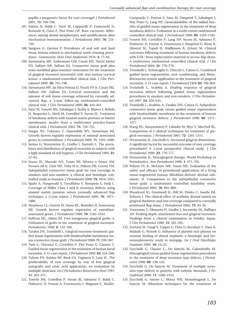
papilla a prognostic factor for root coverage? J Periodontol
2001: 72: 760–766.
107. Saletta D, Baldi C, Nieri M, Ceppatelli P, Franceschi D,
Rotundo R, Cairo F, Pini Prato GP. Root curvature: differ-
ences among dental morphotypes and modifications after
mechanical instrumentation. J Periodontol 2005: 76: 723–
730.
108. Sangnes G, Gjermo P. Prevalence of oral soft and hard
tissue lesions related to mechanical tooth cleaning proce-
dures. Community Dent Oral Epidemiol 1976: 4: 77–83.
109. Santamaria MP, Ambrosano GM, Casati MZ, Nociti Junior
FH, Sallum AW, Sallum EA. Connective tissue graft plus
resin-modified glass ionomer restoration for the treatment
of gingival recession associated with non-carious cervical
lesion: a randomized-controlled clinical trial. J Clin Peri-
odontol 2009: 36: 791–798.
110. Santamaria MP, da Silva Feitosa D, Nociti FH Jr, Casati MZ,
Sallum AW, Sallum EA. Cervical restoration and the
amount of soft tissue coverage achieved by coronally ad-
vanced flap: a 2-year follow-up randomized-controlled
clinical trial. J Clin Periodontol 2009: 36: 434–441.
111. Sanz M, Tonetti MS, Zabalegui I, Sicilia A, Blanco J, Rebelo
H, Rasperini G, Merli M, Cortellini P, Suvan JE. Treatment
of intrabony defects with enamel matrix proteins or barrier
membranes: results from a multicenter practice-based
clinical trial. J Periodontol 2004: 75: 726–733.
112. Saygin NE, Tokiyasu Y, Giannobile WV, Somerman MJ.
Growth factors regulate expression of mineral associated
genes in cementoblasts. J Periodontol 2000: 71: 1591–1600.
113. Serino G, Wennstrom JL, Lindhe J, Eneroth L. The preva-
lence and distribution of gingival recession in subjects with
a high standard of oral hygiene. J Clin Periodontol 1994: 21:
57–63.
114. Souza SL, Macedo GO, Tunes RS, Silveira e Souza AM,
Novaes AB Jr, Grisi MF, Taba M Jr, Palioto DB, Correa VM.
Subepithelial connective tissue graft for root coverage in
smokers and non-smokers: a clinical and histologic con-
trolled study in humans. J Periodontol 2008: 79: 1014–1021.
115. Spahr A, Haegewald S, Tsoulfidou F, Rompola E, Heijl L.
Coverage of Miller Class I and II recession defects using
enamel matrix proteins versus coronally advanced flap
technique: a 2-year report. J Periodontol 2005: 76: 1871–
1880.
116. Strayhorn CL, Garrett JS, Dunn RL, Benedict JJ, Somerman
MJ. Growth factors regulate expression of osteoblast-
associated genes. J Periodontol 1999: 70: 1345–1354.
117. Sullivan HC, Atkins JH. Free autogenous gingival grafts. 3.
Utilization of grafts in the treatment of gingival recession.
Periodontics 1968: 6: 152–160.
118. Tatakis DN, Trombelli L. Gingival recession treatment: gui-
ded tissue regeneration with bioabsorbable membrane ver-
sus connective tissue graft. J Periodontol 2000: 71: 299–307.
119. Tinti C, Vincenzi G, Cortellini P, Pini Prato G, Clauser C.
Guided tissue regeneration in the treatment of human facial
recession. A 12-case report. J Periodontol 1992: 63: 554–560.
120. Tolmie PN, Rubins RP, Buck GS, Vagianos V, Lanz JC. The
predictability of root coverage by way of free gingival
autografts and citric acid application: an evaluation by
multiple clinicians. Int J Periodontics Restorative Dent 1991:
11: 261–271.
121. Tonetti MS, Cortellini P, Suvan JE, Adriaens P, Baldi C,
Dubravec D, Fonzar A, Fourmousis I, Magnani C, Muller-
Campanile V, Patroni S, Sanz M, Vangsted T, Zabalegui I,
Pini Prato G, Lang NP. Generalizability of the added ben-
efits of guided tissue regeneration in the treatment of deep
intrabony defects. Evaluation in a multi-center randomized
controlled clinical trial. J Periodontol 1998: 69: 1183–1192.
122. Tonetti MS, Cortellini P, Lang NP, Suvan JE, Adriaens P,
Dubravec D, Fonzar A, Fourmousis I, Rasperini G, Rossi R,
Silvestri M, Topoll H, Wallkamm B, Zybutz M. Clinical
outcomes following treatment of human intrabony defects
with GTR ⁄ bone replacement material or access flap alone.
A multicenter randomized controlled clinical trial. J Clin
Periodontol 2004: 31: 770–776.
123. Trombelli L, Schincaglia G, Checchi L, Calura G. Combined
guided tissue regeneration, root conditioning, and fibrin-
fibronectin system application in the treatment of gingival
recession. A 15-case report. J Periodontol 1994: 65: 796–803.
124. Trombelli L, Scabbia A. Healing response of gingival
recession defects following guided tissue regeneration
procedures in smokers and non-smokers. J Clin Periodon-
tol 1997: 24: 529–533.
125. Trombelli L, Scabbia A, Tatakis DN, Calura G. Subpedicle
connective tissue graft versus guided tissue regeneration
with bioabsorbable membrane in the treatment of human
gingival recession defects. J Periodontol 1998: 69: 1271–
1277.
126. Wang HL, Bunyaratavej P, Labadie M, Shyr Y, MacNeil RL.
Comparison of 2 clinical techniques for treatment of gin-
gival recession. J Periodontol 2001: 72: 1301–1311.
127. Wennstrom JL, Zucchelli G. Increased gingival dimensions.
A significant factor for successful outcome of root coverage
procedures? A 2-year prospective clinical study. J Clin
Periodontol 1996: 23: 770–777.
128. Wennstrom JL. Mucogingival therapy. World Workshop in
Periodontics. Ann Periodontol 1996: 1: 671–701.
129. Wilson TG Jr, McGuire MK, Nunn ME. Evaluation of the
safety and efficacy of periodontal applications of a living
tissue-engineered human fibroblast-derived dermal sub-
stitute. II. Comparison to the subepithelial connective
tissue graft: a randomized controlled feasibility study.
J Periodontol 2005: 76: 881–889.
130. Woodyard JG, Greenwell H, Hill M, Drisko C, Iasella JM,
Scheetz J. The clinical effect of acellular dermal matrix on
gingival thickness and root coverage compared to coronally
positioned flap alone. J Periodontol 2004: 75: 44–56.
131. Yoneyama T, Okamoto H, Lindhe J, Socransky SS, Haffajee
AD. Probing depth, attachment loss and gingival recession.
Findings from a clinical examination in Ushiku, Japan.
J Clin Periodontol 1988: 15: 581–591.
132. Zechner W, Tangl S, Tepper G, Furst G, Bernhart T, Haas R,
Mailath G, Watzek G. Influence of platelet-rich plasma on
osseous healing of dental implants: a histologic and his-
tomorphometric study in minipigs. Int J Oral Maxillofac
Implants 2003: 18: 15–22.
133. Zucchelli G, Clauser C, De Sanctis M, Calandriello M.
Mucogingival versus guided tissue regeneration procedures
in the treatment of deep recession type defects. J Period-
ontol 1998: 69: 138–145.
134. Zucchelli G, De Sanctis M. Treatment of multiple reces-
sion-type defects in patients with esthetic demands. J Pe-
riodontol 2000: 71: 1506–1514.
135. Zucchelli G, Amore C, Sforza NM, Montebugnoli L, De
Sanctis M. Bilaminar techniques for the treatment of
183
Coronally advanced flap and combination therapy for root coverage
recession-type defects. A comparative clinical study. J Clin
Periodontol 2003: 30: 862–870.
136. Zucchelli G, Cesari C, Amore C, Montebugnoli L, De
Sanctis M. Laterally moved, coronally advanced flap: a
modified surgical approach for isolated recession-type
defects. J Periodontol 2004: 75: 1734–1741.
137. Zucchelli G, De Sanctis M. Long-term outcome following
treatment of multiple Miller class I and II recession defects
in esthetic areas of the mouth. J Periodontol 2005: 76: 2286–
2292.
138. Zucchelli G, Testori T, De Sanctis M. Clinical and ana-
tomical factors limiting treatment outcomes of gingival
recession: a new method to predetermine the line of root
coverage. J Periodontol 2006: 77: 714–721.
139. Zucchelli G, Mounssif I, Stefanini M, Mele M, Monte-
bugnoli L, Sforza NM. Hand and ultrasonic instrumenta-
tion in combination with root-coverage surgery: a
comparative controlled randomized clinical trial. J Period-
ontol 2009: 80: 577–585.
140. Zucchelli G, Mele M, Mazzotti C, Marzadori M, Monte-
bugnoli L, De Sanctis M. Coronally advanced flap with and
without vertical releasing incisions for the treatment of
multiple gingival recessions: a comparative controlled
randomized clinical trial. J Periodontol 2009: 80: 1083–1094.
184
Cortellini & Pini Prato