Embed Size (px)
Citation preview
Citation: Shreya D, Jain Sanjay J, Gupta Sharadha G, Muglikar Sangeeta D and Pathan Danish S . Semilunar Coronally Advanced Flap Technique for Root Coverage in Adjacent Teeth in the Anterior Esthetic Zone – A Case Report. Austin J Dent. 2015;2(2): 1018.
Austin J Dent - Volume 2 Issue 2 - 2015ISSN : 2381-9189 | www.austinpublishinggroup.com Shreya et al. © All rights are reserved
Austin Journal of DentistryOpen Access
Abstract
Background: Gingival recession is a common concern in patients with or without periodontal disease. A variety of periodontal plastic surgery procedures have been attempted in the past to treat the gingival recession deformities with varying degrees of success. This case report describes traditional semilunar coronally advanced flap for the treatment of recession defects on multiple adjacent teeth.
Material and Methods: Probing depth, Clinical Attachment Level (CAL), recession height and width were assessed at baseline and after 3 months. After completion of phase I therapy, the esthetic surgery was planned. The technique involved a semilunar incision made parallel to the free gingival margin of the facial tissue, and coronally positioning the tissue over the denuded root.
Results: A reduction in the recession height, width and CAL was seen after 6 months.
Conclusion: This minimally invasive technique offers the advantage of ease of operation as well as with minimum patient discomfort and improved esthetics.
Keywords: Gingival recession; Esthetics; Semilunar flap
Materials and MethodsA 27–year-old male patient presented for routine dental
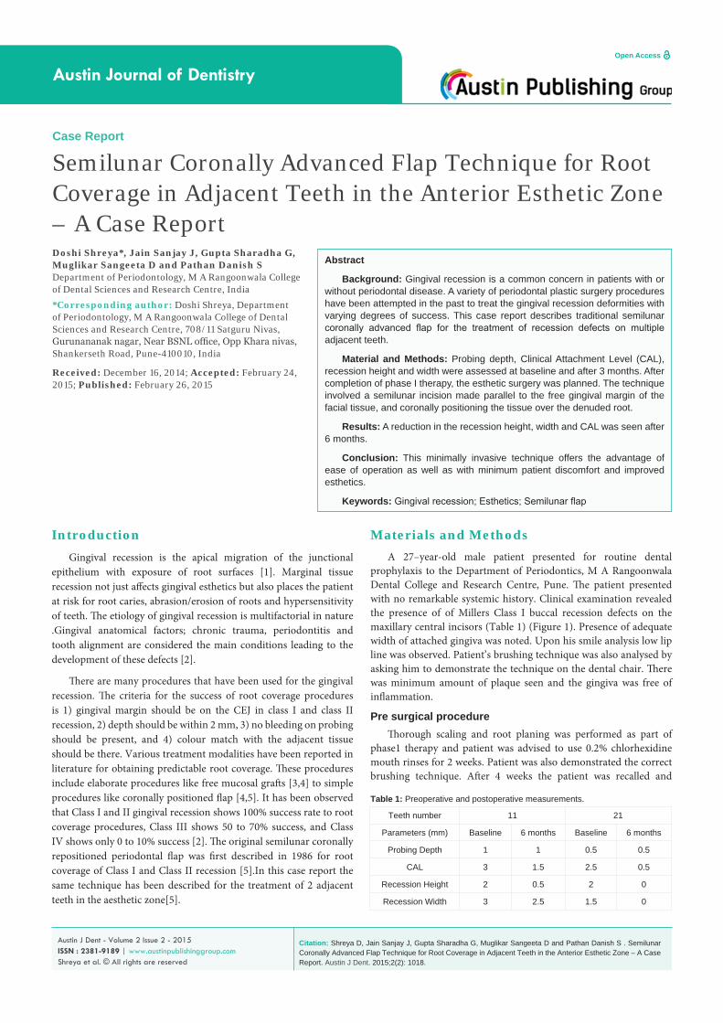
prophylaxis to the Department of Periodontics, M A Rangoonwala Dental College and Research Centre, Pune. The patient presented with no remarkable systemic history. Clinical examination revealed the presence of of Millers Class I buccal recession defects on the maxillary central incisors (Table 1) (Figure 1). Presence of adequate width of attached gingiva was noted. Upon his smile analysis low lip line was observed. Patient’s brushing technique was also analysed by asking him to demonstrate the technique on the dental chair. There was minimum amount of plaque seen and the gingiva was free of inflammation.
Pre surgical procedureThorough scaling and root planing was performed as part of
phase1 therapy and patient was advised to use 0.2% chlorhexidine mouth rinses for 2 weeks. Patient was also demonstrated the correct brushing technique. After 4 weeks the patient was recalled and
IntroductionGingival recession is the apical migration of the junctional
epithelium with exposure of root surfaces [1]. Marginal tissue recession not just affects gingival esthetics but also places the patient at risk for root caries, abrasion/erosion of roots and hypersensitivity of teeth. The etiology of gingival recession is multifactorial in nature .Gingival anatomical factors; chronic trauma, periodontitis and tooth alignment are considered the main conditions leading to the development of these defects [2].
There are many procedures that have been used for the gingival recession. The criteria for the success of root coverage procedures is 1) gingival margin should be on the CEJ in class I and class II recession, 2) depth should be within 2 mm, 3) no bleeding on probing should be present, and 4) colour match with the adjacent tissue should be there. Various treatment modalities have been reported in literature for obtaining predictable root coverage. These procedures include elaborate procedures like free mucosal grafts [3,4] to simple procedures like coronally positioned flap [4,5]. It has been observed that Class I and II gingival recession shows 100% success rate to root coverage procedures, Class III shows 50 to 70% success, and Class IV shows only 0 to 10% success [2]. The original semilunar coronally repositioned periodontal flap was first described in 1986 for root coverage of Class I and Class II recession [5].In this case report the same technique has been described for the treatment of 2 adjacent teeth in the aesthetic zone[5].
Case Report
Semilunar Coronally Advanced Flap Technique for Root Coverage in Adjacent Teeth in the Anterior Esthetic Zone – A Case ReportDoshi Shreya*, Jain Sanjay J, Gupta Sharadha G, Muglikar Sangeeta D and Pathan Danish S Department of Periodontology, M A Rangoonwala College of Dental Sciences and Research Centre, India
*Corresponding author: Doshi Shreya, Department of Periodontology, M A Rangoonwala College of Dental Sciences and Research Centre, 708/11 Satguru Nivas, Gurunananak nagar, Near BSNL office, Opp Khara nivas, Shankerseth Road, Pune-410010, India
Received: December 16, 2014; Accepted: February 24, 2015; Published: February 26, 2015
Teeth number 11 21
Parameters (mm) Baseline 6 months Baseline 6 months
Probing Depth 1 1 0.5 0.5
CAL 3 1.5 2.5 0.5
Recession Height 2 0.5 2 0
Recession Width 3 2.5 1.5 0
Table 1: Preoperative and postoperative measurements.
Austin J Dent 2(2): id1018 (2015) - Page - 02
Doshi Shreya Austin Publishing Group
Submit your Manuscript | www.austinpublishinggroup.com
periodontal therapy for correction of recession was planned. The purpose and design of the procedure was explained to the patient and an informed consent form was signed. The width of attached gingiva was 3 mm and was found to be adequate. The gingiva in relation to 11 and 21 demonstrated thick biotype. The approval of the Local Ethics Committee of M. A. Rangoonwala Dental College and Research Centre was obtained.
Surgical procedure The presurgical measurements were assessed using UNC 15 as
shown in Table 1 (Figure 1, Figure 2) following a pre procedural rinse with 0.2% chlorhexidine. The surgical procedure was performed under local anaesthesia (1:200000 dilutions) §. The incisions were marked on the tissues with a marking pencil. The frenum attachment was gingival, hence frenotomy procedure was carried out before giving the incision Semilunar incisions were made apical to the recession defects of 11 and 21, starting within mucosa extending beyond the mucogingival junction mesio-distally and arching more coronally to terminate apical to the papillae, distal to 11 and 21. Using a number 15c blade, a split thickness dissection is made from the initial incision line coronally connected with an intrasulcular incision, made mid-facially (Figure 3) [5]. The flap was held in its new position for 2 minutes with moist gauze. Sutures were not placed. A periodontal dressing was placed.
The patient was prescribed a combination of Diclofenac Sodium 50mg and Paracetamol 500 mg ¥ twice daily for 3 days and 0.2% chlorhexidine mouth rinse twice daily for 2 weeks. Patient was advised to take soft diet and not to brush at the surgical site for atleast
2 weeks after the day of surgery.
Foot notesClohex Plus (0.2% Chlorhexidine), Dr.Reddys Laboratories Pvt
Ltd, Hyderabad
§ Lignocad ADR (1:200000), Cadila, Ahmedabad
¥ Tablet Enzoflam (Tablet Diclofenac Sodium 50mg, Tablet Paracetamol 500mg) Alkem industries, Bergen
Results A comparison between baseline and 3 months clinical outcomes
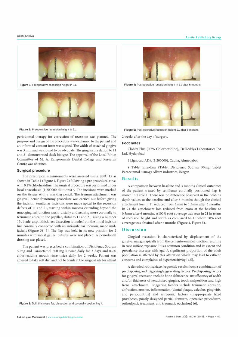
of the patient treated by semilunar coronally positioned flap is shown in Table 1. There was no difference observed in the probing depth values, at the baseline and after 6 months though the clinical attachment loss in 11 reduced from 3 mm to 1.5mm after 6 months. In 21 the attachment loss reduced from 2mm at the baseline to 0.5mm after 6 months. A100% root coverage was seen in 21 in terms of recession height and width as compared to 11 where 50% root coverage was obtained after 6 months (Figure 4, Figure 5).
Discussion Gingival recession is characterized by displacement of the
gingival margin apically from the cemento-enamel junction resulting in root surface exposure. It is a common condition and its extent and prevalence increase with age. A significant proportion of the adult population is affected by this alteration which may lead to esthetic concerns and complaints of hypersensitivity [4,5].
A denuded root surface frequently results from a combination of predisposing and triggering/aggravating factors. Predisposing factors for gingival recession include bone dehiscence, insufficiency of width and/or thickness of keratinised gingiva, tooth malposition and high frenal attachment. Triggering factors include traumatic abrasion, abfraction, erosion, inflammation (dental plaque, calculus, gingivitis, and periodontitis) and iatrogenic factors (inappropriate fixed prostheses, poorly designed partial dentures, operative procedures, orthodontic treatment, and traumatic occlusion) [6].
Figure 1: Preoperative recession height in 11.
Figure 2: Preoperative recession height in 21.
Figure 3: Split thickness flap dissection and coronally positioning it.
Figure 4: Postoperative recession height in 11 after 6 months.
Figure 5: Post operative recession height 21 after 6 months.
Austin J Dent 2(2): id1018 (2015) - Page - 03
Doshi Shreya Austin Publishing Group
Submit your Manuscript | www.austinpublishinggroup.com
The primary goal of reconstructive periodontal therapy is root coverage. Periodontal plastic surgery is defined by World Workshop in Clinical Periodontics 1996 as surgical procedures performed to correct or eliminate anatomic, developmental, or traumatic deformities of the gingival [7]. Many periodontal plastic procedures have been described in the past [8]. There is extensive data in the literature which provides an insight into the various surgical modalities used for the treating gingival recession. But limitations of these procedures are lack of predictability, compromised blood supply, postoperative discomfort, and postoperative morbidity. The traditional semilunar coronally repositioned flap being presented has many advantages like no tension on the flap , no shortening of the vestibule, no sutures are needed because of the lack of tension of the tissue being coronally positioned, minimum discomfort to the patients and single operative site [5]. The main indication for carrying out this technique is adequate width of attached gingiva, Class I buccal/labial defects, where esthetics is affected and cannot be controlled by non surgical therapy. The procedure can also be used where there has been recession around previous full coverage restorations in the anterior section of the mouth, where the patient has a high enough lip line when smiling to show the denuded roots.
In the present case maxillary central incisors presented with unesthetic Class I gingival recession with adequate width of attached gingiva. Hence, semilunar coronally positioned flap technique was the procedure of choice. The gingival biotype also made the technique chosen possible.
In this case report significant improvements in clinical parameters such as recession height and width and clinical attachment level was observed. A100% root coverage was seen in 21 and 50% in 11 after 6 months (Figure 4, Figure 5) (Table 1). Also the technique offers advantages like ease of performing and no need of sutures. Long-term clinical and histological investigations are needed to confirm these results with larger sample size.
ConclusionSemilunar coronally positioned flap procedure is a simple
technique which has high patient acceptance and provides satisfactory results for treating class I labial/buccal recession defects especially in the anterior esthetic zone. Further studies with larger sample size are needed in order to evaluate the long-term stability of the obtained positive results.
References1. Kassab MM, Cohen RE. The etiology and prevalence of gingival recession. J
Am Dent Assoc. 2003; 134: 220-225.
2. Loe H, Anerud A, Boysen H. The natural history of periodontal disease in man: Prevalence, severity and extent of gingival recession. J Periodontol. 1992; 63: 489–495.
3. Nordenram A, Landt H. Evaluation of a surgical technique in the periodontal treatment of maxillary anterior teeth. Acta Odontologica Scandinaviea. 1969; 27: 283-289.
4. American Academy of Periodontology. Glossary of Periodontal Terms. 4th edn. Chicago: American Academy of Periodontology. 2001.
5. Tarnow DP. Semilunar Coronally Repositioned Flap. J Clin Periodontol. 1986; 13: 182–185.
6. Tugnait A, Clerehugh V. Gingival recession: its significance and management. Journal of Dentistry. 2001; 29: 381- 394.
7. Glossary of Periodontal terms. 4th edn. Chicago: American Academy of Periodontology. American Academy of Periodontology. 2001; p. 49.
8. Edel A. The use of free connective tissue graft to increase the width of attached gingiva. Oral Surg Oral Med Oral Pathol. 1975; 39: 341–346.
Citation: Shreya D, Jain Sanjay J, Gupta Sharadha G, Muglikar Sangeeta D and Pathan Danish S . Semilunar Coronally Advanced Flap Technique for Root Coverage in Adjacent Teeth in the Anterior Esthetic Zone – A Case Report. Austin J Dent. 2015;2(2): 1018.
Austin J Dent - Volume 2 Issue 2 - 2015ISSN : 2381-9189 | www.austinpublishinggroup.com Shreya et al. © All rights are reserved