Embed Size (px)
Citation preview

Correlation of Patient Vertical Centering with Radiation Output in
Adult Abdominopelvic CT
Phillip Cheng, MD MS

Disclosures
• None

Objectives
•To illustrate a method for computing the vertical position of a patient from reconstructed CT images
•To determine if vertical position has a significant effect on scanner radiation output in adult abdominopelvic CT

Patient vertical positioning
Philips Brilliance 64 CT(x-ray tube below table for localizer)

Centering and the Localizer Radiograph
Patient
Patient far from x-ray tube
Patient
Patient close to x-ray tube

Prior investigation
AJR 2009; 192: 862-865 (Toshiba Aquilion 64, GE Lightspeed Ultra 16 with Xtream)

Prior investigation
AJR 2014; 203: 123-130(GE Lightspeed VCT Xte)

Coordinate system
(0,0)x
y(xrot, yrot)
(xrot+cx, yrot+cy)

Relevant DICOM Elements
Not available for my scanners!

Other DICOM Elements

Center of Rotation Position
• Let• 𝑑𝑑𝑟𝑟𝑟𝑟𝑟𝑟𝑟𝑟𝑟𝑟 = Reconstruction Diameter (0018, 1100)• 𝑦𝑦𝑡𝑡𝑡𝑡𝑡𝑡𝑡𝑡𝑟𝑟 = Table Height (0018, 1130)• 𝑦𝑦𝑝𝑝𝑟𝑟𝑝𝑝 = Y Coordinate of Image Position (Patient) (0020, 0032)• 𝑦𝑦𝑟𝑟𝑟𝑟𝑡𝑡 = Y Coordinate of Center of Rotation relative to center of image
• Philips Brilliance 64 Scanner• 𝑦𝑦𝑟𝑟𝑟𝑟𝑡𝑡 = 255 − 𝑑𝑑𝑟𝑟𝑟𝑟𝑟𝑟𝑟𝑟𝑟𝑟
2− 𝑦𝑦𝑝𝑝𝑟𝑟𝑝𝑝 − 𝑦𝑦𝑡𝑡𝑡𝑡𝑡𝑡𝑡𝑡𝑟𝑟
• GE Lightspeed 16, Toshiba Aquilion 64• 𝑦𝑦𝑟𝑟𝑟𝑟𝑡𝑡 = −𝑑𝑑𝑟𝑟𝑟𝑟𝑟𝑟𝑟𝑟𝑟𝑟
2− 𝑦𝑦𝑝𝑝𝑟𝑟𝑝𝑝

In-Plane Center of Mass
(xrot+cx, yrot+cy)

Phantom Measurements and Calculations
Actual -114 -65 -17 35 85
cy -91 -49 -3 39 82
Measurements (in mm) expressed as distance below the center of rotation of the scanner.
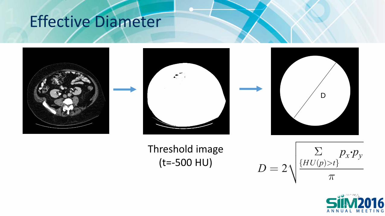
Effective Diameter
D
Threshold image(t=-500 HU)

Patient Calculations
drecon 50 cm 42 cmcy 4.2 -1.3
Eff diam 30.8 30.8CTDIvol 8.75 8.74
Both scans onPhilips Brilliance 64.

All scans on Philips Brilliance 64
Most patientspositioned low

All scans on Philips Brilliance 64

All scans on Philips Brilliance 64

Regression
CTDIvol as a function of effective diameter and vertical positioning

Conclusions
• Automated calculation of vertical center of mass position from reconstructed CT images is feasible
• Patient position may not significantly affect mean CTDIvol for some scanners, depending on the proprietary tube current modulation algorithm


Correcting for the Scanner Table
Patient contribution Table contribution

Phantom Experiments
Actual -114 -65 -17 35 85
cy -91 -49 -3 39 82
cy’ -101 -58 -12 29 71
Measurements (in mm) expressed as distance below the center of rotation of the scanner.

Repeat scans

Philips Brilliance 64 CT(x-ray tube below table for localizer)
Patient Vertical Positioning